millodot kc prevalence 2011
TRANSCRIPT

91
INTRODUCTION
Keratoconus (KC) is a corneal disorder of unknown etiology in which the central portion of the cornea becomes thinner and bulges forward in a cone-shaped fashion as a result of non-inflammatory processes initiated in the anterior part of the cornea.1,2 The disease has its usual onset at puberty and in many cases progresses until the third to fourth decade of life, when it usually arrests1,3,4 The main symptom is a mild to severe visual impairment due to irregular astigmatism, myopia and frequently corneal scarring. However, early in the disease there may not be any symptoms. Although a large proportion of kerato-conic patients can be managed with contact lenses, an average of about 20% of all keratoconic corneas require keratoplasty,5–8 some authors report markedly
different surgical indication rates of 6.5%,9 12%10 to 45%.11
Contact lens fitting for KC is a more complex task than it is for normal corneas and necessitates specially designed lenses.7,12 The impact of keratoplasty and contact lens management poses a heavy financial burden on the providers of health care. Keratoconus has become a major cause of keratoplasty in most countries. For example in Israel 9.2% of patients had keratoplasty for KC in 1961–1970, 18.5% in 1971–1980 and 28.7% in 1981–199013 and this figure was 51% in East Jerusalem in the year 2001–2002, whereas it was 27% in the same period in Sweden14.
A population-based survey can help detect the asymptomatic cases before their condition pro-gresses to the point where they have poor visual acuity and become unproductive members of
Ophthalmic Epidemiology, 18(2), 91–97, 2011Copyright © 2011 Informa Healthcare USA, Inc.ISSN: 0928-6586 print/ 1744-5086 onlineDOI: 10.3109/09286586.2011.560747
Received 02 August 2010; revised 11 January 2011; accepted 01 February 2011
Correspondence: Michel Millodot, OD, PhD, Cardiff University, School of Optometry and Vision Sciences, Maindy Road, Cardiff, CF24 4LU UK. E-mail: [email protected]
02 August 2010
11 January 2011
01 February 2011
© 2011 Informa Healthcare USA, Inc.
2011
Ophthalmic Epidemiology
0928-65861744-5086
10.3109/09286586.2011.560747
18
9197
2
560747
NOPE
ORIGINAL ARTICLE
Prevalence and Associated Factors of Keratoconus in Jerusalem: A Cross-sectional Study
Michel Millodot1, Einat Shneor2, Sophie Albou2, Esther Atlani2, and Ariela Gordon-Shaag2
1School of Optometry and Vision Sciences, Cardiff University, UK, and 2Department of Optometry, Hadassah Academic and Technological College, Jerusalem, Israel
ABSTRACT
Purpose: To determine the prevalence and associated factors for keratoconus in a college student population sample in Jerusalem.
Methods: Volunteers participated in this cross-sectional study. Videokeratography was performed on both eyes of each subject who also completed an anonymous questionnaire. Keratoconus was defined by cone apex ≥ 50D, inferior-superior dioptric difference ≥ 3.5 diopters, as well as positive results from the software indices KISA, KCI and KSI. The association between independent predictors and keratoconus was analyzed using multivariate logistic regression analysis.
Results: Of a total of 987 volunteers, 981 (mean age 24.4) were included. The prevalence of keratoconus among all subjects was 2.34% (95% confidence interval [CI] 1.4–3.3). It was significantly higher in men (4.91%, CI 2.6–7.3) than women (1.07%, CI 0.3–1.9) but not between Israeli Arabs (3.0%, CI 0.6–5.4) and Israeli Jews (2.2%, CI 1.2–3.3). Keratoconus was significantly associated with positive family history of the disease (Odds Ratio [OR] 17.1, CI 5.0–57.8, P < 0.001), male gender (OR 5.4, CI 2.1–14.3, P = 0.001) and atopy (OR 3.0, CI 1.2–7.6, P = 0.02), but not with eye rubbing.
Conclusions: The prevalence of keratoconus in Jerusalem was found to be much higher than that seen in other parts of the world, except India. This may be related to a combination of genetic and environmental factors. Positive family history, male gender and atopy were shown to be significant predictors. The results of this study signal a need for public health outreach and intervention for keratoconus.
Keywords: Keratoconus, Prevalence, Risk factors, Prevalence men, Prevalence women
Oph
thal
mic
Epi
dem
iol D
ownl
oade
d fr
om in
form
ahea
lthca
re.c
om b
y 79
.179
.39.
163
on 0
3/18
/11
For
pers
onal
use
onl
y.

92 M. Millodot et al.
Ophthalmic Epidemiology
society. It could also identify subjects with subclini-cal keratoconus (KC suspects) who should be advised against photorefractive surgery. With the advent of collagen cross-linking therapy early detection is essential to identify KC patients before the corneal steepening significantly affects vision. It may also help in uncovering how genetic and environmental factors can influence the development of KC, a dis-ease whose etiology is still unknown.2,15 Our survey aimed to address this lack of information by report-ing for the first time the prevalence of keratoconus in a cross-sectional sample in Jerusalem, Israel, using videokeratography. This survey is especially perti-nent in view of anecdotal and clinical observations from practitioners and hospital eye clinics of a seem-ingly large proportion of keratoconic patients in this country.
MATERIAL AND METHODS
Site of study
The student population of Hadassah Academic and Technological College was chosen for this survey. This community college located in the center of Jerusalem offers degrees and diplomas in various subjects to approximately 2000 students from a wide spectrum of socioeconomic levels including Arabs and Jews. Approximately one third of the students receive finan-cial aid. All students received an email inviting them to participate in this non-invasive screening test for kera-toconus with a short explanation of the signs and symp-toms of the disease and that in the early stages there may be no visual symptoms. The email stressed that it was important for all students to be tested. Students were also asked to complete an anonymous self-administered questionnaire. The questionnaire asked questions relat-ing to age, gender, domicile, ethnicity (Arab, Jew), fam-ily history of KC, contact lens wear, allergies and eye rubbing. Regarding eye rubbing the question was: do you regularly rub your eyes? Yes/no and followed by a scale ranging from 1 “not at all” to 5 “almost constantly” with 3 and above considered positive. Regarding atopy the questions were: do you have any allergies? Yes/no and followed by a list of environmental allergens, house dust/cat or dog hair/pollen/penicillin/other and asthma/eczema/hay fever. Any one of these conditions and of the allergens was considered positive. Exclusion criteria were subjects who had undergone refractive or corneal surgery (except for keratoconus management), who had been diagnosed with an ocular disease other than KC and who wore hard contact lenses. The nature of the study was explained to the students before sign-ing an informed consent form. The study followed the tenets of the Declaration of Helsinki and was approved by Hadassah College ethics committee. The survey was carried out between January 2009 and April 2010.
Data collection
Videokeratography was performed on both eyes of each subject. Two instruments were used at different times; a Tomey TMS-4 Topographic Modeling System and a Shin-Nippon corneal topographer CT-1000, which pro-duce similar color-coded topographic patterns of the cor-nea. The test was performed by qualified optometrists trained in the correct use of the instrument. Subjects who wore contact lenses (the vast majority wore soft lenses and only a few wore gas permeable lenses) were asked to remove them immediately prior to the examination. Proper fixation and alignment were ascertained when the cross-hair was in the center of the pupil. Two photos of each eye were usually taken. However, in many cases more photos had to be taken if the eye seemed to have moved, or the cornea was partially covered by the eye-lids, or the image appeared to represent a keratoconic cornea; we selected the best topographic image which displayed the largest area of the cornea and had the least distortions. The instruments included KC screen-ing software KISA, KCI and KSI, respectively. These are algorithms designed to detect KC. They are based on various quantitative videokeratography-derived indices and have been shown to have high sensitivity and speci-ficity.16 However, to the best of our knowledge there is not yet any published study evaluating the agreement between these indices. Nevertheless it is generally agreed that the diagnosis of KC needs further confirma-tion. Therefore besides these indices two other assess-ments were employed to arrive at a diagnosis. Firstly the color-coded corneal map of each participant was examined by two investigators who were blind to the results of the questionnaire. Secondly we determined the dioptric power of the corneal apex and the maxi-mum dioptric difference between the corneal apex and a spot in the middle of the superior half of the cornea (IS). On the basis of the videokeratographic indices and the clinical assessments each eye was placed in one of three groups according to the following classification:
1. Normal. KISA <60% and either or both KCI and KSI = 0%; topographic map showing no detectable keratoconus-like pattern; corneal apex < 45D and IS < 1D.
2. KC suspect. KISA > 60% and <100%; suspicious topo-graphic pattern; corneal apex > 45D and <50D, and/or IS >1D and <3.5D. (KCI and KSI indices do not distinguish KC suspects).
3. KC. KISA >100% and both KCI and KSI >0%; clear topographic pattern; corneal apex >50D and/or IS >3.5D.
Data analysis
Prevalence and 95% confidence intervals (CI) of Arabs, Jews, males and females were calculated. If a person
Oph
thal
mic
Epi
dem
iol D
ownl
oade
d fr
om in
form
ahea
lthca
re.c
om b
y 79
.179
.39.
163
on 0
3/18
/11
For
pers
onal
use
onl
y.
Prevalence of Keratoconus 93
© 2011 Informa Healthcare USA, Inc.
had KC in one eye only or both eyes, that person was defined as having the disease. Univariate analyses were performed using standard non-parametric and para-metric tests (Chi-square, Fisher’s exact test if any of the expected frequencies was less than 5, and the t-test) to determine whether gender, ethnicity, family history of KC, atopy or eye rubbing were significantly associated with KC. Multivariate logistic regression analysis was performed to explore the association between KC (the outcome variable) and the independent predictors, which had been found significant in the univariate analysis using a statistical software package (SPSS for Windows). The predictor variables were binary and coded as “1” (KC present) and “0” (KC absent), “1” for male and “0” for female. Adjusted odds ratios (OR) as a measure of association and 95% confidence intervals (CI) were calculated. All tests were two-tailed and P values lower than 0.05 were considered statistically significant.
RESULTS
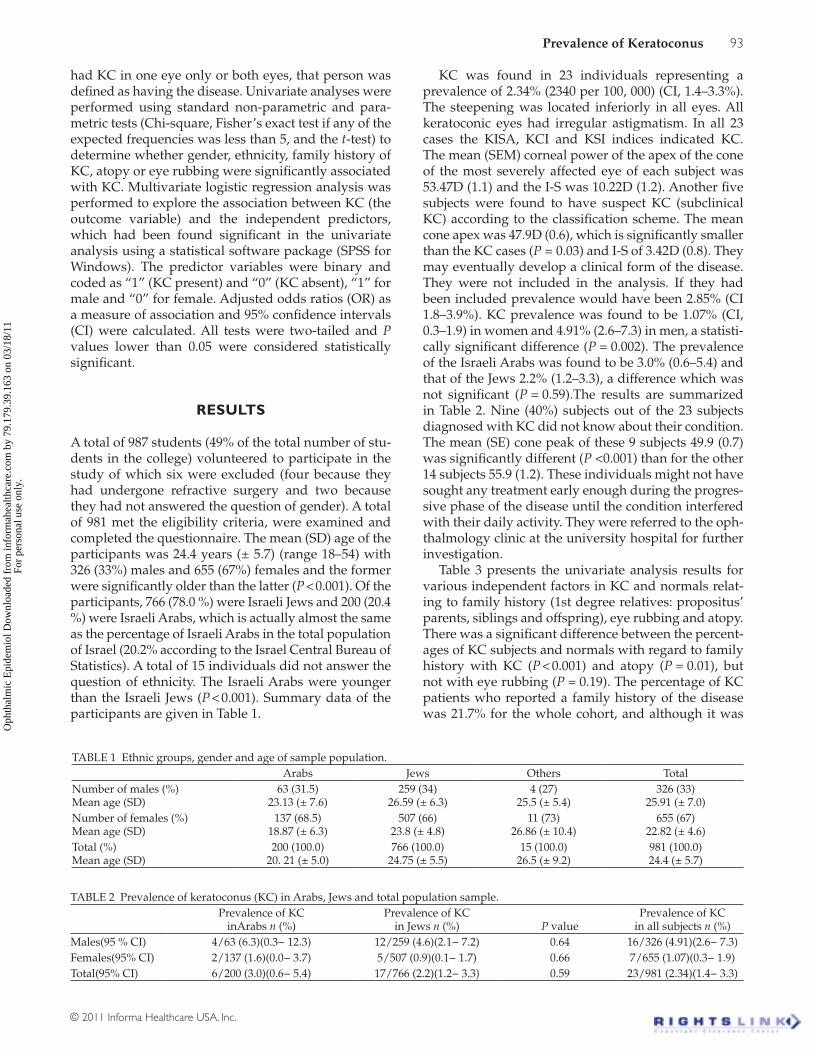
A total of 987 students (49% of the total number of stu-dents in the college) volunteered to participate in the study of which six were excluded (four because they had undergone refractive surgery and two because they had not answered the question of gender). A total of 981 met the eligibility criteria, were examined and completed the questionnaire. The mean (SD) age of the participants was 24.4 years (± 5.7) (range 18–54) with 326 (33%) males and 655 (67%) females and the former were significantly older than the latter (P < 0.001). Of the participants, 766 (78.0 %) were Israeli Jews and 200 (20.4 %) were Israeli Arabs, which is actually almost the same as the percentage of Israeli Arabs in the total population of Israel (20.2% according to the Israel Central Bureau of Statistics). A total of 15 individuals did not answer the question of ethnicity. The Israeli Arabs were younger than the Israeli Jews (P < 0.001). Summary data of the participants are given in Table 1.
KC was found in 23 individuals representing a prevalence of 2.34% (2340 per 100, 000) (CI, 1.4–3.3%). The steepening was located inferiorly in all eyes. All keratoconic eyes had irregular astigmatism. In all 23 cases the KISA, KCI and KSI indices indicated KC. The mean (SEM) corneal power of the apex of the cone of the most severely affected eye of each subject was 53.47D (1.1) and the I-S was 10.22D (1.2). Another five subjects were found to have suspect KC (subclinical KC) according to the classification scheme. The mean cone apex was 47.9D (0.6), which is significantly smaller than the KC cases (P = 0.03) and I-S of 3.42D (0.8). They may eventually develop a clinical form of the disease. They were not included in the analysis. If they had been included prevalence would have been 2.85% (CI 1.8–3.9%). KC prevalence was found to be 1.07% (CI, 0.3–1.9) in women and 4.91% (2.6–7.3) in men, a statisti-cally significant difference (P = 0.002). The prevalence of the Israeli Arabs was found to be 3.0% (0.6–5.4) and that of the Jews 2.2% (1.2–3.3), a difference which was not significant (P = 0.59).The results are summarized in Table 2. Nine (40%) subjects out of the 23 subjects diagnosed with KC did not know about their condition. The mean (SE) cone peak of these 9 subjects 49.9 (0.7) was significantly different (P <0.001) than for the other 14 subjects 55.9 (1.2). These individuals might not have sought any treatment early enough during the progres-sive phase of the disease until the condition interfered with their daily activity. They were referred to the oph-thalmology clinic at the university hospital for further investigation.
Table 3 presents the univariate analysis results for various independent factors in KC and normals relat-ing to family history (1st degree relatives: propositus’ parents, siblings and offspring), eye rubbing and atopy. There was a significant difference between the percent-ages of KC subjects and normals with regard to family history with KC (P < 0.001) and atopy (P = 0.01), but not with eye rubbing (P = 0.19). The percentage of KC patients who reported a family history of the disease was 21.7% for the whole cohort, and although it was
TABLE 1 Ethnic groups, gender and age of sample population. Arabs Jews Others TotalNumber of males (%) Mean age (SD)
63 (31.5) 23.13 (± 7.6)
259 (34) 26.59 (± 6.3)
4 (27) 25.5 (± 5.4)
326 (33) 25.91 (± 7.0)
Number of females (%) Mean age (SD)
137 (68.5)18.87 (± 6.3)
507 (66) 23.8 (± 4.8)
11 (73) 26.86 (± 10.4)
655 (67) 22.82 (± 4.6)
Total (%) Mean age (SD)
200 (100.0) 20. 21 (± 5.0)
766 (100.0) 24.75 (± 5.5)
15 (100.0) 26.5 (± 9.2)
981 (100.0) 24.4 (± 5.7)
TABLE 2 Prevalence of keratoconus (KC) in Arabs, Jews and total population sample.
Prevalence of KC
inArabs n (%)Prevalence of KC
in Jews n (%) P valuePrevalence of KC
in all subjects n (%)Males(95 % CI) 4/63 (6.3)(0.3− 12.3) 12/259 (4.6)(2.1− 7.2) 0.64 16/326 (4.91)(2.6− 7.3)Females(95% CI) 2/137 (1.6)(0.0− 3.7) 5/507 (0.9)(0.1− 1.7) 0.66 7/655 (1.07)(0.3− 1.9)Total(95% CI) 6/200 (3.0)(0.6− 5.4) 17/766 (2.2)(1.2− 3.3) 0.59 23/981 (2.34)(1.4− 3.3)
Oph
thal
mic
Epi
dem
iol D
ownl
oade
d fr
om in
form
ahea
lthca
re.c
om b
y 79
.179
.39.
163
on 0
3/18
/11
For
pers
onal
use
onl
y.

94 M. Millodot et al.
Ophthalmic Epidemiology
33.3% for the Israeli Arabs and 18.7% for the Israeli Jews, the difference was not significant (P = 0.32). Using mul-tivariate logistic regression analysis we determined the adjusted odds ratio as a measure of association between KC (the outcome variable) and the independent pre-dictors, which had been found significantly different in the univariate analysis. The following independent predictors which were found to be significantly associ-ated with KC were family history (OR 17.1, CI 5.0–57.8, P < 0.001), male gender (OR 5.4, CI 2.1–14.3, P < 0.001), and atopy (OR 3.0, CI 1.2–7.6, P = 0.02).
DISCUSSION
To the best of our knowledge, this is the first cross-sectional study on the prevalence of KC in Israel. It confirms the anecdotal observations of a seemingly high prevalence of this disease in this country. Several studies have estimated the prevalence of KC in different parts of the world, though only one in the Middle East.17 The studies are summarized in Table 4. Prevalence figures vary widely from 0.0003% in Russia18 to 2.3 % in Maharashtra, India.19 The first population-based prevalence study was carried out using a Placido disk.20 However, the most commonly cited prevalence is 0.054% found in Minnesota, USA21 in which the diagnosis was based on a mixture of scissors movement in retinoscopy and keratometry. This figure was not dissimilar to the results found in Finland11 or Denmark.22 KC prevalence from French army recruits23 was higher (1.2%) but all the results of the various indices in this only other study using videokeratography were more compatible with suspect than definite cases. In our study the prevalence of KC was found to be 2.34% (2340 per 100,000). This figure represents only definite forms of the disease and possibly advanced cases since the mean steepest cone was 53.74D. There was a significant difference between men and women (prevalence 4.91% vs. 1.07%; P < 0.001). The preponderance of men over women has been noted in the most recent studies,4,11,24–28 however the difference was greater in this study than the other hospital-based studies. Georgiou et al.28 reported a difference of 2.6 times higher in men than women whereas it is 4.6 times higher in our study. The reason for the higher prevalence of KC for men than women is not clear. A possible explanation could be that the mean age of the men (25.91) was higher than that of the women (22.82) and although the second decade of life is the most common period of onset of the disease a few cases may
have developed later, but as we have no knowledge of the incidence rates of the disease in this population we were not able to adjust for the age differences between men and women. The prevalence was higher in Arabs than in Jews (3.0% vs. 2.2%) but this difference was not significant (P = 0.59).
The result of our study represents a far higher esti-mate of prevalence (2.34%) than the commonly cited figures of (0.05–0.23%) for western countries1, 2 and is at least 10 times higher. In a population-based study in India19 the prevalence of KC was also found to be high (2.3%). In that study the diagnosis was made using keratometry. Almost all other studies were based on hospital records, which are likely to underestimate the true prevalence of the disease as patients presenting in hospitals are usually symptomatic and early forms of the disease are missed. Moreover these studies also ignore the number of patients treated by independent optometrists and ophthalmologists.
It is possible that environmental factors may have contributed to the high prevalence found in Jerusalem where the climate is characterized by dry condi-tions for most of the year, hot summers and the city is situated in an area 750 meters above sea level with a mean of 3397 hours of sunshine a year according to the “Climatological information for Jerusalem, Israel” (www.gb.weather.gov.hk). Such weather conditions are not unlike those prevailing in the Nagpur district of Maharashtra in India19, as well as in the Asir Province of Saudi Arabia where a large incidence of the disease was observed.17 On the other hand, in countries with much lower average annual temperature and sun exposure such as Finland,11 Denmark,22 Minnesota,21 Japan29 and the Urals in Russia18 the prevalence of KC is remark-ably lower in comparison (see Table 4). Although to our knowledge, there is no study of the effects of sun exposure on KC in humans, ultraviolet light which is a source of oxidative stress appears to be a compelling risk factor in the development of the disease. Support for this comes from studies showing that keratoconic corneas have an inability to process reactive oxygen species thereby leading to oxidative damage30,31 due to reduced levels of antioxidants such as superoxide dismutase32 and triggering what was referred to as a cascade of events leading to KC, such as an alteration of various corneal proteins, increased enzyme activities and apoptotic cell death.31 Furthermore, animal mod-els of apoptosis as a result of exposure to ultraviolet radiation in rabbit cornea,33 loss of keratocytes and subsequent corneal stromal thinning in mice34 support
TABLE 3 Univariate analyses of KC subjects and normals.Associated factor KC subjects(n / total*; % (95% CI) Normals(n / total*; % (95% CI) P value1st degree relatives with KC 5/23; 21.7 (4.9–38.6) 16/943; 1.7 (0.9–2.5) <0.001Eye rubbing 8/22; 36.4 (16.3–56.4) 224/938; 23.9 (21.2–26.6) 0.19Atopy 9/22; 40.9 (20.4–61.4) 180/949; 19.0 (16.5–21.5) 0.01*Refers to the number of subjects who answered that particular question.
Oph
thal
mic
Epi
dem
iol D
ownl
oade
d fr
om in
form
ahea
lthca
re.c
om b
y 79
.179
.39.
163
on 0
3/18
/11
For
pers
onal
use
onl
y.

Prevalence of Keratoconus 95
© 2011 Informa Healthcare USA, Inc.
the possibility of sun exposure as a risk factor in KC in genetically susceptible individuals. It may be worth not-ing though that UV radiation might provide a benefi-cial effect by inducing cross-linking of corneal collagen thus mitigating the progression of the disease.35 Further research is needed to support these hypotheses.
Ethnic differences may also account for the discrep-ancy in prevalence between the various studies. Tanabe et al.29 reported a prevalence of KC in Japan of less than one third of that seen in white Caucasians. Most impor-tantly the reports of two surveys in the UK indicated a prevalence 4.4 and 7.5 times greater for Asian (Indian, Pakistani and Banglasdeshi) subjects compared to white Caucasians.26,28 These results concur with the higher val-ues of prevalence found in India.19 The results of our study with Israeli Arabs and Jews support the anecdotal observation of a high prevalence of KC in the Middle East. Assiri et al.17 also found a high incidence in Saudi Arabia.
In both UK studies it was noted that most of the Asian subjects were Muslim with a high prevalence of consanguinity, a factor usually associated with a high rate of genetic disease. However, this factor does not appear to have had a lot of influence on the results of our study because Israeli Arabs have a much higher percentage of consanguineous marriages than Israeli Jews,36 yet there was no statistically significant differ-ence between the prevalence of KC in the two groups. However, this possible difference may have been miti-gated by the fact that endogamy is relatively common among Israeli Jews.
Ihalainen11 documented a family history in KC patients of 9% in southern Finland compared to 19% in the north of the country, which she attributed to the more pronounced effect of gene pooling in the larger families of these communities in whom the prevalence of KC was about fourfold higher than in the south. In a survey carried out by Owens and Gamble27 in New
Zealand 23.5% of patients reported having one or more relatives with the condition, which they attributed in part to the larger families of Maori/Polynesian popu-lations, but although they mentioned that there is no recorded prevalence of KC in Maori/Polynesian groups it is a clinical impression that this disease is particularly commonplace in these ethnic groups. In our study the percentage of KC patients who reported at least one first degree relative with the disease was 21.74% for the whole cohort, but it was higher, although not signifi-cantly, for the Israeli Arabs than for the Israeli Jews, pos-sibly reflecting the propensity of Israeli Arabs to have large families compared to Israeli Jews. Moreover, the percentage of normals who reported a family history of KC in first degree relatives was 1.7%. This figure is much higher than in other countries (0.05–0.23%)2 and concurs with the high prevalence of KC found in this sample population. In Bawazeer et al.’s study25 in Canada the control group of subjects without KC had a family his-tory of 0.0%. Most other reports of KC patients indicate lower figures of family history: it was 13.5% in the US Collaborative Longitudinal Evaluation of Keratoconus (CLEK) study37 and 6–10% in other studies.2
The univariate analysis indicated an association between KC and positive family history of the disease in first degree relatives, male gender and atopy, but no significant association with eye rubbing. Many other studies have reported an association between KC and male gender,4,11,24–28 and positive family his-tory of KC2,11,25,27,37 although in a case-control study Bawazer et al.25 found a significant association of the latter in univariate analysis, which was not corrobo-rated in the multivariate analysis. An association with eye rubbing38–40 has also often been reported, although Owens and Gamble27 failed to confirm this associa-tion. Contrary to most other studies we did not find a significant association between eye rubbing and KC. However, it has been shown that it only occurs when
TABLE 4 Epidemiological studies of keratoconus.Author Location Age (years) Sample size Incidence Prevalence SourceHofstetter20 (1959) Indianapolis, USA 1–79 13395 600/100,000 PopulationTanabe29 (1985) Muroran, Japan 10–60 2601-P 9/100,000 HospitalKennedy21 (1986) Minnesota, USA 12–77 64-P 2.0/100,000 54.5/100,000 HospitalIhalainen11 (1986) Finland 15–70 294-P 1.5/100,000 30/100,000 HospitalSantiago23 (1995) France 18–22 670 1190/100,000 Army recruitsGoskova18 (1998) Urals, Russia 0.2–0.4/100,000 HospitalPearson26 (2000) Midlands, UK 10–44 382-P 4.5/100,000-W
19.6/100,000-A57/100,000 229/100,000
Hospital
Ota9 (2002) Tokyo, Japan 325-P 9/100,000 HospitalGeorgiou28 (2004) Yorkshire, UK 74-P 3.3/100,000-W
25/100,000-A Hospital
Assiri17 (2005) Asir, Saudi Arabia 8–28 125-P 20/100,000 HospitalNielsen22 (2007) Denmark 772-P 1.3/100,000 86/100,000 HospitalJonas19 (2009) Maharashtra, India ≥ 30 4667 2300/100,000 PopulationCurrent study Jerusalem, Israel 18–54 981 2340/100,000 College student
populationA, Asian; W, white; P, patient
Oph
thal
mic
Epi
dem
iol D
ownl
oade
d fr
om in
form
ahea
lthca
re.c
om b
y 79
.179
.39.
163
on 0
3/18
/11
For
pers
onal
use
onl
y.

96 M. Millodot et al.
Ophthalmic Epidemiology
high levels of this factor are present39 and this was not the case in our study. Alternatively it could also be that in our dry climate a lot of people rub their eyes irrespective of whether they have KC or not and thus concealed a possible association. A possible association between atopy and KC has been reported in several studies (see25,39). The results of these studies are con-flicting possibly due to the varying criteria for disease classification and the fact that in some studies there was a lack of a control sample. Multivariate logistic regres-sion analysis confirmed the significant association of KC with family history, the strongest predictor with an odds ratio of 17.1, male gender (odds ratio 5.4) and atopy (odds ratio 3.0). Both family history and atopy support a genetic predisposition for KC.
There are limitations to this study. The sample size may be too small to provide a precise estimate of the prevalence of KC, especially of the ethnic groups. Further research is needed to corroborate the results of this study. The sample population is a non-random group of individuals obtained from Hadassah College in Jerusalem who voluntarily presented themselves for a corneal topographic examination and questionnaire. A selection bias may have occurred since individuals who knew they had the disease may have refrained from participating in the study because they were under ophthalmic care, while others with visual prob-lems may have been more likely to volunteer. Only 60% of the KC cases knew about their condition. However, the College student body represents a broad socioeco-nomic spectrum and it may have provided a reason-ably representative sample of the population of this geographic area. As in most studies, the assessment of risk factors depended on the information given in the self-administered questionnaire, which may be biased by the fact that the ability of subjects to report earlier experiences differs.
In conclusion, we found a much higher prevalence of KC in this cross-sectional study than other reported studies, except one in India where the weather is similar to that of Jerusalem; dry, sunny, with hot summers pos-sibly somewhat dusty. The prevalence of KC was also found to be much higher in men than women. Of the independent predictors analyzed only family history, male gender and atopy were found to be significant. Based upon the present data there is a need to focus future public health outreach and intervention for KC, especially as many individuals who have the disease are unaware of it (40% of KC subjects in our study). The high prevalence of the disease found in this study may be accounted for by the influence of environmental fac-tors on genetically susceptible individuals.
ACKNOWLEDGMENTS
The authors are thankful to the students and staff of Hadassah Academic and Technological College who
participated in this study. Dr Einat Shneor is sup-ported by the National Institute of Psychobiology, Israel.
Declaration of interest: The authors report no con-flicts of interest concerning this study. The authors alone are responsible for the content and writing of the paper.
REFERENCES
1. Krachmer JH, Feder RS, Belin MW. Keconratous and related noninflammatory corneal thinning disorders. Surv Ophthalmol 1984;28:293–322.
2. Rabinowitz YS. Keratoconus. Surv Ophthalmol 1998;42:297–319.
3. Tuft SJ, Moodaley, Gregory WM, Davison CR, Buckley RJ. Prognostic factors for the progression of keratoconus. Ophthalmology 1994;101:439–447.
4. Ertan A, Muftuoglu O. Keratoconus clinical findings according to different age and gender groups. Cornea 2008;27:1109–1113.
5. Lass JH, Lembach RG, Park SB, Hom DL, et al. Clinical man-agement of keratoconus. Ophthalmology 1990;97:433–445.
6. Woodward EG, Moodaley LC, O’Hagan A. Predictors for likelihood of corneal transplantation in keratoconus. Eye 1990;4:493–496.
7. Crews MJ, Driebe WT, Stern GA. The clinical manage-ment of keratoconus: a 6 year retrospective study. CLAO J 1994;20:194–197.
8. Zadnik K, Barr JT, Gordon MO. Biomicroscopic signs and disease severity in keratoconus. Cornea 1996;15:139–46.
9. Ota R, Fujiki K, Nakayasu K. Esimation of patient visit rate and incidence of keratoconus in the 23 wards of Tokyo. Nippon Ganka Gakkai Zasshi 2002;106:365–372.
10. Gordon MO, Steger-May K, Szczotka-Flynn, Joslin RC, et al. Baseline factors predictive of incident pen-etrating keratoplasty in keratoconus. Am J Ophthalmol 2006;142:923–930.
11. Ihalainen A. Clinical and epidemiological features of kerato-conus genetic and external factors in the pathogenesis of the disease. Acta Ophthalmol Scand 1986;178(Suppl.):5–64.
12. Lim N, Vogt U. Characteristics and functional outcomes of 130 patients with keratoconus attending a specialist contact lens clinic. Eye 2002;16:54–59.
13. Frucht-Pery J, Shtibel H, Solomon A, Siganos CS, et al. Thirty years of penetrating keratoplasty in Israel. Cornea 1997;16:16–20.
14. Claesson M, Armitage WJ. Corneal grafts at St John Hospital, Jerusalem, January 2001–November 2002. Br J Ophthalmol 2004;88:858–860.
15. Edwards M, McGhee CNJ, Dean S. The genetics of keratoco-nus. Clin Exp Ophthalmol 2001;29:345–351.
16. Rabinowitz YS, Rasheed K. KISA% index: a quantitative vid-eokeratography algorithm embodying minimal topographic critera for diagnosing keratoconus. JCataract Refract Surg 1999;25:1327–1335.
17. Assiri AA, Yousuf BI, Quantok AJ, Murphy PJ. Incidence and severity of keratoconus in Asir province, Saudi Arabia. Br J Ophthalmol 2005;89:1403–1406.
18. Gorskova EN, Sevost’ianov EN. Epidemiology of keratoco-nus in the Urals. Vestn Oftalmol 1998;114:38–40.
19. Jonas JB, Nangia V, Matin A, Kulkarni M, Bhojwani K. Prevalence and associations of keratoconus in rural Maharashtra in central India: the central India Eye Medical Study. Am J Ophthalmol 2009;148:760–765.
Oph
thal
mic
Epi
dem
iol D
ownl
oade
d fr
om in
form
ahea
lthca
re.c
om b
y 79
.179
.39.
163
on 0
3/18
/11
For
pers
onal
use
onl
y.
Prevalence of Keratoconus 97
© 2011 Informa Healthcare USA, Inc.
20. Hofstetter H. A keratoscopic survey of 13,395 eyes. Am J Optom Am Acad Optom 1959;36:3–11.
21. Kennedy RH, Bourne WM, Dyer JA. A 48-year clinical and epidemiological study of keratoconus. Am J Ophthalmol 1986;101:267–273.
22. Nielsen K, Hjortdal J, Aagard NE, Niels E. Incidence and prevalence of keratoconus in Denmark. Acta Ophthalmol Scand 2007;85:890–892.
23. Santiago PY, Assouline M, Ducoussau F, Bazin S, et al. Epidemiology of keratoconus and corneal topography in normal young male subjects. Invest Ophthalmol Vis Sci 1995;36:S307.
24. Millodot M, Owens H. Sensitivity and fragility in keratoco-nus. Acta Ophthalmol Scand 1983;61:908–917.
25. Bawazeer AM, Hodge WG, Lorimer B. Atopy and keratoconus: a multivariate analysis. Br J Ophthalmol 2000;84:834–836.
26. Pearson AR, Soneji B, Sarvananthan N, Sandforth-Smith JH. Does ethnic origin influence the incidence or severity of kera-toconus? Eye 2000;14:625–628.
27. Owens H, Gamble G. A profile of keratoconus in New Zealand. Cornea 2003;22:122–225.
28. Georgiou T, Funnell CL, Cassels-Brown A, O’Connor R. Influence of ethnic origin on the incidence of keratoconus and associated atopic diseases in Asian and white patients. Eye 2004;18:379–383.
29. Tanabe U, Fujiki K, Ogawa A, Ueda S, Kanai A. Prevalence of keratoconus patients in Japan. Nippon Ganka Gakkai Zasshi 1985;89:407–411.
30. Buddi R, Lin B, Atilano SR, Zorapapel NC, Kenney MC, Brown DJ. Evidence of oxidative stress in human corneal diseases. J Histochem Cytochem 2002;50:341–351.
31. Kenney MC, Brown DJ. The cascade hypothesis of keratoco-nus. Contact Lens Anterior Eye 2003;26:139–146.
32. Behndig A, Karlsson K, Johansson BO, Brannstrom T, Marklund SL. Superoxide dismutase isoenzymes in the nor-mal and diseased human cornea. Invest Ophthalmol Vis Sci 2001;42:2293–2296.
33. Podskochy A, Fagerholm P. The expression of Fas ligand protein in ultraviolet-exposed rabbit corneas. Cornea 2002;21:91–94.
34. Newkirk KM, Chandler HL, Parent AE, Young DC, Colitz CMH, Wilkie DA, Kusewitt DF. Ultraviolet radiation-induced corneal degeneration in 129 mice. Toxicologic Pathology 2007;35:817–824.
35. Snibson GR. Collagen cross-linking: a new treatment paradigm in corneal disease− a review. Clin Experiment Ophthalmol 2010;38:141–153.
36. Jaber L, Halpern GJ. Consanguinity among the Arab and Jewish populations in Israel. Ped Endocrinol Rev 2006;3(Suppl.):437–446.
37. Zadnik K, Barr JT, Edrington TB, Everett DF, et al. Baseline findings in the Collaborative Longitudinal Evaluation of Keratoconus (CLEK) study. Invest Ophthalmol Vis Sci 1998;39:2537–2546.
38. Jafri B, Lichter H, Stulting RD. Asymmetric kera-toconus attributed to eye rubbing. Cornea 2004;23: 560–564.
39. McMonnies CW, Boneham GC. Kearatoconus, allergy, itch, eye-rubbing and hand-dominance. Clin Exp Optom 2003;86:376–384.
40. McMonnies CW. Abnormal rubbing and keratectasia. Eye Contact Lens 2007;33:265–271.
Oph
thal
mic
Epi
dem
iol D
ownl
oade
d fr
om in
form
ahea
lthca
re.c
om b
y 79
.179
.39.
163
on 0
3/18
/11
For
pers
onal
use
onl
y.