ministÉrio da educaÇÃo universidade federal do rio …€¦ · antiproliferative activity as...
TRANSCRIPT

MINISTÉRIO DA EDUCAÇÃO
UNIVERSIDADE FEDERAL DO RIO GRANDE DO NORTE
CENTRO DE CIÊNCIAS DA SAÚDE
PROGRAMA DE PÓS-GRADUAÇÃO EM CIÊNCIAS DA SAÚDE
AVALIAÇÃO DAS ATIVIDADES ANTITUMORAL E ANTIOXIDANTE IN VITRO DE
EXTRATOS DE Libidibia ferrea EM CÉLULAS DE CÂNCER COLORRETAL
ANDREZA CONCEIÇÃO VÉRAS DE AGUIAR GUERRA
NATAL/RN
2017

ANDREZA CONCEIÇÃO VÉRAS DE AGUIAR GUERRA
AVALIAÇÃO DAS ATIVIDADES ANTITUMORAL E ANTIOXIDANTE IN VITRO DE
EXTRATOS DE Libidibia ferrea EM CÉLULAS DE CÂNCER COLORRETAL
Dissertação apresentada ao Programa de
Pós-Graduação em Ciências da Saúde da
Universidade Federal do Rio Grande do
Norte como requisito para a obtenção do
título de Mestre em Ciências da Saúde.
Orientador: Prof. Dr. Raimundo Fernandes
Araújo Junior
NATAL/RN
2017
i
Universidade Federal do Rio Grande do Norte - UFRN
Sistema de Bibliotecas - SISBI
Catalogação de Publicação na Fonte. UFRN - Biblioteca Setorial do Centro Ciências da Saúde - CCS
Guerra, Andreza Conceição Véras de Aguiar.
Avaliação das atividades antitumoral e antioxidante in vitro
de extratos de Libidibia ferrea em células de câncer colorretal
/ Andreza Conceição Véras de Aguiar Guerra. - Natal, 2017. 60f.: il.
Dissertação (Mestrado) - Programa de Pós-Graduação em Ciências
da Saúde. Centro de Ciências da Saúde. Universidade Federal do Rio Grande do Norte.
Orientador: Raimundo Fernandes de Araújo Júnior.
1. Neoplasias Colorretais - Dissertação. 2. Libidibia ferrea -
Dissertação. 3. Apoptose - Dissertação. 4. Antioxidante -
Dissertação. I. Araújo Júnior, Raimundo Fernandes de. II. Título.
RN/UF/BS-CCS CDU 616.348-006
ii
MINISTÉRIO DA EDUCAÇÃO
UNIVERSIDADE FEDERAL DO RIO GRANDE DO NORTE
CENTRO DE CIÊNCIAS DA SAÚDE
PROGRAMA DE PÓS GRADUAÇÃO EM CIÊNCIAS DA SAÚDE
Coordenador do Programa de Pós-Graduação em Ciências da Saúde:
Prof. Dr.: Eryvaldo Socrates Tabosa do Egito.
iii

ANDREZA CONCEIÇÃO VÉRAS DE AGUIAR GUERRA
AVALIAÇÃO DAS ATIVIDADES ANTITUMORAL E ANTIOXIDANTE IN VITRO DE
EXTRATOS DE Libidibia ferrea EM CÉLULAS DE CÂNCER COLORRETAL
Aprovada em: 23/06/2017
Banca examinadora:
Presidente da Banca: Prof. Dr. Raimundo Fernandes Araújo Junior (UFRN)
Membros da Banca:
Prof. Dr. Sergio Adriane Bezerra de Moura
Prof. Dr. Jeymesson Raphael Cardoso Vieira
iv

DEDICATÓRIA
Dedico este trabalhoà minha família, em especial, com todo meu amor e
gratidão, aos meus pais Mary e Aldo (in memoriam) e minha avó Fátima por tudo
que fizeram por mim e ao meu esposo Maximiliano, pelo apoio e compreensão
sempre.
v

AGRADECIMENTOS
Agradeço primeiramente à Deus por me fortalecer a cada dia nesta
caminhada e por me permitir alcançar mais uma conquista. À minha família, que
sempre me apoiou, auxiliou e incentivou, sendo minha base e o motivo que me faz
ser melhor a cada dia.
Ao Prof. Dr. Raimundo Fernandes de Araújo Júnior, expresso meu sincero
agradecimento pela confiança, por acreditar em meu potencial e por estar sempre
disponívelem me guiar, ajudar e ensinar.
À Prof.ª Dr.ª Aurigena Antunes de Araújo, pela valorosa colaboração durante
todo o trabalho e pelos ensinamentos que foram essenciais para o desenvolvimento
e êxito obtido.
Aos alunos que fazem parte da equipe do LAICI, que me acolheram e foram
bastante solícitos. Agradeço especialmente às alunas Ana Luiza e Juliana, cuja
amizade ultrapassa os limites do laboratório e sempre estiveram dispostas a ajudar,
ensinar, a oferecer uma palavra de ânimo, consolo ou mesmo um café, criando
momentos descontraídos e deixando mais leve os dias na universidade.
Ao Prof. Dr. Hugo Alexandre Rocha, pela colaboração, atenção e por deixar
sempre seu laboratório e a sala de cultura de células acessível à nossa equipe.
Aos Laboratórios de Imunogenética do Departamento de Bioquímica e de
Microscopia do Instituto do Cérebro pelo auxílio e disponibilidade para realização de
alguns dos ensaios realizados.
Ao Laboratório de Farmacognosia da Universidade Federal de Pernambuco,
em especial ao Prof. Dr. Luiz Alberto Soares e à Dr.ª Magda Rhayanny Ferreira pela
relevante cooperação durante este trabalho.
Ao Programa de Pós-Graduação em Ciências da Saúde, pela oportunidade
de realização deste Mestrado.
À CAPES pelo apoio financeiro por meio da bolsa de estudos de mestrado
que colaborou na conclusão desse projeto.
vi

RESUMO
O câncer colorretal tem se destacado por ser um dos tumores mais freqüentes, com
taxas de morbidade e mortalidade expressivos. Na descoberta de novas drogas,
produtos derivados de plantas se destacam por ser uma fonte segura e capaz de
originar compostos de alta eficiência. Bastante conhecida na medicina popular
brasileira, Libidibia ferrea (Mart. ex Tul.) L.P. Queiroz var. ferrea, tem sido utilizada
no tratamento de um amplo espectro de condições e na prevenção do câncer. Nesse
estudo, extratos etanólicos dos frutos de L. ferrea (a 20T, 40T, 60T e 80T) foram
avaliados por 24 h e 48 h pela capacidade de inibição da proliferação celular;
indução de apoptose através da avaliação de Bcl-2, caspase-3 e Apaf-1; atividade
antioxidante e efeito sobre alvos importantes relacionados a proliferação celular
(EGFR e AKT) na linhagem colorretal humana HT-29, por meio de metodologias que
envolveram ensaios de citometria de fluxo, espectrofotometria e RT-qPCR. Os
resultados demostram que os extratos tiveram atividade antiproliferativa comparado
ao controle, indução de apoptose através da via intrínseca e ação de inibição
tumoral in vitro com a mediação de alvos importantes na tumorigênese. Além disso,
possui efeito antioxidante e anti-peroxidação lipídica, bem como quimioprotetor nas
células saudáveis. Portanto, derivados de L. ferrea possuem importantes efeitos
anticâncer podendo ser considerados candidatos moleculares promissores para o
tratamento do câncer colorretal.
Palavras-chave: Libidibia ferrea. Câncer colorretal. Apoptose. Antioxidante.
vii

ABSTRACT
Colorectal cancer is noted for being one of the most frequent of tumors, with
expressive morbidity and mortality rates. In new drug discovery, plants stand out as a
source capable of yielding safe and high-efficiency products. Well known in Brazilian
popular medicine, Libidibia ferrea (Mart. Ex Tul.) L.P. Queiroz var. ferrea (better
known as "ironwood" or "jucá"), has been used to treat a wide spectrum of conditions
and to prevent cancer. Using methodologies that involved flow cytometry,
spectrophotometry and RT-qPCR assays, ethanolic extracts of the fruits of L. ferrea
(20T, 40T, 60T and 80T) were evaluated at 24 h and 48 h for: their ability to inhibit
cell proliferation; induce apoptosis through Bcl-2, caspase-3 and Apaf-1; their
antioxidant activity and effects on important targets related to cell proliferation (EGFR
and AKT) in the HT-29 human colorectal cancer lineage. The results revealed
antiproliferative activity as compared to the controls, induction of apoptosis through
the intrinsic pathway, and in vitro tumor inhibition activity under the mediation of
important targets in tumorigenesis. In addition, L. ferrea revealed antioxidant, lipid
peroxidation and chemoprotective effects in healthy cells. Thus, L. ferrea derivatives
have important anticancer effects, and may be considered promising candidate for
colorectal cancer therapy.
Key words:Libidibia ferrea. Colorectal cancer. Apoptosis. Antioxidant.
viii

LISTA DE ABREVIATURAS E SIGLAS
20T (Extrato etanólico a 20% do fruto de Libidibia ferrea)
40T (Extrato etanólico a 40% do fruto de Libidibia ferrea)
60T (Extrato etanólico a 60% do fruto de Libidibia ferrea)
80T (Extrato etanólico a 80% do fruto de Libidibia ferrea)
AKT (Proteína Quinase B)
Apaf-1 (Fator Apoptótico de Ativação de Protease 1)
BCL-2 (Célula B de Linfoma 2)
DAPI (4,6-diamidino-2-fenilindol)
DMEM (Meio Eagle Modificado de Dulbecco)
DTNB (Ácido Dithiobisnitrobenzoico)
EDTA (Ácido Etilenodiamino Tetra-acético)
EGFR (Receptor do Fator de Crescimento Epidérmico)
FITC (Isotiocianato de Fluoresceína)
GSH (Glutationa reduzida)
HCl (Ácido Clorídrico)
HEK-293 (Linhagem de células embrionárias de rim humano)
HT-29 (Linhagem de células de câncer colorretal humano)
IRI (Índice de Importância Relativa)
MDA (Malondialdeído)
MTT (Brometo de 3-(4,5-dimetiltiazol-2-il)-2,5-difeniltetrazólio)
PBS (Tampão Fosfato Salino)
PI (Iodeto de Propídio)
RNA (Ácido Ribonucléico)
RT-qPCR (Reação de Transcrição Reversa Quantitativa da Cadeia de Polimerase)
TCA (Ácido Tricloroacético)
ix

LISTA DE FIGURAS
Figura 1 -Libidibia ferrea e suas estruturas ................................................................................... 14
Figura 2 - Mapa global da carcinogênese ..................................................................................... 15
x

SUMÁRIO
1 INTRODUÇÃO ................................................................................................................................ 12
2 JUSTIFICATIVA .............................................................................................................................. 16
3 OBJETIVOS ..................................................................................................................................... 17
3.1 Objetivo geral ......................................................................................................................... 17
3.2 Objetivos específicos ........................................................................................................... 17
4 MÉTODO .......................................................................................................................................... 18
4.1 Obtenção dos extratos de Libidibia ferrea e preparo das soluções ........................ 18
4.2 Linhagem celular e cultivo .................................................................................................. 18
4.3 Ensaio de citotoxicidade pelo método do MTT ............................................................. 18
4.4 Avaliação da morte celular por citometria de fluxo ..................................................... 19
4.5 Imunofluorescência .............................................................................................................. 19
4.6 Dosagem de GSH ................................................................................................................... 20
4.7 Dosagem de MDA .................................................................................................................. 21
4.8 Real Time RT-qPCR ............................................................................................................... 21
4.9 Análise estatística ................................................................................................................. 22
5 ARTIGOS PRODUZIDOS .............................................................................................................. 23
5.1 Full article: Libidibia ferrea presents antiproliferative, apoptotic and antioxidant
effects in a colorectal cancer cell line .................................................................................... 25
6 COMENTÁRIOS, CRÍTICAS E CONCLUSÕES ....................................................................... 52
7 REFERÊNCIAS ............................................................................................................................... 54
xi

12
1 INTRODUÇÃO
Entende-se que o câncer, condição marcada pela proliferação desordenada
de células sutilmente modificadas, é um dos principais problemas de saúde pública
enfrentados neste século e apesar dos avanços, os procedimentos terapêuticos
disponíveis ainda estão aquém do necessário: são altamente invasivos ou não
específicos e muitas vezes estão acompanhados de efeitos secundários e toxicidade
para as células1,2. Enquanto isso, os índices de novos casos continuam a aumentar
no mundo todo e estima-se que seja superior a 20 milhões por ano até 20253.
Dentre os tipos de câncer, o câncer colorretal tem se destacado por ser um
dos tumores sólidos mais frequentes, tanto em homens quanto em mulheres,
representando 10% da incidência global3. Os fatores etiológicos e mecanismos
patogênicos relacionados ao seu desenvolvimento parecem ser complexos e
heterogêneos e contribuem para que seja uma das principais causas de morbidade
e mortalidade no mundo todo, despertando esforços na investigação de novas
estratégias terapêuticas4,5.
Atualmente, o desenvolvimento da terapêutica anticâncer tem sido conduzido
pela identificação de compostos citotóxicos. Esses agentes têm melhorado as taxas
de sobrevivência e a qualidade de vida de pacientes com diferentes tumores,
trazendo certas vantagens sobre os convencionais, como menor tempo de
administração, mecanismos para superar a resistência aos medicamentos e menor
incidência de efeitos adversos6,7. O modelo de triagem convencional é o teste em
linhagens celulares, que são uma ferramenta amplamente utilizada devido, entre
outros fatores, à sua facilidade de manipulação, caracterização molecular e alto grau
de similaridade, sendo excepcionais para o estudo das vias celulares e de genes
críticos envolvidos no câncer8.
As plantas são consideradas uma das principais fontes de novas entidades
químicas biologicamente ativas, com considerável interesse científico e comercial na
busca de potenciais fármacos. Para algumas doenças complexas, representam uma
fonte extremamente valiosa na produção de drogas inovadoras de alta eficiência9-11.
Estima-se que aproximadamente 40% dos medicamentos disponíveis atualmente
foram desenvolvidos, de maneira direta ou indireta a partir de fontes naturais, onde
as plantas são as mais utilizadas12.

13
O Brasil possui a maior diversidade de espécies de plantas no mundo, entre
350.000 a 550.000, porém menos de 10% foram avaliadas no que diz respeito às
suas características biológicas13,14. A vegetação da caatinga, em especial, é uma
fonte de recursos naturais pouco estudados15. Muitas espécies são amplamente
conhecidas, utilizadas empiricamente pela medicina popular, como Libidibia ferrea
Martius L. P. Queiroz, também denominada Caesalpinia ferrea e mais conhecida
como “pau-ferro” ou “jucá”, uma grande árvore nativa de ampla distribuição no norte
e nordeste, pertencente à família Fabaceae (Leguminosae)16.
Esta planta é considerada uma das espécies com maior índice de importância
relativa (IRI), medida quantitativa baseada no número de propriedades médicas
fornecidas por indivíduos de comunidades rurais17. Popularmente, vem sendo usada
no tratamento de afecções bronco-pulmonares, distúrbios gastrointestinais, diabetes,
doenças renais, inflamações e feridas em geral18-20. Em virtude de seu valor
etnomedicinal, o Ministério da Saúde incluiu L. ferrea na lista nacional de plantas
medicinais importantes para o sistema de saúde21.
Muitos componentes botânicos de L. ferrea são aproveitados como, por
exemplo, as folhas, flores, entrecasca e raízes22 (Figura 1). Os frutos, em especial,
são vagens achatadas de casca dura e cor marrom escuro utilizados pela
população, através de infusões aquosas, na prevenção do câncer23. Na literatura, já
foram relatados por possuírem propriedades antimicrobiana24-26, antidiabetes27, além
de ser quimiopreventivo23, não apresentar potencial mutagênico28 ou toxicidade
reprodutiva29. Apresentam ainda alta atividade antioxidante, descrito pela presença
de compostos polifenóis como ácido gálico e epicatequina30.
Sobre a composição fitoquímica dos frutos de L. ferrea, além dos compostos
fenólicos descritos anteriormente, reporta-se ainda etil galato, metil galato e ácido
elágico23,31, bem como a presença de ácidos graxos e terpenoides (ácidos linoleico,
palmítico, elaídico, esteárico, além de gama-sisterol e lupenona)32, que são
responsáveis por muitas das atividades farmacológicas observadas.
Antioxidantes, em especial, produzem uma ação protetora efetiva contra
danos oxidativos que estão frequentemente envolvidos na etiologia e progressão de
muitas doenças humanas, incluindo o câncer33. Entre os sistemas relacionados à
manutenção do equilíbrio redox intracelular, um papel principal é desempenhado
pela glutationa (GSH), que também participa de uma multiplicidade de processos,
incluindo diferenciação celular, proliferação e apoptose, atraindo a atenção de
farmacologistas como um possível alvo de intervenção médica contra o câncer34.

14
Figura 1 -Libidibia ferrea e suas estruturas. Adaptado de Melo18.
Um outro parâmetro importante na avaliação da atividade antioxidante é o
nível de malondialdeído (MDA), produto final da peroxidação lipídica mediada por
radicais livres35. Esse processo é considerado o principal mecanismo de destruição
da membrana e lesão celular, tendo sido relatado em vários tipos de câncer,
inclusive no câncer colorretal, estando intrinsicamente relacionado ao estresse
oxidativo36. Sendo assim, a mensuração da intensidade da peroxidação lipídica é
essencial para a melhor compreensão de seus efeitos moleculares deletérios e
mutagênicos e na avaliação da resposta dos pacientes à terapia37.
A desregulação de vias de sinalização celular relacionadas à apoptose e à
proliferação são fundamentais na sobrevivência das células cancerosas e no
desenvolvimento tumoral, sendo considerada um dos “hallmakers” ou capacidades
biológicas adquiridas38. Há vários fatores na membrana celular que se relacionam
com apoptose e crescimento, além do envolvimento de proteínas citoplasmáticas
como caspases e AKT (serina/treonina quinase ou proteína quinase B), por exemplo,
que por fim atuarão na parada do ciclo celular, inflamação, proliferação, invasão e
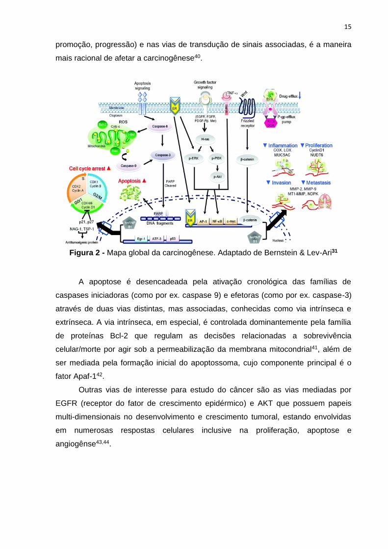
metástase39(Figura 2). Interferindo-se nos passos de modulação (iniciação,
15
promoção, progressão) e nas vias de transdução de sinais associadas, é a maneira
mais racional de afetar a carcinogênese40.
Figura 2 - Mapa global da carcinogênese. Adaptado de Bernstein & Lev-Ari31
A apoptose é desencadeada pela ativação cronológica das famílias de
caspases iniciadoras (como por ex. caspase 9) e efetoras (como por ex. caspase-3)
através de duas vias distintas, mas associadas, conhecidas como via intrínseca e
extrínseca. A via intrínseca, em especial, é controlada dominantemente pela família
de proteínas Bcl-2 que regulam as decisões relacionadas a sobrevivência
celular/morte por agir sob a permeabilização da membrana mitocondrial41, além de
ser mediada pela formação inicial do apoptossoma, cujo componente principal é o
fator Apaf-142.
Outras vias de interesse para estudo do câncer são as vias mediadas por
EGFR (receptor do fator de crescimento epidérmico) e AKT que possuem papeis
multi-dimensionais no desenvolvimento e crescimento tumoral, estando envolvidas
em numerosas respostas celulares inclusive na proliferação, apoptose e
angiogênse43,44.

16
2 JUSTIFICATIVA
Apesar do enorme progresso na compreensão da biologia do câncer, os
procedimentos terapêuticos ainda são uma exceção e até hoje existem poucos
exemplos que levam à cura. Terapias para o câncer colorretal, em especial, são
criticamente necessárias e cientificamente desafiadoras. A intervenção cirúrgica é
considerada a única modalidade de tratamento com potencial curativo, porém
apenas se detectado nos estágios iniciais. Além disso, os pacientes estão sujeitos
ao desenvolvimento de metástase, que ocorre em mais de 60% dos casos, e
recorrência do tumor. A quimioterapia permanece em grande parte de forma
paliativa.45-47
Produtos naturais e, particularmente derivados de plantas, são componentes
essenciais na pesquisa e desenvolvimento de medicamentos novos e econômicos.
Têm sido reconhecidos durante muitos anos como uma fonte importante de
diversidade estrutural, levando a avanços em metodologias sintéticas e promovendo
melhorias nas propriedades farmacológica ou farmacêutica de muitos
agentes9,48,49.Além da ampliação do atendimento médico-farmacêutico na saúde
pública, o estudo de plantas medicinais é importante para adquirir conhecimento
sobre o potencial farmacológico da diversidade vegetal nativa que permanece
subexplorada e auxiliar no desenvolvimento sustentável50.
Requisitos como aplicabilidade versátil e segurança terapêutica apresentadas
por plantas da caatinga tem atraído a atenção para a busca de novas drogas51.
Espécies popularmente usadas como Libidibia ferrea Martius L. P. Queiroz requerem
abordagens etnofarmacológicas que até o momento permanecem escassas. As
propriedades anticâncer e antioxidante in vitro, necessitam de uma avaliação mais
criteriosa no estudo de possíveis mecanismos de ação dos alvos moleculares para
elucidação que conferem tais ações farmacológicas. Diante do exposto, este
trabalho permitirá a investigação da potencialidade dessa planta como candidata à
fármaco antitumoral e contribuir para inovação na terapia do câncer colorretal.

17
3 OBJETIVOS
3.1 Objetivo geral
Avaliar as atividades antitumoral e antioxidante in vitro induzidas por extratos
de Libidibia ferrea frente à linhagem celular de câncer colorretal humano.
3.2 Objetivos específicos
1) Realizar o screening farmacológico da atividade antiproliferativa in vitro dos
extratos etanólicos dos frutos de Libidibia ferrea frente à linhagem de câncer
colorretal (HT-29) e à linhagem de células renais embrionárias (HEK-293), através
do ensaio de viabilidade celular baseado no sal de tetrazólio MTT.
2) Avaliar o comportamento de morte celular das linhagens HT-29 e HEK-293
através da técnica de citometria de fluxo, pela análise da integridade de membrana,
frente aos extratos de L.ferrea.
3) Avaliar a expressão de proteínas mediadoras do processo de morte celular
por apoptose (Bcl-2 e caspase-3) nas células HT-29, após tratamento com os
extratos de L. ferrea, por microscopia de imunofluorescência.
4) Avaliar a atividade antioxidante das células HT-29 e HEK-293 submetidas
ao tratamento pelos extratos de L. ferrea através da dosagem dos níveis de GSH e
MDA por espectrofotometria.
5) Avaliar a expressão de genes associados às vias de sinalização AKT, Apaf-
1 e EGFR nas células HT-29 tratadas com os extratos de L. ferrea através da técnica
de Real Time RT-qPCR.
18
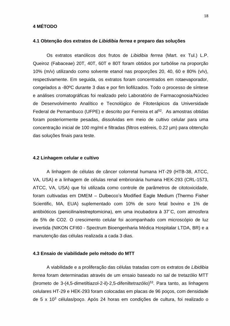
4 MÉTODO
4.1 Obtenção dos extratos de Libidibia ferrea e preparo das soluções
Os extratos etanólicos dos frutos de Libidibia ferrea (Mart. ex Tul.) L.P.
Queiroz (Fabaceae) 20T, 40T, 60T e 80T foram obtidos por turbólise na proporção
10% (m/v) utilizando como solvente etanol nas proporções 20, 40, 60 e 80% (v/v),
respectivamente. Em seguida, os extratos foram concentrados em rotaevaporador,
congelados a -80ºC durante 3 dias e por fim liofilizados. Todo o processo de síntese
e análises cromatográficas foi realizado pelo Laboratório de Farmacognosia/Núcleo
de Desenvolvimento Analítico e Tecnológico de Fitoterápicos da Universidade
Federal de Pernambuco (UFPE) e descrito por Ferreira et al52. As amostras obtidas
foram posteriormente pesadas, dissolvidas em meio de cultivo celular para uma
concentração inicial de 100 mg/ml e filtradas (filtros estéreis, 0.22 µm) para obtenção
das soluções finais para teste.
4.2 Linhagem celular e cultivo
A linhagem de células de câncer colorretal humana HT-29 (HTB-38, ATCC,
VA, USA) e a linhagem de células renal embrionária humana HEK-293 (CRL-1573,
ATCC, VA, USA) que foi utilizada como controle de parâmetros de citotoxicidade,
foram cultivadas em DMEM – Dulbecco’s Modified Eagle Medium (Thermo Fisher
Scientific, MA, EUA) suplementado com 10% de soro fetal bovino e 1% de
antibióticos (penicilina/estreptomicina), em uma incubadora à 37˚C, com atmosfera
de 5% de CO2. O crescimento celular foi acompanhado com microscópio de luz
invertida (NIKON CFI60 - Spectrum Bioengenharia Médica Hospitalar LTDA, BR) e a
manutenção das células realizada a cada 3 dias.
4.3 Ensaio de viabilidade pelo método do MTT
A viabilidade e a proliferação das células tratadas com os extratos de Libidibia
ferrea foram determinadas através de um ensaio baseado no sal de tretazólio MTT
(brometo de 3-(4,5-dimetiltiazol-2-il)-2,5-difeniltetrazólio)53. Para tanto, as linhagens
celulares HT-29 e HEK-293 foram colocadas em placas de 96 poços, com densidade
de 5 x 10³ células/poço. Após 24 horas em condições de cultura, foi realizado o

19
carenciamento e passado o mesmo período, a aplicação dos extratos etanólicos
20T, 40T, 60T e 80T nas concentrações de 12.5 µg/ml, 25 µg/ml, 50 µg/ml e 100
µg/ml. O tratamento foi avaliado em 24 h e 48 h, através da adição de 100 µl/poço
do MTT (1 mg/ml), incubação por 4 h e adição de 100 µl de etanol/poço. As placas
foram agitadas e a absorbância obtida em um leitor de microplacas (Epoch - BioTek
Instruments Inc, VT, EUA) a 570 nm, com o uso do software Gen5 Data Analysis
versão 2.0 (BioTek Instruments Inc, VT, EUA).
4.4 Avaliação da morte celular por citometria de fluxo
O efeito dos extratos de Libidibia ferrea sobre as células HT-29 e HEK-293 foi
determinado por citometria de fluxo com marcação dupla com Anexina V – FITC e
Iodeto de Propídio (PI), que permite a identificação de células apoptóticas e
necróticas através da perda de integridade de membrana. As linhagens celulares
foram dispostas em placas de 6 poços com densidade de 2 x 10⁵ células/poço, com
volume total de 2 ml. Após 24 h de incubação em condições de cultura, as células
foram tratadas com os extratos 40T, 60T e 80T, pois apresentaram melhor efeito sob
a viabilidade das células tumorais. As doses escolhidas para tratamento foram 25
µg/ml e 50 µg/ml, que representaram uma média de inibição da proliferação de 50%
(IC50), após a realização de curva dose-resposta. Após os tempos de 24h e 48h, as
células foram obtidas através da coleta do sobrenadante dos poços, lavagem com
PBS, dissociação enzimática com tripsina e duas etapas de centrifugação a 3000 G
a 4ºC. Por fim foram marcadas conforme instruções do kit de Anexina V-FITC/PI (BD
Pharmigen, CA, EUA) e analisadas com citômetro BD FACSCanto II (BD
Biosciences, CA, EUA) e o software FlowJo, versão 7.6.5 (Tree Star Inc., CA, EUA).
4.5 Imunofluorescência
Para avaliação de alvos de vias de apoptose, as células HT-29 e HEK-293
foram plaqueadas em lamínulas de vidro com densidade celular de 5 x 10⁴
células/poço (para um volume total de 1 ml) e colocadas em placas de 24 poços.
Brevemente, passado o tempo de 24 h, as células foram tratadas com os extratos
40T e 60T, dose 25 µg/ml, com base nos que apresentaram melhor ação no ensaio

20
anterior. Após cada período de tempo (24h e 48h), foram fixadas com
paraformaldeído a 7%, permeabilizadas com Triton X-100/PBS 0.2% e incubadas
durante 1 hora em câmera úmida com os anticorpos policlonais de rato anti-Bcl-2 e
de coelho anti-caspase-3 (Abcam, CA, EUA), uma lamínula para cada anticorpo,
diluídos 1:100 no PBS contendo albumina de soro bovino 5% (Life Technologies,
SP, BR). O anticorpo primário foi detectado com o anticorpo secundário Alexa Fluor
488 anti-rato ou anti-coelho (Abcam, CA, EUA) diluídos 1:500 no PBS contendo
albumina de soro bovino 5 %, e 4,6-diamidino-2-fenilindol (DAPI) (Life Technologies,
SP, BR) diluído 1:200 no PBS contendo albumina de soro bovino 5% foi usado para
coloração nuclear. As lamínulas foram examinadas em um microscópio LSM 510
laser scanning (Carl Zeiss, Jena, DE) na objetiva de 40x. As imagens selecionadas
foram representativas da maioria das células.
4.6 Dosagem de GSH
Para avaliar a atividade antioxidante, o nível total de glutationa (GSH) foi
determinado através do método de Costa et al54. A obtenção inicial das células foi
realizada através do processo descrito por Rahman et. al55. Em resumo, HT-29 foi
disposta em placas de 6 poços, com 1 x 10⁶ células/poço, num volume total de 2 ml.
No dia seguinte foi realizado o tratamento com os extratos etanólicos 40T e 60T na
concentração de 25 µg/ml. Após 24 horas, as células foram removidas,
ressuspendidas em PBS e centrifugadas duas vezes por 5 minutos, em 3000 rpm a
4ºC. Em seguida o pellet foi homogeneizado com uma solução de ácido
etilenodiamino tetra-acético (EDTA) 0.02M (Sigma-Aldrich, SP, Brazil) para
trituração. Após esse processo, 400 µl da suspensão obtida foi então diluída em 80
µl de ácido tricloroacético (TCA) 50% (Vetec, SP, Brazil) e 320 µl de água destilada
para centrifugação por 15 minutos em 3000 rpm a 4ºC. Por fim, em cada poço foi
aplicado 100 µl do sobrenadante celular, 200 µl do tampão Tris 0.4 M (Sigma-
Aldrich, SP, Brazil) e 20 µl de uma solução de ácido dithiobisnitrobenzoico (DTNB)
0.01 M (Sigma-Aldrich, SP, Brazil), em triplicata, para leitura da absorbância de cada
amostra em 412 nm. Os resultados foram expressos em nmol/106 cells.

21
4.7 Dosagem de MDA
Para avaliar a peroxidação lipídica, a produção de malondialdeído (MDA) foi
medida de acordo com o ensaio descrito por Esterbauer e Cheeseman56. As células
passaram pelo mesmo processo inicial de centrifugação descrito no tópico anterior e
em seguida foram homogeneizadas com 500 µl de tampão Tris-HCl 20 mM (Trizma
HCl, Sigma-Aldrich, SP, Brazil) para trituração. Após esse processo, foram
centrifugadas por 20 minutos em 3000 rpm a 4ºC. Então, 375 µl do reagente
cromogênico (10.3 mM 1-metil-2-fenilindol em 3:1 acetonitrila) e 112.5 µl de HCl 37%
foi adicionado a cada 150 µl de sobrenadante da amostra. Após uma etapa de
incubação em banho-maria por 40 min a 45ºC, as amostras foram centrifugadas a
3000 rpm a 4ºC. A absorbância foi medida a 586 nm e os resultados foram
expressos em nmol/106 cells.
4.8 Real Time RT-qPCR
O RNA total foi extraído das células HT-29 tratadas por 24 horas com os
extratos 40T e 60T (dose 25 µg/ml, em duplicata) com o reagente Trizol (Invitrogen,
CA, USA) e SV Total RNA Isolation System (Promega, WI, EUA) segundo as
orientações do fabricante. O RNA total sofreu a ação da transcriptase reversa do kit
ImProm-IITM Reverse Transcriptase System (Promega, WI, EUA) e a RT-qPCR foi
então realizada para a análise quantitativa da expressão de RNA mensageiro
(mRNA) com SYBR Green Mix no sistema Applied Biosystems 7500 FAST (Applied
Biosystems, CA, EUA), de acordo com o protocolo padrão e os seguintes primers
(Integrated DNA Technologies, IA, EUA): AKT (forward: 5’ TCA CCT CTG AGA CCG
ACA CC 3’; reverse: 5’ ACT GGC TGA GTA GGA GAA CTG G 3’, temperatura de
anelamento: 58.3°C), APAF-1 (forward: 5’ CCT CTC ATT TGC TGA TGT CG 3’;
reverse: 5’ TCA CTG CAG ATT TTC ACC AGA 3’, temperatura de anelamento:
56.9°C) e EGF-R (forward: 5’ TGA TAG ACG CAG ATA GTC GCC 3’; reverse: 5’
TCA GGG CAC GGT AGA AGT TG 3’, temperatura de anelamento: 56.9°C).
As concentrações finais dos reagentes no mix foram: 5 µl de SYBR Green, 0.7
µl de cada primer, 1.6 µl de água nuclease e 2 µl de cDNA. As condições de PCR
padrão foram as seguintes: 50 °C durante 2 min e 95 °C durante 10 min, seguido por
quarenta ciclos de 30 s a 95 °C, uma temperatura de anelamento dos primers
22
variável durante 30 s e 72 °C durante 1 min. Os valores médios de Cq foram usados
para calcular a expressão relativa dos níveis dos genes alvos para os grupos
experimentais, em relação aos do grupo controle negativo; os dados de expressão
foram normalizados em relação ao gene β-actina humano usando a fórmula 2-
ΔΔCq57.
4.9 Análise estatística
Todos os experimentos foram realizados em triplicata. A significância das
diferenças entre os grupos foi obtida através da análise de variância (ANOVA) e do
teste de Bonferroni (Nível de significância de p<0,05), com o software GraphPad
Prism versão 5.0 (GraphPad Software Inc., CA, EUA).

23
5 ARTIGOS PRODUZIDOS
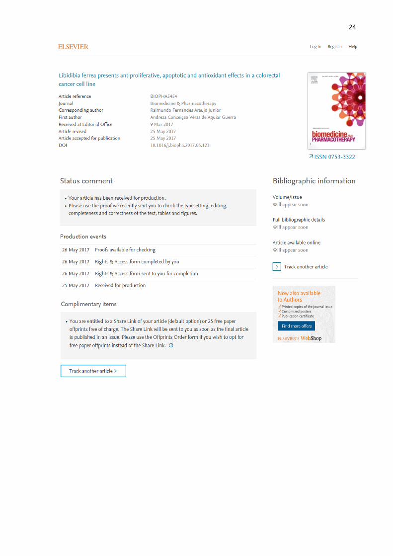
O artigo “Libidibia ferrea presents antiproliferative, apoptotic and
antioxidant effects in a colorectal cancer cell line” foi aceito no periódico
Biomedicine & Pharmacotherapy que possui fator de impacto 2.326 e Qualis B1
da CAPES para área Medicina II.
Acceptance Letter:
24
25
5.1 Full article: Libidibia ferrea presents antiproliferative, apoptotic and
antioxidant effects in a colorectal cancer cell line
Andreza Conceição Véras de Aguiar Guerraa, Luiz Alberto Lira Soaresb, Magda
Rhayanny Assunção Ferreirab, Aurigena Antunes de Araújoc, Hugo Alexandre de
Oliveira Rochad, Juliana Silva de Medeirose, Rômulo dos Santos Cavalcantea,
Raimundo Fernandes de Araújo Júniora,e*
aPost graduation program in Health Science, Federal University of Rio Grande do
Norte (UFRN), Natal, RN, Cep: 59078-970, Brazil
bPost graduation program in Therapeutic Innovation, Department of Pharmaceutical
Sciences, Federal University of Pernambuco (UFPE), Recife, PE, Cep: 50740-530,
Brazil
cPost graduation program in Pharmaceutical Sciences, Department of Biophysics
and Pharmacology, UFRN, Natal, RN, Cep: 59078-970, Brazil
dPost graduation program in Biochemistry, Department of Biochemistry, UFRN,
Natal, RN, Cep: 59078-970, Brazil
ePost graduation program in Functional and Structural Biology, Department of
Morphology, UFRN, Natal, RN, Cep: 59078-970, Brazil
*Corresponding author: Raimundo Fernandes de Araújo Júnior, Department of
Morphology, Centre of Biosciences, UFRN, Av. Senador Salgado Filho, S/N, Campus
Universitário, Lagoa Nova, 59072-970, Natal, RN, Brazil.
E-mail: [email protected]
Abstract
Colorectal cancer is noted for being one of the most frequent of tumors, with
expressive morbidity and mortality rates. In new drug discovery, plants stand out as a
source capable of yielding safe and high-efficiency products. Well known in Brazilian
ACCEPTED ARTICLE
26
popular medicine, Libidibia ferrea (Mart. Ex Tul.) L.P. Queiroz var. ferrea (better
known as "ironwood" or "jucá"), has been used to treat a wide spectrum of conditions
and to prevent cancer. Using methodologies that involved flow cytometry,
spectrophotometry and RT-qPCR assays, crude extracts of the fruits of L. ferrea
(20T, 40T, 60T and 80T) were evaluated at 24 h and/or 48 h for: their ability to inhibit
cell proliferation; induce apoptosis through Bcl-2, caspase-3 and Apaf-1; their
antioxidant activity and effects on important targets related to cell proliferation (EGFR
and AKT) in the HT-29 human colorectal cancer lineage. The results revealed high
antiproliferative potential as compared to the controls, induction of apoptosis through
the intrinsic pathway, and probable tumor inhibition activity under the mediation of
important targets in tumorigenesis. In addition, L. ferrea revealed antioxidant, lipid
peroxidation and chemoprotective effects in healthy cells. Thus, L. ferrea derivatives
have important anticancer effects, and may be considered promising candidate for
colorectal cancer therapy.
Key words: Libidibia ferrea. Colorectal cancer. Apoptosis. Antioxidant.
1. Introduction
It is understood that cancer, a condition marked by the disorderly proliferation
of subtly modified cells, is one of the main public health problems faced in this
century. Despite advances, the therapeutic procedures available are still insufficient,
they are generally highly invasive or non-specific, and are often accompanied by side
effects and cell toxicity [1,2]. Currently, new-case rates continue to rise worldwide
and are estimated to be over 20 million per year by 2025 [3].
Among cancers, colorectal cancer has been noted as one of the most frequent
in solid tumors, in men and women, representing 10% of the global incidence [3]. The
etiological factors and pathogenic mechanisms related to its development are
complex and heterogeneous [4,5], this contributes to its being one of the main
causes of morbidity and mortality in the world.
Natural products, particularly plant derivatives, are essential components in
research and development for safe, innovative, economical and high efficiency drugs
against complex diseases [6-8]. It is estimated that about 60% of the antitumoral
27
drugs available on the market and most of those in the late stages of clinical trials are
derived from natural products, mainly from plants [9].
Over the last two decades, public interest and research efforts from scientific
and medical communities worldwide has increased expressively, generating a large
volume of information including studies on the pharmacological effects, usage, and
the development into future medicines of herbs and derivative medicinal
phytochemicals as anti-tumor and chemoprevention agents. [10] This leads to
growing number of sales of commercialised medicinal herbs and most importantly,
growing number of pharmaceutical companies that involve in the research and
development of plants as a source for modern medicine. [11]
Brazil has one of the largest plant species diversities in the world, yet less than
10% of its species have been evaluated for their biological characteristics [12]. In
particular, the vegetation of the caatinga, an extremely threatened biome, as a
valuable natural resource has been little studied [13]. Many species however are
widely known and used empirically in folk medicine. Thus, we have Libidibia ferrea
(Mart. Ex Tul.) L.P. Queiroz var. ferrea (Fabaceae) known as Caesalpinia ferrea, and
popularly known as "ironwood" or "jucá" [14]. It is used popularly in the treatment of
bronchopulmonary disorders, gastrointestinal disorders, diabetes, rheumatism,
inflammation, wounds in general, and for prevention of cancers [15].
Most of the botanical components of this plant are harnessed, such as flowers,
shells, pods and roots. The fruits have been reported in the literature because they
have antimicrobial [16-18], antioxidant [19] and antidiabetic [20] properties, and
besides being chemopreventive [21], they show no mutagenic potential [22], or
reproductive toxicity [23]. Yet studies exploring their anticancer activity, especially
mechanisms of action in cellular signaling pathways are practically nonexistent.
Dysregulation of apoptosis and cell proliferation signaling pathways is critical
to both the promotion and progression of cancer. Thus, assessment of key points in
these pathways is essential for developing new target molecules with effective
antineoplastic activity [24,25]. Apoptosis is triggered by chronological activation of the
initiator (caspase 9), and effector (caspase-3) families via two distinct but associated
pathways known as the intrinsic and extrinsic pathways. The intrinsic pathway in
particular is dominated by the Bcl-2 family of proteins that regulate cell survival/death
related decisions, acting through permeabilization of the mitochondrial membrane

28
[26], in addition to being mediated by initial apoptosome formation, whose main
component is the Apaf-1 factor [27].
Other pathways of interest in the study of cancer are the EGFR and AKT (AKT
serine/threonine kinase) mediated pathways, which have multidimensional roles in
tumor development and growth, and are involved in numerous cellular responses
including proliferation, apoptosis and angiogenesis [28,29].
The objectives of this work are to investigate the potential of L. ferrea as an
antitumor drug candidate to contribute to colorectal cancer therapy innovation
through in vitro evaluations of cell death, antiproliferative effects, and signaling
pathways related to intrinsic apoptosis (Bcl-2, caspase-3 and Apaf-1), and to AKT
and EGFR expression; and as well, to evaluate L. ferrea’s antioxidant potential. Such
bioactive actions presented by L. ferrea may contribute to the availability of new
anticancer treatments in the clinic.
2. Methodology
2.1 Obtaining Libidibia ferreacrude extracts
The crude extracts of fruits from Libidibia ferrea were obtained at 10% (w/v) by
turbo extraction (four extractive cycles of 30 sec, interspersed with by 5 min of
pause), using ethanol as solvent at 20, 40, 60 and 80% (v/v). The extracts were
filtred and concentrated under reduced pressure at 40°C (RV10 Basic, IKA®) to
remove the ethanol. The aqueous residues were frozen under -80 °C for three days
and then lyophilized (Model L101, Liotop®) to yield the crude extracts. The
quantification of chemical markers (ellagic acid and gallic acid) in the crude extracts
was carried out by high performance liquid chromatography (HPLC) according to the
methodology previously described by Ferreira et al. [30], shown in summary in Table
1.

29
Table 1
Content of ellagic acid and gallic acid (g%) in crude extracts of fruits from L. ferrea.
Sample Ellagic acid (EA) Gallic acid (GA)
20T 2.78 ± 1.227 (0.89) 4.43 ± 0.132 (0.24)
40T 2.89 ± 0.551 (0.39) 3.39 ± 0.268 (0.67)
60T 2.73 ± 2.213 (1.65) 1.61 ± 0.125 (0.66)
80T 2.61 ± 0.381 (0.29) 1.84 ± 0.073 (0.34)
The results were expressed in g%, as mean ± standard deviation (relative standard deviation).
2.2 Cell line and cultivation
The HT-29 human colorectal cancer cell line and the HEK-293 human
embryonic kidney cell line (used as control of cytotoxicity parameters) were obtained
from the American Type Culture Collection (Rockville, MD, USA). All cell lines were
cultivated in Dulbecco's Modified Eagle Medium (Thermo Fisher Scientific, MA, USA)
supplemented with 10% (v/v) fetal bovine serum and 1% antibiotics
(penicillin/streptomycin) in a 37°C incubator with atmosphere of 5% CO2.
2.3 Viability test
Viability and cell proliferation for the Libidibia ferrea extracts were determined
by an assay based on the MTT tretazolium salt (3- (4,5-dimethylthiazol-2-yl) -2,5-
diphenyltetrazolium bromide) [31]. To this end, the HT-29 and HEK-293 cell lines
were plated in 96-well plates, with a density of 5 x 10³ cells/well. After 24 hours under
culture conditions, applications of 20T, 40T, 60T and 80T crude extracts at
concentrations of 12.5 μg/mL, 25 μg/mL, 50 μg/mL and 100 μg/mL were performed.
Treatments were evaluated at 24 h and 48 h by addition of 100 μl/well of MTT (1
mg/mL), incubation for 4 h, and addition of ethanol/well. The plates were shaken and
the absorbance obtained in a microplate reader (Epoch - BioTek Instruments Inc, VT,
USA) at 570 nm. The Gen5 Data Analysis software version 2.0 (BioTek Instruments
Inc, VT, USA) was used.
2.4 Evaluation of in vitro cell death

30
The effect of Libidibia ferrea extracts on HT-29 and HEK-293 cells was
determined by double-labeled flow cytometry with Annexin V-FITC and Propidium
Iodide (PI), which allows the identification of apoptotic and necrotic cells through loss
of membrane integrity. Cell lines were arranged in 6-well plates with a density of 2 x
105 cells/well, at a total volume of 2 mL. After 24 h of incubation under culture
conditions, the cells were treated with the 40T, 60T and 80T crude extracts at doses
of 25 μg/mL and 50 μg/mL, at 24 h and 48 h. After each period, the cells were
obtained by collecting the supernatant from the wells, washing with PBS,
trypsinization, and two steps of centrifugation at 3200 rpm at 4°C. Finally, they were
labeled according to instructions from the Annexin V-FITC / PI kit (BD Pharmigen,
CA, USA), and analyzed with BD FACSCanto II (BD Biosciences, CA, USA) and
FlowJo software, version 7.6.5 (Tree Star Inc., CA, USA).
2.5 Immunofluorescence microscopy
For evaluation of apoptosis pathway targets, HT-29 cells was plated on glass
coverslips with cell density of 5 x 10⁴ cells/well (for a total volume of 1 mL), and
plated in 24-well plates. Briefly, after 24 h the cells were treated with extracts 40T
and 60T, being dosed at 25 μg/mL, for 24 h and 48 h. After each period, they were
fixed with 7% paraformaldehyde, permeabilized with 0.2% Triton X-100/PBS and
incubated for 1 hour in a humid chamber with anti-Bcl-2 and rabbit anti-caspase-3
mouse polyclonal antibodies (Abcam, CA, USA), using one coverslip for each
antibody, diluted 1:100 in PBS containing 5% bovine serum albumin (Life
Technologies, SP, BR). Primary antibody was detected with Alexa Fluor 488 anti-
mouse or anti-rabbit secondary antibody (Abcam, CA, USA) diluted 1:500 in PBS,
containing 5% bovine serum albumin, and 4,6-diamidino-2-phenylindole (DAPI) (Life
Technologies, SP, BR), diluted 1:200 in PBS containing 5% bovine serum albumin
and used for nuclear staining. The coverslips were examined in an Zeiss Observer
Z1 upright microscope for fluorescence (Carl Zeiss, Jena, DE) on the 40x objective.
The selected images were representative of most cells.
2.6 GSH dosage

31
To assess antioxidant activity, the total level of glutathione (GSH) was
determined using the Costa et al [32] method. Initial cell uptake was performed using
the procedure described by Rahman et al [33]. Briefly, HT-29 and HEK-293 cells
were plated in 6-well plates, with 1 x 10⁶ cells/well, in 2 mL total volume. On the
following day, treatment with extracts 40T and 60T in the concentration of 25 μg/mL
was carried out. After 24 hours, the cells were removed, resuspended in PBS and
centrifuged twice for 5 minutes at 3000 rpm, and at 4ºC. The pellet was then
homogenized with a 0.02 M solution of ethylenediamine tetraacetic acid (EDTA)
(Sigma-Aldrich, SP, Brazil) for grinding. After this process, the suspension obtained
was then diluted in 50% trichloroacetic acid (Vetec, SP, Brazil) and distilled water for
centrifugation for 15 minutes at 3000 rpm, and at 4°C. Finally, a mix of the cell
supernatant, 0.4 M Tris buffer (Sigma-Aldrich, SP, Brazil) and 0.01 M
dithiobisnitrobenzoic acid solution (Sigma-Aldrich, SP, Brazil) were applied to each
well, to read the absorbance of each sample at 412 nm. The results were expressed
as nmol /106 cells.
2.7 MDA dosage
To evaluate lipid peroxidation, malondialdehyde (MDA production) was
measured according to an assay described by Esterbauer and Cheeseman [34]. HT-
29 cells underwent the same initial centrifugation procedure described above, and
were then homogenized with a 20 mM Tris-HCl buffer (Trizma HCl, Sigma-Aldrich,
SP, Brazil) for trituration. After this process, they were centrifuged for 20 minutes in
3000 rpm at 4ºC. Afterwards, the chromogenic reagent (10.3 mM 1-methyl-2-
phenylindole in 3:1 acetonitrile), and a 37% solution of HCl were added to each 150
μL of supernatant sample. After an incubation step in a water bath for 40 min at
45°C, the samples were centrifuged at 3000 rpm at 4°C. The absorbance was
measured at 586 nm, and the results were expressed as nmol/106 cells.
2.8 Real time-qPCR
Total RNA was extracted from the HT-29 cells treated for 24 hours with
extracts 40T and 60T (25 μg/mL) with the Trizol reagent (Invitrogen, CA, USA) and
the SV Total RNA Isolation System (Promega, WI, USA) according to the
32
manufacturer's guidelines. First-strand cDNA was synthesized from 1 μg of total RNA
with the ImProm-IITM Reverse Transcriptase System (Promega, WI, USA) and Real
time-qPCR was then performed for the quantitative analysis of messenger RNA
expression (mRNA) with SYBR Green Mix at Applied Biosystems 7500 FAST system
(Applied Biosystems, CA, USA), according to the standard protocol, using the
following primers (Integrated DNA Technologies, IA, USA): AKT (forward: 5’ TCA
CCT CTG AGA CCG ACA CC 3’; reverse: 5’ ACT GGC TGA GTA GGA GAA CTG G
3’, annealing primer temperature: 58.3°C), Apaf-1 (forward: 5’ CCT CTC ATT TGC
TGA TGT CG 3’; reverse: 5’ TCA CTG CAG ATT TTC ACC AGA 3’, annealing primer
temperature: 56.9°C) e EGF-R (forward: 5’ TGA TAG ACG CAG ATA GTC GCC 3’;
reverse: 5’ TCA GGG CAC GGT AGA AGT TG 3’, annealing primer temperature:
56.9°C). The mean Ct values were used to calculate the relative expression of the
target gene levels for the cells treated with the extracts, relative to those untreated
cells control. The expression data were normalized to the β-actin gene using the
formula 2-ΔΔCt [35].
2.9 Statistical analysis
All experiments were performed in triplicate. The significance of the
differences between the groups was obtained through analysis of variance (ANOVA),
and the Bonferroni test (significance level of p <0.05), with GraphPad Prism software
version 7.0 (GraphPad Software Inc., CA, USA).
3. Results
3.1 Cell viability
To assess the antiproliferative and anticancer potential of the extracts of
Libidibia ferrea, HT-29 and HEK-293 cells were treated and evaluated at the 24 hour
and 48 hour periods. As observed in Figure 1, during the first 24 hours in HT-29 cell
line, inhibition of tumor cell proliferation was observed, with significant action of the
40T, 60T and 80T crude extracts. The 40T extract had levels of cell proliferation
inhibition varying between 15% and 25% between doses 25 μg/mL at 100 μg/mL,
whereas for the 60T and 80T extracts these levels were higher ranging from 25% to

33
about 50% (50% inhibition of proliferation at the dose 25 μg/mL in 60T and 43.7% at
the dose of 12.5 μg/mL in 80T). In the same time period, the non-tumor cell line HEK-
293 showed no cell proliferation inhibition, and the cells exposed to the extracts
proliferated in general at rates higher than the untreated cells control (with one
exception: a decrease of 15% at 25 μg/mL in 60T). At 48 hours, there was a certain
reduction in the proliferation inhibitory effect on the tumor cells, mainly in the 40T and
60T extracts, but the 80T extract still presented proliferation inhibition percentages
close to the previous period, ranging from 21.7% to 48.7% between the doses tested.
In the non-tumoral lineage, proliferation was observed as either close to or greater
than the control. Because the half maximal inhibitory concentration (IC50) on
proliferation of the HT-29 cell line was found in the concentration range of 25 μg/mL
at 50 μg/mL of the extracts of L. ferrea, those concentrations were selected for the
flow cytometry assay.

34
Fig 1. Effect of Libidibia ferreaextracts on HT-29 and HEK-293 cell line
proliferation. (A)24 hour treatment time. (B) 48 hour treatment time. *P < 0.05, **P <
0,01 and ***P<0.001 versus control. CTRL (control, untreated cells), 20T (crude
extract 20T), EF40T (crude extract 40T), EF60T (crude extract 60T), EF80T (crude
extract 80T).
3.2 Cell death evaluation
For evaluation of cell line deaths after treatment with L. ferrea extracts, an
assay with flow cytometry was performed by double labeling with Annexin V - FITC
and Propidium Iodide (PI). The selected extracts were the most effective in the cell
viability screening (40T, 60T and 80T), and at the intermediate doses (25 μg/mL and
50 μg/mL). In addition, the antineoplastic agent cisplatin (50 μM) was used as the
standard drug and tested for comparison purposes.
In Figure 2 we observe that during the time period of 24 hours from treatment,
all extracts caused cell death by apoptosis in HT-29 tumor cells, and most of the
apoptotic cells were found in the initial stage. Among the crude extracts, the 40T at
25 μg/mL presented a higher percentage of cells in apoptosis (38.7%), surpassing
even cisplatin, which presented 33.4% of cells in apoptosis. In the non-tumoral
lineage HEK-293, there was no statistical difference between the percentages of
cells in apoptosis presented by the controls as compared to the extracts. Under the
same conditions, the cisplatin antineoplastic presented a significant rate of apoptotic
cells (49%).
According to Figure 3, at the 48 hour time of treatment in the HT-29 line, half
of the extracts tested provoked an increase in the percentage of cells in the
apoptosis process in comparison to the previous period, varying between them at
22.4% for 60T at the 25 μg/mL dose, 3.77% for 60T at 50 μg/mL, and 10.79% for 80T
at 25 μg/mL). Cisplatin also increased apoptotic cell rates over time (up 28.9%). Of
the cells in apoptosis, most were in the early stages. In the non-tumor cell line HEK-
293, all extracts showed concentrations of viable cells close to or above the control,
whereas cisplatin presented viable cells at 43.6%, and an expressive number of cells
in apoptosis (54.6%).

35
The results are most clearly observed in Figure 4, where they were separated
by percentages of cells that were in early apoptosis and late apoptosis in the HT-29
cells and total apoptosis in the HEK-293 cells.
Fig 2. Effect of L. ferrea extracts on HT-29 and HEK-293 cells, after treatment
for 24 hours, evaluated by flow cytometry. (A) Control (untreated cells); (B) 40T -
25 μg/mL; (C) 40T - 50 μg/mL; (D) 60T - 25 μg/mL; (E) 60T - 50 μg/mL; (F) 80T - 25
μg/mL; (G) 80T - 50 μg/mL; (H) Cisplatin - 50 μM. Dot plots are displayed with
Annexin V-FITC (X-axis) e PI (Y-axis). Cells in the upper-left quadrant represent

36
nuclear debris (Q1); upper-right quadrant, late apoptotic cells (Q2); lower-right
quadrant, early apoptotic cells (Q3); lower-left quadrant, viable cells (Q4).
Fig 3. Effect of L. ferrea extracts on HT-29 and HEK-293 cells, after treatment
for 48 hours, evaluated by flow cytometry. (A) Control (untreated cells); (B) 40T -
25 μg/mL; (C) 40T - 50 μg/mL; (D) 60T - 25 μg/mL; (E) 60T - 50 μg/mL; (F) 80T - 25
μg/mL; (G) 80T - 50 μg/mL; (H) Cisplatin - 50 μM. Dot plots are displayed with
Annexin V-FITC (X-axis) e PI (Y-axis). Cells in the upper-left quadrant represent

37
nuclear debris (Q1); upper-right quadrant, late apoptotic cells (Q2); lower-right
quadrant, early apoptotic cells (Q3); lower-left quadrant, viable cells (Q4).
Fig 4. Representation of apoptotic cells concentration treated by the extracts of
L. ferrea at 24 hour and 48 hour times. Left-side: HT-29 cells; Right-side: HEK-293
cells.
3.3 Apoptosis activation analyses
Activation of important targets of the apoptotic pathway (caspase-3 and Bcl-2)
in the HT-29 tumor line, treated with the crude extracts 40T and 60T was performed
to study the mechanisms related to the presented apoptosis induction; 25 μg/mL
dosage, for 24 h and 48 h (chosen because of better performance in the previous
result). The antineoplastic cisplatin was also used for comparison. Representative
images are shown in Figure 5 (caspase-3), and Figure 6 (Bcl-2).
An intense positive labeling of the effector protease of apoptosis caspase-3 is
observed in tumor cells treated with extracts of L. ferrea, as well as with the
antineoplastic cisplatin, indicating that the cell death provoked is mediated by an
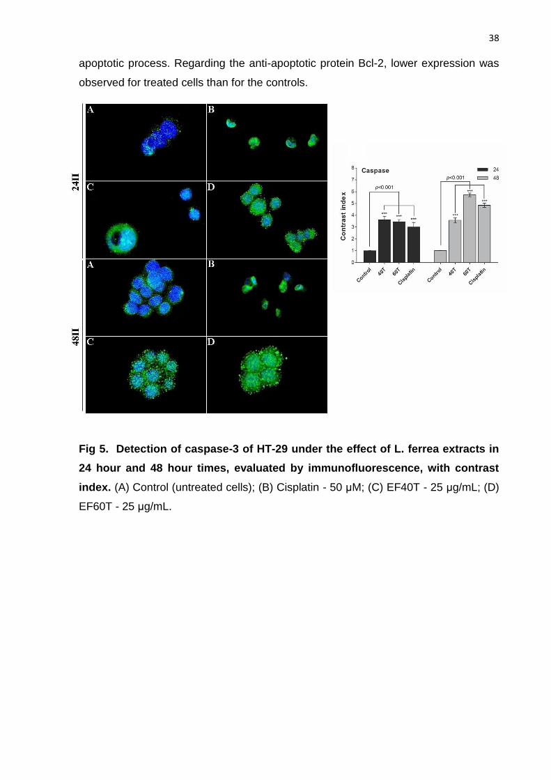
38
apoptotic process. Regarding the anti-apoptotic protein Bcl-2, lower expression was
observed for treated cells than for the controls.
Fig 5. Detection of caspase-3 of HT-29 under the effect of L. ferrea extracts in
24 hour and 48 hour times, evaluated by immunofluorescence, with contrast
index. (A) Control (untreated cells); (B) Cisplatin - 50 μM; (C) EF40T - 25 μg/mL; (D)
EF60T - 25 μg/mL.

39
Fig 6. Detection of Bcl-2 of HT-29 under the effect of L. ferrea extracts in 24
hour and 48 hour times, evaluated by immunofluorescence, with contrast
index. (A) Control (untreated cells); (B) Cisplatin - 50 μM; (C) EF40T - 25 μg/mL; (D)
EF60T - 25 μg/mL.
3.4 GSH and MDA dosages
It is highlighted in Figure 7 that glutathione (GSH) levels, an important marker
of antioxidant activity, increased by 18% in relation to the control in extract EF60T at
25 μg/mL, which was the sample presenting higher dosages of this protein in the HT-
29 tumoral lineage. Regarding extracts EF40T and EF60T at the highest dose (50
μg/mL), they presented reduced levels, and we did not find the same effect.
Regarding the malondialdehyde oxidative stress marker (MDA), we observed in
Figure 8 that all of the extracts at all doses were able to significantly reduce the
levels of the protein, thus indicating a protective effect on lipid peroxidation in the HT-
29 tumor line, being higher than cisplatin.

40
Fig 7. Effect of L. ferrea extracts on total glutathione (GSH) levels of HT-29 and
HEK-293 cells. * P <0.05, ** P <0.01 and *** P <0.001 versus control. CTRL
(untreated cells), CIS (cisplatin), 40T (crude extract 40T), 60T (60T crude extract).
Fig 8. Effect of L. ferrea extracts on malondialdehyde (MDA) levels of HT-29 and
HEK-293 cells. *P <0.05, **P <0.01 and ***P <0.001 versus control. CTRL (untreated
cells), CIS (cisplatin), 40T (crude extract 40T), 60T (crude extract 60T).
3.5 mRNA expression
Based on Figure 9, we see that the AKT gene that is involved in cell survival
and proliferation had a slightly reduced expression in the treatment with EF40T at the

41
extract dose of 25 μg/mL (4%); extract EF60T with the same dose yielded (26%), a
rate still lower than that found with the antineoplastic cisplatin (49%). The Apaf-1
(apoptotic peptidase activating factor 1) gene encoding a cytoplasmic protein that
initiates apoptotic events in the intrinsic pathway was found to be reduced in all
treatments. Epidermal growth factor receptor (EGFR), involved in the pathogenesis
and progression of different carcinomas, showed a reduction in expression as
compared to the control: 29% for the EF40T extract, 48% for the EF60T extract and
53% for cisplatin.
Fig 9.Effect of L. ferrea extracts on the expression of AKT, Apaf-1 and EGFR
mRNAs in HT-29. *P < 0.05, **P < 0,01 e ***P<0.001 versus control. CTRL
(untreated cells), CIS (cisplatin), EF40T (40T crude extract), EF60T (60T crude
extract).
4. Discussion
In addition to expanding medical-pharmacological care in public health, the
study of medicinal plants is important for acquiring knowledge about the
pharmacological potential of a native plant diversity that remains under exploited, and
helps to ensure its rational exploitation [13]. The versatility, applicability and
therapeutic safety presented by caatinga plants have attracted great attention to
them in the search for new drugs [36]. Popular species such as Libidibia ferrea

42
Martius L. P. Queiroz require ethno-pharmacological approaches, and these have so
far remained scarce, especially regarding anticancer activity.
Crude and fractionated extracts obtained from the fruits of L. ferrea tested by
Freitas et al [37] on human cancer cell lines NCI-H292 (mucoepidermoid carcinoma
of the lung), HEP-2 (squamous cell carcinoma of the larynx), and solid tumor
sarcoma 180 presented no significant antitumor activity or inhibition of cell
proliferation. In the present study, for the HT-29 tumoral lineage (human colorectal
adenocarcinoma), proliferation inhibition by the 40T, 60T and 80T crude extracts was
significant in the first observed hours. In addition, during the same time period, the
same extracts did not generally present toxicity in the non-tumor cell line HEK-293
(human embryonic renal cell).
Alterations to apoptosis death process related pathways and consequent cell
resistance to this mechanism are the main factors responsible for the onset of
tumorigenesis, considered one of the fundamental hallmarks of cancer [38]. Despite
causing the problem, apoptosis plays an important role in cancer treatment strategies
through the eradication of transformed cells, and resistance to conventional
treatments [39].
Induction of apoptosis by L. ferrea has been described previously by Nozaki et
al [40], using acetone stem extract in human acute myeloid leukemia (HL-60)
lineage. In this work, all extracts from the L. ferrea fruit tested in the HT-29 line
induced cell death by apoptosis, corroborating their results.
The apoptotic activity exhibited in the present study has a profile suggestive of
affecting the intrinsic apoptosis pathway, a common target of most anticancer agents
[41], through activation of caspase-3 and reduction of Bcl-2 levels. This pathway
results from increased mitochondrial membrane permeability (with inhibition of anti-
apoptotic regulator Bcl-2), and the release of pro-apoptogenic factors in the
cytoplasm, promoting the activation of effector enzymes (such as caspase-3), which
act on a broad spectrum of substrates, resulting in cell death [42].
Most of the apoptotic cells in this work were detected in the initial stage,
characterized by phosphatidylserine exposure (an event that precedes cell
membrane permeabilization), cellular shrinkage, and nuclear condensation. In
addition, the collapse of the mitochondrial transmembrane potential has also been
reported as an early event in the apoptotic process, being present in many cell types

43
in apoptosis that culminate in the release of several apoptotic proteins, independently
of the stimulus [43]. Therapies for colorectal cancer designed to stimulate apoptosis,
as presented here, play a critical role in controlling its development and progression,
as well as improving response to chemotherapy and radiation [44].
To assess antioxidant status, dosages were performed for analysis of GSH
(glutathione) and MDA (malondialdehyde) levels. We observed that the EF60T
extract, at the lowest dose tested presented a significant increase in the level of
GSH, an intracellular peptide that exerts, among other functions, detoxification,
elimination of free radicals, maintenance of the thiol state, and cellular proliferation
modulation [45]. As for the MDA lipid peroxidation marker, all of the extracts at all
doses presented reductions, which is an important sign for prevention of MDA’s
mutagenic and genotoxic activities in tumor cells [46]. All in all, the extract stands out
for presenting potential antioxidant activity. The antioxidant activities of L. ferrea fruits
were first described by Silva et al [47] through several in vitro assays. Later, Barros et
al [19] also described augmented antioxidant activity in addition to hepatoprotection
in mice.
The expression of certain relevant genes to tumor signaling machinery was
evaluated in this work through identification of mRNA-targets (EGFR, AKT and
APAF-1). We found that levels of the EGFR receptor (involved in growth, invasion
and metastasis of epithelial tumors and particularly important in the development of
colorectal tumors) [48] were reduced after treatment with the extracts, indicating that
L. ferrea interferes with this pathway and is effective in tumor inhibition.
Expression of AKT was varied, with reduced expression for EF40T and
increased expression for EF60T. Interestingly, some studies have shown that AKT is
not a single-function kinase and may facilitate, rather than inhibit, cell death under
certain conditions [49], thus being involved both in inducing death and cell survival.
The same scenario of opposite functions in the same protein may also involve Apaf-1
factor, whose expression was reduced in cells treated with L. ferrea. Although this
protein is known for its apoptotic role in the intrinsic pathway, recent work has
suggested additional non-apoptotic functions including a pro-survival role that needs
to be better investigated [50].
Through various mechanisms, cancer itself, through induction of pro-survival
signals such as the AKT activation pathway bypasses activation of the intrinsic

44
pathway as in blockade of apoptosome formation and consequently of Apaf-1. Thus,
strategies to bypass resistance, through therapeutic restoration of apoptotic
pathways as conducted by interactions with the tumor microenvironment, provide
new promise for cancer patients in clinical treatment [51]. Therefore, the differing
expression profiles presented in this study from the extracts of L. ferrea may be
related to different activities in the mediation of important pathways in tumor
development, that culminate in cell death stimuli, as already verified in the previous
results.
Mediation of the AKT pathway, in addition to influencing apoptosis induction
impacts oxidization capacity. Ramos et al [52] showed that apoptosis caused by
treatment with low concentrations of arsenic trioxide in human myelogenous
leukemia cells (HL-60) was potentiated by the use of AKT pathway inhibitors, which
also led to the reduction of levels of GSH causing intracellular oxidation. The results
demonstrated that AKT signaling pathway activity is necessary to maintain normal
GSH metabolism in cells. We observed in the current study that there is also
synchronization between AKT expression levels and intracellular GSH levels:
increased AKT expression is accompanied by increased GSH and vice versa.
The main constituents found in the L. ferrea extracts tested were the
hydrolyzable tannins: gallic acid (GA) and ellagic acid (EA) (30). GA is a phenolic
compound naturally present in a wide variety of fruits and plants exhibiting various
pharmacological activities such as antimicrobial, antitumor, anti-inflammatory,
antioxidant, antidepressant, antidiabetic, anxiolytic, and others [53]. The acid is
associated to inhibition in various cancer cell lines including human colorectal
carcinoma cells through apoptosis induction by different mechanisms such as
regulation of apoptotic and anti-apoptotic proteins [54]. It is also well known for its
efficient protection against oxidative damage, maintenance of endogenous defense,
and chiefly, prevention of lipid peroxidation [55].
EA is a dimeric derivative of gallic acid present in the plant vacuole and, like
other polyphenols; it has a wide range of biological activities such as antioxidant,
anti-inflammatory, prebiotic and anticancer functions, and the latter by selectively
inhibiting growth in differring tumor types, including inducing apoptosis in human
colon adenocarcinoma cells via the mitochondrial pathway [56].
45
Among the results reported, it is important to highlight the protective effect of
L. ferrea extracts on non-tumor HEK-293 cells, which presented high rates of cell
proliferation and viable cells. In vitro evaluations by Nakamura et al [57] have
demonstrated the chemopreventive effects of L. ferrea through the presence of
ellagic acid in its composition: three adjacent hydroxyl phenolic groups were
responsible for cytotoxicity, and the carboxyl group appeared to be implicated in the
distinction between normal and tumor cells. These results are consistent with studies
by Araújo Júnior et al [58,59], in the sense that many plant derivatives may act both
in inhibiting the proliferation and viability of tumor cells, and in mediating a protective
effect on healthy cells.
5. Conclusions
The present results demonstrate that L. ferrea has antiproliferative activity in
HT-29 cells with induction of apoptosis in association with mitochondrial effects in the
intrinsic pathway (via caspase-3 activation, negative regulation of Bcl-2 and
Reduction of Apaf-1), this as well as acting in the expression of important targets in
the process of colorectal cancer development such as EGFR and AKT, with probable
tumor inhibition. L. ferrea also has antioxidant and lipid peroxidation inhibiting effects,
together with chemopreventive action in healthy renal cells. Taken as such, L. ferrea
is a promising candidate for cancer therapy with both efficacy and selectivity as
potential characteristics.
Acknowledgments
The authors thank the Pharmacognosy Laboratory of Pernambuco Federal University
(UFPE); the cell culture room of the Department of Biochemistry, the Health Sciences
Graduate Program, and the Brain Institute Microscopy Laboratory at Rio Grande do
Norte Federal University (UFRN).
References
[1] V. Pavet, M.M. Portal, J.C. Moulin, R. Herbrecht, H. Gronemeyer, Towards novel
paradigms for cancer therapy, Oncogene 30 (2011) 01-20.

46
[2] Y.A. Luqmani, Mechanisms of drug resistance in cancer chemotherapy, Med.
Princ. Pract. 14 (2005) 35-48.
[3] B.W. Stewart, C.P. Wild, World Cancer Report 2014, International Agency for
Research on Cancer, Lyon, 2014.
[4] E.R. Fearon, Molecular genetics of colorectal cancer, Annu. Rev. Pathol. Mech.
Dis. 6 (2011) 479-507.
[5] M. De Rosa, U. Pace, D. Rega, V. Costabile, F. Duraturo, P. Izzo, P. Delrio,
Genetics, diagnosis and management of colorectal cancer, Oncol. Rep. 34 (2015)
1087-1096.
[6] M.J. Balunas, A.D. Kinghorn, Drug Discovery from medicinal plants, Life Sci. 78
(2005) 431-441.
[7] A. Paul, Manjula, Cytotoxic and antiproliferative activity of indian medicinal plants
in cancer cells, IJSR 3 (2014) 88-93.
[8] P.M.P. Ferreira, D.F. Farias, M. Viana, T.M. Souza, I.M. Vasconcelos, B.M.
Soares, C. Pessoa, L.V. Costa-Lotufo, M.O. Moraes, A.F.U. Carvalho, Study of
antiproliferative potential of seed extracts from northeastern brazilian plants, An.
Acad. Bras. Ciênc. 83 (2011) 1045-1058.
[9] J.B. Calixto. Efficacy, safety, quality control, marketing and regulatory guidelines
for herbal medicines (phytotherapeutic agents). Braz. J. Med. Biol. Res. 33 (2000)
179-189.
[10] S.Y. Yin, W.C. Wei, F.Y. Jian, N.S. Yang. Therapeutic applications of herbal
medicines for cancer patients. Evid. Based Complement. Alternat. Med. 2013 (2013)
01-15.
[11] I. Mohamed, A. Shuid, B. Borhanuddin, N. Fozi. The application of
phytomedicine in modern drug development. The Internet Journal of Herbal and
Plant Medicine 1 (2013) 01-9.
[12] U.P. Albuquerque, P.M. Medeiros, A.L.S. Almeida, J.M. Monteiro, E.M.F. Lins,
J.G. Melo, J.P. Santos, Medicinal plants of the caatinga (semi-arid) vegetation of NE
Brazil: a quantitative approach, J. Ethnopharmacol. 114 (2007) 325-354.

47
[13] C.P. Benedito, M.F.B. Coelho, I.P. Guimarães, V.P. Amaral, S.S.S. Maia, P.F.
Batista, Emergence and early growth of Caesalpinia ferrea Mart. ex Tul. var. ferrea
seedlings on different substrates, Rev. Bras. Ciênc. Agrár. 7 (2012) 508-513.
[14] M.R.A. Ferreira, L.A.L. Soares, Libidibia ferrea (Mart. ex Tul.) L. P. Queiroz: A
review of the biological activities and phytochemical composition, J. Med. Plants Res.
9 (2015) 140-150.
[15] L.M. Costa, F. Guilhon-Simplicio, T.P. Souza, Libidibia ferrea (Mart. Ex Tul) L. P.
Queiroz var. ferrea: Pharmacological, phytochemical and botanical aspects, Int. J.
Pharm. Pharm. Sci. 7 (2015) 48-53.
[16] F.C. Sampaio, M.S.V. Pereira, C.S. Dias, V.C.O. Costa, N.C.O. Conde, M.A.R.
Buzalaf, In vitro antimicrobial activity of Caesalpinia ferrea Martius fruits against oral
pathogens, J. Ethnopharmacol. 124 (2009) 289-294.
[17] R.O. Marreiro, M.F.C.L. Bandeira, T.P. Souza, M.C. Almeida, K. Bendaham,
G.N. Venancio, I.C. Rodrigues, C.N. Coelho, P.S.L.L. Milério, G.P. Oliveira GP,
N.C.O. Conde, Evaluation of the stability and antimicrobial activity of an ethanolic
extract of Libidibia ferrea, Clin. Cosmet. Investig. Dent. 6 (2014) 9-13.
[18] L.C. Da Silva, J.M. Sandres, M.M. De Paiva, J.M. De Araújo, R.C. De
Figueiredo, M.V. Da Silva, M.T. Correia, Anti-staphylococcus aureus action of three
Caatinga fruits evaluated by electron microscopy, Nat. Prod. Res. 27 (2013) 1492-
1496.
[19] A.O. Barros, R.S. Souza, E.S.P. Aranha, L.M. Costa, T.P. Souza, M.C.
Vasconcellos, E.S. Lima, Antioxidant and Hepatoprotective Activities of Libidibia
ferrea bark and fruit extracts, Int. J. Pharm. Pharm. Sci. 6 (2014) 71-76.
[20] H. Ueda, Y. Tachibana, M. Moriyasu, K. Kawanishi, S.M. Alves, Aldose
reductase inhibitors from the fruits of Caesalpinia ferrea Mart, Phytomedicine 8
(2001) 377-381.
[21] E.S. Nakamura, F. Kurosaki, M. Arisawa, T. Mukainaka, J. Takayasu, M. Okuda,
H. Tokuda, H. Nishino, F. Pastore, Cancer chemopreventive effects of a Brazilian folk
medicine Juca on in vivo two-stage skin carcinogenesis, J. Ethnopharmacol. 81
(2002) 135-137.

48
[22] A.B. Souza, L.M.S. Souza, J.C.T. Carvalho, E.L. Maistro, No clastogenic activity
of Caesalpinia ferrea Mart. (Leguminosae) extract on bone marrow cells of Wistar
rats, Genet. Mol. Biol. 29 (2006) 380-383.
[23] V.M. Peters, S.O. Souza, J.C.T. Carvalho, L.V. Borges, M.O. Guerra MO,
Evaluation of reproductive toxicity of aqueous extract of the fruits from Caesalpinia
ferrea Mart in rats, B. Latinoam. Caribe Pl. 7 (2008) 268-272.
[24] F. Hua, M.G. Cornejo, M.H. Cardone, C.L. Stokes, D.A. Lauffenburger, Effects
on fas signaling-induced caspase-3 activation: molecular genetic tests of
computational model predictions, J. Immunol. 175 (2005) 985-995.
[25] J. Polivka Jr, F. Janku, Molecular targets for cancer therapy in the
PI3K/AKT/mTOR pathway, Pharmacol. Ther. 142 (2014) 164-175.
[26] S. Baig, I. Seevasant, J. Mohamad, A. Mukheem, H.Z. Huri, T. Kamarul,
Potential of apoptotic pathway-targeted cancer therapeutic research: Where do we
stand?, Cell Death Dis. 7 (2016) 01-11.
[27] A. Gortat, M. Sancho, L. Mondragón, A. Messeguer, E. Pérez-Payá, M. Orzáez,
Apaf1 inhibition promotes cell recovery from apoptosis, Protein Cell 6 (2015) 833-
843.
[28] P. Seshacharyulu, M.P. Ponnusamy, D. Haridas, M. Jain, A.K. Ganti, S.K.
Batra,Targeting the EGFR signaling pathway in cancer therapy, Expert Opin. Ther.
Targets 16 (2012) 15-31.
[29] G.M. Nitulescu, D. Margina, P. Juzenas, Q. Peng, O.T. Olaru, E. Saloustros, C.
Fenga, D.A. Spandidos, M. Libra, A.M. Tsatsakis, Akt inhibitors in cancer treatment:
The long journey from drug discovery to clinical use, Int. J. Oncol. 48 (2016) 869-885.
[30] M.R.A. Ferreira, M.T.M. Fernandes, W.A.V. Silva, I.C.F. Bezerra, T.P. Souza,
M.F. Pimentel, L.A.L. Soares LAL, Chromatographic and Spectrophotometric
Analysis of Phenolic Compounds from Fruits of Libidibia ferrea Martius, Pharmacogn.
Mag. 12 (2016) 285-291.
[31] T. Mosman, Rapid colorimetric assay for cellular growth and survival: application
to proliferation and cytotoxicity assays, J. Immunol. Methods 65 (1983) 55-63.

49
[32] C.M. Costa, R.C.C. Santos, E.S. Lima, A simple automated procedure for thiol
measurement in human serum samples, J. Bras. Patol. Med. Lab. 42 (2006) 345-
350.
[33] I. Rahman, A. Kode, S.K. Biswas, Assay for quantitative determination of
gluthathione and gluthathione disulfide levels using enzymatic recycling method, Nat.
Protoc. 1 (2006) 3159-3165.
[34] H. Esterbauer H, K.H. Cheeseman, Determination of aldehydic lipid peroxidation
products: malonaldehyde and 4-hydroxynonenal, Methods Enzymol. 186 (1990) 407-
421.
[35] K.J. Livak, T.D. Schmittgen, Analysis of relative gene expression data using real-
time quantitative PCR and the 2-ΔΔCt method, Methods 25 (2001) 402-408.
[36] N.L. Alencar, F.R. Santoro, U.P. Albuquerque, What is the role of exotic
medicinal plants in local medical systems? A study from the perspective of utilitarian
redundancy, Braz. J. Pharmacog. 24 (2014) 506-515.
[37] A.C.C. Freitas, N.C.A. Ximenes, J.S. Aguiar, S.C. Nascimento, T.U.L. Lins, L.R.
Magalhães, L.C.B.B. Coelho, M.G. Carneiro-da-Cunha, T. Gonçalves-Silva, M.T.S.
Correia, Biological activities of Libidibia (Caesalpinia) ferrea var. parvifolia (Mart. ex
Tul.) L. P. Queiroz pod preparations, Evid. Based Complement. Alternat. Med. 2012
(2012) 01-07.
[38] D. Hanahan, R.A. Weinberg, Hallmarks of cancer: the next generation, Cell 144
(2011) 646-674.
[39] M. Hassan, H. Watari, A. AbuAlmaaty, Y. Ohba, N. Sakuragi, Apoptosis and
molecular targeting therapy in cancer, BioMed. Res. Int. 2014 (2014) 01-23.
[40] H. Nozaki, K. Hayashi, M. Kido, K. Kakumoto, S. Ikeda, N. Matsuura, H. Tani, D.
Takaoka, M. Iinuma, Y. Akao Y, Pauferrol A - a novel chalcone trimer with a
cyclobutane ring from Caesalpinia ferrea mart exhibiting DNA topoisomerase II
inhibition and apoptosis-inducing activity, Tetrahedron Lett. 48 (2007) 8290-8292.
[41] S. Fulda, K.M. Debatin, Extrinsic versus intrinsic apoptosis pathways in
anticancer chemotherapy, Oncogene 25 (2006) 4798-4811.
[42] R.S. Wong, Apoptosis in cancer: from pathogenesis to treatment, J. Exp. Clin.
Cancer Res. 30 (2011) 01-14.

50
[43] G. Denecker, H. Dooms, G.V. Loo, D. Vercammen, J. Grooten, W. Fiers, W.
Declercq, P. Vandenabeele, Phosphatidyl serine exposure during apoptosis
precedes release of cytochrome c and decrease in mitochondrial transmembrane
potential, FEBS Letters 465 (2000) 47-52.
[44] A.M. Abraha, E.B. Ketema, Apoptotic pathways as a therapeutic target for
colorectal cancer treatment, World J. Gastrointest. Oncol. 8 (2016) 583-591.
[45] S.C. Lu, Regulation of glutathione synthesis, Mol. Aspects Med. 30 (2009) 42-
59.
[46] D. Grotto, L.S. Maria, J. Valentini, C. Paniz, G. Schmitt, S.C. Garcia, V.J.
Pomblum, J.B.T. Rocha, M. Farina, Importance of the lipid peroxidation biomarkers
and methodological aspects for malondialdehyde quantification, Quím. Nova 32
(2009) 169-174.
[47] L.C.N. Silva, C.A. Silva Júnior, R.M. Souza, A.J. Macedo, M.V. Silva, M.T.S.
Correia, Comparative analysis of the antioxidante and DNA protection capacities of
Anadenanthera colubrina, Libidibia ferrea and Pityrocarpa moniliformis fruits, Food
Chem. Toxicol. 49 (2011) 2222-2228.
[48] T. Sasaki, K. Hiroki, Y. Yamashita, The role of epidermal growth factor receptor
in cancer metastasis and microenvironment, Biomed. Res. Int. 2013 (2013) 01-08.
[49] M. Los, S. Maddika, B. Erb, K. Schulze-Osthoff, Switching Akt: from survival
signaling to deadly response, Bioessays 31 (2009) 492-495.
[50] M. Sancho, A. Gortat, A.E. Herrera, V. Andreu-Fernández, E. Ferraro, F.
Cecconi, M. Orzáez, E. Pérez-Payá, Altered mitochondria morphology and cell
metabolism in Apaf-1 deficient cells, PLos One 9 (2014) 01-08.
[51] M.V. Fiandalo, N. Kyprianou, Caspase control: protagonists of cancer cell
apoptosis, Exp. Oncol. 34 (2012) 165-175.
[52] A.M. Ramos, C. Fernández, D. Amrán, P. Sancho, E. Blas, P. Aller,
Pharmacologic inhibitors of PI3K/Akt potentiate the apoptotic action of the
antileukemic drug arsenic trioxide via glutathione depletion and increased peroxide
accumulation in myeloid leukemia cells, Blood 105 (2005) 4013-4020.
[53] N. Nayeem, A. Smb, H. Salem, S. Ahei-Alfqy, Gallic acid: a promising lead
molecule for drug development, J. App. Pharm. 8 (2016) 01-4.

51
[54] A.P. Subramanian, A.A. John, M.V. Vellayappan, A. Balaji, S.K. Jaganathan, E.
Supriyanto, Gallic acid: prospects and molecular mechanisms of its anticancer
activity, RSC Adv. 5 (2015) 35608-35621.
[55] B. Badhani, N. Sharma, R. Kakkar, Gallic acid: a versatile antioxidante with
promising therapeutic and industrial applications, RSC Adv. 5 (2015) 27540-27557.
[56] J.M. Landete, Ellagitannins, ellagic acid and their derived metabolites: a review
about source, metabolism, functions and health, Food Res. Int. 44 (2011) 1150-1160.
[57] E.S. Nakamura, F. Kurosaki, M. Arisawa, T. Mukainaka, M. Okuda, H. Tokuda,
H. Nishino, F. Pastore, Cancer chemopreventive effects of constituents of
Caesalpinia ferrea and related compounds, Cancer Lett 177 (2002) 119-124.
[58] R.F. Araújo Júnior, A.L.C.S.L. Oliveira, J.B. Pessoa, V.B. Garcia, G.C.B. Guerra,
L.A.L. Soares, T.P. Souza, P.R. Petrovick, A.A. Araújo, Maytenus ilicifolia dry extract
protects normal cells, induces apoptosis and regulates Bcl-2 in human cancer cells,
Exp. Biol. Med. 238 (2013) 1251-1258.
[59] R.F. Araújo Júnior, T.P. Souza, J.G. Pires, L.A. Soares, A.A. Araújo, P.R.
Petrovick, H.D. Macêdo, A.L.S.L. Oliveira, G.C. Guerra, A dry extract of Phyllanthus
niruri protects normal cells and induces apoptosis in human liver carcinoma cells,
Exp. Biol. Med. 237 (2012) 1281-1288.
___________________________________________________________________

52
6 COMENTÁRIOS, CRÍTICAS E CONCLUSÕES
O projeto de pesquisa inicial intitulava-se “Avaliação da Atividade Citotóxica e
dos Mecanismos de Morte Celular In vitro de Extratos de Libidibia ferrea em Células
de Câncer Colorretal”, porém no decorrer dos experimentos e dos resultados
obtidos, decidimos expandir o estudo e avaliar também a atividade antioxidante, em
parceria com o Laboratório Farmol sob orientação da Prof. Aurigena Antunes, onde
obtivemos dados bastante satisfatórios e contribuiu significativamente para a
elucidação do potencial farmacológico dos extratos em questão. Outros alvos de
vias de sinalização de interesse para o câncer também foram acrescentadas à
metodologia inicial (EGFR e Apaf-1), decorrentes também de parcerias e
financiamentos de projetos desenvolvidos pelo nosso grupo de pesquisa.
As concentrações utilizadas para os testes in vitro dos extratos de L. férrea
precisaram sofrer alterações do que foi inicialmente sugerido (doses de mg/mL para
µg/mL), decorrentes de adequações metodológicascom base nos primeiros
resultados de screening da atividade antitumoral. Tais modificações foram
prontamente identificadas e executadas, realizando-se um estudo da curva dose-
resposta dos extratos, o que não comprometeu o desenvolvimento do trabalho.
Ao longo do desenvolvimento desta pesquisa, o cronograma de atividades
passou por mudanças, devido a situações inerentes da pesquisa experimental e do
trabalho científico, relacionadas ao cultivo celular, repetições de resultados,
disponibilidade de equipamentos e material, entre outros. Porém o trabalho seguiu
às metas e ao desenho do estudo planejado, adaptando-se às pequenas limitações
encontradas.
Este projeto contribuiu bastante como ponto de partida para a consolidação
de novas metodologias científicas para o Laboratório LAICI, principalmente quanto à
inserção de técnicas para avaliação da atividade antioxidante in vitro que foram
desenvolvidas após muito estudo e experimentação, para chegarmos ao método
ideal (desde a obtenção das células à leitura). Outra técnica também que foi
estabelecida e aperfeiçoada foi o Real Time RT-qPCR. Esse processo proporcionou
o avanço científico dos alunos e a melhoria, como um todo, dos projetos executados.

53
Os resultados apresentados possuem grande valor para esclarecer o
potencial bioativo que a diversidade vegetal nativa fornece, em especial de L.
ferreaque carece de abordagens etnofarmacológicas, trazendo conhecimento à
população e auxiliando à comunidade científica na busca de novas terapias para o
câncer colorretal. Além disso, este trabalho faz parte de um projeto maior
desenvolvido por nosso grupo de pesquisa que possui resultados preliminares
indicando também atividades anti-inflamatórias e antimicrobianas in vivo, que
constituirão na publicação de artigos e no avanço científico em geral.
Durante a trajetória do mestrado pude progredir intelectualmente, através do
desenvolvimento de novas habilidades e competências que foram necessárias para
dar seguimento ao trabalho, além das aulas e discussões científicas promovidas
pelo Programa de Pós-Graduação em Ciências da Saúde da UFRN, que foram
bastante enriquecedoras e permitiram o contato com diversos profissionais
proporcionando um ambiente de muito aprendizado e conhecimento.
Após a conclusão dessa etapa acadêmica, há a perspectiva futura de serem
realizadas outras abordagens, com o uso de nanotecnologia para avaliação dos
extratos de L. ferrea associado à quimioterápicos, envolvendo novas investigações
de suas aplicações na área da oncologia molecular. A finalidade desta trajetória
seguirá com a busca constante de aprimoramento e inovação científica.

54
7 REFERÊNCIAS
1. Pavet V, Portal MM, Moulin JC, Herbrecht R, Gronemeyer H. Towards novel
paradigms for cancer therapy. Oncogene 2011; 30(1):01-20.
2. Luqmani YA. Mechanisms of drug resistance in cancer chemotherapy. Med. Princ. Pract. 2005; 14(1):(2005).
3. Stewart BW, Wild CP.World Cancer Report 2014. International Agency for
Research on Cancer; 2014.
4. Bogaert J, Prenen H. Molecular genetics of colorectal cancer. AnnGastroenterol.
2014; 27(1):9-14.
5. De Rosa M, Pace U, Rega D, Costabile V, Duraturo F, Izzo P, Delrio P. Genetics,
diagnosis and management of colorectal cancer (Review). Oncol Rep.2015;
34(3):1087-96.
6. Ismael GFV, Rosa DD, Mano MS, Awada A. Novel cytotoxic drugs: old challenges,
new sollutions. Cancer Treat Ver. 2008; 34(1):81-91.
7. Lu Y, Mahato RI. Pharmaceutical Perspectives of Cancer Therapeutics. 1ª edition.
New York: Springer Science; 2009.
8. López-Camarillo C, Aréchaga-Ocampo E. Oncogenomics and cancer proteomics –
Novel approaches in biomarkers discovery and therapeutic targets in cancer. 1ª
edition. Rijeka: InTech; 2013.
9. Lahlou M. The success of natural products in drug discovery. Pharmacol Pharm.
2013; 4:17-31.
10. Paul A, Manjula. Cytotoxic and antiproliferative activity of indian medicinal plants
in cancer cells. IJSR 2014; 3:88-93.
11. Ferreira PMP, Farias DF, Viana MP, Souza TM, Vasconcelos IM, Soares BM,
Pessoa C, Lotufo LVC, Moraes MO, Carvalho AFU. Study of antiproliferative
potential of seed extracts from northeastern brazilian plants. An Acad Bras Cienc.
2011; 83:1045-58.
12. Calixto JB. Efficacy, safety, quality control, marketing and regulatory guidelines
for herbal medicines (Phytotherapeuthic agents). Braz J Med Biol Research. 2000;
33: 179-87.

55
13. Albuquerque UP, Medeiros PM, Almeida ALS, Monteiro JM, Lins EMF, Melo JG,
Santos JP. Medicinal plants of the caatinga (semi-arid) vegetation of NE Brazil: a
quantitative approach. J Ethnopharmacol. 2007; 114(3):325-354.
14. Ribeiro DA, Macedo DG, Oliveira LGS, Saraiva ME, Oliveira SF, Souza MMA,
Menezes IRA. Potencial terapêutico e uso de plantas medicinais em uma área de
Caatinga no estado do Ceará, nordeste do Brasil. Rev Bras PI Med. 2014; 16(4):
912-30.
15. Silva MIG, Melo CTV, Vasconcelos LF, Carvalho AMR, Sousa FCF. Bioactivity
and potential therapeutic benefi ts of some medicinal plants from the Caatinga (semi-
arid) vegetation of Northeast Brazil: a review of the literature. Rev. Bras. Farmacogn.
2012; 22(1): 193-207.
16. National Tropical Botanical Garden. Caesalpinia ferrea [Internet]. Kalaheo:
National Tropical Botanical Garden; 2017 [citado em 2017 May 01]. Disponível em:
<http://www.ntbg.org/plants/index.php>.
17. Ferreira Júnior WS, Ladio AH, Albuquerque UP. Resilience and adaptation in the
use of medicinal plants with suspected anti-inflammatory activity in the Brazilian
Northeast. J Ethnopharmacol. 2011; 138(1):238-52.
18. Frasson APZ, Bittencourt CF, Heinzmann BM. Caracterização físico-química e
biológica do caule de Caesalpinia ferrea Mart. Rev Bras Farmacogn. 2003; 13(1):35-
39.
19. Roque AA, Rocha RM, Loiola MIB. Uso e diversidade de plantas medicinais da
caatinga na comunidade rural de Laginhas, município de Caicó, Rio Grande do Norte
(nordeste do Brasil). RevBras Plan Med. 2010; 12(1):31-42.
20.Hassan SK, El-Sammad NM, Mousa AM, Mohammed MH, Farrag ARH, Hashim
ANE, Werner V, Lindequist U, Nawwar MAE. Hypoglycemic and antioxidant activities
of Caesalpinia ferrea Martius leaf extract in streptozotocin-induced diabetic rats.
Asian Pac J Trop Biomed. 2015; 5(6):462-71.
21. Vasconcelos CF, Maranhão HM, Batista TM, Carneiro EM, Ferreira F, Costa J,
Soares LAL, Sá MDC, Souza TP, Wanderley AG. Hypoglycaemic activity and
molecular mechanisms of Caesalpinia ferrea Martius bark extract on streptozotocin-
induced diabetes in wistar rats. J Ethnopharmacol. 2011; 137(3):1533-41.

56
22. Melo EA [homepage na internet]. Pau ferro – Caesalpinia ferrea [acesso em 27
mai 2017]. Disponível em: http://www.arvores.brasil.nom.br/esq.htm.
23. Nakamura ES, Kurosaki F, Arisawa M, Mukainaka T, Okuda M, Tokuda H,
Nishino H, Jr Pastore F. Cancer chemopreventive effects of constituents of
Caesalpinia ferrea and related compounds. Cancer Lett. 2002;177:119-24.
24. Sampaio FC, Pereira MSV, Dias CS, Costa VCO, Conde NCO, Buzalaf MAR. In
vitro antimicrobial activity of Caesalpinia ferrea Martius fruits against oral pathogens.
J Ethnopharmacol. 2009; 124:289-94.
25. Marreiro RO, Bandeira MFCL, Souza TP, Almeida MC, Bendaham K, Venancio
GN, Rodrigues IC, Coelho CN, Milério PSLL, Oliveira GP, Conde NCO. Evaluation of
the stability and antimicrobial activity of an ethanolic extract of Libidibia ferrea. Clin
Cosmet Investig Dent. 2014; 6:9-13.
26. Da Silva LC, Sandres JM, De Paiva MM, De Araújo JM, De Figueiredo RC, Da
Silva MV, Correia MT. Anti-staphylococcus aureus action of three Caatinga fruits
evaluated by electron microscopy. Nat Prod Res. 2013; 27(16):1492-96.
27. Ueda H, Tachibana Y, Moriyasu M, Kawanishi K, Alves SM. Aldose reductase
inhibitors from the fruits of Caesalpinia ferrea Mart. Phytomedicine. 2001; 8(5):377-
81.
28. Souza AB, Souza LMS, Carvalho JCT, Maistro EL. No clastogenic activity of
Caesalpinia ferrea Mart. (Leguminosae) extract on bone marrow cells of Wistar rats.
Genet Mol Biol. 2006; 29(2):380-83.
29. Peters VM, Souza SO, Carvalho JCT, Borges LV, Guerra MO. Evaluation of
reproductive toxicity of aqueous extract of the fruits from Caesalpinia ferrea Mart in
rats. B Latinoam Caribe Pl. 2008; 7:268-72.
30. Barros AO, Souza RS, Aranha ESP, Costa LM, Souza TP, Vasconcellos MC,
Lima ES. Antioxidant and Hepatoprotective Activities of Libidibia ferrea bark and fruit
extracts. Int J Pharm Pharm Sci. 2014; 6(11):71-6.
31. Reboredo MM, Lucinda LMF, Rocha CB, Queiroz GT, Faria VC, Vieira VA.
Avaliação da toxicidade do extrato aquoso de Caesalpinia ferrea em órgãos vitais,
no sistema reprodutor e na produção de espermatozóides de ratos Wistar
submetidos a tratamento subagudo. Rev Int Est Exp. 2006; 25:17-29.

57
32. Dias AMA, Rey-Rico A, Oliveira RA, Marceneiro S, Alvarez-Lorenzo C, Concheiro
A, Júnior RNC, Braga MEM, de Sousa H. Wound dressings loaded with an anti-
inflammatory jucá (Libidibia ferrea) extract using supercritical carbon dioxide
technology. J. Supercrit. Fluids 2013; 74: 34-45.
33. Traverso N, Ricciarelli R, Nitti M, Marengo B, Furfaro AL, Pronzato MA, Marinari
UM, Domenicotti C. Role of glutathione in cancer progression and chemoresistance.
Oxid. Med. Cell. Longev. 2013; 2013: 01-10.
34. Bitla AR, Reddy EP, Sambasivaih K, Suchitra MM, Reddy VS, Rao PVLNS.
Evaluation of plasma malondialdehyde as a biomarker in patients with carcinoma of
stomach. Biomed. Res. 2011; 22(1):63-8.
35. Mahmood NA. Glutathion-S- transferase Enzyme and Malondialdehyde (MDA) in
colorectal cancer and in healthy control. IJCMG 2010; 3(1):21-6.
36. Barrera G. Oxidative stress and lipid peroxidation products in cancer progression
and therapy. ISRN Oncol. 2012; 2012:01-21.
37. Hanahan D. Hallmarks of cancer: the next generation. Cell 2011; 144(5):646-74.
38. Bernstein LH, Lev-Ari A [homepage na internet]. Cancer signalling pathways and
tumor progression [acesso em 27 mai 2017]. Disponível em:
https://pharmaceuticalintelligence.com.
39. Fresco P, Borges F, Diniz C, Marques MPM. New insights on the anticancer
properties of dietary polyphenols. Med Res Rev. 2006; 26(6):747-66.
40. Baig S, Seevasant I, Mohamad J, Mukheem A, Huri HZ, Kamarul T. Potential of
apoptotic pathway-targeted cancer therapeutic research: Where do we stand?. Cell
Death Dis. 2016; 7(1):01-11.
41. Gortat A, Sancho M, Mondragón L, Messeguer A, Pérez-Payá E, Orzáez M.
Apaf1 inhibition promotes cell recovery from apoptosis. Protein Cell. 2015; 6(11):833-
43.
42. Seshacharyulu P, Ponnusamy MP, Haridas D, Jain M, Ganti AK, Batra SK.
Targeting the EGFR signaling pathway in cancer therapy. Expert. Opin. Ther.
Targets 2012; 16(1):15-31.

58
43. Nitulescu GM, Margina D, Juzenas P, Peng Q, Olaru OT, Saloustros E, Fenga C,
Spandidos DA, Libra M, Tsatsakis AM. Akt inhibitors in cancer treatment: The long
journey from drug discovery to clinical use. Int. J. Oncol. 2016; 48(3): 869-85.
44. Pavet V, Portal MM, Moulin JC, Herbrecht R, Gronemeyer H. Towards novel
paradigms for cancer therapy. Oncogene 2011; 30(1):01-20.
45. Janne PA, Mayer RJ. Chemoprevention of colorectal cancer. N Engl J Med 2000;
342:1960-8.
46. Edwards MS, Chadda SD, Zhao Z, Barber BL, Sykes DP. A systematic review of
treatment guidelines for metastatic colorectal cancer. Colorectal Dis 2011; 14(2):31-
47.
47. Nicum S, Midgley R, Kerr DJ. Chemotherapy for colorectal cancer. JR Soc. Med.
2000; 93(8):416-19.
48. Mateescu I, Paun L, Popescu S, Roata G, Sidoroff M. Medicinal and aromatic
plants – a statistical study on the role of phytotherapy in human health. Bulletin
UASVM Animal Science and Biotechnologies 2014; 71(1):14-9.
49. Harvey AL. Natural products in drug discovery. Drug Discov Today 2008; 13(19-
20):895-901.
50.Benedito CP, Coelho MFB, Guimarães IP, Amaral VP, Maia SSS, Batista PF.
Emergência e crescimento inicial de plântulas de Caesalpinia ferrea Mart. ex. Tul.
var. ferrea em diferentes substratos. Rev Bras Ciên Agrár. 2012; 7(3):508-13.
51. Alencar NL, Santoro FR, Albuquerque UP. What is the role of exotic medicinal
plants in local medical systems? A study from the perspective of utilitarian
redundancy. Braz. J. Pharmacog. 2014; 24(5):506-15
52. Ferreira MRA, Fernandes MTM, Silva WAV, Bezerra ICF, Souza TP, Pimentel
MF, Soares LAL. Chromatographic and spectrophotometric analysis of phenolic
compounds from fruits of Libidibia ferrea Martius. Pharmacogn. Mag. 2016;
12(2):285-291.
53. Mosman T. Rapid colorimetric assay for cellular growth and survival: application
to proliferation and cytotoxicity assays. J. Immunol. Methods 1983; 65(1-2):55-63.

59
54. Costa CM, Santos RCC, Lima ES. A simple automated procedure for thiol
measurement in human serum samples. J. Bras. Patol. Med. Lab. 2006; 42(5):345-
50.
55. Rahman I, Kode A, Biswas SK. Assay for quantitative determination of
gluthathione and gluthathione disulfide levels using enzymatic recycling method. Nat.
Protoc. 2006; 1(6):3159-65.
56. Esterbauer H, Cheeseman KH. Determination of aldehydic lipid peroxidation
products: malonaldehyde and 4-hydroxynonenal. Methods Enzymol. 1990; 186:407-
21.
57. Livak KJ, Schmittgen TD. Anaylisis of relative gene expression. Data using real-
time quantitative PCR and the 2(-Delta Delta C(T)) Method. Methods. 2001;
25(4):402-8