mir-214-3p promotes proliferation and inhibits estradiol
TRANSCRIPT

RESEARCH Open Access
MiR-214-3p promotes proliferation andinhibits estradiol synthesis in porcinegranulosa cellsShengjie Shi1,2, Xiaoge Zhou1,2, Jingjing Li1,2, Lutong Zhang1,2, Yamei Hu1,2, Yankun Li1,2, Gongshe Yang1,2 andGuiyan Chu1,2*
Abstract
Background: Granulosa cells (GCs) proliferation and estradiol synthesis significantly affect follicular development.The miR-214-3p expression in the ovarian tissues of high-yielding sows is higher than that in low-yielding sows,indicating that miR-214-3p may be involved in sow fertility. However, the functions and mechanisms of miR-214-3pon GCs are unclear. This study focuses on miR-214-3p in terms of the effects on GCs proliferation and estradiolsynthesis.
Results: Our findings revealed that miR-214-3p promotes proliferation and inhibits estradiol synthesis in porcineGCs. MiR-214-3p can increase the percentage of S-phase cells, the number of EdU labeled positive cells, and cellviability. However, E2 concentration was reduced after miR-214-3p agomir treatment. We also found that miR-214-3p up-regulates the expression of cell cycle genes including cell cycle protein B (Cyclin B), cell cycle protein D(Cyclin D), cell cycle protein E (Cyclin E), and cyclin-dependent kinase 4 (CDK4) at the transcription and translationlevels, but down-regulates the mRNA and protein levels of cytochrome P450 family 11 subfamily A member 1(CYP11A1), cytochrome P450 family 19 subfamily A member 1 (CYP19A1), and steroidogenic acute regulatory protein(StAR) (i.e., the key enzymes in estradiol synthesis). On-line prediction, bioinformatics analysis, a luciferase reporterassay, RT-qPCR, and Western blot results showed that the target genes of miR-214-3p in proliferation and estradiolsynthesis are Mfn2 and NR5A1, respectively.
Conclusions: Our findings suggest that miR-214-3p plays an important role in the functional regulation of porcineGCs and therefore may be a target gene for regulating follicular development.
Keywords: Estradiol synthesis, Granulosa cells, MiR-214-3p, Proliferation
© The Author(s). 2020 Open Access This article is licensed under a Creative Commons Attribution 4.0 International License,which permits use, sharing, adaptation, distribution and reproduction in any medium or format, as long as you giveappropriate credit to the original author(s) and the source, provide a link to the Creative Commons licence, and indicate ifchanges were made. The images or other third party material in this article are included in the article's Creative Commonslicence, unless indicated otherwise in a credit line to the material. If material is not included in the article's Creative Commonslicence and your intended use is not permitted by statutory regulation or exceeds the permitted use, you will need to obtainpermission directly from the copyright holder. To view a copy of this licence, visit http://creativecommons.org/licenses/by/4.0/.The Creative Commons Public Domain Dedication waiver (http://creativecommons.org/publicdomain/zero/1.0/) applies to thedata made available in this article, unless otherwise stated in a credit line to the data.
* Correspondence: [email protected] Laboratory of Animal Genetics, Breeding and Reproduction of ShaanxiProvince, Yangling 712100, China2Laboratory of Animal Fat Deposition & Muscle Development, College ofAnimal Science and Technology, Northwest A&F University, Yangling 712100,China
Shi et al. Journal of Animal Science and Biotechnology (2020) 11:94 https://doi.org/10.1186/s40104-020-00500-y

IntroductionGranulosa cells (GCs), as the largest cell population in ma-ture follicles, are the body’s primary source of estrogen andprogesterone. The morphology and function of GCs are al-tered by primordial follicle growth initiation, proliferation,differentiation, atresia, ovulation, and luteum formation.GCs also can regulate the development of oocytes and folli-cles by secreting cytokines and hormones, which furtheraffect female reproductive performance [1, 2]. Thus, theproliferation and hormone secretion of GCs are closely re-lated to the growth and development of follicles [3].Follicle development in the ovary requires recruitment,
selection and dominance processes. The original folliclesgradually develop into primary follicles, secondary folli-cles, antral follicles, and preovulatory follicles [4] accom-panied by the transformation of GCs from a monolayerto a cubic shape of 2–3 layers, followed by multiplelayers and cavities [5]. Follicle growth is, to this effect,inseparable from GCs proliferation [6].There are three types of estrogen, the most active of which
is estradiol [7]. During the synthesis of estradiol, FSH (fol-licle-stimulating hormone) receptors produced by GCs bindto FSH from the pituitary gland, which activates the FSH sig-naling pathway and increases the expression of related en-zymes (e.g., CYP11A1, a cytochrome P450) and promotesestradiol synthesis [8, 9]. FSH can interact with receptors inthe surface membranes of GCs, activate adenylyl cyclase andsubsequently increase intracellular cAMP levels. The expres-sion of aromatase (CYP19A1) corresponds to the increase ofE2 secretion. In addition, StAR can transport the cholesterolfrom the outer to the inner mitochondrial membrane, whereit is converted to pregnenolone by CYP11A1. Estradiol pro-motes the formation of follicles and gonadotropin receptorsin the ovary [10, 11], inhibits the apoptosis of GCs [12], facil-itates the formation of corpus luteum, and maintains the cor-pus luteum and regulates steroid synthesis.MicroRNA (miRNA) is a short (20-24 nt) non-coding
RNA, which mainly binds to the 3’UTR of the targetgenes’ mRNA sequence to stimulate degradation ofmRNA, to regulate mRNA expression at the post-transcriptional level and inhibit its translation [13, 14].Many previous studies have demonstrated that miRNAregulates the biological function of GCs by its targets.For example, in mouse GCs, miRNA-746-3p targets ste-roidogenic factor-1 (SF-1) to regulate 17β-estradiol syn-thesis [15]. MiR-202-5p induces apoptosis in goat GCsby targeting TGFβR2 [14]. Another research proved thatmiR-1275 controls GCs apoptosis and estradiol synthesisby impairing LRH-1/CYP19A1 axis [16]. However, cer-tain phenotypes and mechanisms that other miRNAsregulate porcine ovarian GC proliferation and estradiolsynthesis yet merit further research.MiR-214 is transcribed from Dynamin 3 and forms a
vertebrate-specific conserved cluster with miR-199 [17, 18].
Research on miR-214-3p tends to center on oncology, skel-etal muscle development, adipogenesis, and similar applica-tions [19–21]. Sequencing results from the ovarian tissue ofYorkshire pigs have shown that the expression of miR-214-3p in ovary tissues of high-yielding sows is higher than thatin low-yielding sows [22]. Studies have also shown that miR-214 may regulate steroids by targeting low-density lipopro-tein receptor genes in rat GCs [23]. We used KyotoEncyclopedia of Genes and Genomes (KEGG) pathway andGene Ontology (GO) analyses to find that miR-214-3p is in-volved in the physiological processes of cell proliferation andestradiol synthesis. In short, the literature suggests that miR-214 is involved in the biological functions of GCs. However,the specific effects of miR-214 on GCs remain unclear andare worth further analysis.According to the above analysis, we suspect that miR-
214-3p may be involved in the biological functions ofGCs. In this study, we sought to detect whether miR-214-3p affects cell proliferation and estradiol synthesis by tar-geting functional genes in the GCs. The results presentedhere may provide new insight into the mechanisms bywhich miR-214-3p regulates biological functions of GCs.
Materials and methodsIdentification of high-yielding and low-yielding sowsWe collected and collated litter size records (a total of 8,657 parity) from Hanshiwei Food Ltd., Co. (Dahua,Guangxi, China) from 2016 to 2018 and used SPSS25.0to perform normal distribution processing on the data.After normal transformation and testing, the total littersize (12.9 ± 2.17) was found to be approximately nor-mally distributed, with a critical value of 15% right tailprobability (14.7 head/litter) and a critical value of 15%left tail probability of (9.3 head/litter). Therefore, we de-fined the lower litter sizes as smaller litter sizes (SLS)below 9.3 head/litter and the higher yield groups above14.7 head/litter as larger litter sizes (LLS). The ovariantissues of three sows in each of the two groups wereused as shown in Fig. 1a.
Granulosa cells isolation and cultureLandrace ovaries (n = 20) from cyclic sows (Sus scrofa)were obtained immediately after slaughter, soaked in sa-line solution, and stored at 37 °C. The ovaries wereshipped back to the laboratory within 2 h and were dis-sected and cleaned in thermostatic saline solution. Theantral follicles (3–5mm diameter) situated on the ovar-ian surface were punctured by needles to release the fol-licular fluid and flushed with Dulbecco’s modifiedEagle’s medium/Nutrient Mixture F12 (DMEM/F12)medium (Cytiva, Buckinghamshire, England) containing3% BSA, 1 IU FSH and 1 IU LH [24]. The culturemedium with GCs and cumuluse oocyte complexes wasfiltered through a 70-mm cell strainer. The cumuluse
Shi et al. Journal of Animal Science and Biotechnology (2020) 11:94 Page 2 of 13

oocyte complexes were filtered out and the filtrate withGCs was centrifuged at 1,000×g for 10 min. The GCswere then suspended with DMEM/F12 containing 3%BSA, inoculated in a cell culture well, and cultured in acell incubator with 5% CO2 at 37 °C [3].
Transfection of miRNA agomir and antagomirAn agomir is a type of specially labeled and chemicallymodified double-stranded microRNA which can regulatethe biological function of a target gene by mimicking en-dogenous microRNA. An antagomir is a type of speciallylabeled and chemically modified single-stranded micro-RNA, designed based on the mature microRNA sequence,which can inhibit the expression of endogenous microRNA.The miR-214-3p agomir, antagomir, and respective nonspe-cific control (NC) materials used in this study were pur-chased from GenePharma (GenePharma, Shanghai, China)and were transfected into GCs with X-treme GENE HPDNA Transfection Reagent (Roche, Mannheim, Germany)at a final concentration of 50 nmol according to the manu-facturer’s protocol. The medium was changed once after24 h of transfection [21].
RNA isolation and quantitative real-time PCRTotal RNA samples were isolated using Trizol (Takara,Otsu, Japan). The final concentrations were measured byNanoDrop 2000 (Thermo, Waltham, MA, USA). ThecDNA was synthesized using a reverse transcription kit(Takara, Otsu, Japan). We used quantitative real-timePCR (RT-qPCR) for mRNA analysis. Every reaction wasperformed in triplicate with SYBR Premix (Vazyme,Nanjing, China) on a StepOne Real-Time PCR Machine(ABI, Carlsbad, CA, USA) [25]. The relative mRNA levelwas normalized to that of Gapdh and calculated usingthe 2-ΔΔCt algorithm. The primer sequences we used forthe RT-qPCR are listed in Table 1.
Western blot analysisThe cell total protein was isolated using RIPA (ApplygenTechnologies Inc., Beijing, China). Protease inhibitor(CWBIO, Shanghai, China) was added into the RIPA ata ratio of 1:100. After adding RIPA to the cell cultureplate, we collected the cells and centrifuged (12,000 r/min)the material at 4 °C for 10min [26]. Protein concentra-tions were measured on a Thermo Scientific Pierce BCAprotein assay kit (Thermo Fisher, Massachusetts, USA) with
Fig. 1 Expression level of miR-214-3p in porcine ovarian tissue (high-litter, low-litter) and bioinformatics analysis. a RT-qPCR analysis of miR-214-3pexpression; b Sequence of mature miR-214-3p highly conserved across species; c GO term analysis of miR-214-3p; d KEGG pathway analysisof miR-214-3p
Shi et al. Journal of Animal Science and Biotechnology (2020) 11:94 Page 3 of 13

1/4 volume of 5 × loading buffer added to the supernatant. Atotal of 20 μL of protein was blotted using 10% SDS-polyacrylamide gel, then transferred to a polyethylenedifluoride (PVDF) membrane (CST, Boston, MA, USA).After blocking with 5% defatted milk for 2 h, the mem-
branes were incubated overnight at 4 °C with antibodies(1:1,000) against StAR, CYP19A1, CYP11A1, Mfn2,NR5A1/SF-1 (Abcam, Cambridge, UK) and against cyclinB, cyclin D, cyclin E, CDK4 (Santa Cruz, TX, USA). Themembrane HRP goat anti-mouse IgG, goat anti-rabbitIgG, and rabbit anti-goat IgG secondary antibodies (BOS-TER, Wuhan, China) were diluted 1:3,000 according tothe instructions and incubated for 1 h. Detection was per-formed using chemiluminescence Western blotting sub-strate (Santa Cruz, CA, USA) in Image Lab analysissoftware Image Lab™, (Bio-Rad, Berkeley, CA, USA).
Flow cytometryPorcine GCs were cultured in a 6-well culture plate at adensity of 4 × 10⁵ per well. The cells were treated withmiR-214-3p-agomir or antagomir for 48 h. The cells weredigested with 0.25% trypsin and terminated with DMEMcontaining 10% FBS, then collected and fixed in cold 70%ethanol overnight at 4 °C [27]. The cells were then washedtwice and stained with 50mg/mL propidium iodide (PI)for 30min. Finally, the cell cycles of the porcine subcuta-neous preadipocytes were analyzed by flow cytometry(Becton Dickinson, Franklin Lakes, NJ, USA).
EdU stainingGCs were seeded in 96-well plates at a concentration of2 × 103 per well. The GCs were then treated with miR-214-3p agomir and antagomir for 48 h and incubated with50 μmol EDU (RiboBio, Guangzhou, China) for 2 h. Thecells were washed twice with PBS, fixed with 4% parafor-maldehyde for 30min, neutralized with 2mg/mL glycinefor 5 min, then permeabilized with 0.5% Trixon-100 for 5min. At the end of each step, the cells were washed twicewith PBS for 5min. According to the kit, the cells were
incubated in a mixture of Reagents B, C, D, and E for 30min. The cells were then washed three times with 0.5%Trixon-100, then twice with methanol. The nuclei werestained with Hoechst for 30min. The stained cells were fi-nally observed on a Nikon TE2000 microscope (Nikon,Tokyo, Japan) and the data were analyzed in Image J.
Cell counting kit-8Porcine GCs were seeded in 96-well plates with 2000cells per well. After 48 h of rHhip treatment, 10 mLCCK8 reagent (Vazyme, Nanjing, China) was added intoeach well away from light, then the cells were incubatedat 37 °C for 2–4 h. Finally, the plate absorbance wasmeasured at 450 nm.
Luciferase reporter assayLuciferase reporter plasmids (psi-CHECK2) containingthe wild-type 3’UTRs of Mfn2/NR5A1 (WT-Mfn2/NR5A1)and mutant 3’UTRs of Mfn2/NR5A1 (Mut-Mfn2/NR5A1)were obtained as manufactured by General BiosystemsCo., Ltd. (General Biosystems, Anhui, China). HEK293Twas seeded in a 48-well plate. X-treme GENE HP DNATransfection Reagent was used to co-transfect the HEK293Tcells with the wild-type or mutant 3’UTR luciferase re-porter plasmids [28] and the miR-214-3p agomir or thenegative control, respectively. The cells were harvested24 h after transfection. Luciferase activities were measuredon a Dual-Glo Luciferase AssaySystem (Promega;Madison, WI, USA) following the manufacturer’s instruc-tions. Firefly luciferase was used as a normalization control.
ELISAE2 existing in the follicular fluid and medium supernatantwas detected using a porcine E2 ELISA Kit (NanjingJiancheng Bioengineering Institute, Nanjing, China)operated according to the manufacturer’s instructions(tolerance within batch: CV < 10%; tolerance betweenbatches: CV < 12%; sensitivity: 20–6,000 ng/L). The
Table 1 Primer sequences used in this study (Sus scrofa)
Gene name Forward Reverse
Cyclin B 5’-AATCCCTTCTTGTGGTTA-3’ 5’-CTTAGATGTGGCATACTTG-3’
Cyclin D 5’-TACACCGACAACTCCATCCG-3’ 5’-GAGGGCGGGTTGGAAATGAA-3’
Cyclin E 5’-AGAAGGAAAGGGATGCGAAGG-3’ 5’-CCAAGGCTGATTGCCACACT-3’
Star 5’-CGTTTAAGCTGTGTGCTGGG-3’ 5’-TCCATGACCCTGAGGTTGGA-3’
Cyp11a1 5’-GGGCAACCCATTTCCTACCA-3’ 5’-CGAGCACTGGTGGTACAGAC-3’
Cyp19a1 5’-TCCGCAATGACTTGGGCTAC-3’ 5’-GCCTTTTCGTCCAGTGGGAT-3’
Mfn2 5’-AGCGGCTGCGGTTTATC-3’ 5’-TCTATATGGCGATGCAGTTCA-3’
NR5A1 5’-CTGCCTCAAGTTCCTCATTCTC-3’ 5’-GGTAGTGGCACAGGGTGTAATC-3’
Gapdh 5’-AGGTCGGAGTGAACGGATTTG-3’ 5’-CCATGTAGTGGAGGTCAATGAAG-3’
Shi et al. Journal of Animal Science and Biotechnology (2020) 11:94 Page 4 of 13

ELISA kit is coated with monoclonal antibodies andthere is basically no cross reaction.
Bioinformatics methodWe performed a bioinformatics analysis using TargetS-can, miRBase and miRTarBase. Many thousands of
potential target genes were predicted. The common tar-get gene associated with myogenes was predicted by atleast these three programs. We also used KOBAS 3.0 tocomplete a Gene Ontology (GO) analysis and the KyotoEncyclopedia of Genes and Genomes (KEGG) for furtheranalysis.
Fig. 2 Overexpression of miR-214-3p promotes porcine GC proliferation. MiR-214-3p agomir or negative control (NC) transfected into cellsharvested after 24 h. a Overexpression efficiency of miR-214-3p after transfection with miR-214-3p agomir compared to NC; b Flow cytometrydetermines cell percentages in different cycle phases; c Cell cycle analysis statistical results; d EdU staining assay of proliferous cell quantities.Positive cells stained by EdU (red) and total cell nucleus stained with Hoechst (blue); e Results presented as red/blue cell nuclei; f CCK-8 assaydetects cell viability after 24-h transfection as absorbance value at 450 nm after incubation with 10% CCK-8 solution for 4 h; g RT-qPCR detectscell cycle genes, Cyclin B, Cyclin E, Cyclin D, CDK4 after 24-h transfection; h Western blot analysis of cell cycle genes; i Quantification of Westernblot analysis of Cyclin B, Cyclin D, Cyclin E, CDK4. Note: Data are mean ± SEM of three independent experiments; * P < 0.05, ** P < 0.01
Shi et al. Journal of Animal Science and Biotechnology (2020) 11:94 Page 5 of 13
Statistical analysisStatistical analyses were performed in GraphPad Prism6 software. One-way analysis of variance (ANOVA)and a Newman-Keuls test were used to compare thegroups. A paired Student’s test was used for compari-son between any two groups. The data is presentedhere as the mean ± SEM of at least three independent
experiments with statistical significance of * = P < 0.05;** = P < 0.01.
ResultsBiological characteristics of miR-214-3pWe detected the expression level of miR-214-3p in theovarian tissue of Yorkshire × Landrace sows with high-
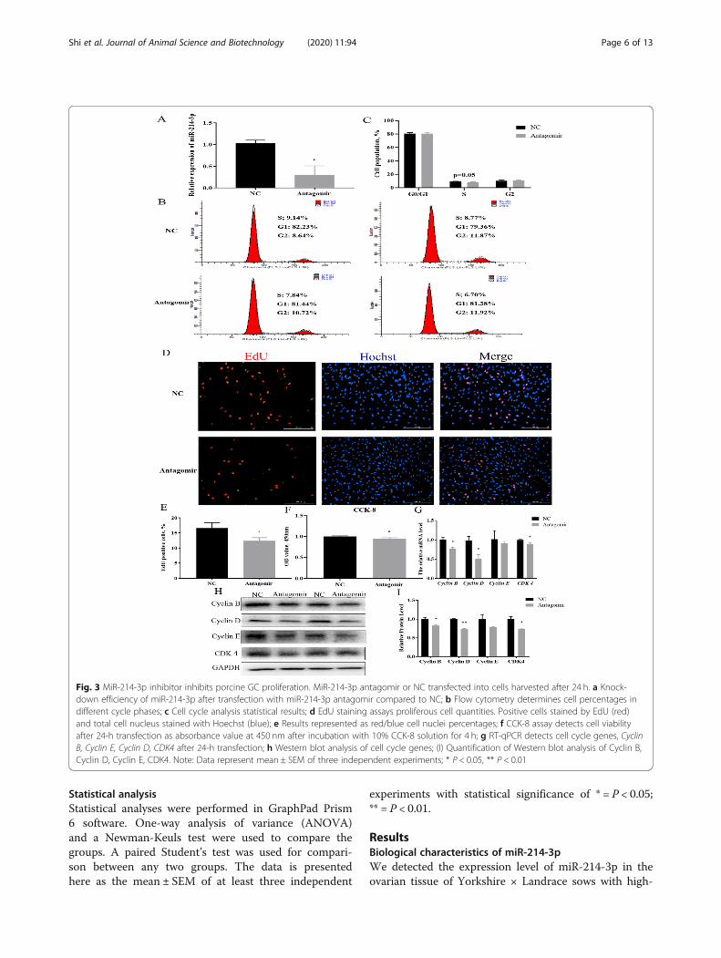
Fig. 3 MiR-214-3p inhibitor inhibits porcine GC proliferation. MiR-214-3p antagomir or NC transfected into cells harvested after 24 h. a Knock-down efficiency of miR-214-3p after transfection with miR-214-3p antagomir compared to NC; b Flow cytometry determines cell percentages indifferent cycle phases; c Cell cycle analysis statistical results; d EdU staining assays proliferous cell quantities. Positive cells stained by EdU (red)and total cell nucleus stained with Hoechst (blue); e Results represented as red/blue cell nuclei percentages; f CCK-8 assay detects cell viabilityafter 24-h transfection as absorbance value at 450 nm after incubation with 10% CCK-8 solution for 4 h; g RT-qPCR detects cell cycle genes, CyclinB, Cyclin E, Cyclin D, CDK4 after 24-h transfection; h Western blot analysis of cell cycle genes; (I) Quantification of Western blot analysis of Cyclin B,Cyclin D, Cyclin E, CDK4. Note: Data represent mean ± SEM of three independent experiments; * P < 0.05, ** P < 0.01
Shi et al. Journal of Animal Science and Biotechnology (2020) 11:94 Page 6 of 13

litter and low-litter characteristics in this study. We ob-served a higher expression in high-litter sows than inlow-litter sows (Fig. 1a). The mature sequence of miR-214-3p is highly conserved across multiple species (e.g.,mouse, pig, human, rat) (Fig. 1b). We also performedGO analysis on the targets of miR-214-3p to find that it
may indeed be involved in follicular growth processessuch as cell proliferation and steroid synthesis (Fig. 1c).The TGF-beta and mTOR signaling pathways play im-portant roles in the process of follicular growth. OurKEGG pathway analysis showed that miR-214-3p partici-pates in these signaling pathways (Fig. 1d).
Fig. 4 MiR-214-3p targets Mfn2 during GC proliferation. a miR-214-3p binding site within the Mfn2 3′-UTR predicted by RNAhybrid; b Target siteof miR-214-3p within porcine Mfn2 mRNA 3’UTR and mutational site of Mfn2 3’UTR; c Dual luciferase assay by co-transfection of miR-214-3pagomir and wild-type vectors or mutant vectors. Relative luciferase activity represented by Renilla Luciferase/Firefly Luciferase (RLUC/FLUC); dRelative Mfn2 mRNA expression levels after treatment with miR-214-3p agomir; e Relative Mfn2 mRNA expression levels after treatment with miR-214-3p antagomir; f Western blot analysis of Mfn2 protein expression after treatment with miR-214-3p agomir; h Western blot analysis of Mfn2protein expression after treatment with miR-214-3p antagomir; g,i Mfn2 protein level quantifications. Note: Data are mean ± SEM of threeindependent experiments. * P < 0.05, ** P < 0.01
Shi et al. Journal of Animal Science and Biotechnology (2020) 11:94 Page 7 of 13

miR-214-3p overexpression promotes granulosa cellproliferationIn order to determine the effect of miR-214-3p on theproliferation of porcine ovarian GCs, we transfected theGCs samples with miR-214-3p agomir, antagomir, and thenegative control. The expression of miR-214-3p increasedsignificantly after transfection into agomir (Fig. 2a). Flowcytometry analysis indicated that miR-214-3p increasedthe percentage of S-phase cells (Fig. 2b,c). The EdU stain-ing assay showed that the number of EdU labeled positivecells increased in the miR-214-3p agomir group, unlike inthe negative control group (Fig. 2d,e). The CCK-8 assayalso up-regulated cell viability (Fig. 2f). In addition, cellcycle-related genes (Cyclin B, Cyclin E, and CDK4)showed remarkably higher mRNA and protein levels butthere was no such effect in cyclin D (Fig. 2g-i).To further explore the effect of miR-214-3p on GC
proliferation, we next treated the cells with antagomir-NC and antagomir. The expression of miR-214-3p in thetreatment group was dramatically reduced below thenegative control group (Fig. 3a). The flow cytometry re-sults indicated down-regulation of the S-phase cells aftersuppressing the expression of miR-214-3p (Fig. 3b,c).Our EdU staining assay showed that inhibition of miR-214-3p can markedly decrease the number of EdU
labeled positive cells (Fig. 3d,e). Our CCK-8 assay alsoverified the knock-down of miR-214-3p induced cell via-bility (Fig. 3f). RT-qPCR and Western blot data showedthat miR-214-3p inhibition depressed the expression ofcell cycle genes (Fig. 3g-i). In summary, miR-214-3p wasfound to promote GC proliferation.
MiR-214-3p targets Mfn2 in GCsThe experiments described above showed that miR-214-3p can promote porcine GC proliferation(Figs. 2 and 3). To better understand the regulatorymechanism of this process, we used TargetScan7.2 andmiRTarBase to predict potential target genes. We detectedMfn2 as a candidate gene from thousands of target genes(Fig. 4a) and constructed wild-type Mfn2 3’UTR and mu-tant Mfn2 3’UTR dual luciferase reporter vectors accord-ingly (Fig. 4b). We found that the dual-luciferase activityof wild-type Mfn2 3’UTR and agomir co-transfected intoGCs was higher than that of co-transfected wild-typeMfn2 3’UTR and NC, while mutant dual-luciferase activitywith NC and agomir appears to have no effect (Fig. 4c).Our RT-qPCR and Western blot data also suggest
that Mfn2 mRNA and protein levels were reducedand increased, respectively, in the miR-214-3p agomirand antagomir groups (Fig. 4d-i). Altogether, our tests
Fig. 5 Overexpression of miR-214-3p inhibits porcine GC estradiol synthesis. MiR-214-3p agomir or NC transfected into cells harvested after 24 h.a Overexpression efficiency of miR-214-3p after transfection with miR-214-3p agomir compared to NC; b Estradiol concentration detected by ELISA. Culture supernatants collected 24 h after miR-214-3p agomir and NC treatment; c RT-qPCR detects E2 synthesis-related genes including Star,Cyp11a1, and Cyp19a1 after 24-h transfection; d Western blot analysis of E2 synthesis-related genes; (E) Quantification of Western blot analysis ofStAR, CYP11A1, CYP19A1. Note: Data are mean ± SEM of three independent experiments; * P < 0.05, ** P < 0.01
Shi et al. Journal of Animal Science and Biotechnology (2020) 11:94 Page 8 of 13

demonstrated that miR-214-3p promotes GC prolifer-ation by directly targeting Mfn2.
Correlation of miR-214-3p with GC estradiol synthesisOne of the most important functions of GCs is the se-cretion of estradiol. We detected the E2 concentration inour culture medium accordingly. The expression ofmiR-214-3p increased or decreased sharply after trans-fection with agomir or antagomir (Figs. 5 and 6a). TheELISA results demonstrated that E2 concentration wasmarkedly down-regulated or up-regulated in differenttreatment groups (Figs. 5 and 6b). E2 synthesis-relatedgenes including Star, Cyp11a1, and Cyp19a1 were alsosuppressed in mRNA and protein levels in the agomirgroup (Fig. 5c-e). The results in the antagomir treatmentgroup were consistent with this (Fig. 6c-e). We infer thatmiR-214-3p inhibits GC estradiol synthesis.
MiR-214-3p directly inhibits NR5A1/SF-1 in GCsNR5A1 is also referred to as “steroidogenic factor 1” (SF-1) and is known to regulate estradiol synthesis by regu-lating the transcription of Cyp11a1 and Cyp19a1 genesvia binding to the nuclear receptor motifs. To explore
the mechanism by which miR-214-3p regulates estradiolsynthesis, we forecasted the target genes of miR-214-3pwith TargetScan7.2 and miRTarBase.Coincidentally, NR5A1/SF-1 is one of the candidate
target genes of miR-214-3p. This caught our attentionover the course of our analysis, so we tested it specific-ally as a target gene of miR-214-3p (Fig. 7a). Similar tothe results reported in Section 3.3, we constructed a dualluciferase reporter vector for assay (Fig. 7b), the assayrevealed that miR-214-3p markedly suppressed the dual-luciferase activity (Fig. 7c). NR5A1/SF-1 mRNA and pro-tein levels were also attenuated and increased in themiR-214-3p agomir and antagomir groups (Fig. 7d-i).These observations suggest that miR-214-3p inhibits GCestradiol synthesis by targeting NR5A1/SF-1.
DiscussionIn this study, we found that miR-214-3p plays an im-portant role in GC proliferation and estradiol synthesis.Specifically, miR-214-3p promotes GC proliferation andinhibits estradiol synthesis. Our findings represent work-able information regarding the regulation of GCs func-tions by miR-214-3p. GC functions such as proliferationand estradiol synthesis are affected by many regulatory
Fig. 6 Inhibitor of miR-214-3p promotes porcine GC estradiol synthesis. MiR-214-3p agomir or NC were transfected into cells which wereharvested after 24 h. a Overexpression efficiency of miR-214-3p after transfection with miR-214-3p antagomir compared to NC; b Estradiolconcentration detected by ELISA. Culture supernatants collected 24 h after miR-214-3p antagomir and NC treatment; c RT-qPCR detects E2synthesis-related genes including Star, Cyp11a1, and Cyp19a1 after 24-h transfection; d Western blot analysis of E2 synthesis-related genes; eQuantification of Western blot analysis of StAR, CYP11A1, CYP19A1. Note: Data are mean ± SEM of three independent experiments; * P < 0.05,** P < 0.01
Shi et al. Journal of Animal Science and Biotechnology (2020) 11:94 Page 9 of 13

factors [29–31]; miRNA plays an important part in theseprocesses [32, 33] and miR-214-3p is expressed to great-est extent in porcine ovarian tissue among other bodytissues [23]. We used high-yield (> 14.7 head/litter) andlow-yield (< 9.3 head/litter) ovary tissues of Yorkshire×-Landrace to verify that miR-214-3p is expressed higherin high-yield sows but not only in Yorkshire [22], whichindicates that miR-214-3p is important for reproduction.Bioinformatics analysis and conservative prediction alsoindicate that miR-214-3p plays a role in regulating GCfunction.
Our experimental results further indicate that miR-214-3p promotes proliferation by upregulating themRNA and protein levels of Cyclin B, Cyclin D, CyclinE, and CDK4 (Figs. 2 and 3g-i). Cyclin B is a marker ofimmunohistochemical proliferation [34] and CDK4 is akinase that regulates the transition from the G1 to Sphases of the cell cycle [35]. We found that due to miR-214-3p agomir and antagomir, compared to our NC,Cyclin B and CDK4 had the most significant differentialexpression of mRNA and protein levels. Flow cytometry,EdU staining, and CCK-8 assays also proved that miR-
Fig. 7 MiR-214-3p targets NR5A1 during GC estradiol synthesis. a miR-214-3p binding site within NR5A1 3′-UTR predicted by RNAhybrid; b targetsite of miR-214-3p within porcine NR5A1 mRNA 3’UTR and mutational site of NR5A1 3’UTR; c Dual luciferase assay via co-transfection of miR-214-3p agomir and wild-type vectors or mutant vectors. Relative luciferase activity represented by RLUC/FLUC; d Relative NR5A1 mRNA expressionlevels after treatment with miR-214-3p agomir; e Relative NR5A1 mRNA expression levels after treatment with miR-214-3p antagomir; f Westernblot analysis of NR5A1 protein expression after treatment with miR-214-3p agomir; h Western blot analysis of NR5A1 protein expression aftertreatment with miR-214-3p antagomir; g, i Quantification of NR5A1 protein levels. Note: Data are mean ± SEM of three independent experiments;* P < 0.05, ** P < 0.01
Shi et al. Journal of Animal Science and Biotechnology (2020) 11:94 Page 10 of 13

214-3p promotes proliferation in GCs (Fig. 2 and Fig. 3),which is consistent with previous research results. Forexample, miR-214-3p regulates the proliferation ofbreast cancer cells by targeting survivin protein [36] andcan promote smooth muscle cell proliferation [37],Mfn2 is regarded as a proliferation inhibitor because it
can limit the expression of Cyclin D protein to inhibit theproliferation process [38]. By RNAhybrid prediction, miR-214-3p binds to the 3′-UTR region of Mfn2 (Fig. 4a).Accordingly, Mfn2 can be used as a candidate target gene ofmiR-214-3p. In the present study, we found that miR-214-3pcan repress the mRNA and protein levels of Mfn2 (Fig. 4d-i).This indicates that Mfn2 is a direct target gene of miR-214-3p via dual-luciferase reporter assay (Fig. 4c) and that Mfn2can perform as a target gene for miR-214-3p to regulate cellproliferation. These results are consistent with previousreports, for instance, where Feng et al. [39] reported thatmiR-93 regulates vascular smooth muscle cell proliferationby targeting Mfn2. Additionally, miR-497 promotes cardio-myocyte proliferation by downregulating the expression ofMfn2 [40].There have been no such results regarding the synthe-
sis of estradiol by miR-214-3p published previously. Ourfindings suggest, however, that miR-214-3p does inhibitthe synthesis of estradiol (Fig. 5 and Fig. 6). During thesynthesis of E2, StAR can transport cholesterol from the
outer to the inner mitochondrial membrane, where it isconverted to pregnenolone by CYP11A1 [41]. Aromatase(CYP19A1) in GCs transforms testosterone into estradiol[41, 42]. We found that miR-214-3p attenuated the tran-scription and translation levels of Star, Cyp11a1, andCyp19a1 (Figs. 5 and 6c-e). These results enrich theexisting knowledge of miR-214-3p in terms of the regu-lation of GC functions.In order to further study the molecular mechanism of
miR-214-3p regulating E2 synthesis in GCs, we selectedNR5A1/SF-1 as the target gene because it can bind toSF-1 response elements on the promoter of target genessuch as Star, Cyp11a1, and Cyp19a1 to regulate theirtranscription activity [43, 44]. NR5A1/SF-1 also ispresent in fetal and adult steroidogenic tissues and partici-pates in the regulation of ovarian function [45]. Therefore,NR5A1/SF-1 may play an important role in E2 synthesis.Our results proved that miR-214-3p attenuates the mRNAand protein levels of NR5A1/SF-1 (Fig. 7d-i), which sug-gests that NR5A1/SF-1 may be a target gene of miR-214-3p in GCs.Our double luciferase reporter assay indicates that
NR5A1/SF-1 is the direct target gene of miR-214-3p(Fig. 7c). These data suggest that miR-214-3p inhibits E2synthesis through NR5A1/SF-1 in GCs. It is worth not-ing that many previous researchers have reached
Fig. 8 Schematic diagram of miR-214-3p regulation on porcine GC proliferation and estradiol synthesis. a MiR-214-3p promotes porcine GCproliferation by targeting Mfn2. b MiR-214-3p inhibits GC estradiol synthesis by targeting NR5A1. Note: Green upward arrows indicate promotionof a given process; red downward arrows indicate that this process is inhibited
Shi et al. Journal of Animal Science and Biotechnology (2020) 11:94 Page 11 of 13

conclusions consistent with ours. For example, in mouseovaries, miR-320 and miR-764-3p regulate estradiol syn-thesis by targeting SF-1 [15, 46, 47].
ConclusionsIn summary, as shown in Fig. 8, our results show thatmiR-214-3p promotes GC proliferation by targetingMfn2 and inhibits GC estradiol synthesis by targetingNR5A1/SF-1. The results presented here may provideworkable insight into regulating the GCs functions, fol-licular growth and development.
AbbreviationsGCs: Granulosa cells; StAR: Steroidogenic acute regulatory protein;CYP11A1: Cytochrome P450 family 11 subfamily A member 1;CYP19A1: Aromatase; Cyclin B: Cell cycle protein B; Cyclin D: Cell cycleprotein D; Cyclin E: Cell cycle protein E; CDK4: Cyclin-dependent kinase 4;mmu: Mus musculus; ssc: Sus scrofa; hsa: Homo sapiens; rno: Rattus norvegicus;mml: Macaca mulatta; mdo: Monodelphis domestica; oan: Ornithorhynchusanatinus; tgu: Taeniopygia guttata; aca: Anolis carolinensis
AcknowledgementsThe authors gratefully acknowledge all the teachers and students inLaboratory of Animal Fat Deposition & Muscle Development.
Authors’ contributionsSSJ and CGY conceived and designed the experiments; SSJ, ZXG and LJJperformed the experiments; HYM and ZLT contributed reagents/materials/analysis tools; YGS managed the project; SSJ wrote the manuscript and CGYmodified the manuscript. The authors read and approved the finalmanuscript.
FundingThis work was supported by grants from the National Natural ScienceFoundation (No.31802047), the National Science and Technology MajorProject of China (No. 2016ZX08006003) and Shaanxi Provincial Key Researchand Development Project (CN)(No. 2018ZDXM-NY-035).
Availability of data and materialsThe data sets used and analysed during the current study are available fromthe corresponding author on reasonable request.
Ethics approval and consent to participateThese studies were approved by Northwest Agriculture and ForestryUniversity Animal Research Ethics Committee (Yangling, Shaanxi, China).
Consent for publicationNot applicable.
Competing interestsThe authors declare they have no competing interest.
Received: 29 March 2020 Accepted: 16 July 2020
References1. Boyer A, Lapointe E, Zheng X, Cowan RG, Li H, Quirk SM, et al. WNT4 is
required for normal ovarian follicle development and female fertility. FasebJ. 2010;24(8):3010–25.
2. Pangas SA, Matzuk MM. Genetic models for transforming growth factorbeta superfamily signaling in ovarian follicle development. Mol CellEndocrinol. 2004;225(1–2):83–91.
3. Wang W, Yin L, Bai L, Ma G, Zhao C, Xiang A, et al. Bmal1 interferenceimpairs hormone synthesis and promotes apoptosis in porcine granulosacells. Theriogenology. 2017;99:63–8.
4. Bai L, Chu G, Mai Y, Zheng J, Wang W, Zhang Q, et al. Identification andexpression analyses of BAMBI mediated by FSH in swine luteinizinggranulosa cells. Theriogenology. 2014;82(8):1094–101.
5. Mani AM, Fenwick MA, Cheng Z, Sharma MK, Singh D, Wathes DC. IGF1induces up-regulation of steroidogenic and apoptotic regulatory genes viaactivation of phosphatidylinositol-dependent kinase/AKT in bovinegranulosa cells. Reproduction. 2010;139(1):139.
6. Zhao GS, Wei HK, Yan JZ, Shan Y, Ning Lu JI, Zheng GU, et al. A novelubiquitin carboxyl terminal hydrolase is involved in toad oocyte maturation.Cell Res. 2002;12(3):199–206.
7. Chen S, Wu RF, Su L, Zhou WD, Zhu MB, Chen QH. Lipoxin A4 regulatesexpression of the estrogen receptor and inhibits 17beta-estradiol inducedp38 mitogen-activated protein kinase phosphorylation in humanendometriotic stromal cells. Fertil Steril. 2014;102(1):264–71.
8. Umer S, Sammad A, Zou H, Khan A, Weldegebriall SB, Hao H, et al.Regulation of AMH, AMHR-II, and BMPs (2,6) genes of bovine granulosa cellstreated with exogenous FSH and their association with protein hormones.Genes (Basel). 2019;10(12):1038.
9. Hershlag A, Lesser M, Montefusco D, Lavy G, Kaplan P, Liu HC, et al.Interinstitutional variability of follicle-stimulating hormone and estradiollevels. Fertil Steril. 1992;58(6):1123–6.
10. Dewailly D, Robin G, Peigne M, Decanter C, Pigny P, Catteau-Jonard S.Interactions between androgens, FSH, anti-Mullerian hormone and estradiolduring folliculogenesis in the human normal and polycystic ovary. HumReprod Update. 2016;22(6):709–24.
11. Tian C, Liu L, Ye X, Fu H, Sheng X, Wang L, et al. Functional oocytes derivedfrom granulosa cells. Cell Rep. 2019;29(13):4256–67.
12. Peluso JJ, Delidow BC, Lynch J, White BA. Follicle-stimulating hormone andinsulin regulation of 17 beta-estradiol secretion and granulosa cellproliferation within immature rat ovaries maintained in perifusion culture.Endocrinology. 1991;128(1):191–6.
13. Shen G, Sun Q, Yao Y, Li S, Liu G, Yuan C, et al. Role of ADAM9 and miR-126 in thedevelopment of abdominal aortic aneurysm. Atherosclerosis. 2020;297:47–54.
14. Ding Q, Jin M, Wang Y, Liu J, Kalds P, Wang Y, et al. Transactivation of miR-202-5p by steroidogenic factor 1 (SF1) induces apoptosis in goat granulosacells by targeting TGFbetaR2. Cells-Basel. 2020;9(2):445.
15. Wang L, Li C, Li R, Deng Y, Tan Y, Tong C, et al. MicroRNA-764-3p regulates17beta-estradiol synthesis of mouse ovarian granulosa cells by targetingsteroidogenic factor-1. In Vitro Cell Dev Biol Anim. 2016;52(3):365–73.
16. Liu J, Li X, Yao Y, Li Q, Pan Z, Li Q. miR-1275 controls granulosa cellapoptosis and estradiol synthesis by impairing LRH-1/CYP19A1 axis. BiochimBiophys Acta Gene Regul Mech. 2018;1861(3):246–57.
17. Desvignes T, Contreras A, Postlethwait JH. Evolution of the miR199–214cluster and vertebrate skeletal development. RNA Biol. 2014;11(4):281–94.
18. Lee YB, Bantounas I, Lee DY, Phylactou L, Caldwell MA, Uney JB. Twist-1regulates the miR-199a/214 cluster during development. Nucleic Acids Res.2009;37(1):123–8.
19. Chen Y, Du H, Bao L, Liu W. LncRNA PVT1 promotes ovarian cancerprogression by silencing miR-214. Cancer Biol Med. 2018;15(3):238–50.
20. Wang J, Yang LZ, Zhang JS, Gong JX, Wang YH, Zhang CL, et al. Effects ofmicroRNAs on skeletal muscle development. GENE. 2018;668:107–13.
21. Xi FX, Wei CS, Xu YT, Ma L, He YL, Shi XE, et al. MicroRNA-214-3p targetingCtnnb1 promotes 3T3-L1 Preadipocyte differentiation by interfering withthe Wnt/beta-catenin signaling pathway. Int J Mol Sci. 2019;20(8).
22. Huang L, Yin ZJ, Feng YF, Zhang XD, Wu T, Ding YY, et al. Identification anddifferential expression of microRNAs in the ovaries of pigs (Sus scrofa) withhigh and low litter sizes. Anim Genet. 2016;47(5):543-51.
23. Tian M, Zhang X, Ye P, Tao Q, Zhang L, Ding Y, et al. MicroRNA-21 andmicroRNA-214 play important role in reproduction regulation duringporcine estrous. Anim Sci J. 2018;89(10):1398–405.
24. Yin L, Wang W, Wei H, Xi F, Chu G, Yang G. Localization and expression ofCTRP6 in ovary and its regulation by FSH in porcine granulosa cells.Theriogenology. 2019;127:56–65.
25. Chu G, Zhou X, Hu Y, Shi S, Yang G. Rev-erbalpha inhibits proliferation andpromotes apoptosis of Preadipocytes through the agonist GSK4112. Int JMol Sci. 2019;20(18).
26. Wang J, Ge J, Cao H, Zhang X, Guo Y, Li X, et al. Leptin promotes White adipocyteBrowning by inhibiting the Hh signaling pathway. Cells-Basel. 2019;8(4):372.
27. Wei H, Li J, Shi S, Zhang L, Xiang A, Shi X, et al. Hhip inhibits proliferationand promotes differentiation of adipocytes through suppressing hedgehogsignaling pathway. Biochem Biophys Res Commun. 2019;514(1):148–56.
28. Huang K, Shi X, Wang J, Yao Y, Peng Y, Chen X, et al. UpregulatedmicroRNA-106a promotes porcine Preadipocyte proliferation anddifferentiation by targeting different genes. Genes (Basel). 2019;10(10).
Shi et al. Journal of Animal Science and Biotechnology (2020) 11:94 Page 12 of 13

29. Wang J, Qiu J, Bo L, Wu Z, Zhou A, Xu W, et al. WT1 influences apoptosisand proliferation of immature mice granular cells through regulation of thewnt/beta-catenin signal pathway. Cell Mol Biol (Noisy-le-Grand). 2019;65(7):138–45.
30. Qin Y, Tang T, Li W, Liu Z, Yang X, Shi X, et al. Bone morphogenetic protein15 knockdown inhibits porcine ovarian follicular development andovulation. Front Cell Dev Biol. 2019;7:286.
31. Liu Y, Yang Y, Li W, Ao H, Zhang Y, Zhou R, et al. Effects of melatonin onthe synthesis of estradiol and gene expression in pig granulosa cells. JPineal Res. 2019;66(2):e12546.
32. Pande HO, Tesfaye D, Hoelker M, Gebremedhn S, Held E, Neuhoff C, et al.MicroRNA-424/503 cluster members regulate bovine granulosa cellproliferation and cell cycle progression by targeting SMAD7 gene throughactivin signalling pathway. J Ovarian Res. 2018;11(1):34.
33. Li Q, Du X, Pan Z, Zhang L, Li Q. The transcription factor SMAD4 and miR-10b contribute to E2 release and cell apoptosis in ovarian granulosa cells bytargeting CYP19A1. Mol Cell Endocrinol. 2018;476:84–95.
34. Koliadi A, Nilsson C, Holmqvist M, Holmberg L, de La Torre M, Warnberg F,et al. Cyclin B is an immunohistochemical proliferation marker which canpredict for breast cancer death in low-risk node negative breast cancer.Acta Oncol. 2010;49(6):816–20.
35. Grison A, Gaiser C, Bieder A, Baranek C, Atanasoski S. Ablation of cdk4 andcdk6 affects proliferation of basal progenitor cells in the developing dorsaland ventral forebrain. Dev Neurobiol. 2018;78(7):660–70.
36. Han LC, Wang H, Niu FL, Yan JY, Cai HF. Effect miR-214-3p on proliferationand apoptosis of breast cancer cells by targeting survivin protein. Eur RevMed Pharmacol Sci. 2019;23(17):7469–74.
37. Xing XQ, Li B, Xu SL, Liu J, Zhang CF, Yang J. MicroRNA-214-3p regulateshypoxia-mediated pulmonary artery smooth muscle cell proliferation andmigration by targeting ARHGEF12. Med Sci Monit. 2019;25:5738–46.
38. Liu X, Sun J, Yuan P, Shou K, Zhou Y, Gao W, et al. Mfn2 inhibitsproliferation and cell-cycle in Hela cells via Ras-NF-kappaB signal pathway.Cancer Cell Int. 2019;19:197.
39. Feng S, Gao L, Zhang D, Tian X, Kong L, Shi H, et al. MiR-93 regulatesvascular smooth muscle cell proliferation, and neointimal formationthrough targeting Mfn2. Int J Biol Sci. 2019;15(12):2615–26.
40. Qin L, Yang W, Wang YX, Wang ZJ, Li CC, Li M, et al. MicroRNA-497promotes proliferation and inhibits apoptosis of cardiomyocytes throughthe downregulation of Mfn2 in a mouse model of myocardial ischemia-reperfusion injury. Biomed Pharmacother. 2018;105:103–14.
41. Hebert-Schuster M, Rotta BE, Kirkpatrick B, Guibourdenche J, Cohen M. Theinterplay between glucose-regulated protein 78 (GRP78) and steroids in thereproductive system. Int J Mol Sci. 2018;19(7).
42. Bai L, Chu G, Wang W, Xiang A, Yang G. BAMBI promotes porcine granulosa cellsteroidogenesis involving TGF-beta signaling. Theriogenology. 2017;100:24–31.
43. Lai WA, Yeh YT, Fang WL, Wu LS, Harada N, Wang PH, et al. Calcineurin andCRTC2 mediate FSH and TGFbeta1 upregulation of Cyp19a1 and Nr5a inovary granulosa cells. J Mol Endocrinol. 2014;53(2):259–70.
44. Wang J, Gong Y. Transcription of CYP19A1 is directly regulated by SF-1 in thetheca cells of ovary follicles in chicken. Gen Comp Endocrinol. 2017;247:1–7.
45. Mlynarczuk J, Wrobel MH, Rekawiecki R, Kotwica J. The expression ofsteroidogenic factor-1 and its role in bovine steroidogenic ovarian cellsduring the estrus cycle and first trimester of pregnancy. Anim Reprod Sci.2013;138(1–2):74–81.
46. Yin M, Lu M, Yao G, Tian H, Lian J, Liu L, et al. Transactivation of microRNA-383 by steroidogenic factor-1 promotes estradiol release from mouseovarian granulosa cells by targeting RBMS1. Mol Endocrinol. 2012;26(7):1129–43.
47. Yin M, Wang X, Yao G, Lu M, Liang M, Sun Y, et al. Transactivation ofmicroRNA-320 by microRNA-383 regulates granulosa cell functions bytargeting E2F1 and SF-1 proteins. J Biol Chem. 2014;289(26):18239–57.
Shi et al. Journal of Animal Science and Biotechnology (2020) 11:94 Page 13 of 13