mirna-106a directly targeting rarb associates with the ... · regulating ask1-p38 pathway....
TRANSCRIPT
RESEARCH Open Access
miRNA-106a directly targeting RARBassociates with the expression of Na+/I−
symporter in thyroid cancer by regulatingMAPK signaling pathwayChen-Tian Shen, Zhong-Ling Qiu*, Hong-Jun Song, Wei-Jun Wei and Quan-Yong Luo*
Abstract
Background: Serum miRNAs profiles between papillary thyroid carcinoma (PTC) patients with non-131I and 131I-avidlung metastases are differentially expressed. These miRNAs have to be further validated and the role of these miRNAsin the molecular function level of thyroid cancer cell lines has not been investigated.
Methods: Expression levels of six identified miRNAs were assessed via quantitative real-time PCR (qRT-PCR) in theserum of eligible patients. Dual-luciferase reporter assay was used to determine the potential target of miR-106a.Cell viability and apoptosis were evaluated by MTT assay and flow cytometry analysis, respectively. The change ofgene expression was detected by qRT-PCR and western blotting analysis. In vitro iodine uptake assay wasconducted by a γ-counter.Results: Compared to PTC patients with 131I-avid lung metastases, miR-106a was up-regulated in the serum of patientswith non-131I-avid lung metastases. The results of dual-luciferase reporter assay demonstrated that miR-106a directlytargeted retinoic acid receptor beta (RARB) 3′-UTR. miR-106a-RARB promoted viability of thyroid cancer cells byregulating MEKK2-ERK1/2 and MEKK2-ERK5 pathway. miR-106a-RARB inhibited apoptosis of thyroid cancer cells byregulating ASK1-p38 pathway. Moreover, miR-106a-RARB could regulate the expression of sodium iodide symporter,TSH receptor and alter the iodine uptake function of thyroid cancer cells.
Conclusions: miRNA-106a, directly targeting RARB, associates with the viability, apoptosis, differentiation and theiodine uptake function of thyroid cancer cell lines by regulating MAPK signaling pathway in vitro. These findingsin the present study may provide new strategies for the diagnosis and treatment in radioiodine-refractorydifferentiated thyroid carcinoma.
Keywords: miR-106a, RARB, Sodium-iodide symporter, Thyroid cancer, MAPK signaling pathway
BackgroundDifferentiated thyroid cancer (DTC) is increasing all overthe world [1], as an indolent tumor, DTC patients haveexcellent prognosis following conventional treatmentsbased on adequate surgical management, radioactiveiodine (RAI) ablation and thyroid-stimulating hormone(TSH) suppression [2, 3]. Radioiodine is considered to
be the initial systemic and efficient treatment formetastatic DTC patients. Unfortunately, approxi-mately 30 % of patients with advanced, metastaticDTC have radioiodine-refractory disease [4]. Losingthe ability to concentrate radioiodine in metastaticsites from DTC most likely owns to less differenti-ated types transformation (dedifferentiation) [5]. Thisproblem creates a major obstacle in radioiodinetreatment for those patients while the mechanismsunderlying the dedifferentiation transformation ofDTC are still not well understood.
* Correspondence: [email protected]; [email protected] of Nuclear Medicine, Shanghai Jiao Tong University AffiliatedSixth People’s Hospital, 600 Yishan Road, Shanghai 200233, People’s Republicof China
© 2016 The Author(s). Open Access This article is distributed under the terms of the Creative Commons Attribution 4.0International License (http://creativecommons.org/licenses/by/4.0/), which permits unrestricted use, distribution, andreproduction in any medium, provided you give appropriate credit to the original author(s) and the source, provide a link tothe Creative Commons license, and indicate if changes were made. The Creative Commons Public Domain Dedication waiver(http://creativecommons.org/publicdomain/zero/1.0/) applies to the data made available in this article, unless otherwise stated.
Shen et al. Journal of Experimental & Clinical Cancer Research (2016) 35:101 DOI 10.1186/s13046-016-0377-0
It is well accepted that constitutive activation of mitogen-activated protein kinase (MAPK) signaling pathway playsa significant role in the tumorigenesis of thyroid carcin-oma and it also could promote the dedifferentiation ofthyroid-cancer cells. Regarding to this fact, disease-specific molecular targets of therapy is studied muchpopularly [6]. MAP3K2 (MEKK2) is a serine/threoninekinase that belongs to the MEKK/STE11 family ofMAP kinase kinase kinases, which can activate JNK1/2[7], p38 [8], ERK5 [9] and ERK1/2 [10] pathways. How-ever, its role in thyroid cancer has not been clearlystudied by now.Recently, the involvement of miRNAs in proliferation,
differentiation and apoptosis has been defined and sev-eral reports have displayed the changes in miRNA pro-files in differentiated thyroid cancers as compared tonormal thyroid tissues [11–15]. But the role of miRNAsin the differentiation/dedifferentiation of DTC, especiallyin the expression of sodium iodide symporter (NIS) andNIS-mediated iodine uptake, is not clearly understood.Lakshmanan et al. found that miR-339-5p directly boundto hNIS-3′UTR and miR-339-5p overexpression de-creased NIS-mediated radioiodine uptake in HEK293cells expressing exogenous hNIS [16].One of our previous studies analyzed the differentially
expressed serum miRNAs profiles between papillary thy-roid carcinoma (PTC) patients with non-131I and 131I-avid lung metastases [17]. But the function of these miR-NAs in thyroid cancer has not been reported. In thecurrent study, the role of miRNA-106a in the viability,apoptosis, migration, invasion and differentiation (fo-cused on the expression of NIS and TSH receptor andthe ability of iodine uptake) of thyroid cancer cell lineswas investigated.
MethodsSerum samples and cell cultureThe serum samples of PTC patients with 131I-avid andnon-131I-avid lung metastases were collected fromSeptember 2010 to July 2014 at our department. The in-clusion/exclusion criteria and the method to process thesamples have been described before [17]. Total RNAfrom serum sample was extracted and purified using themiRNeasy Mini Kit (Qiagen, Valencia, CA) according tothe manufacturer’s instructions. The study was approvedby the Institutional Ethics Review Board of our hospitaland informed consent was obtained from each patient.Human PTC cell line CGTH-W3 (Institute of Bio-
chemistry and Cell Biology, SIBS, CAS, Shanghai, China)and anaplastic thyroid carcinoma (ATC) cell line 8505C(Institute of Biochemistry and Cell Biology, SIBS, CAS,Shanghai, China) were cultured in RPMI-1640 (Gibco,Cat. # 11875–093) supplemented with 10 % fetal bovineserum (Gibco, Cat. # 16000–044). HEK 293 T (Institute
of Biochemistry and Cell Biology, SIBS, CAS, Shanghai,China) was maintained in Deulbecco’s Modified Eaglemedium (Gibco, Cat. # 10565–018) supplemented with10 % fetal bovine serum (Gibco, Cat. # 16000–044). Allthe cells were incubated at 37 °C in a humidified cham-ber supplemented with 5 % CO2. U0126 and SB203580were obtained from Selleck Chemicals (Houston, TX,Cat. # S1102 and S1076). All-trans retinoic acid (RA)was obtained from Sigma-Aldrich (Cat. # R2625).
Different managements of cells8505C-miR106a(−) and 8505C-scrambled controlmiArrest™ miRNA inhibitor (GeneCopoeia, Cat. # HmiR-AN0026-AM03), scrambled control (GeneCopoeia, Cat. #CmiR-AN0001-AM03) and Lenti-Pac™ HIV ExpressionPackaging Kit (GeneCopoeia, Cat. # HPK-LvTR-20) wereused to construct Lenti-Pac-miRNA-106a inhibitor andLenti-Pac- scrambled control. After transfected to pack-aging cells (HEK 293T), the medium was collected andcentrifuged at 4,000 × g for 10 min at room temperatureto pellet cell debris, and then filtered through a 0.45 μmfilter. Target cells (8505C) were transfected with Lenti-Pac-miRNA-106a inhibitor [8505C-miR106a(−)] or Lenti-Pac-scrambled control (8505C-scrambled control).
8505C-miR106a(−) + RARB(−)8505C-miR106a(−) cells were transfected with Humanretinoic acid receptor beta (RARB) Silencer® SelectsiRNA (Ambion, target sequence: TCAGACGGCCT-TACCCTAAAT, Cat. # 4392420) using Lipofectamine2000 (Invitrogen, USA, Cat. # 11668019) following themanufacturer’s instructions.
8505C-miR106a(−) + ASK1(−)Selective inhibitor of ASK1, (2,7-dihydro-2,7-dioxo-3H-naphtho [1,2,3-de] quinoline-1-carboxylic acid ethyl ester;NQDI-1) (Sigma, Cat. # SML0185), was used to inhibitASK1 by pretreating the 8505C-miR106a (−) cells.
8505C-miR106a(−) + MAP3K2(+)8505C-miR106a(−) cells were transiently transfectedwith MAP3K2 (NM_006609) Human cDNA ORF Clone(OriGene, Cat. # RC223988) using 100 μl Opti-MEM I(Gibco, Cat. # 51985091) and 3 μL Turbofectin 8.0 (Ori-Gene, Cat. # TF81001).
CGTH-W3-miR106a(+) and CGTH-W3-control vectorThe amplified human miR-106a gene was digested withBamHI and EcoRI restriction enzymes and was clonedinto the lentiviral vector pCDH-CMV-MCS-EF1-copGFP(System Biosciences, Cat. # CD511B-1). The pCDH-miR106a lentiviral vector was transformed into E. coli DH5α,then pCDH-miR106a was purified using a plasmid kit(Qiagen, Cat. # 12143) according to the manufacturer’s
Shen et al. Journal of Experimental & Clinical Cancer Research (2016) 35:101 Page 2 of 12
instructions. Then pCDH-miR106a was packaged intoHEK 293 T cells with the pPACK packaging mix using alentivector expression system (System Biosciences) accord-ing to the manufacturer’s instructions. After transfection topackaging cells for 48 h, the medium was collected andcentrifuged at 4,000 × g for 10 min at room temperature topellet cell debris, and then filtered through a 0.45 μm filter.Target cells (CGTH-W3) were transfected with Lenti-pCDH-miR106a [CGTH-W3-miR106a(+)] or Lenti-pCDH(CGTH-W3-control vector).
CGTH-W3-RARB(−)CGTH-W3 cells were transfected with Human RARB Si-lencer® Select siRNA (Ambion, target sequence: TCA-GACGGCCTTACCCTAAAT) using Lipofectamine 2000(Invitrogen, Cat. # 11668019) following the manufac-turer’s instructions.
Dual-luciferase reporter assayA fragment of 3′UTR of RARB (NM_000965) containingthe putative miR-106a binding sites was amplified byPCR using the following primers:
wt- RARB (forward): 5′- GGGTACCCCTACTTCAAACATTCCCCAG-3′;wt- RARB (reverse): 5′-CCCTCGAGGGTGAGAACTAAGAAACTGACA-3′;3′UTR of RARB with a mutant seed sequence ofmiR-106a was synthesized using the following:mut-RARB (forward): 5′-GGGTACCCTTCAAACATTCCCCAGTACCTTCAGT-3′;mut-RARB(reverse): 5′-CCCTCGAGGGTTTTAATTTAAGCGCACATTAACAAT-3′;
Then pGL3-RARB 3′UTR–wt, pGL3-RARB 3′UTR-mut vectors were constructed. For the reporter assay,HEK 293T cells were plated into 24-well plates and co-transfected with the above constructs and miR-106amimics/miR-negative controls using the Lipofectamine2000 reagent (Invitrogen, Cat. # 11668019). After 48 h,the cells were harvested and assayed using the dual-Luciferase Reporter Assay system (Promega, Cat. #E1910) according to the manufacturer’s instructions.
MTT: cell viability assaysThe cell viability were evaluated using 3-(4, 5-dimethylthiazol-2-yl) 22, 5-diphenyltetrazolium brom-ide (MTT) assay. Cells were seeded in sextuplicate in96-well microtiter plates at a density of 1 × 10⁴ cells/well in 100 μL medium. The plates were incubated in a37 °C humidified incubator for adherence overnight.Then after 0, 24, 48, 72 and 96 h culture, 20 μL of 5 g/L MTT (Amresco, Cat. # 0793-500MG) was added, re-spectively. The medium was removed after 4 h, and the
reaction was then stopped by the addition of DMSOand measured at A570 in a Microplate spectrophotom-eter (Spectra Max Plus, Molecular Devices, Sunnyvale,CA). The results were expressed as percentage, basedon the ratio of the absorbance between the treated cellsand the controls (100 %). Experiments were repeatedthree times.
Apoptosis flow cytometry analysisApoAlert Annexin V-FITC kit (Clontech, Cat. # 630109)was used to assess the cell apoptosis. Parental CGTH-W3 and 8505C cells and transfected sublines wereseeded in 6-well plates at 1 × 10⁵ per well. Cells wereharvested 72 h later and stained with Annexin V-FITCand propidium iodide according to the manufacturer’sprotocol. Cell samples were analyzed on a FACScanAnalyzer and apoptotic fractions were determined. Ex-periments were repeated three times.
Measurement of caspase-3 activityThe caspase-3 activity in parental CGTH-W3 and 8505Ccells and transfected sublines were measured by usingCaspase-3 Activity Assay Kit (Beyotime Biotech, Cat. #C1116) according to the manufacturer’s instructions. Theassay is based on the hydrolysis of the peptide substrateacetyl-Asp-Glu-Val-Asp p-nitroanilide (Ac-DEVD-pNA)by caspase-3, resulting in the release of a pNA moiety. Ab-sorbance values were measured at 405 nm. Results wereadjusted to the total protein content, and activity wasexpressed as μmol pNA/h/mg of total protein.
Scratch-wound migration and transwell invasion assaysWound healing assays were used to determine cell mi-gration. Briefly, cells grown in 6-well plates as confluentmonolayers were mechanically scratched by using a200 μL pipette tip and then washed with PBS to removethe debris. Cells were cultured for 24 h to allow woundhealing. Each scratch-wound area was calculated usingthe ImageProPlus 6.0 program (Media Cybernetics Inc.,Bethesda, MD). Transwell invasion assays were per-formed with Matrigel (BD Biosciences) coated on theupper surface of the transwell chamber (Corning).Twenty four hours later, cells invaded through theMatrigel membrane were fixed with 4 % paraformalde-hyde and stained with crystal violet. The number of in-vaded cells was counted for analysis.
RNA extraction and quantitative real-time polymerasechain reaction (qRT-PCR) analysisTotal RNA in cultured cells was isolated using Trizol re-agent (Invitrogen, Cat. # 15596–026) following the manu-facturer’s instructions, and stored at −80 °C. RevertAidTM
First Strand cDNA Synthesis Kit (Fermentas, Cat. #K1622) was used for reverse transcription. qRT-PCR was
Shen et al. Journal of Experimental & Clinical Cancer Research (2016) 35:101 Page 3 of 12
performed in the ABI PRISM 7500 Sequence DetectionSystem (Applied Biosystems, Foster City, CA) using theSYBR Green RT-PCR kit (Qiagen, Cat. # 204147). Allvalues were normalized using an internal reference (U6,for miRNAs; and GAPDH, for mRNAs). Relative expres-sion was estimated by the comparative Ct method (2-ΔΔCt)[18]. A 2-ΔΔCt >3 or < 0.3 was deemed to indicate statis-tical significance.
Western blot analysisEqual amounts of cell lysates were separated by 12 % SDS-PAGE, and electrophoretically transferred to PVDF mem-brane. The membrane was blocked and probed with pri-mary antibody (anti-ASK1, anti-phospho-ASK1, anti-p38,anti-phospho-p38, anti-ERK1/2, anti-phospho-ERK1/2,anti-ERK5, anti-phospho- ERK5, anti-JNK, anti-phospho-JNK from Cell Signaling Technology; anti-MEKK2, anti-NIS, anti-TSHR from Santa Cruz; anti-β-actin from Sigma)followed by HRP (horseradish peroxidase)-labeled goatanti-mouse IgG (Abcam) or HRP-labeled goat anti-rabbitIgG (Abcam). Chemiluminescence was used to analyze pro-tein levels and β-actin was used as a protein loading con-trol. Semi-quantitative analysis was conducted by usingImageJ 1.49v (National Institutes of Health, USA).
In vitro iodine uptake assayParental CGTH-W3 and 8505C cells and transfected sub-lines were seeded in 24-well plates at 5 × 10⁴ per well overnight. After washed with 1 mL HBSS twice, 1 mL HBSScontaining 0.1 μCi Na125I and 1 μmol/L NaI was added.After 30 min at 37 °C in a humid atmosphere, cells werecollected and washed with ice-cold HBSS. Radioactivitywas counted in a γ-counter.
Statistics analysisComparisons of continuous variables between two groupswere performed using the Student’s t test while categoricalvariables were performed using the Chi-square test. (a P <0.05 was considered a statistically significant difference).Analyses were performed using the Statistical Package forthe Social Sciences, version 20.0 (SPSS, Chicago, IL, USA)and Graph Pad prism Version 5.0 (GraphPad Software,Inc, USA).
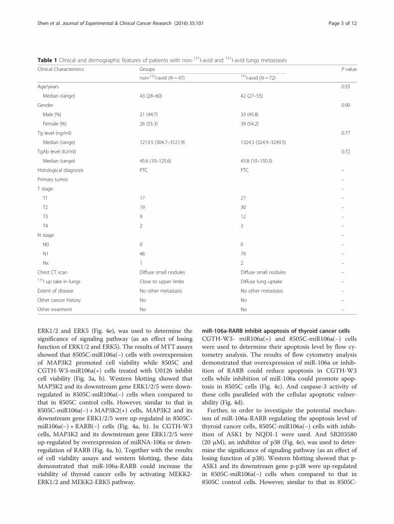
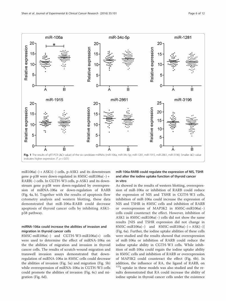
ResultsmiR-106a is up-regulated in the serum of patients withnon-131I-avid lung metastasesForty seven PTC patients with non-131I-avid lung metas-tases (A) and 72 with 131I-avid lung metastases (B) wereidentified in our institution. Demographic and clinicalfeatures of these patients are summarized in Table 1. Sixcandidate miRNAs (miR-106a, miR-34c-5p, miR-1281,miR-1915, miR-2861, miR-3196) which were most chan-ged between the serum of patients with non-131I-avid
lung metastases or 131I-avid lung metastases [12] werevalidated in the current study. The results of qRT-PCRconfirmed the up-regulation of miRNA-106a (P < 0.01).However, the expressions of the other five miRNAs hadno significant differences between the two categories ofpatents (Fig. 1).
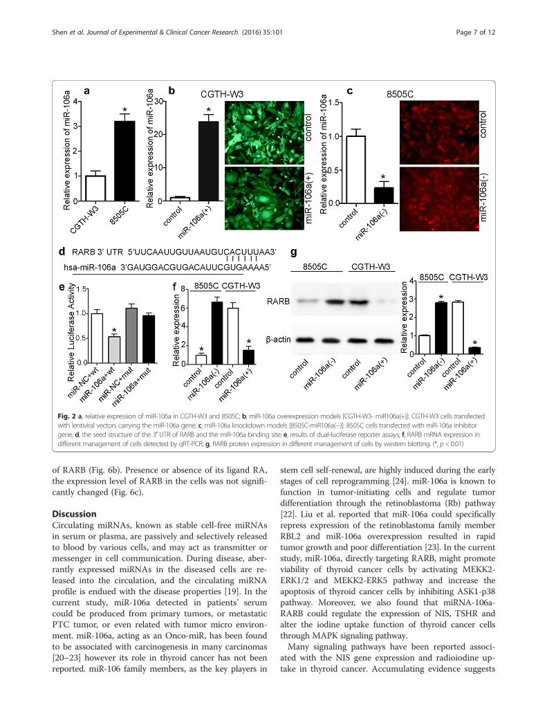
miR-106a is differently expressed in different thyroidcancer cell lines and altered after transfectionExpression levels of miR-106a were detected in CGTH-W3 and 8505C cells by qRT-PCR. The result showedthat the level of miR-106a in 8505C was up-regulatedwhen compared to CGTH-W3 (Fig. 2a). miR-106a over-expression models in CGTH-W3 cells [CGTH-W3- miR106a(+)] and miR-106a knockdown models in 8505Ccells [8505C-miR106a(−)] were established by infectedwith lentiviral vectors carrying the miR-106a gene andmiR-106a inhibitor gene, respectively (Fig. 2b-c). The dataof qRT-PCR indicated that levels of miR-106a in CGTH-W3- miR106a(+) cells was significantly up-regulated whilethe level of miR-106a in 8505C-miR106a(−) cells was sig-nificantly down-regulated.
miR-106a directly targets RARB 3′-UTR (miR-106a-RARB)The 3′-UTR of RARB mRNA contains a complementarysite for the seed region of miR-106a (Fig. 2d). To deter-mine whether RARB is a direct target of miR-106a, theRARB 3′-UTR and the mutant containing the miR-106abinding sites were subcloned into a reporter vectordownstream of the luciferase gene. Dual-luciferase re-porter assays showed that the relative luciferase activityof the reporter that contained wild-type 3′-UTR ofRARB mRNA was significantly decreased in miR-106a-overexpressing cells compared with control cells. How-ever, mutation of the predicted binding site of miR-106aon the RARB 3′-UTR rescued the luciferase activity(Fig. 2e). Furthermore, the results of qRT-PCR and west-ern blotting showed that overexpression of miR-106asignificantly decreased the expression level of RARB,whereas inhibition of miR-106a induced reduction ofRARB mRNA and protein (Fig. 2f, g).
miR-106a-RARB promote the viability of thyroid cancerin vitroThe results of MTT assays demonstrated that overex-pression of miR-106a or inhibition of RARB could pro-mote cell viability in CGTH-W3 cells while inhibition ofmiR-106a could suppress cell viability in 8505C cells(Fig. 3a, b) and the reduced proliferation effect could re-sult from cell cycle arrest (Fig. 3c).In order to investigate the potential mechanism of miR-
106a-RARB regulating the viability of thyroid cancer cells,8505C-miR106a(−) cells with overexpression of MAP3K2(MEKK2) were used. And U0126 (10 μM), an inhibitor of
Shen et al. Journal of Experimental & Clinical Cancer Research (2016) 35:101 Page 4 of 12
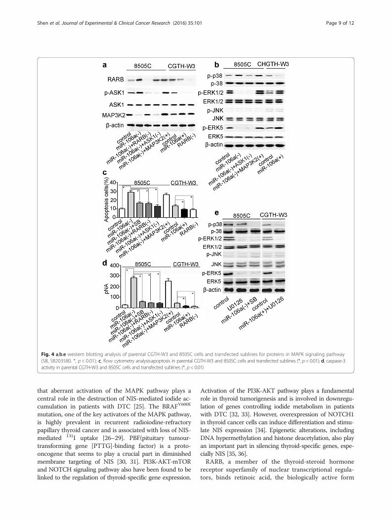
ERK1/2 and ERK5 (Fig. 4e), was used to determine thesignificance of signaling pathway (as an effect of losingfunction of ERK1/2 and ERK5). The results of MTTassaysshowed that 8505C-miR106a(−) cells with overexpressionof MAP3K2 promoted cell viability while 8505C andCGTH-W3-miR106a(+) cells treated with U0126 inhibitcell viability (Fig. 3a, b). Western blotting showed thatMAP3K2 and its downstream gene ERK1/2/5 were down-regulated in 8505C-miR106a(−) cells when compared tothat in 8505C control cells. However, similar to that in8505C-miR106a(−) +MAP3K2(+) cells, MAP3K2 and itsdownstream gene ERK1/2/5 were up-regulated in 8505C-miR106a(−) + RARB(−) cells (Fig. 4a, b). In CGTH-W3cells, MAP3K2 and its downstream gene ERK1/2/5 wereup-regulated by overexpression of miRNA-106a or down-regulation of RARB (Fig. 4a, b). Together with the resultsof cell viability assays and western blotting, these datademonstrated that miR-106a-RARB could increase theviability of thyroid cancer cells by activating MEKK2-ERK1/2 and MEKK2-ERK5 pathway.
miR-106a-RARB inhibit apoptosis of thyroid cancer cellsCGTH-W3- miR106a(+) and 8505C-miR106a(−) cellswere used to determine their apoptosis level by flow cy-tometry analysis. The results of flow cytometry analysisdemonstrated that overexpression of miR-106a or inhib-ition of RARB could reduce apoptosis in CGTH-W3cells while inhibition of miR-106a could promote apop-tosis in 8505C cells (Fig. 4c). And caspase-3 activity ofthese cells paralleled with the cellular apoptotic vulner-ability (Fig. 4d).Further, in order to investigate the potential mechan-
ism of miR-106a-RARB regulating the apoptosis level ofthyroid cancer cells, 8505C-miR106a(−) cells with inhib-ition of ASK1 by NQDI-1 were used. And SB203580(20 μM), an inhibitor of p38 (Fig. 4e), was used to deter-mine the significance of signaling pathway (as an effect oflosing function of p38). Western blotting showed that p-ASK1 and its downstream gene p-p38 were up-regulatedin 8505C-miR106a(−) cells when compared to that in8505C control cells. However, similar to that in 8505C-
Table 1 Clinical and demographic features of patients with non-131I-avid and 131I-avid lungs metastases
Clinical Characteristics Groups P value
non-131I-avid (N = 47) 131I-avid (N = 72)
Age/years 0.55
Median (range) 43 (28–60) 42 (27–55)
Gender 0.90
Male (%) 21 (44.7) 33 (45.8)
Female (%) 26 (55.3) 39 (54.2)
Tg level (ng/ml) 0.77
Median (range) 1213.5 (304.7–3121.9) 1324.3 (324.9–3249.5)
TgAb level (IU/ml) 0.72
Median (range) 45.6 (10–125.6) 43.8 (10–150.3)
Histological diagnosis PTC PTC –
Primary tumor –
T stage –
T1 17 27 –
T2 19 30 –
T3 9 12 –
T4 2 3 –
N stage –
N0 0 0 –
N1 46 70 –
Nx 1 2 –
Chest CT scan Diffuse small nodules Diffuse small nodules –131I up take in lungs Close to upper limbs Diffuse lung uptake –
Extent of disease No other metastasis No other metastasis –
Other cancer history No No –
Other treatment No No –
Shen et al. Journal of Experimental & Clinical Cancer Research (2016) 35:101 Page 5 of 12
miR106a(−) + ASK1(−) cells, p-ASK1 and its downstreamgene p-p38 were down-regulated in 8505C-miR106a(−) +RARB(−) cells. In CGTH-W3 cells, p-ASK1 and its down-stream gene p-p38 were down-regulated by overexpres-sion of miRNA-106a or down-regulation of RARB(Fig. 4a, b). Together with the results of apoptosis flowcytometry analysis and western blotting, these datademonstrated that miR-106a-RARB could decreaseapoptosis of thyroid cancer cells by inhibiting ASK1-p38 pathway.
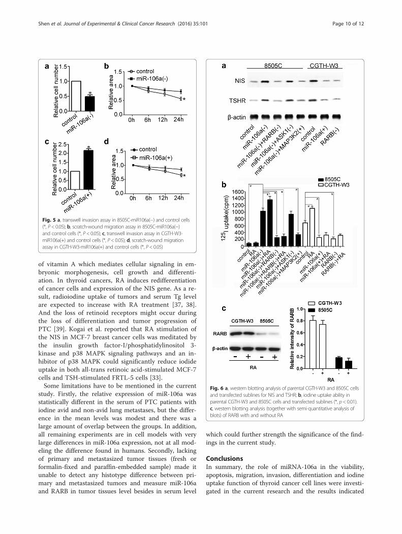
miRNA-106a could increase the abilities of invasion andmigration in thyroid cancer cells8505C-miR106a(−) and CGTH-W3-miR106a(+) cellswere used to determine the effect of miRNA-106a onthe the abilities of migration and invasion in thyroidcancer cells. The results of scratch-wound migration andtranswell invasion assays demonstrated that down-regulation of miRNA-106a in 8505C cells could decreasethe abilities of invasion (Fig. 5a) and migration (Fig. 5b)while overexpression of miRNA-106a in CGTH-W3 cellscould promote the abilities of invasion (Fig. 6c) and mi-gration (Fig. 6d).
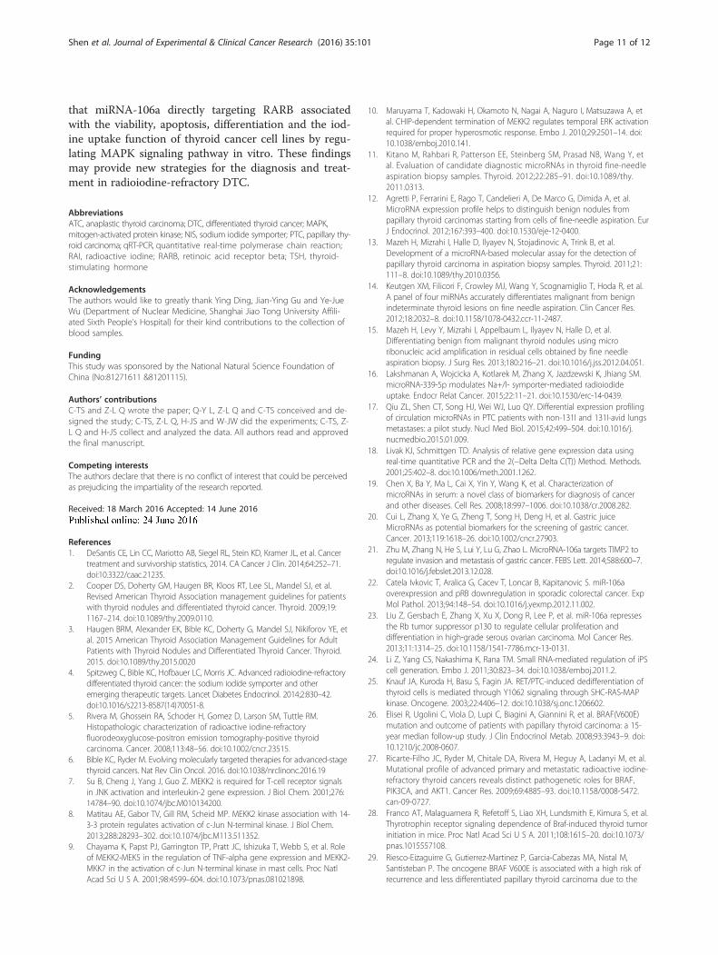
miR-106a-RARB could regulate the expression of NIS, TSHRand alter the iodine uptake function of thyroid cancerin vitroAs showed in the results of western blotting, overexpres-sion of miR-106a or inhibition of RARB could reducethe expression of NIS and TSHR in CGTH-W3 cells,inhibition of miR-106a could increase the expression ofNIS and TSHR in 8505C cells and inhibition of RARBor overexpression of MAP3K2 in 8505C-miR106a(−)cells could counteract the effect. However, inhibition ofASK1 in 8505C-miR106a(−) cells did not show the sameresults [NIS and TSHR expression did not change in8505C-miR106a(−) and 8505C-miR106a(−) + ASK(−)](Fig. 6a). Further, the iodine uptake abilities of these cellswere studied and the results showed that overexpressionof miR-106a or inhibition of RARB could reduce theiodine uptake ability in CGTH-W3 cells. While inhib-ition of miR-106a could regain the iodine uptake abilityin 8505C cells and inhibition of RARB or overexpressionof MAP3K2 could counteract the effect (Fig. 6b). Inaddition, the influence of RA, the ligand of RARB, on125I uptake in these models was also studied and the re-sults demonstrated that RA could increase the ability ofiodine uptake in thyroid cancer cells under the existence
Fig. 1 The results of qRT-PCR (ΔCt value) of the six candidate miRNAs (miR-106a, miR-34c-5p, miR-1281, miR-1915, miR-2861, miR-3196). Smaller ΔCt valueindicates higher expression. (*, p< 0.01)
Shen et al. Journal of Experimental & Clinical Cancer Research (2016) 35:101 Page 6 of 12
of RARB (Fig. 6b). Presence or absence of its ligand RA,the expression level of RARB in the cells was not signifi-cantly changed (Fig. 6c).
DiscussionCirculating miRNAs, known as stable cell-free miRNAsin serum or plasma, are passively and selectively releasedto blood by various cells, and may act as transmitter ormessenger in cell communication. During disease, aber-rantly expressed miRNAs in the diseased cells are re-leased into the circulation, and the circulating miRNAprofile is endued with the disease properties [19]. In thecurrent study, miR-106a detected in patients’ serumcould be produced from primary tumors, or metastaticPTC tumor, or even related with tumor micro environ-ment. miR-106a, acting as an Onco-miR, has been foundto be associated with carcinogenesis in many carcinomas[20–23] however its role in thyroid cancer has not beenreported. miR-106 family members, as the key players in
stem cell self-renewal, are highly induced during the earlystages of cell reprogramming [24]. miR-106a is known tofunction in tumor-initiating cells and regulate tumordifferentiation through the retinoblastoma (Rb) pathway[22]. Liu et al. reported that miR-106a could specificallyrepress expression of the retinoblastoma family memberRBL2 and miR-106a overexpression resulted in rapidtumor growth and poor differentiation [23]. In the currentstudy, miR-106a, directly targeting RARB, might promoteviability of thyroid cancer cells by activating MEKK2-ERK1/2 and MEKK2-ERK5 pathway and increase theapoptosis of thyroid cancer cells by inhibiting ASK1-p38pathway. Moreover, we also found that miRNA-106a-RARB could regulate the expression of NIS, TSHR andalter the iodine uptake function of thyroid cancer cellsthrough MAPK signaling pathway.Many signaling pathways have been reported associ-
ated with the NIS gene expression and radioiodine up-take in thyroid cancer. Accumulating evidence suggests
Fig. 2 a, relative expression of miR-106a in CGTH-W3 and 8505C; b, miR-106a overexpression models [CGTH-W3- miR106a(+)]: CGTH-W3 cells transfectedwith lentiviral vectors carrying the miR-106a gene; c, miR-106a knockdown models [8505C-miR106a(−)]: 8505C cells transfected with miR-106a inhibitorgene; d, the seed structure of the 3′ UTR of RARB and the miR-106a binding site; e, results of dual-luciferase reporter assays; f, RARB mRNA expression indifferent management of cells detected by qRT-PCR; g, RARB protein expression in different management of cells by western blotting. (*, p< 0.01)
Shen et al. Journal of Experimental & Clinical Cancer Research (2016) 35:101 Page 7 of 12
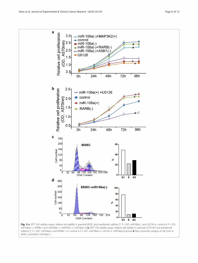
Fig. 3 a, MTT Cell viability assays: relative cell viability in parental 8505C and transfected sublines [*, P< 0.01, miR106a(−) and U0126 vs. control; #, P< 0.01,miR106a(−) + RARB(−) and miR106a(−) +MAP3K2 vs. miR106a(−)]; b, MTT Cell viability assays: relative cell viability in parental CGTH-W3 and transfectedsublines [*, P< 0.01, miR106a(+) and RARB(−) vs. control; #, P< 0.01, miR106a(+) + U0126 vs. miR106a(+)]; c and d, flow cytometry analysis of cell cycle in8505C and 8505C-miR106a(−)
Shen et al. Journal of Experimental & Clinical Cancer Research (2016) 35:101 Page 8 of 12
that aberrant activation of the MAPK pathway plays acentral role in the destruction of NIS-mediated iodide ac-cumulation in patients with DTC [25]. The BRAFV600E
mutation, one of the key activators of the MAPK pathway,is highly prevalent in recurrent radioiodine-refractorypapillary thyroid cancer and is associated with loss of NIS-mediated 131I uptake [26–29]. PBF(pituitary tumour-transforming gene [PTTG]-binding factor) is a proto-oncogene that seems to play a crucial part in diminishedmembrane targeting of NIS [30, 31]. PI3K-AKT-mTORand NOTCH signaling pathway also have been found to belinked to the regulation of thyroid-specific gene expression.
Activation of the PI3K-AKT pathway plays a fundamentalrole in thyroid tumorigenesis and is involved in downregu-lation of genes controlling iodide metabolism in patientswith DTC [32, 33]. However, overexpression of NOTCH1in thyroid cancer cells can induce differentiation and stimu-late NIS expression [34]. Epigenetic alterations, includingDNA hypermethylation and histone deacetylation, also playan important part in silencing thyroid-specific genes, espe-cially NIS [35, 36].RARB, a member of the thyroid-steroid hormone
receptor superfamily of nuclear transcriptional regula-tors, binds retinoic acid, the biologically active form
Fig. 4 a,b,e western blotting analysis of parental CGTH-W3 and 8505C cells and transfected sublines for proteins in MAPK signaling pathway(SB, SB203580. *, p < 0.01); c, flow cytometry analysis:apoptosis in parental CGTH-W3 and 8505C cells and transfected sublines (*, p< 0.01); d, caspase-3activity in parental CGTH-W3 and 8505C cells and transfected sublines (*, p< 0.01)
Shen et al. Journal of Experimental & Clinical Cancer Research (2016) 35:101 Page 9 of 12
of vitamin A which mediates cellular signaling in em-bryonic morphogenesis, cell growth and differenti-ation. In thyroid cancers, RA induces redifferentiationof cancer cells and expression of the NIS gene. As a re-sult, radioiodine uptake of tumors and serum Tg levelare expected to increase with RA treatment [37, 38].And the loss of retinoid receptors might occur duringthe loss of differentiation and tumor progression ofPTC [39]. Kogai et al. reported that RA stimulation ofthe NIS in MCF-7 breast cancer cells was meditated bythe insulin growth factor-I/phosphatidylinositol 3-kinase and p38 MAPK signaling pathways and an in-hibitor of p38 MAPK could significantly reduce iodideuptake in both all-trans retinoic acid-stimulated MCF-7cells and TSH-stimulated FRTL-5 cells [33].Some limitations have to be mentioned in the current
study. Firstly, the relative expression of miR-106a wasstatistically different in the serum of PTC patients withiodine avid and non-avid lung metastases, but the differ-ence in the mean levels was modest and there was alarge amount of overlap between the groups. In addition,all remaining experiments are in cell models with verylarge differences in miR-106a expression, not at all mod-eling the difference found in humans. Secondly, lackingof primary and metastasized tumor tissues (fresh orformalin-fixed and paraffin-embedded sample) made itunable to detect any histotype difference between pri-mary and metastasized tumors and measure miR-106aand RARB in tumor tissues level besides in serum level
which could further strength the significance of the find-ings in the current study.
ConclusionsIn summary, the role of miRNA-106a in the viability,apoptosis, migration, invasion, differentiation and iodineuptake function of thyroid cancer cell lines were investi-gated in the current research and the results indicated
Fig. 6 a, western blotting analysis of parental CGTH-W3 and 8505C cellsand transfected sublines for NIS and TSHR; b, iodine uptake ability inparental CGTH-W3 and 8505C cells and transfected sublines (*, p< 0.01).c, western blotting analysis (together with semi-quantitative analysis ofblots) of RARB with and without RA
Fig. 5 a, transwell invasion assay in 8505C-miR106a(−) and control cells(*, P< 0.05); b, scratch-wound migration assay in 8505C-miR106a(−)and control cells (*, P< 0.05); c, transwell invasion assay in CGTH-W3-miR106a(+) and control cells (*, P< 0.05); d, scratch-wound migrationassay in CGTH-W3-miR106a(+) and control cells (*, P< 0.05)
Shen et al. Journal of Experimental & Clinical Cancer Research (2016) 35:101 Page 10 of 12
that miRNA-106a directly targeting RARB associatedwith the viability, apoptosis, differentiation and the iod-ine uptake function of thyroid cancer cell lines by regu-lating MAPK signaling pathway in vitro. These findingsmay provide new strategies for the diagnosis and treat-ment in radioiodine-refractory DTC.
AbbreviationsATC, anaplastic thyroid carcinoma; DTC, differentiated thyroid cancer; MAPK,mitogen-activated protein kinase; NIS, sodium iodide symporter; PTC, papillary thy-roid carcinoma; qRT-PCR, quantitative real-time polymerase chain reaction;RAI, radioactive iodine; RARB, retinoic acid receptor beta; TSH, thyroid-stimulating hormone
AcknowledgementsThe authors would like to greatly thank Ying Ding, Jian-Ying Gu and Ye-JueWu (Department of Nuclear Medicine, Shanghai Jiao Tong University Affili-ated Sixth People’s Hospital) for their kind contributions to the collection ofblood samples.
FundingThis study was sponsored by the National Natural Science Foundation ofChina (No:81271611 &81201115).
Authors’ contributionsC-TS and Z-L Q wrote the paper; Q-Y L, Z-L Q and C-TS conceived and de-signed the study; C-TS, Z-L Q, H-JS and W-JW did the experiments; C-TS, Z-L Q and H-JS collect and analyzed the data. All authors read and approvedthe final manuscript.
Competing interestsThe authors declare that there is no conflict of interest that could be perceivedas prejudicing the impartiality of the research reported.
Received: 18 March 2016 Accepted: 14 June 2016
References1. DeSantis CE, Lin CC, Mariotto AB, Siegel RL, Stein KD, Kramer JL, et al. Cancer
treatment and survivorship statistics, 2014. CA Cancer J Clin. 2014;64:252–71.doi:10.3322/caac.21235.
2. Cooper DS, Doherty GM, Haugen BR, Kloos RT, Lee SL, Mandel SJ, et al.Revised American Thyroid Association management guidelines for patientswith thyroid nodules and differentiated thyroid cancer. Thyroid. 2009;19:1167–214. doi:10.1089/thy.2009.0110.
3. Haugen BRM, Alexander EK, Bible KC, Doherty G, Mandel SJ, Nikiforov YE, etal. 2015 American Thyroid Association Management Guidelines for AdultPatients with Thyroid Nodules and Differentiated Thyroid Cancer. Thyroid.2015. doi:10.1089/thy.2015.0020
4. Spitzweg C, Bible KC, Hofbauer LC, Morris JC. Advanced radioiodine-refractorydifferentiated thyroid cancer: the sodium iodide symporter and otheremerging therapeutic targets. Lancet Diabetes Endocrinol. 2014;2:830–42.doi:10.1016/s2213-8587(14)70051-8.
5. Rivera M, Ghossein RA, Schoder H, Gomez D, Larson SM, Tuttle RM.Histopathologic characterization of radioactive iodine-refractoryfluorodeoxyglucose-positron emission tomography-positive thyroidcarcinoma. Cancer. 2008;113:48–56. doi:10.1002/cncr.23515.
6. Bible KC, Ryder M. Evolving molecularly targeted therapies for advanced-stagethyroid cancers. Nat Rev Clin Oncol. 2016. doi:10.1038/nrclinonc.2016.19
7. Su B, Cheng J, Yang J, Guo Z. MEKK2 is required for T-cell receptor signalsin JNK activation and interleukin-2 gene expression. J Biol Chem. 2001;276:14784–90. doi:10.1074/jbc.M010134200.
8. Matitau AE, Gabor TV, Gill RM, Scheid MP. MEKK2 kinase association with 14-3-3 protein regulates activation of c-Jun N-terminal kinase. J Biol Chem.2013;288:28293–302. doi:10.1074/jbc.M113.511352.
9. Chayama K, Papst PJ, Garrington TP, Pratt JC, Ishizuka T, Webb S, et al. Roleof MEKK2-MEK5 in the regulation of TNF-alpha gene expression and MEKK2-MKK7 in the activation of c-Jun N-terminal kinase in mast cells. Proc NatlAcad Sci U S A. 2001;98:4599–604. doi:10.1073/pnas.081021898.
10. Maruyama T, Kadowaki H, Okamoto N, Nagai A, Naguro I, Matsuzawa A, etal. CHIP-dependent termination of MEKK2 regulates temporal ERK activationrequired for proper hyperosmotic response. Embo J. 2010;29:2501–14. doi:10.1038/emboj.2010.141.
11. Kitano M, Rahbari R, Patterson EE, Steinberg SM, Prasad NB, Wang Y, etal. Evaluation of candidate diagnostic microRNAs in thyroid fine-needleaspiration biopsy samples. Thyroid. 2012;22:285–91. doi:10.1089/thy.2011.0313.
12. Agretti P, Ferrarini E, Rago T, Candelieri A, De Marco G, Dimida A, et al.MicroRNA expression profile helps to distinguish benign nodules frompapillary thyroid carcinomas starting from cells of fine-needle aspiration. EurJ Endocrinol. 2012;167:393–400. doi:10.1530/eje-12-0400.
13. Mazeh H, Mizrahi I, Halle D, Ilyayev N, Stojadinovic A, Trink B, et al.Development of a microRNA-based molecular assay for the detection ofpapillary thyroid carcinoma in aspiration biopsy samples. Thyroid. 2011;21:111–8. doi:10.1089/thy.2010.0356.
14. Keutgen XM, Filicori F, Crowley MJ, Wang Y, Scognamiglio T, Hoda R, et al.A panel of four miRNAs accurately differentiates malignant from benignindeterminate thyroid lesions on fine needle aspiration. Clin Cancer Res.2012;18:2032–8. doi:10.1158/1078-0432.ccr-11-2487.
15. Mazeh H, Levy Y, Mizrahi I, Appelbaum L, Ilyayev N, Halle D, et al.Differentiating benign from malignant thyroid nodules using microribonucleic acid amplification in residual cells obtained by fine needleaspiration biopsy. J Surg Res. 2013;180:216–21. doi:10.1016/j.jss.2012.04.051.
16. Lakshmanan A, Wojcicka A, Kotlarek M, Zhang X, Jazdzewski K, Jhiang SM.microRNA-339-5p modulates Na+/I- symporter-mediated radioiodideuptake. Endocr Relat Cancer. 2015;22:11–21. doi:10.1530/erc-14-0439.
17. Qiu ZL, Shen CT, Song HJ, Wei WJ, Luo QY. Differential expression profilingof circulation microRNAs in PTC patients with non-131I and 131I-avid lungsmetastases: a pilot study. Nucl Med Biol. 2015;42:499–504. doi:10.1016/j.nucmedbio.2015.01.009.
18. Livak KJ, Schmittgen TD. Analysis of relative gene expression data usingreal-time quantitative PCR and the 2(−Delta Delta C(T)) Method. Methods.2001;25:402–8. doi:10.1006/meth.2001.1262.
19. Chen X, Ba Y, Ma L, Cai X, Yin Y, Wang K, et al. Characterization ofmicroRNAs in serum: a novel class of biomarkers for diagnosis of cancerand other diseases. Cell Res. 2008;18:997–1006. doi:10.1038/cr.2008.282.
20. Cui L, Zhang X, Ye G, Zheng T, Song H, Deng H, et al. Gastric juiceMicroRNAs as potential biomarkers for the screening of gastric cancer.Cancer. 2013;119:1618–26. doi:10.1002/cncr.27903.
21. Zhu M, Zhang N, He S, Lui Y, Lu G, Zhao L. MicroRNA-106a targets TIMP2 toregulate invasion and metastasis of gastric cancer. FEBS Lett. 2014;588:600–7.doi:10.1016/j.febslet.2013.12.028.
22. Catela Ivkovic T, Aralica G, Cacev T, Loncar B, Kapitanovic S. miR-106aoverexpression and pRB downregulation in sporadic colorectal cancer. ExpMol Pathol. 2013;94:148–54. doi:10.1016/j.yexmp.2012.11.002.
23. Liu Z, Gersbach E, Zhang X, Xu X, Dong R, Lee P, et al. miR-106a repressesthe Rb tumor suppressor p130 to regulate cellular proliferation anddifferentiation in high-grade serous ovarian carcinoma. Mol Cancer Res.2013;11:1314–25. doi:10.1158/1541-7786.mcr-13-0131.
24. Li Z, Yang CS, Nakashima K, Rana TM. Small RNA-mediated regulation of iPScell generation. Embo J. 2011;30:823–34. doi:10.1038/emboj.2011.2.
25. Knauf JA, Kuroda H, Basu S, Fagin JA. RET/PTC-induced dedifferentiation ofthyroid cells is mediated through Y1062 signaling through SHC-RAS-MAPkinase. Oncogene. 2003;22:4406–12. doi:10.1038/sj.onc.1206602.
26. Elisei R, Ugolini C, Viola D, Lupi C, Biagini A, Giannini R, et al. BRAF(V600E)mutation and outcome of patients with papillary thyroid carcinoma: a 15-year median follow-up study. J Clin Endocrinol Metab. 2008;93:3943–9. doi:10.1210/jc.2008-0607.
27. Ricarte-Filho JC, Ryder M, Chitale DA, Rivera M, Heguy A, Ladanyi M, et al.Mutational profile of advanced primary and metastatic radioactive iodine-refractory thyroid cancers reveals distinct pathogenetic roles for BRAF,PIK3CA, and AKT1. Cancer Res. 2009;69:4885–93. doi:10.1158/0008-5472.can-09-0727.
28. Franco AT, Malaguarnera R, Refetoff S, Liao XH, Lundsmith E, Kimura S, et al.Thyrotrophin receptor signaling dependence of Braf-induced thyroid tumorinitiation in mice. Proc Natl Acad Sci U S A. 2011;108:1615–20. doi:10.1073/pnas.1015557108.
29. Riesco-Eizaguirre G, Gutierrez-Martinez P, Garcia-Cabezas MA, Nistal M,Santisteban P. The oncogene BRAF V600E is associated with a high risk ofrecurrence and less differentiated papillary thyroid carcinoma due to the
Shen et al. Journal of Experimental & Clinical Cancer Research (2016) 35:101 Page 11 of 12
impairment of Na+/I- targeting to the membrane. Endocr Relat Cancer.2006;13:257–69. doi:10.1677/erc.1.01119.
30. Smith VE, Sharma N, Watkins RJ, Read ML, Ryan GA, Kwan PP, et al.Manipulation of PBF/PTTG1IP phosphorylation status; a potential newtherapeutic strategy for improving radioiodine uptake in thyroid and othertumors. J Clin Endocrinol Metab. 2013;98:2876–86. doi:10.1210/jc.2012-3640.
31. Smith VE, Franklyn JA, McCabe CJ. Expression and function of the novelproto-oncogene PBF in thyroid cancer: a new target for augmentingradioiodine uptake. J Endocrinol. 2011;210:157–63. doi:10.1530/joe-11-0064.
32. Liu YY, Zhang X, Ringel MD, Jhiang SM. Modulation of sodium iodidesymporter expression and function by LY294002, Akti-1/2 and Rapamycin inthyroid cells. Endocr Relat Cancer. 2012;19:291–304. doi:10.1530/erc-11-0288.
33. Kogai T, Sajid-Crockett S, Newmarch LS, Liu YY, Brent GA. Phosphoinositide-3-kinase inhibition induces sodium/iodide symporter expression in ratthyroid cells and human papillary thyroid cancer cells. J Endocrinol. 2008;199:243–52. doi:10.1677/joe-08-0333.
34. Ferretti E, Tosi E, Po A, Scipioni A, Morisi R, Espinola MS, et al. Notchsignaling is involved in expression of thyrocyte differentiation markers andis down-regulated in thyroid tumors. J Clin Endocrinol Metab. 2008;93:4080–7. doi:10.1210/jc.2008-0528.
35. Xing M, Usadel H, Cohen Y, Tokumaru Y, Guo Z, Westra WB, et al.Methylation of the thyroid-stimulating hormone receptor gene in epithelialthyroid tumors: a marker of malignancy and a cause of gene silencing.Cancer Res. 2003;63:2316–21.
36. Kitazono M, Robey R, Zhan Z, Sarlis NJ, Skarulis MC, Aikou T, et al. Lowconcentrations of the histone deacetylase inhibitor, depsipeptide(FR901228), increase expression of the Na(+)/I(−) symporter and iodineaccumulation in poorly differentiated thyroid carcinoma cells. J ClinEndocrinol Metab. 2001;86:3430–5. doi:10.1210/jcem.86.7.7621.
37. Kogai T, Ohashi E, Jacobs MS, Sajid-Crockett S, Fisher ML, Kanamoto Y, et al.Retinoic acid stimulation of the sodium/iodide symporter in MCF-7 breastcancer cells is mediated by the insulin growth factor-I/phosphatidylinositol3-kinase and p38 mitogen-activated protein kinase signaling pathways. JClin Endocrinol Metab. 2008;93:1884–92. doi:10.1210/jc.2007-1627.
38. Schmutzler C, Schmitt TL, Glaser F, Loos U, Kohrle J. The promoter of thehuman sodium/iodide-symporter gene responds to retinoic acid. Mol CellEndocrinol. 2002;189:145–55.
39. Tang W, Nakamura Y, Zuo H, Yasuoka H, Yang Q, Wang X, et al.Differentiation, proliferation and retinoid receptor status of papillarycarcinoma of the thyroid. Pathol Int. 2003;53:204–13.
• We accept pre-submission inquiries
• Our selector tool helps you to find the most relevant journal
• We provide round the clock customer support
• Convenient online submission
• Thorough peer review
• Inclusion in PubMed and all major indexing services
• Maximum visibility for your research
Submit your manuscript atwww.biomedcentral.com/submit
Submit your next manuscript to BioMed Central and we will help you at every step:
Shen et al. Journal of Experimental & Clinical Cancer Research (2016) 35:101 Page 12 of 12