mirror neuron and theory of mind mechanisms involved in...
TRANSCRIPT

Mirror Neuron and Theory of Mind MechanismsInvolved in Face-to-Face Interactions: A FunctionalMagnetic Resonance Imaging Approach to Empathy
Martin Schulte-Ruther1,2, Hans J. Markowitsch3, Gereon R. Fink1,4,and Martina Piefke1,2
Abstract
& Empathy allows emotional psychological inference aboutother person’s mental states and feelings in social contexts. Weaimed at specifying the common and differential neural mech-anisms of ‘‘self’’- and ‘‘other’’-related attribution of emotionalstates using event-related functional magnetic resonanceimaging. Subjects viewed faces expressing emotions with director averted gaze and either focused on their own emotionalresponse to each face (self-task) or evaluated the emotionalstate expressed by the face (other-task). The common networkactivated by both tasks included the left lateral orbito-frontaland medial prefrontal cortices (MPFC), bilateral inferior frontalcortices, superior temporal sulci and temporal poles, as wellas the right cerebellum. In a subset of these regions, neuralactivity was significantly correlated with empathic abilities. Theself- (relative to the other-) task differentially activated the
MPFC, the posterior cingulate cortex (PCC)/precuneus, and thetemporo-parietal junction bilaterally. Empathy-related process-ing of emotional facial expressions recruited brain areasinvolved in mirror neuron and theory-of-mind (ToM) mecha-nisms. The differential engagement of the MPFC, the PCC/precuneus, and temporo-parietal regions in the self-task indi-cates that these structures act as key players in the evaluationof one’s own emotional state during empathic face-to-face in-teraction. Activation of mirror neurons in a task relying onempathic abilities without explicit task-related motor compo-nents supports the view that mirror neurons are not only in-volved in motor cognition but also in emotional interpersonalcognition. An interplay between ToM and mirror neuron mech-anisms may hold for the maintenance of a self–other distinctionduring empathic interpersonal face-to-face interactions. &
INTRODUCTION
A key aspect of social interaction is the inference ofother persons’ emotional states by evaluating their facialexpressions (Ekman, 1982). These can help us to gainaccess to someone’s feelings and may act as triggers ofempathy. Empathy is based upon processes of psycho-logical inferences about other persons’ mental andemotional states occurring within a specific social con-text. This context may provide the frame of reference forthe integration of emotion and cognition to yield in-sights into the intentions and feelings of others (Decety& Jackson, 2004). Three core aspects are likely to play arole in the emergence and maintenance of empathy: (i)an intuitive feeling of having something in common withthe other person which relies on socially shared emo-tional experiences; (ii) cognitive mechanisms of per-spective-taking; and (iii) the ability to maintain a self–
other distinction during interpersonal interaction. Em-pathic feelings allow for socially appropriate emotionalresponses, be it a shared or reactive emotional state.Therefore, empathy is different from pure emotionalcontagion as the resulting emotional state can be dif-ferent from the one being observed (see, for example,Davis, 1996; Stotland, 1969).
It has been suggested that shared emotional feelingsmight be traced back to mirror neuron mechanisms(Gallese, 2003), which had initially been proposed inthe field of motor representations. Mirror neurons havebeen discovered in the monkey brain in frontal areaF5 and parietal area PF (see, e.g., Rizzolatti, Fogassi,& Gallese, 2001, for a review). There is increasing evi-dence from neuroimaging studies that core componentsof a human mirror neuron system (hMNS) compriseparts of the inferior frontal cortex and the posteriorparietal cortex (e.g., Koski, Iacoboni, Dubeau, Woods, &Mazziotta, 2003; Iacoboni et al., 1999). The mirror neu-ron account has recently been adapted to processes op-erating during interpersonal cognition (Gallese, Keysers,& Rizzolatti, 2004; however, see Jacob & Jeannerod,2005 for a critique). It has been suggested that the
1Research Center Julich, Germany, 2University Hospital Aachen,Rheinisch-Westfalische Technische Hochschule (RWTH) Aachen,Germany, 3University of Bielefeld, Germany, 4University HospitalCologne, Germany
D 2007 Massachusetts Institute of Technology Journal of Cognitive Neuroscience 19:8, pp. 1354–1372

brain networks controlling motor behavior and socialinteraction share (at least in part) computational parallels(Wolpert, Doya, & Kawato, 2003), and might be imple-mented in similar or overlapping brain regions. Recentstudies indeed indicate that not only motor plans but alsomore abstract intentions (Iacoboni et al., 2005) or emo-tional states (Carr, Iacoboni, Dubeau, Mazziotta, & Lenzi,2003) recruit the same areas in the inferior frontal cortexwhich are supposed to contain mirror neurons.
Carr et al. (2003) demonstrated that components ofthe hMNS (including the inferior frontal cortex) are in-volved in the imitation and observation of facial expres-sions of emotions. Reports on hMNS involvement in themere perception of facial expressions of emotions, how-ever, are yet rare. This might be due to the fact that mostprevious studies focused on the implicit processing ofemotional facial expressions using distractor tasks suchas gender detection or conditioning paradigms (Winston,O’Doherty, & Dolan, 2003; Critchley et al., 2000), and/orexplicit tasks such as emotion categorization or judgmentof emotion intensity (e.g., Winston et al., 2003; Critchleyet al., 2000; George et al., 1993). All these tasks can beaccomplished by the use of perceptual strategies, forexample, pattern matching. The above studies may thushave failed to detect hMNS activity because the percep-tual processing of emotional facial expressions was notembedded in an interpersonal context.
For pain and disgust, functional neuroimaging studiesshowed evidence that the processing of one’s own emo-tional experience and the observation of another personhaving a similar emotional experience may engage over-lapping brain networks (Singer et al., 2004; Wicker et al.,2003). Interestingly, in the study by Singer et al. (2004),the relative engagement of these networks was corre-lated with individual differences in empathic abilities.
The cognitive aspects of empathic interpersonal per-spective taking are closely related to theory of mind(ToM). The ToM concept depicts meta-cognitive abilitiessuch as the inference of beliefs, intentions, and desiresof other people (Premack & Woodruff, 1978). Impor-tantly, ToM also requires the distinction between one’sown thoughts and intentions and those of others, whichis a key component of interpersonal interactions.
The neural network supporting cognitive aspects ofToM include temporo-parietal areas, the medial prefron-tal cortex (MPFC), and the temporal poles (e.g., Vogeleyet al., 2001; see Frith & Frith, 2003; Vogeley & Fink, 2003for reviews). Functional neuroimaging data on emotionalToM, however, remain scarce (but see Vollm et al., 2006).Few studies directly addressed the functional neuroanat-omy of taking the self- or other-perspective in the contextof emotional experience (Ochsner et al., 2004; Ruby &Decety, 2004). For example, Ochsner et al. (2004) askedtheir subjects to evaluate emotional responses to affectivephotographs from their own (i.e., first person) perspectiveor from the perspective of a character depicted in thephotos (i.e., third-person perspective). This approach,
however, did not take into account that the empathicswitching between the self- and the other-perspective goesbeyond the ‘‘classic’’ first-person/third-person conceptadopted by studies on perspective-taking (e.g., Vogeley& Fink, 2003; Vogeley et al., 2001). Rather, empathy relieson a dyadic social situation where the inferred intentionsand feelings of the other directly affect the self as the tar-get of the other’s emotion. This account is also supportedby studies of cognitive aspects of interpersonal interac-tions, suggesting that ‘‘on-line’’ social reasoning is mostefficiently triggered in a context where the observedbehavior of others is highly relevant to one’s own inten-tions and actions (Kampe, Frith, & Frith, 2003; Gallagher,Jack, Roepstorff, & Frith, 2002). To our knowledge, nofunctional neuroimaging data are currently available onthe neural circuits supporting empathic self–other inter-actions unfolding in the dynamics of emotional interper-sonal contexts.
We used functional magnetic resonance imaging(fMRI) to investigate empathy-related neural processes,applying a paradigm which required both the decod-ing of other persons’ emotional states from facial cues(‘‘other’’-condition) and the evaluation of one’s ownemotional response to these faces (‘‘self’’-condition).Subjects evaluated either the emotional state of theobserved person or their own emotional reaction whenviewing that person. Responses were given by choosingappropriate emotional descriptions from a list of adjec-tives depicting emotional states. The experimental setupthus differed from previous approaches in that it didnot focus on a perceptual decision on the emotion ex-pressed by a face (e.g., a decision between basic emotioncategories), but rather assessed the interactive switchingbetween the self- and the other-perspective during theattribution of emotion. Using this paradigm, we wereable to experimentally construct an interpersonal con-text in which empathic social cognition could emerge. Inaddition, we manipulated the head direction of thestimulus faces such that they were either directed tothe observer or averted by �458. An age/gender decisiontask on neutral faces was used as a high-level baseline.Standard empathy scales were administered to assessindividual differences in empathic abilities. Individualscores on these scales were used to determine whethermeasures of empathic capabilities may predict the am-plitude of empathy-related brain activity, especially inareas belonging to the hMNS.
We expected that brain regions supporting ToM (e.g.,MPFC, superior temporal regions) and structures previ-ously associated with the hMNS (e.g., inferior frontalgyrus, superior temporal sulcus [STS], inferior parietalcortex) would be activated during both the attributionof emotion to oneself and the other person. However,we also hypothesized that some of these areas wouldshow differential responses depending upon the in-volvement of either the self- or the other-perspective.hMNS engagement was expected to predominate in the
Schulte-Ruther et al. 1355

self-condition, whereas we considered ToM mechanismsto be specifically implicated in the other-condition.Moreover, we hypothesized that empathy would betriggered more easily in the direct gaze condition (whenthe self is the target of the other’s emotion) becausedirect eye contact may signal communicative intent andis typically cueing inferences on the emotional andmental state of others (Kleinke, 1986). Also, it has beenshown that direct (compared to averted) gaze is ableto drive ToM areas in the brain (Kampe et al., 2003).
METHODS
Participants
Twenty-six subjects (12 men, mean age ± SD = 24.4 ±3.0 years; 14 women, mean age ± SD = 24.8 ± 3.7 years)participated in the study which was approved by thelocal ethics committee. All participants (mean age ±SD = 24.6 ± 3.4 years) were right-handed, as as-sessed by the Edinburgh inventory (Oldfield, 1971), andwere native speakers of German language. They werescreened medically to rule out the use of medicationaffecting the central nervous system, a history of neuro-logical or psychiatric disorders, head trauma, substanceabuse, and other serious medical conditions. To controlfor sufficient abilities of verbalizing emotional states,subjects were screened for alexithymia using the Ger-man version of the Toronto Alexithymia Scale (TAS-26;Kupfer, Brosig, & Brahler, 2001). Subjects underwentneuropsychological testing, including standard mea-sures of IQ, working memory, and attention. Moreover,they completed questionnaires on emphatic abilities(Balanced Emotional Empathy Scale [BEES], Mehrabian,1997; Empathic Concern Scale [ECS], Davis, 1983) forlater analysis of a possible relationship between individ-ual degrees of empathic capabilities and regional brainactivation. The 30-item BEES extends the EmotionalEmpathic Tendency Scale (Mehrabian & Epstein, 1972)in that it is designed to balance ‘‘acquiescence bias’’(i.e., the tendency of the majority of people to generallyagree or disagree with statements included in a ques-tionnaire). The BEES allows for a measure of one’stendency to empathize with the emotional experiencesof others (‘‘emotional empathy’’). The ECS, a subscaleof the Interpersonal Reactivity Index developed by Davis(1983), taps emotional aspects of empathic abilities in asimilar way. For example, items of both scales includequestions concerning one’s own emotional state whenobserving the misfortune of another person or one’stendency to get caught up in the emotions of a crowd.Both scales can be easily administered as a paper-and-pencil test and have previously been used in functionalneuroimaging studies to predict empathy-related brainactivation (e.g., Singer et al., 2004).
Mean TAS-26, BEES, and ECS scores of participantsinvolved in the present study and population norms are
summarized in Table 1. Individual scores on these scalesand all individual neuropsychological data were withinthe range of reference population norms for all testsadministered.
Stimuli
Facial stimuli were constructed using commerciallyavailable software for the generation of synthetic faces(FaceGen 3.0, Singular Inversions, Vancouver, Canada).With this software, faces can be rotated freely in 3-Dspace and morphed on diverse dimensions (e.g., emo-tion categories, gender, age) to create specific faces withdifferential degrees of distinct emotional expressions,thus allowing for a higher degree of control of stimulusmaterials than in studies where real faces are used.There were 318 faces preselected according to thenaturalness of their emotional expressions by threeindependent judges from a pool of more than 1500faces created randomly by the software. Validity ofemotional expressions was confirmed in a behavioralstudy. Preselected faces were rated by volunteers (n =9) according to the procedure devised by Ekman andFriesen (1976). Faces were presented with either anangry, fearful, sad, disgusted, happy, or neutral expres-sion, and participants were asked to indicate whichemotion category was displayed in a given face. Applyingthis procedure, 192 face stimuli were finally selected forthe fMRI study (64 angry, 64 fearful, 64 neutral faces; halfmale and half female in each category). A minimum of65% correct choices was required for the selection ofa face. The average percentage (±SD) of correct choiceswas 86.4% (10.4%). This degree of agreement in cate-gorical judgments of facial expressions of emotion is inaccordance with behavioral studies in which the Ekmanand Friesen face stimuli were used (Ekman, 1984). Themean percentage of correct choices (i.e., correct rec-ognition of the intended emotional expression of aface) was counterbalanced across the stimulus catego-ries (angry and fearful faces). A subset of the resultingstimulus set (30 stimulus faces) was rated by volunteers(n = 19) for emotional arousal (as described by Bradley
Table 1. Group Mean Scores and Respective PopulationNorms for TAS-26, BEES, and ECS
BEES ECS TAS-26
Group mean± SD
41.58 ± 30.23 18.31 ± 4.00 2.38 ± 0.47
Mean (norms)± SD
45 ± 24a 20.36 ± 4.02a,b 2.42 ± 0.53
aNorms for ECS and BEES refer to the original English version. In thepresent study, a German translation was used. For this German ver-sion, population norms are unavailable.bMean scores of norms for the ECS are estimated from separate scoresfor male and female samples provided by Davis (1980).
1356 Journal of Cognitive Neuroscience Volume 19, Number 8
& Lang, 1999) to ensure that the synthetic stimulus facesevoked emotional responses sufficient for empathicreactions. A univariate analysis of variance (ANOVA) con-firmed a significant effect of emotion category on thearousal ratings [F(2, 36) = 98.33, p < .001]. Post hoct tests revealed that this was due to a significant strongerarousal in the fearful ( p < .001) and the angry faces( p < .001) each compared to neutral faces.
The present study was restricted to (i) two emotioncategories to retain statistical power sufficient to detectpotential differences between emotion categories and (ii)negatively valenced emotions because the switchingbetween negative and positive emotional valence mighthave confounded the experimental design. We did notuse positively valenced emotional facial expressions forour experiment as it is difficult to find two positive basicemotions which can easily be distinguished. The selec-tion of fear and anger for a study on empathy is alsowell in accordance with recent studies investigating face-based emotion recognition in brain-damaged patients.Paired deficits in recognition and production of thesame emotion could be revealed for three emotions,namely, fear, disgust, and anger (see Goldman & Sripada,2005 for a review).
Experimental Tasks
Subjects were asked to either concentrate on their ownfeelings that emerged when they were looking at eachemotional facial expression (self-task), or on the emo-tional state expressed by the stimulus face (other-task).Besides the factor task (self-/other-perspective), two ad-ditional factors were varied systematically. These wereemotion (angry/fearful faces) and head direction (direct-ed toward the observer/averted by �458). This experi-mental setup resulted in a 2 � 2 � 2 factorial design. Inaddition, a gender and age decision task on neutral faces(also with direct or averted head direction) was includedas a high-level baseline condition. Neutral instead ofemotional faces were used in the baseline conditionbecause emotion processing is known to comprise un-conscious, preconceptual stages such that empathic re-sponses might implicitly emerge in the mere presenceof an emotional face (Coricelli, 2005).
Each face stimulus was presented for 2 sec. Thereaf-ter, a list of four 1- or 2-word descriptions appeared onthe screen for 3 sec. The subject’s task was to choosefrom this list of words the description which bestdepicted either the emotional state expressed by thepreceding face (other-task) or the subject’s own emo-tional response to the stimulus face (self-task). Theemotional adjectives used for these judgments wererandomly drawn from four sets of words, dependingon the experimental condition (self-task with angry orfearful faces; other-task with angry or fearful faces).Choice words had been generated in a pilot study:subjects (n = 6) performed the same paradigm but
generated the words by themselves instead of choosingthem from a given list. Four sets of 15 emotional ad-jectives were extracted from these responses corre-sponding to the dimensions self versus other, and fearversus anger (see below). Written word frequency (esti-mated by deriving frequencies from a collection oflinguistic databases of German written language [COS-MAS2, www.ids-mannheim.de/cosmas2, Institut fur deut-sche Sprache, Mannheim]), word length, arousal, andpositivity were kept equivalent across tasks. In a separatepilot experiment, each of these words was rated onemotional scales (arousal and positivity) by 19 partic-ipants, as described by Bradley and Lang (1999). A two-factorial multivariate analysis of variance (MANOVA)including these measures as dependent variables yieldedno significant effect of the factors emotion [i.e., angerversus fear; Rao’s R(5,52) = 0.95, p = .74], task [i.e., selfversus other; Rao’s R(5,52) = 0.97, p = .92], or theirinteraction [Rao’s R(5,52) = 0.93, p = .53]. Similarly,univariate two-factorial ANOVAs for each measure didnot yield significant differences between the factors ofinterest.
For the high-level baseline, the following combina-tions of age and gender descriptions were employed:‘‘older female,’’ ‘‘older male,’’ ‘‘younger female,’’ ‘‘youngermale,’’ with the order of descriptions randomly varyingfrom trial to trial.
During the fMRI measurement, subjects made theirchoices using four fingers of their right hand (exclud-ing the thumb) and a four-button response device.The word lists were followed by an intertrial intervalwith a duration jittered between 0.2 and 1.2 sec. Ran-dom jitter was included to prevent correlation of eventregressors. The two experimental tasks (attributionof emotion to oneself or the other) and the high-levelbaseline task (age/gender decision) alternated blockwise in a pseudorandomized counterbalanced order.Each block consisted of a series of four trials andwas cued by a preceding verbal instruction (duration:2.5 sec) (see Figure 1). For stimulus presentation andresponse collection, the software Presentation 9.0 (Neu-robehavioral Systems, Albany, CA, USA; www.neurobs.com) was used. During the fMRI experiment, the stimu-lus display was back-projected onto a screen mountedon the head coil.
Magnetic Resonance Technical Parameters
Subjects were scanned with a Siemens MagnetomSONATA whole-body system (Erlangen, Germany) at1.5 T using a standard radio-frequency head coil. Thirtyslices (0.4 mm interslice gap) were individually positionedto cover the whole brain with reference to a midsagittalscout image. During task performance, gradient-echo,echo-planar T2*-weighted images (EPI) were acquiredusing blood-oxygen-level-dependent (BOLD) contrast(TE = 66 msec, TR = 3020 msec, a = 908, FOV =
Schulte-Ruther et al. 1357

200 mm, voxel size = 3.1 � 3.1 � 4 mm3, matrix size =64 � 64). Scanning was performed continuously duringone single session lasting �25 min. After functional neuro-imaging, high-resolution anatomical images were acquiredusing a T1-weighted 3-D magnetization-prepared, rapid ac-quisition gradient-echo (MP-RAGE) pulse sequence (TE =3.93 msec, TR = 2200 msec, TI = 1200 msec, a = 158,FOV = 240 mm, matrix size = 179 � 256, 128 sagittalslices, slice thickness = 1.33 mm).
Image Processing and Data Analysis
Preprocessing of imaging data and statistical analysis wereaccomplished using SPM2 (Wellcome Department ofImaging Neuroscience, London, UK; www.fil.ion.ucl.ac.uk/spm) implemented in MATLAB 6.5 (The Mathworks,Natick, MA, USA). The first four images of each func-tional time-series were discarded to allow for T1 equil-ibration effects. The remaining volumes were slice-timecorrected, realigned (Friston, Ashburner, et al., 1995),and corrected for movement-by-field-inhomogeneityinteractions using the realign and unwarp module ofSPM2 (Andersson, Hutton, Ashburner, Turner, & Friston,2001). Individual differences in brain anatomy were
accounted for by normalizing each subject’s datasetto an EPI template of the Montreal National Institute(MNI) series implemented in SPM2, transforming all im-ages to ‘‘MNI-space,’’ a standard stereotactic referencespace (which is approximately equivalent to the atlas ofTalairach & Tournoux, 1988). Subsequently, functionalvolumes were spatially smoothed with an 8 � 8 � 8 mmGaussian kernel (full width half maximum) to compen-sate for remaining variation in gyral and sulcal anatomyacross subjects, and to meet the requirements of theGaussian random fields theory for subsequent statisticalanalysis. Individual T1 anatomical scans were coregis-tered to the corresponding mean image of the EPI time-series. Thereafter they were also normalized into MNIspace, applying the same linear transformation parame-ters obtained by the normalization procedure of thefunctional volumes described above. Boxcar functions of2 sec duration (corresponding to the on-time of eachface stimulus) were convolved with a model of the he-modynamic response function (Friston, Holmes, et al.,1995), which thus comprised the presentation of facialstimuli, but not the word-choice procedure. Instructioncues were modeled in analogous manner using boxcarfunctions of 2.5 sec length, respectively. Parameter
Figure 1. Experimental paradigm. (A) Time course of stimulus presentation. Trials were presented blockwise for the self-, other-, and
high-level baseline tasks with each block consisting of four trials. The instruction cue was given only at the beginning of each block. Thefactors emotion category and gaze direction were pseudorandomized on a trial-by-trial basis. Choice words and task cues were originally
presented in German language. (B) Experimental factors. We employed a 2 � 2 � 2 factorial design with the factors task (self vs. other),
head direction (direct vs. averted), emotion category (fear vs. anger), and a high-level baseline task with neutral faces and varying head
direction (direct vs. averted).
1358 Journal of Cognitive Neuroscience Volume 19, Number 8

estimates of the resulting general linear model wereobtained for each subject and each voxel, correspondingto a mass-univariate approach. For population inference,a second level analysis was performed, using the param-eter estimates for each task-related regressor in a within-subject ANOVA model (with subjects treated as randomeffects). Departures from sphericity assumptions wereaccommodated by applying the nonsphericity correctionimplemented in SPM2 (Glaser & Friston, 2003). Specificeffects at each voxel were tested by applying appropriatelinear contrasts to the parameter estimates. Differentialcontrasts of interest were calculated according to theexperimental factors task (self-task vs. other-task, andvice versa), emotion (fear vs. anger, and vice versa), andhead direction (directed to the observer vs. averted,and vice versa) to assess differential modulation of theBOLD signal induced by each factor. In addition, a con-junction analysis was performed to detect for activationscommon to the self- and the other-task (relative to thehigh-level baseline).
We report results that survive a statistical heightthreshold of p < .05, family wise error (FWE) correctedfor multiple comparisons, and an extent threshold of atleast 15 voxels. This conservative threshold was adoptedto reduce the risk of false-positive activations (type IIerror). Additionally, for those brain regions targetedby a priori hypotheses (see Introduction), a region-of-interest (ROI) approach was applied, using a heightthreshold of p < .05 (FWE corrected across this partic-ular region). To assess correlations between brain acti-vations and individual measures of emphatic abilities (asassessed by the BEES and the ECS; see above), individ-ual parameter estimates of regressors coding for the self-and other-tasks were extracted for regions previouslyimplicated in the hMNS at voxels showing peak activa-tion in the group conjunction analysis of the ‘‘self’’- andthe ‘‘other’’-tasks (each relative to the high-level base-line). Individual means of these parameter estimateswere correlated with individual scores on the BEESand ECS scales.
Localization of Activations
SPMT maps resulting from the group analysis were super-imposed onto a group mean MR image which was cal-culated from the normalized anatomical T1-images ofeach subject (see above). Standard stereotactic coor-dinates of the local maxima within areas of significantrelative changes in neural activity associated with dif-ferent experimental conditions were determined andanatomically localized by comparing activation mapssuperimposed on the anatomical group mean brain witha standard atlas of brain anatomy (Duvernoy, 1999).In addition, an anatomical SPM2 toolbox developed byEickhoff et al. (2005) was applied, which allows for theintegration of probabilistic cytoarchitectonic maps of thebrain and functional neuroimaging data.
Prescanning and Postscanning Procedures
To accommodate subjects to the experimental tasks,each subject accomplished six training blocks prior tothe fMRI measurement outside the scanner. Trainingstimuli were not included into the stimulus set used forthe fMRI experiment. None of the subjects reportedproblems with the task or the required reaction times ofless than 3 sec. Immediately after scanning, subjectsrated all stimulus faces for the intensity of feelings (i)expressed by the face stimulus and (ii) evoked in the par-ticipants. In addition, participants completed a debrief-ing questionnaire on the naturalness of stimulus faces,as well as the levels of difficulty of the self-task, other-task, and high-level baseline task for each emotion andhead direction condition.
RESULTS
Behavioral Data
Response Times during Scanning
A 3 � 2 ANOVA with the factors head direction and taskshowed a significant main effect of task [F(2, 50) = 33.94;p < .001]. Paired post hoc t tests revealed that subjectsresponded significantly faster in the high-level baselinetask than both experimental tasks ( p < .001 for eachcomparison). A 2 � 2 � 2 ANOVA with the factors task,head direction, and emotion showed that reaction timeswere significantly faster in the other- than in the self-task[F(1, 25) = 16.930; p < .001]. Differences in reactiontimes between directed and averted head direction aswell as angry and fearful emotion were nonsignificant.The interaction between the factors task and head direc-tion just failed to reach significance [F(1, 25) = 4.012;p = .056]. This interaction resulted from faster responsesto faces directed toward the observer (relative to avertedfaces) in the other-task [t(25) = 2.003, p = .056, two-sided], but not in the self-task [t(25) = �0.55, 0.587;post hoc t tests for matched pairs].
Postscanning Debriefing Questionnaire
Comparisons between groups of different stimuli (direct/averted head direction; angry/fearful emotion) did notyield statistically significant differences for the ratings oftask difficulty and naturalness of faces. The respectivedata are provided in Table 2. Ratings of task difficulty dif-fered significantly between tasks [Friedman ANOVA, n =26; x2(df = 2) = 15.31, p < .001]. The self-task was con-sidered more difficult than both the other-task (WilcoxonTest, n = 26, Z = �3.856; p < .001) and the high-levelbaseline task (Wilcoxon Test, n = 26, Z = �2.97; p <.01), whereas the ratings of levels of difficulty did notdiffer significantly between the other- and the high-levelbaseline task (Wilcoxon Test, n = 26, Z = �0.41;p = .678). Also, there was no significant difference in
Schulte-Ruther et al. 1359

the mean ratings of neutral and emotional faces’ natural-ness (Wilcoxon Test, n = 23, Z = �0.112; p = .911).
Correlations between Ratings of Difficultyand Reaction Times
To clarify whether increased difficulty ratings were re-lated to longer reaction times, respective correlationanalyses (Spearman rank-order correlation) were per-formed. Individual postscanning ratings of difficultyfor each of the three tasks (self, other, high-level base-line) were compared to the mean reactions times re-lated to performance of each task during scanning.There were no positive correlations for self (r = .136,p = .506) or other conditions (r = �.164, p = .424), andan inverse correlation for the high-level baseline task(r = �.432, p < .05). Furthermore, the differencebetween individual ratings of difficulty for the self- andthe other-task was compared to the difference betweenthe mean reaction times associated with the same con-ditions. This correlation analysis did not yield a signifi-cant result as well (r = .012, p = .955).
Postscanning Rating of Stimulus Faces
The mean ratings of emotion intensity for each stimulusface were compared by two-way 2 � 2 ANOVAs with thefactors emotion and head direction. The intensity ofemotion induced by the face stimuli in the subjects (self-task) was rated significantly higher in direct relative toaverted head direction trials [F(1, 25) = 7.859; p < .01].This was not paralleled by respective differential ratingsof the intensity of the emotions expressed in the facestimuli (i.e., in the other task) [F(1, 25) = 2.333; p = .139].Concerning the factor emotion, angry faces were rated
significantly higher for emotion intensity than fearfulfaces in the other-task [F(1, 25) = 11.695; p < .01], butnot in the self-task [F(1, 25) = 3.458; p = .075]. Inter-actions between emotion and head direction were non-significant for either experimental task.
fMRI Data
Common Effects of the Self- and the Other-task(Relative to the High-level Baseline)
Areas of significant differential activation revealed bya conjunction analysis of the self- and the other-task(each compared to the high-level baseline) were locatedin the left STS and the left lateral orbito-frontal cor-tex (BA 47, extending into the inferior frontal cortex,BA 44 and 45), left MPFC, the left middle frontal gy-rus, the left presupplementary motor area (pre-SMA),and the right cerebellum (see Table 3 and Figure 2A).Using an ROI approach (see Methods), activations werealso significant in the right STS, the right inferior fron-tal gyrus (BA 45), and the temporal pole ( p < .05,FWE corrected across ROI; see Table 3 and Figures 3and 5).
Main Effect of the Self- versus the Other-task
The main effect of the self- (relative to the other-) taskrevealed significant differential bilateral activations of thetemporo-parietal junction (TPJ), the MPFC, the posteriorcingulate cortex (PCC)/precuneus, the middle frontal gy-rus, the left inferior frontal gyrus, the pre-SMA, and theright cerebellum. The reverse contrast (other- vs. self-task) did not show significant differential activations, evenat a lower statistical threshold ( p < .001, uncorrected).Data are displayed in Table 4 and Figure 2B.
Table 2. Results of the Postscanning Questionnaire
nDirect
Mean ± SDAverted
Mean ± SD pFear
Mean ± SDAnger
Mean ± SD pAll
Mean ± SD
Difficulty emotionalexpression
26 2.48 ± 0.57 2.90 ± 0.79 .032* 2.88 ± 0.91 2.65 ± 0.82 .234 2.73 ± 0.50
Difficulty own emotion 26 3.19 ± 0.71 3.33 ± 0.90 .565 3.35 ± 0.77 3.04 ± 0.90 .145 3.22 ± 0.60
Difficulty age/genderdetection
26 2.67 ± 0.70 2.68 ± 0.69 .700 2.68 ± 0.61
Naturalness ofemotional faces
26 2.92 ± 0.92 2.96 ± 0.79 .778 2.92 ± 0.77 3.15 ± 0.81 .04* 2.99 ± 0.73
Naturalness of neutralfaces
22 2.93 ± 0.82 2.93 ± 0.84 .952 2.93 ± 0.76
Comparison of mean ratings (±SD) given by the subjects on items of the postscanning questionnaire for the different experimental conditions.Rating scales ranging from 1 to 5 were used for the naturalness (1 = very unnatural, 2 = unnatural, 3 = moderately natural, 4 = natural, 5 = verynatural) and the difficulty score (1 = very easy, 2 = easy, 3 = moderate, 4 = difficult, 5 = very difficult). Mean ratings were compared by means of aWilcoxon matched-pairs test; p values for each test are given. n differs in one instance due to missing data.
SD = standard deviation; * = trend of statistical significance ( p < .05, uncorrected).
1360 Journal of Cognitive Neuroscience Volume 19, Number 8

Table 3. Conjunction Analysis of the Self- and the Other-task (Each Compared to the High-level Baseline)
MNI coordinates
Anatomical Region Side Brodmann’s Area x y z t
Inferior frontal gyrus pars orbitalis L 47 �46 28 �8 12.70
Inferior frontal gyrus pars triangularis/opercularis# L 44/45 �54 24 14 10.92
Inferior frontal cortex pars triangularis*a R 45 56 32 0 4.82
Inferior frontal cortex pars triangularis*a R 45 60 28 16 4.18
Cerebellum R 28 �78 �40 10.54
Superior temporal sulcus L 21 �62 �46 2 10.22
Superior temporal sulcus*b R 21 52 �32 �2 5.04
Superior frontal gyrus pars medialis L 9 �8 54 42 6.95
Superior frontal gyrus pars medialis# L 9/10 �8 58 34 6.32
Middle frontal gyrus L 6/9 �44 8 52 6.03
Supplementary motor area L 6/8 �4 20 50 5.65
Temporal pole*#,c R 38/20 46 16 �36 5.21
Temporal pole*#,c L 38/20 �48 12 �36 4.30
Group activations for the conjunction of the self- and the other-task (each compared to the high-level baseline), p < .05, FWE corrected (except*p < .05, corrected for multiple comparisons across a small volume of interest.#Local submaxima within a bigger cluster of activation.
ROIs: aright inferior frontal cortex pars triangularis/opercularis; bright superior temporal gyrus, right middle temporal gyrus; cbilateral temporal poles.
L = left hemisphere; R = right hemisphere; Coordinates are given in MNI space.
Figure 2. (A) Statistical parametric map (SPM) showing the group activations for the conjunction of the self- and the other-tasks (each vs.the high-level baseline). (B) SPM of group activations for the contrast between the self- and the other-task. SPMs depict statistically significant
relative increases in neural activity at p < .05, FWE corrected for multiple comparisons across the whole brain volume (see Tables 3 and 4
for details).
Schulte-Ruther et al. 1361

To extract neurofunctional components which weredifferentially implicated in the self-related processing offaces, but not the other-related task, the self versusother comparison was exclusively masked with the other
versus high-level baseline contrast (at p < .05, uncor-rected). Masking corroborated activations in the regionsindicated above, except in the left inferior frontal cortexand the right cerebellum.
Figure 3. Covariation of mirror neuron activation and empathic ability. (A) Increase of brain activation in regions associated with the hMNS,
as revealed in the conjunction analysis of self- and other-tasks (each vs. high-level baseline). Activations are superimposed on the anatomical
group mean image (see Methods), depicting statistically significant relative increases in neural activity at p < .05, FWE corrected across thewhole brain volume or (if indicated) at p < .05, FWE corrected for ROI. (B) Corresponding significant correlations (*p < .05, **p < .01)
between individual empathy scores on the BEES (Mehrabian, 1997) and activation level (mean parameter estimates) in peak voxels of the
depicted regions (see Table 5 for details). Coordinates are given in millimeters (mm). Solid lines represent the linear best fit. r = correlation
coefficient. (C) Plots show the mean and standard errors of condition-specific parameter estimates, which ref lect the relative contributionof each condition to the amplitude of the adjusted BOLD signal relative to the fitted mean in the respective brain region. Note, the scaling
of mean parameter estimates is in arbitrary units. As the experimental design did not include a low-level baseline, absolute values cannot be
interpreted, for example, as deviations from a physiological baseline. In particular, negative values do not indicate a deactivation in comparison
to a resting state baseline (as described, for example, by Gusnard et al., 2001). S = self-task; O = other-task; B = high-level baseline task.
1362 Journal of Cognitive Neuroscience Volume 19, Number 8

Direct versus Averted Head Direction
The main effect of emotional facial expressions withdirect (relative to averted) head direction revealed bi-lateral activations of the calcarine sulcus. The reversecontrast (averted vs. direct head direction) did not showany significant differential activation.
Fearful versus Angry Emotion Expressed by the Faces
At p < .05 (FWE corrected), no significant differentialactivations were associated with the contrasts betweenfearful and angry emotional facial expressions.
Interactions
At p < .05 (FWE corrected), there were no significantinteractions between the experimental factors.
Covariance of Individual Empathy Scores and LocalBrain Activation
Table 5 summarizes the covariance between local brainactivity and measures of individual differences in em-pathic abilities for regions implicated in the hMNS whichwere activated in the conjunction analysis of the self-and the other-task (each relative to the high-level base-line). The inferior frontal gyrus (bilaterally) and the leftSTS showed a significant correlation with empathyscores on the BEES (Mehrabian, 1997). Correlations be-tween the ECS (Davis, 1983) and brain activation wereless pronounced, albeit still significant for the left STSand right inferior frontal gyrus. Correlations are illus-trated in Figure 3.
DISCUSSION
The present study aimed at identifying the neural sys-tems mediating key components of empathy. Using
Table 4. Contrast between the Self- and the Other-Task
MNI coordinates
Anatomical Region Side Brodmann’s Area x y z t
Temporo-parietal junction L 21/22/39 �56 �60 20 9.52
Supramarginal gyrus# L 40 �56 �48 30 7.51
Temporo-parietal junction R 21/22 62 �54 20 6.15
Inferior frontal cortex pars triangularis/opercularis L 44/45 �54 24 10 5.73
Superior frontal gyrus pars medialis R 10 2 58 20 8.00
Superior frontal gyrus pars medialis# L 10 �8 62 30 7.66
Superior frontal gyrus# L 9 �18 46 40 7.28
Superior frontal gyrus pars medialis L 8/9 �10 36 56 5.49
Gyrus rectus*a L 11 �6 56 �18 5.06
Middle orbital gyrus*a R 11 2 40 �8 4.41
Anterior cingulate cortex*a M 24 0 28 18 4.34
Supplementary motor area L 6/8 �10 20 64 5.45
Supplementary motor area# R 6/8 2 20 64 5.44
Superior frontal gyrus pars medialis# L 8 �4 30 60 5.34
Middle frontal gyrus L 9 �38 22 50 6.06
Posterior cingulate cortex L 23 �6 �48 32 7.41
Precuneus# L 7 �6 �62 38 6.88
Precuneus/posterior cingulate cortex# R 7 8 �52 30 5.52
Cerebellum R 30 �86 �34 6.48
Group activations for the contrast between the self- and the other-task (the main effect of task), p < .05, FWE corrected (except *p < .05, correctedfor multiple comparisons across a small volume of interest.#Local submaxima within a bigger cluster of activation.
ROIs: amedial prefrontal cortex.
L = left hemisphere; R = right hemisphere; Coordinates are given in MNI space.
Schulte-Ruther et al. 1363

fMRI, we demonstrated that the attribution of feelingseither to an emotional facial expression or to oneself (inresponse to a face) recruits brain areas involved in emo-tion processing, mirror neuron mechanisms, and ToM.More importantly, we were able to disentangle brainactivations related to the self- or the other-perspective indyadic face-to-face interactions by directly contrastingthe self- and other-related attribution of emotions.
An important point of the current experiment was theattempt to create an interactive and interpersonal con-text in which empathy could emerge. Although inter-personal interaction is experimentally limited (e.g., thesubject is lying in the MR scanner, perceives emotionalfacial expressions, and is instructed to evaluate thesesocial cues with respect to the ‘‘agents’’ coded by thestimulus faces or with respect to themselves), the cur-rent paradigm is well designed to study components ofemotional interpersonal interaction. It accounts for akey aspect of empathy, namely, that it relies on a dyadicsocial situation where the inferred emotional state ofanother person directly affects oneself as the target ofthe other’s emotion. Previous studies have successfullyused such virtual social settings to study ‘‘on-line’’ socialinteraction (e.g., Schilbach et al., 2005).
Self- and Other-perspectives and Empathy
Mirror Neuron Mechanisms Involved in Empathy
Both the self- and the other-task (compared to thehigh-level baseline) activated the inferior frontal gyrusbilaterally, including Broca’s area (BA 44/45) and itshomologue in the right hemisphere, which have previ-ously been implicated in the hMNS. Furthermore, thisarea was differentially activated in the self- relative to theother-task, indicating that mirror neuron mechanismsmay play a key role in the attribution of emotions tooneself during processes of empathic interpersonal cog-nition. This assumption is in accordance with the adap-tation of the mirror neuron approach (see Introduction)to mental processes occurring in social interactions, for
example, the detection of intentions (Iacoboni et al.,2005), emotional states (Carr et al., 2003), empathy, ormind-reading (Gallese, 2003). The operation of mirrorneuron mechanisms in emotional social interactionsmay constitute the neural basis of the observer’s reso-nance of the emotional state of another individual, forexample, by means of some kind of inverse mappingof the underlying neural representations. Further evi-dence for such a mechanism comes from electromyo-graphic studies of covert facial muscle activity during thepassive viewing of emotional faces (Dimberg, Thunberg,& Elmehed, 2000; Dimberg & Thunberg, 1998). In thesestudies, muscles, which are involved in the creation ofemotional facial expressions, were also activated duringthe mere observation of the faces.
The mirror neuron account of imitation postulatesthat visual representations of observed movements areformed in the STS and relayed via the parietal cortex toinferior frontal areas, thereby converting visual informa-tion into a motor plan (Iacoboni et al., 1999). When anaction is executed, activity in frontal areas is relayed backto the STS, which then transforms the motor plan backinto some predicted visual representation (Iacoboniet al., 2001). The inferior frontal gyrus may thus consti-tute a ‘‘mirror’’ interface for the ability to imitate otherpeople’s actions. It is conceivable that such mechanismsof interpersonal motor behavior are involved in socialcommunication in a more general sense, especially inthe context of face-to-face interaction. A facial expres-sion of emotion can be referred to as a goal-directedsocial action used to intentionally affect the mental stateof others (Wolpert et al., 2003). Similar to motor actions,the observation of emotions expressed by a face mightbe ‘‘mirrored’’ in the brain to help understand theunderlying social meaning by facial mimicry. Functionalneuroimaging data demonstrating inferior frontal activa-tion during both imitation and passive viewing of emo-tional faces (although less pronounced during passiveobservation) are in line with this view (Dapretto et al.,2006; Carr et al., 2003). Our data corroborate and extendthese previous findings in that they show mirror mecha-
Table 5. Correlations between Mirror Neuron Activation and Individual Empathy Scores
MNI coordinates BEES ECS
Anatomical Region x y z r p r p
Left inferior frontal gyrus pars triangularis �54 24 14 .44* <.05 .29# .078
Right inferior frontal gyrus pars triangularis1 60 28 16 .50** <.01 .37* <.05
Left superior temporal sulcus �62 �46 2 .34* <.05 .41* <.05
Right superior temporal sulcus 52 �32 �2 .13 .54 .16 .42
Covariation of measures of empathic abilities and brain activation for regions activated in the conjunction analysis of the self- and the other-task(each compared to the high-level baseline).
r = correlation coefficient, # = trend toward significance ( p < .1); BEES = Balanced Emotional Empathy Scale; ECS = Empathic Concern Scale(see text for references). All regions were significantly activated in the conjunction analysis ( p < .05, FWE corrected; except 1p < .05, FWE correctedacross a small volume of interest).
1364 Journal of Cognitive Neuroscience Volume 19, Number 8

nisms acting as key players in the decoding of sociallymeaningful emotional expressions and the interpersonalattribution of emotions. This hypothesis is also supportedby a study of Lawrence et al. (2006), who reported activa-tion of Broca’s area and motor/premotor regions duringa task which required the decoding of mental and emo-tional states from video clips showing gestures, bodypostures, and facial expressions. Moreover, a study byDecety and Chaminade (2003) showed mirror neuronactivation (in both emotion processing areas and fronto-parietal networks including the inferior frontal gyrus)during the passive viewing of actors telling a story incases where an actor’s emotional facial expression corre-sponded to the emotional content of a story.
A number of neuroimaging experiments on the de-coding of emotion from facial displays failed to revealinferior frontal or other mirror neuron activation (cp.Adolphs, 2002 for a recent review; but see, for example,Gorno-Tempini et al., 2001; George et al., 1993). Wesuggest that this may depend on the fact that percep-tual tasks not embedded in an interpersonal contextwere used in these studies. As a result, emotional facialexpressions might not have been perceived as (target-directed) interpersonal actions. In contrast, by introduc-ing a task which required the evaluation of both otherpersons’ emotional facial expressions and one’s ownreaction to the observed emotional faces, we explicitlyfocused on the dyadic social nature of emotional face-to-face communication.
Although the subjects’ choice options for the evalua-tion of faces were more varied in the emotional task thanthe high-level baseline, it is unlikely that the observeddifferential brain activations (e.g., in the inferior frontalcortex) could be attributed to increased task difficultyresulting from a higher ‘‘effort’’ of reading the choicewords in the emotional face conditions. The trainingsession prior to the fMRI measurement can be assumedto sufficiently have familiarized participants with thepossible response options for all conditions. Condition-specific differences in the variability of choice words arethus not likely to have caused differences in readingeffort as could have been the case, for example, with newwords in every trial. Moreover, postscanning ratings oftask difficulty differed significantly between the self- andthe other-task (where choice word variability was com-parable), but not the other- and the high-level baselinetask. This agrees well with the finding of significantlylonger reaction times in the self- relative to the other-task. Postscanning data and reaction times both convergeon the view that something inherent in the self-conditionrendered the task more difficult for the volunteers thanthe other-task, but not a confound. The demands of self-reflection and the self–other distinction which weredifferentially associated with the self-condition may wellhave caused an increased level of difficulty, as reflectedby the related longer reaction times and higher ratings ofdifficulty. Reaction time differences between the emo-
tional tasks and the high-level baseline are also likely toreflect the operation of additional cognitive processesrequired by the evaluation of emotional facial expres-sions which we intended to measure. Moreover, reactiontimes and postscanning ratings of task difficulty were notpositively correlated for any of the conditions includedin the experiment, also suggesting that the condition-specific differences in behavioral performance dependedon the controlled experimental manipulation, ratherthan on confounding variables.
Although the portion of the inferior frontal gyrus andthe pre-SMA, which were activated by our experimentaltask, are also involved in language processing (Grezes &Decety, 2001; Poldrack et al., 1999), it is unlikely thatactivations in this area were related to overt languageproduction in the present study: Participants were silentlylooking at the stimulus display and reading was involvedin all conditions. Moreover, the significant covariation ofactivation in the inferior frontal cortex and individualempathic abilities renders a language-dependent involve-ment of this region unlikely (see Table 5), in particular,as there was a corresponding correlation between em-pathic abilities and neural activity also in BA 44/45 inthe right hemisphere. This is also in good accordancewith the notion of a bilateral representation of the hMNS(Aziz-Zadeh, Koski, Zaidel, Mazziotta, & Iacoboni, 2006).
Motor-related regions activated in the present studyinclude the SMA and the cerebellum. The maximum ofSMA activation was located in the pre-SMA, a subregionwhich has specifically been implicated in more cognitivefunctions than the SMA proper. In particular, pre-SMAactivation has been related to motor imagery and the ob-servation of movements with the aim to imitate (Grezes& Decety, 2001). There is also evidence that neural ac-tivity in the pre-SMA can be differentially modulated byemotionally triggered movements (Oliveri et al., 2003),suggesting a role of this area in the transformation ofemotional experiences into motor actions. These find-ings converge with our data showing differential pre-SMA engagement during the self-referential decoding ofemotion from facial expressions, that is, during a taskwhich does not rely only on emotional ‘‘contagion’’(possibly mediated by mirror neuron mechanisms), butalso on higher cognitive functions such as ToM andmental imagery.
Taken together, activations revealed by the conjunc-tion analysis of the self- and the other-task are inaccordance with our suggestion that mirror neuronmechanisms operate during the self- and other-relateddecoding of emotional facial expressions. Moreover,the stronger mirror neuron activation during the self-compared to the other-condition and the accompanyingspecific involvement of areas involved in higher cogni-tion suggest specific roles of mirror mechanisms andcognitive control in the self-attribution of emotion.
As expected, subjects did not always attribute thesame emotion to themselves which was displayed by a
Schulte-Ruther et al. 1365

stimulus face in the self-condition. Rather, reactiveresponses were highly frequent (see Appendix). Thisagrees with previous studies demonstrating that facialmimicry to observed emotional expressions also occursin the absence of a contagious emotional response (e.g.,Hess & Blairy, 2001) and may be correlated with individ-ual differences in empathic ability (Sonnby-Borgstrom,2002). The exact neural mechanisms of contagious andreactive conscious emotional reactions and their rela-tionship to unconscious mimicry of emotional facial ex-pressions during face-to-face interaction are unknown,to date. These questions, however, go beyond the con-cerns of the present study.
Individual Empathy Scores and Brain Activation
For several regions of the hMNS which were activatedin the conjunction analysis of the self- and the other-task, a significant covariation of the intensity of localactivation with individual empathy scores was observed(see Figure 3). Subjects who scored higher on the BEES(Mehrabian, 1997) showed stronger activations in theleft and right inferior frontal cortices, as well as in theleft STS. These activations may reflect unconscious mim-icry of observed facial expressions supporting the abilityto infer feelings from other persons’ faces. Findings frombehavioral studies which demonstrate that individualswith high scores on empathy scales are more likely toexhibit such facial mimicry corroborate this assumption(Sonnby-Borgstrom, 2002).
To our knowledge, the present study is the first toreport activation of the inferior frontal cortex (includingBA 44/45) in a task where subjects explicitly reflectedeither on their own or someone else’s emotional state(represented by our stimulus materials showing emotion-al facial expressions), in the absence of any instructionto consciously imitate the observed face. Previous studieswhich reported inferior frontal activation (Dapretto et al.,2006; Leslie, Johnson-Frey, & Grafton, 2004; Carr et al.,2003) used experimental settings with alternating blocksof imitation and observation. It is conceivable that brainactivation during observation was influenced by the sub-jects’ ‘‘preparedness’’ to imitate (or, alternatively, the sup-pression thereof ), thus introducing motor componentsinto the observation task. Our finding of hMNS activationin a paradigm, which required the ‘‘on-line’’ self- andother-related decoding of emotions from faces withoutany imitation condition, strongly supports the importanceof mirror neuron mechanisms in emotional interpersonalcommunication.
The ‘‘Self’’ in Shared Emotional Experience
If it is true that similar neural mechanisms are recruitedduring the execution and perception of diverse types ofbehavior (e.g., body movements, emotional behavior,social interaction), the question then arises how the brain
distinguishes between self- and other-related processing.In this respect, empathizing cannot be considered only asthe sharing of an emotional state. Rather, it also requiresthe maintenance of a distinction between the self and theother (Decety & Jackson, 2004), especially when anemotional response occurs that is not similar to the ob-served emotion. In the present study, we observed dif-ferential self-related activation of the MPFC, that is, in abrain area involved in ToM (Gallagher & Frith, 2003), so-cial cognition (cp. Ochsner et al., 2004 for a review), anddiverse types of self-related processing (Kampe et al.,2003; Ruby & Decety, 2003; Vogeley & Fink, 2003). Evi-dence suggests that social cognitive judgments primar-ily rely on the dorsal MPFC (dMPFC) regions, whereasself-referential cognitive and emotional processing addi-tionally involve the more ventral aspects (vMPFC; roughlydefined by z < 10) of the MPFC. It is consistent with thishypothesis that in our study, self-related MPFC activationswere not restricted to the dMPFC, but rather extendedinto the ventral aspect of the MPFC (see Figure 4). In-terestingly, a similar pattern of results was recently re-ported in the work of Mitchell, Macrae, and Banaji (2006)(see also Mitchell, Banaji, & Macrae, 2005 for a relatedstudy). The authors report a double dissociation ofventral and dorsal aspects of the MPFC when perceiversmade ‘‘mental state inferences’’ (in the sense of prefer-ence statements) about themselves or another personwho either resembles themselves, or not. The vMPFC wassignificantly stronger activated in that study when sub-jects were mentalizing about similar than dissimilarothers, whereas the dMPFC showed the opposite pat-tern. In the vMPFC, activations were comparable forjudgments referring to oneself or a similar other (com-pared to a dissimilar other), whereas the dMPFC area didnot show significant differences in activations associatedwith self- relative to other-conditions. Our study corrob-orates and extends these findings by showing evidencethat the dissociation of self- and other-related mentalizingalso occurs during the ‘‘on-line’’ interpersonal attribu-tion of emotions cued by angry and fearful facial expres-sions. Anatomical differences in the MPFC also supporta ventral–dorsal functional differentiation: The vMPFC isstrongly interconnected with the amygdala, ventral stria-tum, and orbito-frontal cortex (thereby relating the ven-tral region to emotion processing), whereas the dMPFClacks such interconnections (indicating more cognitivefunctions of the dorsal area) (Ongur, Ferry, & Price,2003). Our data thus contribute to the current knowledgeof vMPFC involvement in self-referential and emotionalprocessing, in that they demonstrate differential activa-tions in this region when one introspectively assesses hisor her feelings in response to emotional facial expressions.
It is conceivable that the ability to attribute emotions tooneself and to others is at least, to some degree, based onmemories, in particular, on episodic emotional autobio-graphical memory. This assumption could explain activa-tion of memory-related areas in our paradigm. The basal
1366 Journal of Cognitive Neuroscience Volume 19, Number 8

Figure 4. MPFC activations
for the conjunction of self and
other versus the high-level
baseline and for the contrastself versus other. Activations
are superimposed on the
anatomical group mean image(see Methods), depicting
statistically significant relative
increases in neural activity at
p < .05, FWE corrected for ROI(see Table 4 for the exact MNI
coordinates, and Figure 3 for
an explanation of BOLD signal
change plots).
Figure 5. Group activations for the conjunction of the self- and the other-tasks (each vs. the high-level baseline) in regions associated withToM. Activations are superimposed on the anatomical group mean image (see Methods), depicting statistically significant relative increases in
neural activity at p < .05, FWE corrected across the whole brain volume or (if indicated) at p < .05, FWE corrected for ROI (see Table 3 for
the exact MNI coordinates and Figure 3 for an explanation of BOLD signal change plots).
Schulte-Ruther et al. 1367

temporal cortices including the temporal poles (whichwere activated in both the self- and the other-task) havebeen implicated in emotional autobiographical memoryretrieval (Piefke, Weiss, Zilles, Markowitsch, & Fink, 2003;Fink et al., 1996), and the recollection of familiar faces andscenes (Sugiura, Shah, Zilles, & Fink, 2005; Nakamuraet al., 2000). One might thus speculate that our subjectsused their own personal past experiences to gain access toother persons’ thoughts and emotional states.
The medial parietal and adjacent posterior cingulatecortices have been implicated in emotional and non-emotional types of social cognition, for example, in mak-ing forgivabilty judgments in social scenarios (Farrowet al., 2001) and watching social interactions (Iacoboniet al., 2004). Furthermore, an important role of the an-terior precuneus in self-related information processinghas been suggested (e.g., Shah et al., 2001; see Cavanna& Trimble, 2006 for a review). These data are consistentwith our finding of precuneus/PCC involvement in emo-tional aspects of self-related cognition, specifically, inthe evaluation of one’s own emotional state during in-terpersonal face-to-face interaction.
ToM Mechanisms and Perspective Taking
In the present study, the MPFC, the TPJ, and basaltemporal cortices, including the temporal poles, wereactivated in the conjunction of the self- and the other-tasks (each compared to the high-level baseline; seeFigure 5). This is consistent with the idea that ToMabilities are crucial for empathic understanding, in par-ticular, for the more cognitive aspects of empathy.However, circumscribed TPJ and MPFC areas were alsoexclusively involved in the self-task. This pattern of acti-vation suggests the differential operation of distinct sub-modules in the medial prefrontal and temporo-parietalcortices in self- or other-related processing within abroader network supporting mentalizing abilities.
There is a current debate on the question whether areasat the TPJ are involved in the ‘‘preprocessing’’ of socialsignals that ‘‘aid’’ ToM reasoning (Gallagher & Frith, 2003)or in ToM itself (Saxe & Kanwisher, 2003). Our datasuggest that the two functions may be implemented inseparate TPJ regions. The conjunction analysis of the self-and the other-task showed STS/TPJ activations in a re-gion which has been implicated in the processing ofsocially relevant cues (Frith & Frith, 2003). Such process-ing of social cues occurred during the decoding of facialfeatures signaling an emotional state in both of ourexperimental tasks. A more posterior TPJ area (extend-ing into the inferior parietal cortex) was activated dif-ferentially during the self-related attribution of emotion.This region has been implicated in agency ( Jeannerod,2001), self-related visuospatial perspective-taking (Vogeley& Fink, 2003; Ruby & Decety, 2001), but also in the other-perspective (e.g., Ruby & Decety, 2003). The posteriorTPJ and the inferior parietal cortex could thus play a role
in the self–other distinction (Decety & Sommerville,2003). Following this hypothesis, we suggest that theseregions act as mediators between the self- and the otherperspective, thereby allowing us to mentally separatethe observer’s view from the perspective of ourselves.Although both perspectives may sometimes overlap, weare usually able to disentangle the two points of view. Itis in good accordance with such an account, that themisattribution of self-generated actions to an externalsource in schizophrenic patients has been related to theabnormal function of the inferior parietal cortex (Brunelinet al., 2006; Spence et al., 1997).
The hypothesis of a temporo-parietal mechanism forthe differentiation between the self- and the other-perspective may also help explain TPJ and inferior pa-rietal activations in ToM studies using false-belief tasks(Saxe & Kanwisher, 2003). To successfully answer ques-tions on typical false-belief stories (as used, for example,by Gallagher et al., 2000), simultaneous access to one’sown and someone else’s knowledge about a specificsituation is required, but both perspectives also need tobe distinguished correctly.
Direct versus Averted Head Direction
Although of minor importance with regard to the aim ofthe present study, the effect of direct (versus averted)head direction showed a significant differential effect inthe postscanning rating of emotion intensity: As ex-pected, faces directed toward the observer (relative tofaces with averted head direction) enhanced one’s ownemotional response (self-task), but did not affect theintensity of emotion perceived in the facial expressions(other-task). These results suggest that direct gazeaffects empathic reactions on the behavioral level. How-ever, this behavioral effect was not paralleled by differ-ential activations beyond early visual processing areas.We suggest that this discrepancy may result from the factthat the effect might have been too small for causingsignificant changes in activation patterns. Instead, weobserved activation related to direct (relative to averted)head direction in the striate cortex only. This differencein the early perceptual domain is likely to reflect visualdifferences in the stimuli.
Fearful versus Angry Facial Expressions
We did not observe differential activations related to theprocessing of angry versus fearful stimulus faces. Thisfinding suggests that our paradigm tapped a generalmechanism involved in the interpersonal decoding of atleast negatively valenced emotional faces rather thanspecific responses to distinct emotion categories. Fur-ther studies are needed to investigate brain mechanismsunderlying the interpersonal evaluation of faces express-ing other negatively and positively valenced emotions tocorroborate this assumption.
1368 Journal of Cognitive Neuroscience Volume 19, Number 8

Conclusions
Our present data support the notion that mirror neuronand ToM mechanisms are involved in empathy. Impor-tantly, we show evidence for an hMNS engagement in anempathy-related experimental paradigm without explicitmotor component (e.g., imitation), corroborating the viewthat mirror neuron mechanisms are not only involved inmotor behavior but also act as key players in emotional
interpersonal cognition. The differential engagement ofthe MPFC, the PCC/precuneus, and temporo-parietal sub-regions in the self-related attribution of emotion indicatesa neurofunctional segregation within the brain circuits sub-serving emotional face-to-face interactions (which includeprefrontal, temporal, and parietal areas). Furthermore, ourdata suggest that posterior TPJ and inferior parietal regionsmediate the distinction between the self- and the other-perspective in emotional interpersonal cognition.
APPENDIX
I. List of Words Used as Response Alternatives in the Emotional Conditions and the AssociatedFrequency of Choices (German)
Self-task Other-task
Fearful Faces Angry Faces Fearful Faces Angry Faces
besorgt 90 angegriffen 102 erschuttert 79 angiffslustig 98
beunruhigt 90 unbehaglich 98 hilf los 79 aggressiv 98
betroffen 87 unangenehm 85 hilfesuchend 73 hasserfullt 74
mitleidig 81 gereizt 71 erschrocken 66 verargert 67
mitleidend 78 verunsichert 67 geschockt 65 wutend 66
gefordert 73 unsicher 64 angstvoll 61 zornig 65
hilf los 67 angstlich 49 besorgt 56 sauer 57
verunsichert 49 verargert 49 ratlos 55 rachsuchtig 56
verstort 42 verangstigt 48 schockiert 53 erbost 48
angespannt 41 verachtet 43 angstlich 52 grimmig 45
traurig 31 aggressiv 38 unsicher 46 gehassig 43
angstlich 26 genervt 32 verwirrt 46 misslaunig 41
schuldig 25 verletzt 28 verletzt 40 angegriffen 33
nervos 22 wutend 25 uberrumpelt 37 hamisch 19
uberfordert 22 zornig 23 angespannt 19 genervt 13
II. List of Words Used as Response Alternatives in the Emotional Conditions and the AssociatedFrequency of Choices (English Translation)
Self-task Other-task
Fearful Faces Angry Faces Fearful Faces Angry Faces
afraid 90 attacked 102 appalled 79 belligerent 98
alarmed 90 uneasy 98 helpless 79 aggressive 98
concerned 87 uncomfortable 85 defenseless 73 hating 74
pitiful 81 testy 71 scared 66 annoyed 67
compassionate 78 uncertain 67 startled 65 angry 66
required 73 insecure 64 anxious 61 irate 65
helpless 67 fearful 49 afraid 56 sulky 57
Schulte-Ruther et al. 1369
Acknowledgments
M. P. was supported by the Hans-Lungwitz-Foundation (Berlin)and the START program of the RWTH Aachen. H. J. M. is currentlya fellow at the Hanse Institute of Advanced Study. G. R. F. issupported by the Deutsche Forschungsgemeinschaft (DFG; KFO112) and the Bundesministerium fur Bildung und Forschung(BMBF; Brain Imaging Center West; Forderkennzeichen01GO0509). We thank our colleagues in the MR and CognitiveNeurology groups at the Institute of Medicine (Research Cen-ter Julich) for their support and helpful advice.
Reprint requests should be sent to Martin Schulte-Ruther, In-stitute of Neurosciences and Biophysics—Medicine, ResearchCenter Julich, Leo-Brand Str. 5, 52425 Julich, Germany, or viae-mail: [email protected].
REFERENCES
Adolphs, R. (2002). Recognizing emotion from facial expressions:Psychological and neurological mechanisms. Behavioraland Cognitive Neuroscience Reviews, 1, 21–62.
Andersson, J. L., Hutton, C., Ashburner, J., Turner, R., &Friston, K. (2001). Modeling geometric deformationsin EPI time series. Neuroimage, 13, 903–919.
Aziz-Zadeh, L., Koski, L., Zaidel, E., Mazziotta, J., & Iacoboni, M.(2006). Lateralization of the human mirror neuronsystem. Journal of Neuroscience, 26, 2964–2970.
Bradley, M. M., & Lang, P. J. (1999). Affective norms forEnglish words (ANEW): Instruction manual and affectiveratings (Rep. No. Technical Report C-1). The Center forResearch on Psychophysiology, University of Florida.
Brunelin, J., Poulet, E., Bediou, B., Kallel, L., Dalery, J.,D’amato, T., et al. (2006). Low frequency repetitivetranscranial magnetic stimulation improves sourcemonitoring deficit in hallucinating patients withschizophrenia. Schizophrenia Research, 81, 41–45.
Carr, L., Iacoboni, M., Dubeau, M. C., Mazziotta, J. C., &Lenzi, G. L. (2003). Neural mechanisms of empathy inhumans: A relay from neural systems for imitation tolimbic areas. Proceedings of the National Academy ofSciences, U.S.A., 100, 5497–5502.
Cavanna, A. E., & Trimble, M. R. (2006). The precuneus:
A review of its functional anatomy and behaviouralcorrelates. Brain, 129, 564–583.
Coricelli, G. (2005). Two-levels of mental states attribution:From automaticity to voluntariness. Neuropsychologia,43, 294–300.
Critchley, H., Daly, E., Phillips, M., Brammer, M., Bullmore, E.,Williams, S., et al. (2000). Explicit and implicit neuralmechanisms for processing of social information fromfacial expressions: A functional magnetic resonanceimaging study. Human Brain Mapping, 9, 93–105.
Dapretto, M., Davies, M. S., Pfeifer, J. H., Scott, A. A.,Sigman, M., Bookheimer, S. Y., et al. (2006). Understandingemotions in others: Mirror neuron dysfunction in childrenwith autism spectrum disorders. Nature Neuroscience, 9,28–30.
Davis, M. H. (1980). A multidimensional approach toindividual differences in empathy. JSAS Catalog of SelectedDocuments in Psychology, 10, 85.
Davis, M. H. (1983). Measuring individual-differences inempathy—Evidence for a multidimensional approach.Journal of Personality and Social Psychology, 44,113–126.
Davis, M. H. (1996). Empathy—A social psychologicalapproach. Boulder, CO: Westview.
Decety, J., & Chaminade, T. (2003). Neural correlates offeeling sympathy. Neuropsychologia, 41, 127–138.
Decety, J., & Jackson, P. L. (2004). The functionalarchitecture of human empathy. Behavioral andCognitive Neuroscience Reviews, 3, 71–100.
Decety, J., & Sommerville, J. A. (2003). Shared representationsbetween self and other: A social cognitive neuroscienceview. Trends in Cognitive Sciences, 7, 527–533.
Dimberg, U., & Thunberg, M. (1998). Rapid facial reactionsto emotional facial expressions. Scandinavian Journal ofPsychology, 39, 39–45.
Dimberg, U., Thunberg, M., & Elmehed, K. (2000).Unconscious facial reactions to emotional facialexpressions. Psychological Science, 11, 86–89.
Duvernoy, H. M. (1999). The human brain: Surface,three-dimensional sectional anatomy with MRI, andblood supply (2nd ed.). New York: Springer.
Eickhoff, S. B., Stephan, K. E., Mohlberg, H., Grefkes, C.,Fink, G. R., Amunts, K., et al. (2005). A new SPMtoolbox for combining probabilistic cytoarchitectonic
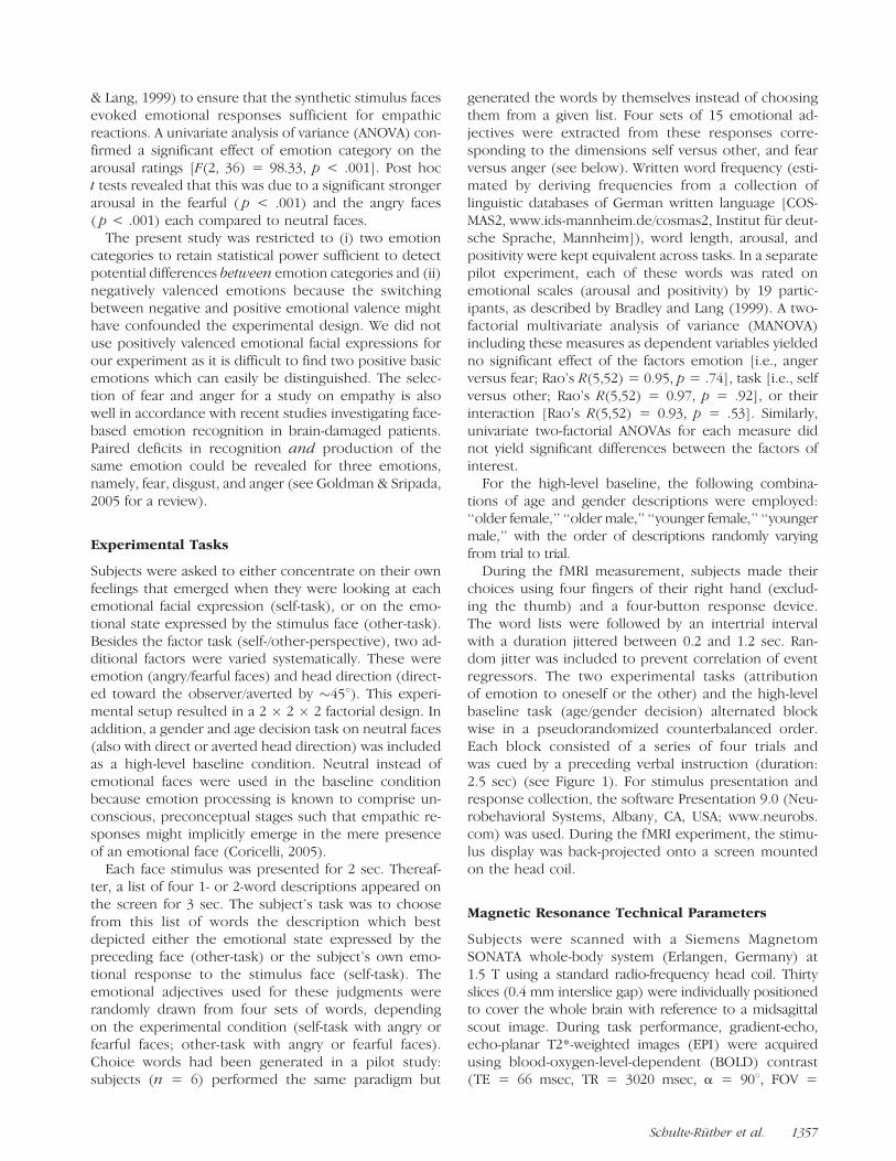
APPENDIX II. (continued)
Self-task Other-task
Fearful Faces Angry Faces Fearful Faces Angry Faces
insecure 49 annoyed 49 perplexed 55 revengeful 56
deranged 42 scared 48 shocked 53 furious 48
tense 41 disdained 43 fearful 52 fierce 45
sad 31 aggressive 38 insecure 46 nasty 43
fearful 26 irritated 32 confused 46 grumpy 41
guilty 25 harmed 28 harmed 40 attacked 33
nervous 22 angry 25 upset 37 malicious 19
overstrained 22 irate 23 tense 19 irritated 13
In the tables, numbers given after each word indicate how often the respective word was chosen by the subjects for responses in each experimentalcondition.
1370 Journal of Cognitive Neuroscience Volume 19, Number 8

maps and functional imaging data. Neuroimage, 25,1325–1335.
Ekman, P. (1982). Emotion in the human face (2nd ed.).New York: Cambridge University Press.
Ekman, P. (1984). Expression and the nature of emotion.In K. R. Scherer & P. Ekman (Eds.), Approaches toemotion (pp. 319–344). Hillsdale, NJ: Erlbaum.
Ekman, P., & Friesen, W. V. (1976). Pictures of facial affect.Palo Alto, CA: Consulting Psychologists Press.
Farrow, T. F. D., Zheng, Y., Wilkinson, I. D., Spence, S. A.,Deakin, J. F. W., Tarrier, N., et al. (2001). Investigatingthe functional anatomy of empathy and forgiveness.NeuroReport, 12, 2433–2438.
Fink, G. R., Markowitsch, H. J., Reinkemeier, M.,Bruckbauer, T., Kessler, J., & Heiss, W. D. (1996).Cerebral representation of one’s own past: Neuralnetworks involved in autobiographical memory. Journalof Neuroscience, 16, 4275–4282.
Friston, K. J., Ashburner, J., Frith, C. D., Poline, J.-B., Heather,J. D., & Frackowiak, R. (1995). Spatial registration andnormalization of images. Human Brain Mapping, 2,165–189.
Friston, K. J., Holmes, A. P., Poline, J. B., Grasby, P. J.,Williams, S. C., Frackowiak, R. S., et al. (1995). Analysisof fMRI time-series revisited. Neuroimage, 2, 45–53.
Frith, U., & Frith, C. D. (2003). Development andneurophysiology of mentalizing. Philosophical Transactionsof the Royal Society of London, Series B, Biological Sciences,358, 459–473.
Gallagher, H. L., & Frith, C. D. (2003). Functional imagingof ‘‘theory of mind’’. Trends in Cognitive Sciences, 7,77–83.
Gallagher, H. L., Happe, F., Brunswick, N., Fletcher, P. C.,Frith, U., & Frith, C. D. (2000). Reading the mind incartoons and stories: An fMRI study of ‘‘theory ofmind’’ in verbal and nonverbal tasks. Neuropsychologia,38, 11–21.
Gallagher, H. L., Jack, A. I., Roepstorff, A., & Frith, C. D.(2002). Imaging the intentional stance in a competitivegame. Neuroimage, 16, 814–821.
Gallese, V. (2003). The manifold nature of interpersonalrelations: The quest for a common mechanism.Philosophical Transactions of the Royal Society ofLondon, Series B, Biological Sciences, 358, 517–528.
Gallese, V., Keysers, C., & Rizzolatti, G. (2004). A unifyingview of the basis of social cognition. Trends in CognitiveSciences, 8, 396–403.
George, M. S., Ketter, T. A., Gill, D. S., Haxby, J. V., Ungerleider,L. G., Herscovitch, P., et al. (1993). Brain regions involvedin recognizing facial emotion or identity: An oxygen-15PET study. Journal of Neuropsychiatry and ClinicalNeurosciences, 5, 384–394.
Glaser, D. E., & Friston, K. J. (2003). Variance components.In R. S. Frackowiak, K. J. Friston, C. D. Frith, R. J. Dolan,C. J. Price, S. Zeki, et al. (Eds.), Human brain function(2nd ed.). San Diego, CA: Academic Press.
Goldman, A. I., & Sripada, C. S. (2005). Simulationist modelsof face-based emotion recognition. Cognition, 94, 193–213.
Gorno-Tempini, M. L., Pradelli, S., Serafini, M., Pagnoni, G.,Baraldi, P., Porro, C., et al. (2001). Explicit and incidentalfacial expression processing: An fMRI study. Neuroimage,14, 465–473.
Grezes, J., & Decety, J. (2001). Functional anatomy ofexecution, mental simulation, observation, and verbgeneration of actions: A meta-analysis. Human BrainMapping, 12, 1–19.
Gusnard, D. A., Akbudak, E., Shulman, G. L., & Raichle,M. E. (2001). Medial prefrontal cortex and self-referential
mental activity: Relation to a default mode of brain function.Proceedings of the National Academy of Sciences, U.S.A.,98, 4259–4264.
Hess, U., & Blairy, S. (2001). Facial mimicry and emotionalcontagion to dynamic emotional facial expressions andtheir influence on decoding accuracy. InternationalJournal of Psychophysiology, 40, 129–141.
Iacoboni, M., Koski, L. M., Brass, M., Bekkering, H., Woods,R. P., Dubeau, M. C., et al. (2001). Reafferent copies ofimitated actions in the right superior temporal cortex.Proceedings of the National Academy of Sciences, U.S.A.,98, 13995–13999.
Iacoboni, M., Lieberman, M. D., Knowlton, B. J., Molnar-Szakacs,I., Moritz, M., Throop, C. J., et al. (2004). Watching socialinteractions produces dorsomedial prefrontal and medialparietal BOLD fMRI signal increases compared to a restingbaseline. Neuroimage, 21, 1167–1173.
Iacoboni, M., Molnar-Szakacs, I., Gallese, V., Buccino, G.,Mazziotta, J. C., & Rizzolatti, G. (2005). Grasping theintentions of others with one’s own mirror neuronsystem. PLoS Biology, 3, e79.
Iacoboni, M., Woods, R. P., Brass, M., Bekkering, H.,Mazziotta, J. C., & Rizzolatti, G. (1999). Corticalmechanisms of human imitation. Science, 286, 2526–2528.
Jacob, P., & Jeannerod, M. (2005). The motor theory ofsocial cognition: A critique. Trends in Cognitive Sciences,9, 21–25.
Jeannerod, M. (2001). Neural simulation of action:A unifying mechanism for motor cognition. Neuroimage,14, S103–S109.
Kampe, K. K., Frith, C. D., & Frith, U. (2003). ‘‘Hey John’’:Signals conveying communicative intention toward theself activate brain regions associated with ‘‘mentalizing,’’regardless of modality. Journal of Neuroscience, 23,5258–5263.
Kleinke, C. L. (1986). Gaze and eye contact: A researchreview. Psychological Bulletin, 100, 78–100.
Koski, L., Iacoboni, M., Dubeau, M. C., Woods, R. P., &Mazziotta, J. C. (2003). Modulation of cortical activityduring different imitative behaviors. Journal ofNeurophysiology, 89, 460–471.
Kupfer, J., Brosig, B., & Brahler, E. (2001). Toronto-Alexithymie-Skala-26 (TAS-26) Deutsche Version.Gottingen: Hogrefe.
Lawrence, E. J., Shaw, P., Giampietro, V. P., Surguladze, S.,Brammer, M. J., & David, A. S. (2006). The role of‘‘shared representations’’ in social perception andempathy: An fMRI study. Neuroimage, 29, 1173–1184.
Leslie, K. R., Johnson-Frey, S. H., & Grafton, S. T. (2004).Functional imaging of face and hand imitation—Towardsa motor theory of empathy. Neuroimage, 21, 601–607.
Mehrabian, A. (1997). Relations among personality scales ofaggression, violence, and empathy: Validational evidencebearing on the risk of eruptive violence scale. AggressiveBehavior, 23, 433–445.
Mehrabian, A., & Epstein, N. (1972). A measure of emotionalempathy. Journal of Personality, 40, 525–543.
Mitchell, J. P., Banaji, M. R., & Macrae, C. N. (2005). Thelink between social cognition and self-referential thoughtin the medial prefrontal cortex. Journal of CognitiveNeuroscience, 17, 1306–1315.
Mitchell, J. P., Macrae, C. N., & Banaji, M. R. (2006). Dissociablemedial prefrontal contributions to judgments of similarand dissimilar others. Neuron, 50, 655–663.
Nakamura, K., Kawashima, R., Sato, N., Nakamura, A.,Sugiura, M., Kato, T., et al. (2000). Functional delineationof the human occipito-temporal areas related to face andscene processing. A PET study. Brain, 123, 1903–1912.
Schulte-Ruther et al. 1371

Ochsner, K. N., Knierim, K., Ludlow, D. H., Hanelin, J.,Ramachandran, T., Glover, G., et al. (2004). Reflectingupon feelings: An fMRI study of neural systems supportingthe attribution of emotion to self and other. Journal ofCognitive Neuroscience, 16, 1746–1772.
Oldfield, R. C. (1971). Assessment and analysis ofhandedness—Edinburgh Inventory. Neuropsychologia,9, 97–113.
Oliveri, M., Babiloni, C., Filippi, M. M., Caltagirone, C.,Babiloni, F., Cicinelli, P., et al. (2003). Influence of thesupplementary motor area on primary motor cortexexcitability during movements triggered by neutral oremotionally unpleasant visual cues. Experimental BrainResearch, 149, 214–221.
Ongur, D., Ferry, A. T., & Price, J. L. (2003). Architectonicsubdivision of the human orbital and medial prefrontalcortex. Journal of Comparative Neurology, 460, 425–449.
Piefke, M., Weiss, P. H., Zilles, K., Markowitsch, H. J., &Fink, G. R. (2003). Differential remoteness and emotionaltone modulate the neural correlates of autobiographicalmemory. Brain, 126, 650–668.
Poldrack, R. A., Wagner, A. D., Prull, M. W., Desmond,J. E., Glover, G. H., & Gabrieli, J. D. (1999). Functionalspecialization for semantic and phonological processingin the left inferior prefrontal cortex. Neuroimage, 10,15–35.
Premack, D., & Woodruff, G. (1978). Does the chimpanzeehave a theory of mind. Behavioral and Brain Sciences, 1,515–526.
Rizzolatti, G., Fogassi, L., & Gallese, V. (2001).Neurophysiological mechanisms underlying theunderstanding and imitation of action. Nature ReviewsNeuroscience, 2, 661–670.
Ruby, P., & Decety, J. (2001). Effect of subjective perspectivetaking during simulation of action: A PET investigation ofagency. Nature Neuroscience, 4, 546–550.
Ruby, P., & Decety, J. (2003). What you believe versus whatyou think they believe: A neuroimaging study of conceptualperspective-taking. European Journal of Neuroscience,17, 2475–2480.
Ruby, P., & Decety, J. (2004). How would you feel versushow do you think she would feel? A neuroimaging studyof perspective-taking with social emotions. Journal ofCognitive Neuroscience, 16, 988–999.
Saxe, R., & Kanwisher, N. (2003). People thinking aboutthinking people—The role of the temporo-parietaljunction in ‘‘theory of mind’’. Neuroimage, 19, 1835–1842.
Schilbach, L., Wohlschlaeger, A. M., Kraemer, N. C.,Newen, A., Shah, N. J., Fink, G. R., et al. (2005). Beingwith virtual others: Neural correlates of social interaction.Neuropsychologia, 44, 718–730.
Shah, N. J., Marshall, J. C., Zafiris, O., Schwab, A., Zilles, K.,Markowitsch, H. J., et al. (2001). The neural correlates ofperson familiarity. A functional magnetic resonance imagingstudy with clinical implications. Brain, 124, 804–815.
Singer, T., Seymour, B., O’Doherty, J., Kaube, H., Dolan,R. J., & Frith, C. D. (2004). Empathy for pain involves theaffective but not sensory components of pain. Science,303, 1157–1162.
Sonnby-Borgstrom, M. (2002). Automatic mimicry reactionsas related to differences in emotional empathy.Scandinavian Journal of Psychology, 43, 433–443.
Spence, S. A., Brooks, D. J., Hirsch, S. R., Liddle, P. F.,Meehan, J., & Grasby, P. M. (1997). A PET study ofvoluntary movement in schizophrenic patients experiencingpassivity phenomena (delusions of alien control).Brain, 120, 1997–2011.
Stotland, E. (1969). Exploratory investigations of empathy.In L. Berkowitz (Ed.), Advances in experimental socialpsychology (pp. 271–314). New York: Academic Press.
Sugiura, M., Shah, N. J., Zilles, K., & Fink, G. R. (2005).Cortical representations of personally familiar objects andplaces: Functional organization of the human posteriorcingulate cortex. Journal of Cognitive Neuroscience, 17,183–198.
Talairach, J., & Tournoux, P. (1988). Co-planar stereotaxicatlas of the human brain. New York: Thieme MedicalPublishers.
Vogeley, K., Bussfeld, P., Newen, A., Herrmann, S., Happe, F.,Falkai, P., et al. (2001). Mind reading: Neural mechanismsof theory of mind and self-perspective. Neuroimage, 14,170–181.
Vogeley, K., & Fink, G. R. (2003). Neural correlates of thefirst-person-perspective. Trends in Cognitive Sciences, 7,38–42.
Vollm, B. A., Taylor, A. N., Richardson, P., Corcoran, R.,Stirling, J., McKie, S., et al. (2006). Neuronal correlatesof theory of mind and empathy: A functional magneticresonance imaging study in a nonverbal task. Neuroimage,29, 90–98.
Wicker, B., Keysers, C., Plailly, J., Royet, J. P., Gallese, V., &Rizzolatti, G. (2003). Both of us disgusted in My Insula:The common neural basis of seeing and feeling disgust.Neuron, 40, 655–664.
Winston, J. S., O’Doherty, J., & Dolan, R. J. (2003). Commonand distinct neural responses during direct and incidentalprocessing of multiple facial emotions. Neuroimage, 20,84–97.
Wolpert, D. M., Doya, K., & Kawato, M. (2003). A unifyingcomputational framework for motor control and socialinteraction. Philosophical Transactions of the Royal Societyof London, Series B, Biological Sciences, 358, 593–602.
1372 Journal of Cognitive Neuroscience Volume 19, Number 8