mitochondria detection
TRANSCRIPT

Mitochondria Detection in 3D Brain Images
Joy Patel and Courtney Smith
July 22, 2014
Joy Patel and Courtney Smith Mitochondria Detection in 3D Brain Images

Introduction
How can we identify mitochondria in 3Dimage sets of a brain?
Joy Patel and Courtney Smith Mitochondria Detection in 3D Brain Images

Introduction
Data Sets We are Working With
1Supervoxel-Based Segmentation of Mitochondria in EM Image Stacks withLearned Shape Features
Joy Patel and Courtney Smith Mitochondria Detection in 3D Brain Images

Introduction
Data Sets We are Working With
Joy Patel and Courtney Smith Mitochondria Detection in 3D Brain Images

Introduction
Outline
(i) Why mitochondria?(ii) Methods we used and their results(iii) Conclusion and moving forward
Joy Patel and Courtney Smith Mitochondria Detection in 3D Brain Images

Introduction
What is the Open Connectome project?
1Volumetric Exploitation of Synaptic Information using Context Localizationand Evaluation
Joy Patel and Courtney Smith Mitochondria Detection in 3D Brain Images

Mitochondria
How does mitochondria detection play arole in the Open Connectome project?
1Supervoxel-Based Segmentation of Mitochondria in EM Image Stacks withLearned Shape Features
Joy Patel and Courtney Smith Mitochondria Detection in 3D Brain Images

Mitochondria
Characteristics of Mitochondria
Joy Patel and Courtney Smith Mitochondria Detection in 3D Brain Images

Mitochondria Detection
GoalsCreate a program that detects mitochondria in brain images
1 Properly segment the images2 Identify features to train and to classify mitochondria3 Improve boundaries of detected mitochondria
Joy Patel and Courtney Smith Mitochondria Detection in 3D Brain Images

Ground Truth
Ground Truth
Figure : Highlighted Mitochondria
Joy Patel and Courtney Smith Mitochondria Detection in 3D Brain Images

Earlier Stages
Earlier Methods
Joy Patel and Courtney Smith Mitochondria Detection in 3D Brain Images

Earlier Stages
Hough Transform
Joy Patel and Courtney Smith Mitochondria Detection in 3D Brain Images

Earlier Stages
Edge Histogram
Figure : Left: Slide Used, Right: Masked Edges
Joy Patel and Courtney Smith Mitochondria Detection in 3D Brain Images

Earlier Stages
Edge Histogram
Figure : Histograms of Object Edges
Joy Patel and Courtney Smith Mitochondria Detection in 3D Brain Images

Earlier Stages
Mitochondria Histograms
Figure : GUI with Image Histograms
Joy Patel and Courtney Smith Mitochondria Detection in 3D Brain Images

Earlier Stages
Gabor Filters
Figure : Gabor Atoms
Joy Patel and Courtney Smith Mitochondria Detection in 3D Brain Images

Earlier Stages
Gabor Filters
Joy Patel and Courtney Smith Mitochondria Detection in 3D Brain Images

Image Find
SURF Detection
Figure : Left: SURF Feature Points in Object, Right: SURF FeaturePoints in Mitochondria
Joy Patel and Courtney Smith Mitochondria Detection in 3D Brain Images
Earlier Stages
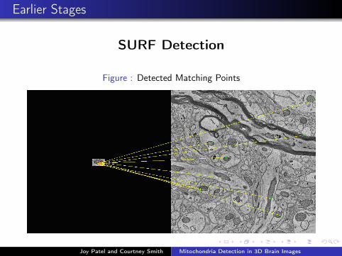
SURF Detection
Figure : Detected Matching Points
Joy Patel and Courtney Smith Mitochondria Detection in 3D Brain Images

Earlier Stages
Normalized Cross Correlation
Joy Patel and Courtney Smith Mitochondria Detection in 3D Brain Images
Mitochondria Detection
Best Working Pipeline of MitochondriaDetection
Joy Patel and Courtney Smith Mitochondria Detection in 3D Brain Images
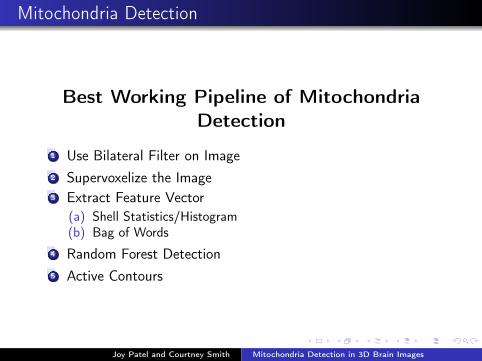
Mitochondria Detection
Best Working Pipeline of MitochondriaDetection
1 Use Bilateral Filter on Image2 Supervoxelize the Image3 Extract Feature Vector
(a) Shell Statistics/Histogram(b) Bag of Words
4 Random Forest Detection5 Active Contours
Joy Patel and Courtney Smith Mitochondria Detection in 3D Brain Images

Bilateral Filter
Bilateral Filter
Joy Patel and Courtney Smith Mitochondria Detection in 3D Brain Images

Supervoxels
Supervoxelizing the whole image
Figure : Left: Full Slide, Right: Zoomed In
Joy Patel and Courtney Smith Mitochondria Detection in 3D Brain Images

Supervoxels
Supervoxels
1SLIC Superpixels Compared to State-of-the-art Superpixel MethodsJoy Patel and Courtney Smith Mitochondria Detection in 3D Brain Images
Machine Learning
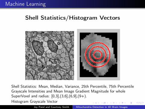
Shell Statistics/Histogram Vectors
Shell Statistics: Mean, Median, Variance, 25th Percentile, 75th PercentileGrayscale Intensities and Mean Image Gradient Magnitude for wholeSuperVoxel and radius: [0,3],(3,6],(6,9],(9+).Histogram Grayscale Vector
Joy Patel and Courtney Smith Mitochondria Detection in 3D Brain Images

Machine Learning
Bag of WordsMaking CodeBook:
Segment image into 10-by-10 patches.Convert patches into a R100 vectors.Concatenate all R100 vectors into a k-by-100 matrix.Do k-means with k=100; output from k-means is CodeBook.
Joy Patel and Courtney Smith Mitochondria Detection in 3D Brain Images

Machine Learning
Bag of Words
Figure : Codebook
Joy Patel and Courtney Smith Mitochondria Detection in 3D Brain Images

Machine Learning
Bag of Words
Joy Patel and Courtney Smith Mitochondria Detection in 3D Brain Images

Machine Learning
Use information gain to decide splits
Ij = H(Sj)−∑
i∈{L,R}
|S ij ||Sj |
H(S ij )
1www.cs.ubc.ca/ nando/540-2013/lectures.l9.pdfJoy Patel and Courtney Smith Mitochondria Detection in 3D Brain Images

Machine Learning
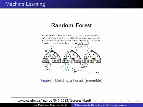
Random Forest
1www.cs.ubc.ca/ nando/540-2013/lectures.l9.pdfJoy Patel and Courtney Smith Mitochondria Detection in 3D Brain Images
Machine Learning
Random Forest
Figure : Building a Forest (ensemble)
1www.cs.ubc.ca/ nando/540-2013/lectures.l9.pdfJoy Patel and Courtney Smith Mitochondria Detection in 3D Brain Images

Supervoxels
Machine Learning
Joy Patel and Courtney Smith Mitochondria Detection in 3D Brain Images
Mitochondria Detection
Active Contour
1Image Segmentation Using the Chan-Vese Algorithm - Robert CrandallJoy Patel and Courtney Smith Mitochondria Detection in 3D Brain Images

Active Contour
Selected Centerpoints vs. Supervoxels
Joy Patel and Courtney Smith Mitochondria Detection in 3D Brain Images

Active Contour
Selected Centerpoints vs. Supervoxels
Joy Patel and Courtney Smith Mitochondria Detection in 3D Brain Images

Active Contour
Selected Centerpoints vs. Supervoxels
Joy Patel and Courtney Smith Mitochondria Detection in 3D Brain Images

Results
Results
Joy Patel and Courtney Smith Mitochondria Detection in 3D Brain Images

Ground Truth
3D Visualization of ResultsWith Connected Components
Joy Patel and Courtney Smith Mitochondria Detection in 3D Brain Images
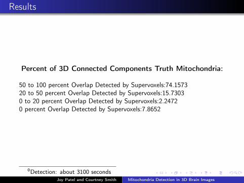
Results
Percent of 3D Connected Components Truth Mitochondria:
50 to 100 percent Overlap Detected by Supervoxels:74.157320 to 50 percent Overlap Detected by Supervoxels:15.73030 to 20 percent Overlap Detected by Supervoxels:2.24720 percent Overlap Detected by Supervoxels:7.8652
0Detection: about 3100 secondsJoy Patel and Courtney Smith Mitochondria Detection in 3D Brain Images

Supervoxels
Machine Learning
Percent of 2d Supervoxels Detected:True Positives: 86.6092False Positives: 13.3908
Joy Patel and Courtney Smith Mitochondria Detection in 3D Brain Images
Conclusion
(i) Why mitochondria plays a vital role in the Open Connectomeproject
(ii) Methods we have used and their results
We have seen that supervoxels and random forest used with activecontours have given us the best results for mitochondria detection.Through our research we have noticed that others have beenpublishing papers where they are implementing similar methodsthat we have used.
Joy Patel and Courtney Smith Mitochondria Detection in 3D Brain Images

ConclusionFurther Research
Implement Supervoxel process in 3D and compare results.Determine a better feature space for machine learning.
Joy Patel and Courtney Smith Mitochondria Detection in 3D Brain Images

A special thanks to:
1 Duke Math RTG2 Dr. Guillermo Sapiro3 Dr. Paul Bendich4 Dr. Joshua Vogelstein5 Dr. John Harer6 Dr. Rann Bar-On7 Dr. Robert Calderbank8 Chris Tralie9 William R. Gray10 Dean Kleissas11 Kathy Peterson
Joy Patel and Courtney Smith Mitochondria Detection in 3D Brain Images