mitochondrial disease: genetics and management...inheritance patterns of mutations in nuclear genes...
TRANSCRIPT

NEUROLOGICAL UPDATE
Mitochondrial disease: genetics and management
Yi Shiau Ng1 • Doug M. Turnbull1
Received: 25 June 2015 / Revised: 18 August 2015 / Accepted: 18 August 2015 / Published online: 28 August 2015
� The Author(s) 2015. This article is published with open access at Springerlink.com
Abstract Mitochondrial disease is one of the most
common groups of genetic diseases with a minimum
prevalence of greater than 1 in 5000 in adults. Whilst
multi-system involvement is often evident, neurological
manifestation is the principal presentation in most cases.
The multiple clinical phenotypes and the involvement of
both the mitochondrial and nuclear genome make mito-
chondrial disease particularly challenging for the clinician.
In this review article we cover mitochondrial genetics and
common neurological presentations associated with adult
mitochondrial disease. In addition, specific and supportive
treatments are discussed.
Keywords Mitochondrial disease � Mitochondrial DNA
(mtDNA) � Nuclear genes � Acute and chronic neurological
presentations � Treatment
Introduction
Mitochondrial disease is a collective term that encom-
passes the genetically and clinically heterogeneous group
of diseases due to defects in mitochondrial oxidative
phosphorylation. It is one of the most common groups of
genetic disease and can be caused by mutation in either
mitochondrial DNA or nuclear genes that directly or indi-
rectly interfere with the mitochondrial respiratory chain
function. To date, mitochondrial proteomics analysis
reveals that in addition to the 13 proteins encoded by the
mitochondrial genome, around 1500 proteins [50] are
linked to various mitochondrial functions and so far more
than 200 genes have been implicated in the development of
human disease [40].
A number of syndromes have been described in mito-
chondrial disease but often patients present with non-syn-
dromic presentation of which nervous system is most
commonly affected [49]. In addition to the diagnostic
challenge, clinicians also encounter difficulty in the man-
agement of mitochondrial disease due to lacking of effec-
tive disease-modifying therapy and, until recently, best
practice guidelines on various complications associated
with the disease [59].
In this review article, we discuss the genetics and epi-
demiology of mitochondrial disease, neurological presen-
tations and their management, genetic counselling and
reproductive options for patients.
Mitochondrial genetics
Mitochondria are cellular organelles found in all nucleated
human cells. A crucial function of mitochondria is to
generate energy in the form of ATP (adenosine triphos-
phate) via oxidative phosphorylation using predominantly
carbohydrates and fatty acids as fuel. The oxidative phos-
phorylation system (OXPHOS) is located in the inner
membrane and it consists of five multimeric protein com-
plexes: complex I-IV form the respiratory chain and com-
plex V (ATP synthase). In addition, there are two mobile
electron carriers (co-enzyme Q10 and cytochrome c).
Mitochondria are under dual genetic control of the
mitochondrial and nuclear genomes. The mitochondrial
genome consists of multiple copies of 16,569 bp, double
& Doug M. Turnbull
1 Wellcome Trust Centre for Mitochondrial Research, Institute
of Neuroscience, The Medical School, Newcastle University,
Framlington Place, Newcastle upon Tyne NE2 4HH, UK
123
J Neurol (2016) 263:179–191
DOI 10.1007/s00415-015-7884-3
stranded mitochondrial DNA (mtDNA) molecules and
located adjacent to the OXPHOS system in the matrix.
Only thirty-seven genes (22 transfer RNAs, 2 ribosomal
RNAs and 13 polypeptides that form structural subunits of
OXPHOS system) [86] are encoded by mtDNA. The
remaining mitochondrial proteins, including the majority of
respiratory chain subunits (79 out of 92), assembly factors
of the respiratory chain, those involved in maintenance and
expression of mtDNA, mtDNA transcription and transla-
tion, and control the mitochondrial dynamics are nuclear
encoded [16], synthesised in the cytosol and imported to
the mitochondria [51].
There are several unique properties associated with the
mitochondrial genome that are important in understanding
the primary mitochondrial DNA disease: (1) there are
multiple copies (up to thousands) of mtDNA in each cell;
(2) mtDNA is maternally inherited; (3) the phenomenon of
homoplasmy and heteroplasmy. Homoplasmy implies all
mtDNA are identical which could be all wild type or
mutated. Heteroplasmy is a mixture of mutated and wild
type mtDNA. In the presence of heteroplasmy there is a
threshold effect and clinical expression can vary between
different tissues and mtDNA mutations. In women with
heteroplasmic mtDNA mutations there is a bottleneck in
the female germline which means that the transmission of
heteroplasmy level from mother to offspring is often ran-
dom and unpredictable. This explains the heterogeneity in
heteroplasmy level, clinical phenotype and severity fre-
quently observed within the same pedigree.
Multiple mtDNA deletions and mtDNA depletion (re-
duced copy number of mtDNA) are secondary changes in
mtDNA due to mutations in the mtDNA replication and/or
maintenance genes such as POLG, PEO1, ANT1, DGUOK,
TYMP [72]. Mitochondrial depletion syndrome is associ-
ated with infantile/early childhood onset, multi-system
disease with fatal outcome and multiple deletions generally
result in later onset (child- or adulthood) and milder disease
burden.
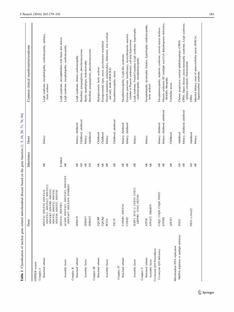
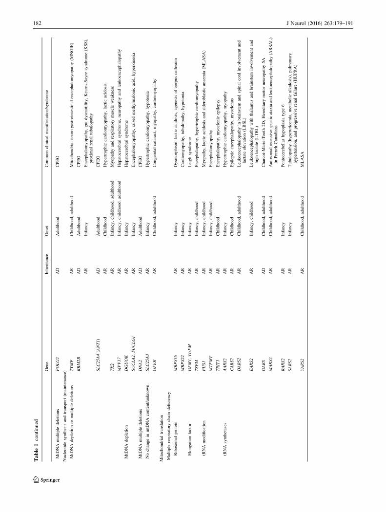
The emergence of next generation sequencing is lead-
ing to rapid discovery of new nuclear genes linked to
mitochondrial disease and the classification of nuclear
gene related mitochondrial disease is summarised in
Table 1. There is also increased recognition of mito-
chondrial dysfunction and genetic mutation in various
other genetic neurological disorders, for examples SPG7
gene in hereditary spastic paraplegia [58] and ataxia [61]
and GDAP1 gene in Charcot-Marie-Tooth disease type 4A
[56, 96].
The inheritance pattern of mitochondrial disease is
dependent on the genetic mutations. Point mutations in the
primary mitochondrial DNA such as m.3243A[G, three
common LHON mutations, m.8344A[G, m.8993T[G/C,
m.1555A[G and others are maternally inherited but
sporadic mutations exist [53]. Single, large deletions in
mtDNA are a common cause mitochondrial disease and
they occur sporadically with rare exceptions [15]. The
inheritance patterns of mutations in nuclear genes causing
mitochondrial disease include autosomal recessive, domi-
nant or X-linked. Both recessive and dominant forms exist
in several mtDNA maintenance genes, for examples
POLG, PEO1 and RRM2B genes.
Epidemiology of mitochondrial disease
The overall prevalence of mitochondrial disease is com-
parable to other neurogenetic diseases such as Charcot-
Marie-Tooth (CMT) disease, myotonic dystrophy and
muscular dystrophy. The prevalence of adult mitochondrial
disease, both affected patients and those at risk, has
recently been reported to be approximately 1 in 4300 in
North East England [28]. Primary mutations in the mtDNA
are more prevalent in the adult patients compared to
mutations in the nuclear genes, and vice versa in the pae-
diatric population where there is a much higher incidence
of autosomal recessive disease particularly in consan-
guineous families. Although several hundreds of mutations
have been reported in mtDNA since 1988, a handful of
mutations are far more common than the others, for
examples m.1555A[G (associated with aminoglycoside
induced deafness), m.3243A[G (associated with MELAS
syndrome and MIDD), m.3460G[A and m.11778A[G
(associated with LHON), have an estimated prevalence of
0.19, 0.14, 0.11 and 0.11 %, respectively, in the population
[11, 21]. However, individuals with these common muta-
tions may remain clinically asymptomatic throughout their
life if not exposed to relevant toxin or if they have a low
mutation load.
Clinical diagnosis
Mitochondria are ubiquitous and therefore mitochondrial
disease can affect any organ, although organs with high
energy demand such as brain, skeletal muscle and heart, are
more commonly affected than the others. The clinical
features are heterogeneous and often can mimic many
neurological or other systemic diseases. Multi-system
involvement is often evident with detailed clinical exami-
nation and investigations in most cases although there are
exceptions such as Leber hereditary optic neuropathy
(LHON). Paediatric onset disease is associated with more
severe multi-systemic involvement, relentless progression
and poorer prognosis, however, there are rare exceptions
such as reversible respiratory chain deficiency caused by
the m.14674T[C mutation [33].
180 J Neurol (2016) 263:179–191
123
Table
1Classificationofnucleargenerelatedmitochondrial
disease
based
onthegenefunction[1,5,16,20,71,76,94]
Gene
Inheritance
Onset
Commonclinical
manifestation/syndrome
OXPHOSsystem
ComplexI
Structuralsubunit
NDUFA2,NDUFA9,NDUFA10,
NDUFA11,NDUFA12,NDUFA13,
NDUFB3,NDUFB9,NDUFS1,
NDUFS2,NDUFS3,NDUFS4,
NDUFS6,NDUFS7,NDUFS8
AR
Infancy
Leighsyndrome,
encephalopathy,cardiomyopathy,epilepsy,
lactic
acidosis
NDUFA1,NDUFB11
X-linked
Leighsyndrome,
microphthalmia
withlinearskin
defects
Assem
bly
factor
ACAD9,NDUFAF1,NDUFAF2,NDUFAF4,
NDUFAF5,NDUFAF6,FOXRED1
AR
Leighsyndrome,
encephalopathy,cardiomyopathy
ComplexII
Structuralsubunit
SDHA-D
AR
Infancy,childhood
Leighsyndrome,
dilated
cardiomyopathy
AD
Childhood,adulthood
Hereditaryparaganglioma,
pheochromocytoma
Assem
bly
factor
SDHAF1
AR
Infancy
Spasticquadriplegia,leukodystrophy
SDHAF2
AD
Adulthood
Hereditaryparaganglioma,
pheochromocytoma
ComplexIII
Structuralsubunit
UQCRB
AR
Childhood
Hypoglycaem
ia,lactic
acidosis
UQCRQ
AR
Childhood
Extrapyramidal
signs,ataxia,psychomotorretardation
Assem
bly
factor
BCS1L
AR
Infancy
Growth
retardation,am
inoaciduria,
cholestasis,ironoverload,
andearlydeath
(GRACILE)
TTC19
AR
Childhood,adulthood
Encephalomyopathy,ataxia
ComplexIV
Structuralsubunit
COX6B1,NDUFA4
AR
Infancy,childhood
Encephalomyopathy,Leigh-likesyndrome
COX4I2
AR
Infancy,childhood
Exocrinepancreaticinsufficiency,dyserythropoetic
anaemia
(sim
ilar
toPearsonsyndrome),calvarialhyperostosis
Assem
bly
factor
SURF1,SCO1-2,COX10,COX15,
LRPPRC,COA5,PET100
AR
Infancy
Leighsyndrome,
French-CanadianLeighsyndrome,
hypertrophic
cardiomyopathy,encephalomyopathy
ComplexV
Structuralsubunit
ATP5E
AR
Infancy
Encephalopathy,dysm
orphic
features,hypertrophic
cardiomyopathy,
lactic
acidosis
Assem
bly
factor
ATPAF2,TMEM70
AR
Co-enzymeQ10biosynthesis
Co-enzymeQ10deficiency
COQ2,COQ4,COQ6,PDSS2
AR
Infancy,childhood
Encephalomyopathy,nephroticsyndrome,
sensori-neuraldeafness
ETFDH
AR
Infancy,childhood,adulthood
Glutaricacidaemia
IIC
(multiple
acyl-CoA
dehydrogenasedeficiency,
MADD),Myopathy
ADCK3
AR
Childhood
Cerebellarataxia
Mitochondrial
DNA
replication
MtDNA
depletionormultiple
deletions
POLG
AD
Adulthood
Chronic
progressiveexternal
ophthalmoplegia
(CPEO)
AR
Infancy,childhood,adulthood
CPEO,Alpersdisease,ataxia-neuropathysyndrome,
Leighsyndrome,
epilepsy
(occipital),Parkinsonism
PEO1(c10orf2)
AD
Adulthood
CPEO
AR
Infancy
Inherited
Infancy
onsetofspinocerebellarataxia
(IOSCA),
hepatocerebralsyndrome
J Neurol (2016) 263:179–191 181
123
Table
1continued
Gene
Inheritance
Onset
Commonclinical
manifestation/syndrome
MtDNA
multiple
deletions
POLG2
AD
Adulthood
CPEO
Nucleotidesynthesis
andtransport(m
aintenance)
MtDNA
depletionormultiple
deletions
TYMP
AR
Childhood,adulthood
Mitochondrial
neuro-gastrointestinal
encephalomyopathy(M
NGIE)
RRM2B
AD
Adulthood
CPEO
AR
Infancy
Encephalomyopathy,gutdysm
otility,Kearns-Sayre
syndrome(K
SS),
proxim
alrenal
tubulopathy
SLC25A4(ANT1)
AD
Adulthood
CPEO
AR
Childhood
Hypertrophic
cardiomyopathy,lactic
acidosis
TK2
AR
Infancy,childhood,adulthood
Myopathyandrespiratory
muscle
weakness
MPV17
AR
Infancy,childhood,adulthood
Hepatocerebralsyndrome,
neuropathyandleukoencephalopathy
MtDNA
depletion
DGUOK
AR
Infancy
Hepatocerebralsyndrome
SUCLA2,SUCLG1
AR
Infancy
Encephalomyopathy,raised
methylm
alonic
acid,hyperkinesia
MtDNA
multiple
deletions
DNA2
AD
Adulthood
CPEO
Nochangein
mtDNA
content/unknown
SLC25A3
AR
Infancy
Hypertrophic
cardiomyopathy,hypotonia
GFER
AR
Childhood,adulthood
Congenital
cataract,myopathy,cardiomyopathy
Mitochondrial
translation
Multiple
respiratory
chaindeficiency
Ribosomal
protein
MRPS16
AR
Infancy
Dysm
orphism,lactic
acidosis,agenesis
ofcorpuscallosum
MRPS22
AR
Infancy
Cardiomyopathy,tubulopathy,hypotonia
Elongationfactor
GFM1,TUFM
AR
Infancy
Leighsyndrome
TSFM
AR
Infancy,childhood
Encephalopathy,hypertrophic
cardiomyopathy
tRNA
modification
PUS1
AR
Infancy,childhood
Myopathy,lactic
acidosisandsideroblastic
anaemia
(MLASA)
MTFMT
AR
Infancy,childhood
Encephalomyopathy
TRIT1
AR
Childhood
Encephalopathy,myoclonic
epilepsy
tRNA
synthetases
AARS2
AR
Infancy
Hypertrophic
cardiomyopathy,myopathy
CARS2
AR
Childhood
Epilepticencephalopathy,myoclonus
DARS2
AR
Childhood,adulthood
Leukoencephalopathyin
brainstem
andspinal
cord
involvem
entand
lactateelevation(LBSL)
EARS2
AR
Infancy,childhood
Leukoencephalopathywiththalam
usandbrainstem
involvem
entand
highlactate(LTBL)
GARS
AD
Childhood,adulthood
Charcot-Marie-Tooth
2D,Hereditarymotorneuropathy5A
MARS2
AR
Childhood,adulthood
Autosomal
recessivespasticataxiaandleukoencephalopathy(A
RSAL)
inFrench
Canadians
RARS2
AR
Infancy
Pontocerebellarhypoplasiatype6
SARS2
AR
Infancy
Tubulopathy(hyperuricemia,metabolicalkalosis),pulm
onary
hypertension,andprogressiverenal
failure
(HUPRA)
YARS2
AR
Childhood,adulthood
MLASA
182 J Neurol (2016) 263:179–191
123

Many classic syndromes have been described over the
last few decades. The examples of clinical syndromes
associated with adolescence and adulthood include mito-
chondrial encephalomyopathy, lactic acidosis with stroke-
like episode (MELAS), myoclonic epilepsy with ragged
red fibres (MERRF), mitochondrial neuro-gastrointestinal
involvement and encephalopathy (MNGIE), neuropathy,
ataxia and retinitis pigmentosa (NARP), chronic progres-
sive external ophthalmoplegia (CPEO). In contrast, syn-
dromes with neonatal and childhood onset include Alpers
disease, Pearson syndrome, Leigh disease, Sengers syn-
drome and Kearns-Sayre syndrome.
However, clinicians are more commonly confronted by
the non-specific constellation of clinical features. Many
symptoms associated with mitochondrial disease such as
deafness, diabetes, myopathy, gastrointestinal symptoms
and others are also common on their own in the population
but the ‘unusual’ combination of these symptoms in the
same individual should alert the clinicians about the pos-
sibility of mitochondrial disease. Detailed system-based
examination coupled with extended investigations is nec-
essary to identify other system involvement such as short
stature, sensori-neural deafness, pigmentary retinopathy,
optic atrophy, diabetes mellitus and/or other endocrine
dysfunction, cardiac involvement, renal tubulopathy and
others. This systemic involvement may be subtle and
asymptomatic in the early phase of disease. Disease rating
scales have been utilised to document the extent of system
involvement, disease burden and progression in paediatric
and adult patients [62, 69].
Family history can be informative and often reveals
what appear to be seemingly unrelated diseases among the
maternal family members in primary mtDNA disease and
the m.3243A[G mutation is a prime example [54]. It is
important to ascertain history of consanguinity when
recessive disease is suspected. Late adulthood presentation
and/or lacking of apparent family history should not deter
testing for mitochondrial disease because sporadic muta-
tions or late presenting autosomal dominant diseases are
not uncommon.
Acute neurological presentations
Stroke-like episodes and acute symptomatic seizure
MELAS syndrome is a severe, multi-system disease char-
acterised by recurrent metabolic strokes with typical onset
of below 40 years [31] although patients at the older age
have been reported [6, 92]. Headache and prominent visual
disturbance (both positive and negative symptoms) are
often the prodrome of acute stroke-like episodes and can
occur days or weeks before the development of focalTable
1continued
Gene
Inheritance
Onset
Commonclinical
manifestation/syndrome
Mitochondrial
dynam
icnetwork
(mitochondrial
mem
branebiogenesis
andmaintenance)
Fusion
MFN2
AD
Childhood,adulthood
Charcot-Marie-Tooth
2A
(CMT2A)(m
ultiple
deletions)
OPA1
AD
Childhood
Opticatrophy(m
ultiple
deletions)
OPA3
AD
Adulthood
Opticatrophy
AR
Infancy,childhood
TypeIII3-m
ethylglutaconic
aciduria,
Costeffsyndrome
PINK1
AR
Childhood,adulthood
JuvenileParkinsonDisease
Fission
DNM1L(D
RP1)
AR
Infancy
Microcephaly,lactic
acidosis,opticatrophy
ARautosomal
recessive,
AD
autosomal
dominant
J Neurol (2016) 263:179–191 183
123

neurological deficit or motor seizure. These visual symp-
toms can masquerade as migranous visual aura but in fact
is the onset of occipital seizure [24, 35]. The severity of
neurological deficits are related to the extent of parietal,
temporal and occipital lobe involvement such as dysphasia,
dyspraxia, heminanopia, cortical blindness, mild hemi-
paresis and psychosis. Epilepsia partialis continua and less
commonly generalised status epilepticus occur during the
stroke-like episodes in some patients.
The key imaging findings are cortical and subcortical
lesions that cross the vascular territories of middle cerebral
artery and posterior cerebral arteries and bilateral, asym-
metrical changes are not infrequent (Fig. 1). The most
common cause of MELAS phenotype is m.3243A[G
mutation which accounts for 80 % of cases but mutations
in the nuclear gene POLG, encoding for the catalytic
subunit of DNA polymerase c (pol c), can cause similar
stroke-like lesions [14, 19]. However, the POLG-related
disease often has more aggressive disease course with
explosive onset of focal seizure and status epilepticus that
is highly refractory to pharmacological treatments in chil-
dren and young adults and the outcome is very poor.
Administration of sodium valproate is recognised to trigger
fulminant hepatic failure among patients with POLG dis-
ease [73, 80]. In contrast, patients who have stroke-like
episodes associated with the m.3243A[G mutation tend to
have pre-existing, and often subtle, multi-system involve-
ment and they often make good recovery in conjunction
with the partial or complete resolution of imaging changes
within few weeks or months at the early course of disease if
appropriately managed (refer to Treatment section). Nev-
ertheless, recurrence of stroke-like episodes leads to
cumulative neuronal loss and results in severe cognitive
impairment.
Given the high prevalence of m.3243A[G mutation and
common carrier status of several pathogenic variants in
POLG gene (p. A467T, p. W748S and p. G848S) in pop-
ulations of European descent [29], clinicians should pri-
oritise mitochondrial disease as a main differential
diagnosis to atypical, evolving posterior circulation stroke,
recurrent ‘encephalitis/encephalopathy’ with negative
infective screen, auto-antibodies and ‘atypical/recurrent’
posterior reversible encephalopathy syndrome (PRES).
Securing the diagnosis of mitochondrial disease early can
Fig. 1 Axial FLAIR (a, b) andDWI (c, d) sequences of MRI
head. a and c were performed
on admission whilst b and
d were performed 8 days later.
The stroke-like lesion ‘spread’
from the right occipital lobe to
the right temporal lobe and
thalamus
184 J Neurol (2016) 263:179–191
123
avert the patients having invasive diagnostic procedure
such as brain biopsy or potentially harmful treatment such
as immune-suppressants.
Subacute visual loss
The classic presentation of LHON is subacute, evolving
painless visual loss in young adults with male predomi-
nance (4:1) [43]. Majority of the cases have the con-
tralateral eye affected within a year and visual loss is often
irreversible. Although there is a conventional belief that
LHON mutations only affect eyes, other neurological fea-
tures such as dystonia [48], myoclonus [41], sensori-neural
deafness [66] may occur which broaden the spectrum of the
clinical phenotype in these mutations [48]. It is clear that
therefore LHON should be considered as a differential
diagnosis to the ‘atypical’ optical neuritis that shows no
recovery and Susac’s syndrome [97]. A link between
LHON mutations and multiple sclerosis was speculated for
a long time and a recent study has suggested that such
association may occur by chance even though mechanistic
interaction is possible [57].
Dominant optic neuropathy is caused by mutations in
the nuclear gene such as OPA1 and the irreversible visual
loss often occurs in the childhood [95] although late-onset
optic neuropathy after 4th decade [2] and adult-onset
CPEO and parkinsonism with subclinical optic neuropathy
have recently been described [12].
Chronic neurological presentations
Chronic progressive external ophthalmoplegia
and ptosis
More than half of adult patients have external ophthalmo-
plegia and/or ptosis in our large cohort of patients (un-
published). Ptosis and ophthamoplegia can be
asymmetrical at the outset but most cases become sym-
metrical with time. CPEO is one of the main presenting
features in adult patients with mitochondrial disease [77]
and ptosis often imposes more functional limitation than
the restricted eye movement because the extra-ocular
paresis occurs insidiously allowing cerebral adaptation and
symptomatic diplopia is less common. Some patients with
CPEO phenotype are occasionally misdiagnosed as other
conditions such as seronegative myasthenia gravis.
Myopathy
Many adult patients experience fatigue, exercise intoler-
ance and muscle weakness. The degree of proximal
myopathy is often mild on clinical testing and it progresses
slowly. Early loss of ambulation due to muscle weakness is
not a typical feature of adult mitochondrial disease with a
few exceptions such as patients with Kearns–Sayre syn-
drome caused by single deletion in mtDNA and TK2
mutation [4]. There is a risk of aspiration when facial and
oropharyngeal weakness is present in addition to the res-
piratory muscle weakness. Some patients may have normal
muscle strength and EMG study but complain of limited
exercise capacity with recurrent nausea and/or vomiting on
exertion due to lactic acidosis.
Ataxia
Cerebellar ataxia is often subtle at the onset and typically
progress with time and becomes debilitating in many
genotypes. Cerebellar atrophy is a common imaging find-
ing. Sensory ataxia due to dorsal root ganglionopathy is
one of the defining features in SANDO phenotype (sensory
ataxia, neuropathy, dysarthria and ophthalmoplegia) caused
by POLG mutations [42].
Neuropathy
Diminished or absent long tendon reflexes are a common
clinical finding and axonal neuropathy is the most common
finding in nerve conduction studies in adult patients with
mitochondrial disease with a few exceptions such as
demyelinating neuropathy described in patients with MNGIE
[25].A recent study showed that neuropathy is a useful feature
toguidemolecular diagnosis in adult patientswithCPEO[32].
Diagnostic approach
Comprehensive diagnostic criteria and guidelines have
been published to provide a framework for clinicians when
investigating patients with suspected mitochondrial disease
[8, 85, 86]. Patients who present with classic syndromes
such as MELAS, MERRF, LHON and Alpers disease can
be diagnosed by direct sequencing of mitochondrial genes
or POLG gene in blood. It is important to consider that
blood heteroplasmy (leucocytes) declines with age in
m.3243A[G [65] and ‘false negative result’ is possible in
older adults therefore concomitant testing of additional
tissue such as urinary epithelium is recommended. Some
nuclear gene mutations have been found to have associa-
tion with distinctive radiological appearances which can
expedite the candidate gene sequencing, these include
leukoencephalopathy with brainstem and spinal cord
involvement and lactate elevation (LBSL) caused by
DARS2 mutations [89] and leukoencephalopathy with
thalamus and brainstem involvement and high lactate
(LTBL) caused by EARS2 mutations [78].
J Neurol (2016) 263:179–191 185
123
However, many mitochondrial diseases do not have
pathognomonic features that point towards a particular
genetic diagnosis and muscle biopsy remains important in
current clinical practice. The findings of muscle biopsy that
are supportive of mitochondrial disease include: ragged red
fibres (RRF), COX-negative fibres, individual complex or
multiple respiratory chain deficiency, qualitative (multiple
deletions) and quantitative (depletion) abnormalities in
mtDNA.
Next generation sequencing is a new and high-
throughput technique that allows sequencing of multiple
candidate genes simultaneously leading to a more rapid
diagnosis and increase the diagnostic yield [88] especially
in the well-phenotyped cohort of patients [84]. Clinical
exome sequencing is likely to become part of standard
clinical care for undiagnosed patients.
Treatment and long term management
Specific treatment in mitochondrial disease
Currently, there remains no effective and specific treatment
for vast majority of patients with mitochondrial disease.
Various treatments (mostly nutritional supplements) such
as co-enzyme Q10, carnitine, creatine, dichloroacetate and
vitamin ‘cocktails’ have been widely used based upon
anecdotal data and individual case reports, however,
Cochrane systemic review of treatments that were tested in
randomised-control trials concluded that none of these
treatment showed meaningful clinical efficacy [59]. Since
then, another randomised, controlled trial using idebenone
in LHON mutations found no significant result in the pri-
mary end point defined as the best recovery in visual acuity
but post hoc interaction analysis suggested benefits in those
with discordant visual acuities [38]. EPI-743, a novel anti-
oxidant has been reported to show clinical improvement in
small number of patients with Leigh syndrome [47] and
LHON [67] in the open-label clinical trials and phase 2B
randomized-control trial is currently in progress.
Although there is no formal clinical trial, supplement of
high dose co-enzyme Q10 (up to 2400 mg in three divided
doses in adults; 30 mg/kg in paediatric cases) in primary
co-enzyme Q10 biosynthetic defect has been reported to
show variable clinical improvement across different phe-
notypes [23].
L-arginine has been reported to be effective in treating
acute stroke-like episodes associated with m.3243A[G
mutation [39]. However, this result is yet to be replicated
by other research groups.
Allogenic haematopoietic stem cell transplant has
emerged as a promising therapeutics to restore the enzy-
matic function in patients with MNGIE caused by TYMP
mutations but this treatment is associated with high mor-
bidity and mortality [25, 75].
Supportive treatment and surveillance
for complications
Symptomatic treatment and screening for associated com-
plications remain fundamental to the management of
mitochondrial disease.
Acute seizure and stroke-like episodes
Early recognition and prompt, aggressive seizure man-
agement are crucial to mitigate the cellular metabolic crisis
perpetuated by the ictal activities [10]. The seizure man-
agement should follow the guidelines on status epilepticus
except sodium valproate is absolutely contra-indicated in
POLG mutations. Phenytoin was implicated in causing
paralytic ileus in a patient with stroke-like episodes [17],
however, this is likely to be a co-incidental finding as
pseudo-obstruction is a common complication associated
with MELAS syndrome [74] and we use this drug regularly
to control seizures. Anecdotal evidence on the use of
magnesium infusion [91], ketamine [64], ketogenic diet
[34, 46, 79], folinic acid supplement [30] in termination of
status epilepticus associated with POLG and m.3243A[G
has been reported.
Pseudo-obstruction
Several genetic mutations (TYMP, m.3243A[G and
POLG) have been associated with intestinal pseudo-ob-
struction [9, 63, 83, 90] involving small and/or large
intestine. Distinguishing this from mechanical obstruction
is of paramount importance as they tend to resolve with
conservative management alone and surgery has little role
and could exacerbate the metabolic crisis. Serum lactate is
not a reliable marker for tissue ischaemia in patients with
mitochondrial disease because some patients have persis-
tently raised lactate even when they are well. Aggressive
medical management of acute episodes is important as is
prevention by using regular laxatives.
Cardiac involvement
Cardiac involvement is often part of the multi-system
manifestation in adult mitochondrial DNA disease [7]
although isolated cardiomyopathy has been reported in rare
mtDNA mutations [26]. Cohort studies have shown that
hypertrophic cardiomyopathy and pre-excitation syndrome
are prevalent in m.3243A[G [37, 45] and m.8344A[G [13,
44] mutations, whereas conduction defect necessitating
pacemaker is associated with single, large scale deletion
186 J Neurol (2016) 263:179–191
123

particularly among those who have Kearns-Sayre syn-
drome. Currently, there is limited longitudinal data study-
ing the prevalence of cardiac involvement in various
nuclear genes in adults [60]. Baseline cardiac assessment
with electrocardiogram (ECG) and echocardiogram should
be performed in all patients and cardiac magnetic reso-
nance in selected cases. Subsequent cardiac surveillance
should be tailored according to the initial findings but the
recommended interval is every 12 to 24 months [22, 55].
Diabetes mellitus
Diabetes mellitus is common in patients with m.3243A[G
and single, large scale mtDNA deletions [70]. Metformin is
best avoided because of the theoretical risk of lactic aci-
dosis. Treatment of mitochondrial diabetes is otherwise
similar to the usual form of diabetes although it appears
that it is associated with a more rapid progression to insulin
therapy [93].
Ptosis
A proportion of patients with CPEO develop significant
ptosis that obscures visual field. Corrective ptosis surgery
such as frontalis sling operation improves the functional
and cosmetic outcome in selected patients [3].
Deafness
Young onset, bilateral sensori-neural deafness is prevalent
in mitochondrial disease. The quality of life of many
patients can simply be improved with digital hearing aid
and cochlear implant can be reserved for those with severe
hearing loss [36, 81].
Genetic counselling and reproductive options
Similarly to other genetic disorders, screening for family
members at risk and offering genetic counselling is
essential in mitochondrial disease. For patients with
nuclear gene disorders, genetic counselling and reproduc-
tive options are identical to other nuclear defects. For
women with mtDNA mutations genetic counselling is a
challenging area. Patients should be reassured if they har-
bour sporadic mutation, such as single, large scale mtDNA
deletion, because risks of transmission are low. For
mtDNA point mutations, accurate elucidation of the risk of
transmission and prediction of disease status is extremely
challenging due to the genetic bottleneck effect and uneven
tissue segregation associated with some point mutations. It
is estimated that there are approximately 152 births per
year in the UK of children who carry potentially
pathogenic mitochondrial DNA mutations [27]. In view of
the complexity of mtDNA genetics, referral of child-
bearing age female patients to specialist centres for dis-
cussion of reproductive options is recommended. The
available options are chorionic villous sampling (CVS),
amniocentesis [52] and preimplantation genetic diagnosis
(PGD) [68, 87]. CVS and amniocentensis are performed at
different stages of pregnancy, 10–12 and 14–20 weeks,
respectively. PGD is an IVF procedure that involves
embryo biopsy and the selection of embryos with the
lowest mutation load. However, PGD will not benefit
carriers with homoplasmic mtDNA mutation.
Mitochondrial donation, either pronuclear transfer [18]
or metaphase II spindle transfer [82], is emerging as a
potential reproductive option to prevent the transmission of
mtDNA mutations. In the UK after many years of debate
and scientific scrutiny, Mitochondrial Donation Regula-
tions, were passed by both Houses of Parliament, making
mitochondrial donation legal for the first time in the UK.
The Human Fertilisation and Embryology Authority will
now develop a licencing framework through which appli-
cations can be considered on a case by case basis.
Conclusions
Over recent years there have been important advances in
mitochondrial disease, particularly in terms of diagnosis
and reproductive options available. The role of the clini-
cian remains crucial since a high index of clinical suspicion
and prompt recognition of complications remain essential
to make an earlier diagnosis and instigate a better man-
agement. Currently, the management of mitochondrial
disease is largely supportive; however, with the improved
understanding of disease mechanisms, ongoing treatment
trials and discovery of new therapeutic agents should give
hope for patients with mitochondrial disease.
Acknowledgments We would like to thank Charlotte Alston, clin-
ical scientist, for her input on preparing Table 1.
Compliance with ethical standards
Funding Work in the authors laboratory is supported by The
Wellcome Trust (074454/Z/04/Z), Newcastle University Centre for
Ageing and Vitality [supported by the Biotechnology and Biological
Sciences Research Council and Medical Research Council
(M501700)], MRC Centre for Neuromuscular Disease, UK NIHR
Biomedical Research Centre for Ageing and Age-related disease
award to the Newcastle upon Tyne Hospitals NHS Foundation Trust,
Lily Foundation and the UK NHS Specialist Commissioners which
funds the ‘‘Rare Mitochondrial Disorders of Adults and Children’’
Clinical Service in Newcastle upon Tyne. This work also received
infrastructure support from the Newcastle NIHR Biomedical
Research Centre, Newcastle and North Tyneside Comprehensive
Local Research Network.
J Neurol (2016) 263:179–191 187
123
Conflicts of interest On behalf of all authors, the corresponding
author states that there is no conflict of interest.
Open Access This article is distributed under the terms of the
Creative Commons Attribution 4.0 International License (http://crea
tivecommons.org/licenses/by/4.0/), which permits unrestricted use,
distribution, and reproduction in any medium, provided you give
appropriate credit to the original author(s) and the source, provide a
link to the Creative Commons license, and indicate if changes were
made.
References
1. Online Mendelian Inheritance in Man, OMIM�. McKusick-
Nathans Institute of Genetic Medicine, Johns Hopkins University
(Baltimore, MD). http://omim.org/. Accessed 18 Jun 2015
2. Ahmad KE, Davis RL, Sue CM (2015) A novel OPA1 mutation
causing variable age of onset autosomal dominant optic atrophy
plus in an Australian family. J Neurol, pp 1–6
3. Ahn J, Kim NJ, Choung HK, Hwang SW, Sung M, Lee MJ,
Khwarg SI (2008) Frontalis sling operation using silicone rod for
the correction of ptosis in chronic progressive external ophthal-
moplegia. Br J Ophthalmol 92:1685–1688
4. Alston CL, Schaefer AM, Raman P, Solaroli N, Krishnan KJ,
Blakely EL, He L, Craig K, Roberts M, Vyas A, Nixon J, Horvath
R, Turnbull DM, Karlsson A, Gorman GS, Taylor RW (2013)
Late-onset respiratory failure due to TK2 mutations causing
multiple mtDNA deletions. Neurology 81:2051–2053
5. Angelini C, Bello L, Spinazzi M, Ferrati C (2009) Mitochondrial
disorders of the nuclear genome. Acta Myologica Myopathies
Cardiomyopathies 28:16–23
6. Aurangzeb S, Vale T, Tofaris G, Poulton J, Turner MR (2014)
Mitochondrial encephalomyopathy with lactic acidosis and
stroke-like episodes (MELAS) in the older adult. Pract Neurol
14:432–436
7. Bates MG, Bourke JP, Giordano C, d’Amati G, Turnbull DM,
Taylor RW (2012) Cardiac involvement in mitochondrial DNA
disease: clinical spectrum, diagnosis, and management. Eur Heart
J 33:3023–3033
8. Bernier FP, Boneh A, Dennett X, Chow CW, Cleary MA,
Thorburn DR (2002) Diagnostic criteria for respiratory chain
disorders in adults and children. Neurology 59:1406–1411
9. Betts J, Barron MJ, Needham SJ, Schaefer AM, Taylor RW,
Turnbull DM (2008) Gastrointestinal tract involvement associ-
ated with the 3243A[G mitochondrial DNA mutation. Neurology
70:1290–1292
10. Bindoff LA, Engelsen BA (2012) Mitochondrial diseases and
epilepsy. Epilepsia 53(Suppl 4):92–97
11. Bitner-Glindzicz M, Pembrey M, Duncan A, Heron J, Ring SM,
Hall A, Rahman S (2009) Prevalence of mitochondrial
1555A?G mutation in European children. N Engl J Med
360:640–642
12. Carelli V, Musumeci O, Caporali L, Zanna C, La Morgia C, Del
Dotto V, Porcelli AM, Rugolo M, Valentino ML, Iommarini L,
Maresca A, Barboni P, Carbonelli M, Trombetta C, Valente EM,
Patergnani S, Giorgi C, Pinton P, Rizzo G, Tonon C, Lodi R,
Avoni P, Liguori R, Baruzzi A, Toscano A, Zeviani M (2015)
Syndromic parkinsonism and dementia associated with OPA1
missense mutations. Ann Neurol 78:21–38
13. Catteruccia M, Sauchelli D, Della Marca G, Primiano G, Cuc-
cagna C, Bernardo D, Leo M, Camporeale A, Sanna T, Cianfoni
A, Servidei S (2015) ‘‘Myo-cardiomyopathy’’ is commonly
associated with the A8344G ‘‘MERRF’’ mutation. J Neurol.
263(3):701–710
14. Cheldi A, Ronchi D, Bordoni A, Bordo B, Lanfranconi S, Bellotti
MG, Corti S, Lucchini V, Sciacco M, Moggio M, Baron P, Comi
GP, Colombo A, Bersano A, on behalf of Lombardia Gc (2013)
POLG1 mutations and stroke like episodes: a distinct clinical
entity rather than an atypical MELAS syndrome. BMC Neurol-
ogy 13:8
15. Chinnery PF, DiMauro S, Shanske S, Schon EA, Zeviani M,
Mariotti C, Carrara F, Lombes A, Laforet P, Ogier H, Jaksch M,
Lochmuller H, Horvath R, Deschauer M, Thorburn DR, Bindoff
LA, Poulton J, Taylor RW, Matthews JNS, Turnbull DM (2004)
Risk of developing a mitochondrial DNA deletion disorder.
Lancet 364:592–596
16. Chinnery PF, Hudson G (2013) Mitochondrial genetics. Br Med
Bull 106:135–159
17. Chiyonobu T, Noda R, Yoshida M, Fujiki A, Ishii R, Nukina S,
Fujita K, Goto Y, Morimoto M (2008) Intestinal pseudo-ob-
struction in a patient with mitochondrial myopathy,
encephalopathy, lactic acidosis, and stroke-like episodes
(MELAS) associated with phenytoin therapy. Brain Dev
30:430–433
18. Craven L, Tuppen HA, Greggains GD, Harbottle SJ, Murphy JL,
Cree LM, Murdoch AP, Chinnery PF, Taylor RW, Lightowlers
RN, Herbert M, Turnbull DM (2010) Pronuclear transfer in
human embryos to prevent transmission of mitochondrial DNA
disease. Nature 465:82–85
19. Deschauer M, Tennant S, Rokicka A, He L, Kraya T, Turnbull
DM, Zierz S, Taylor RW (2007) MELAS associated with muta-
tions in the POLG1 gene. Neurology 68:1741–1742
20. El-Hattab AW, Scaglia F (2013) Mitochondrial DNA depletion
syndromes: review and updates of genetic basis, manifestations,
and therapeutic options. Neurotherapeutics 10:186–198
21. Elliott HR, Samuels DC, Eden JA, Relton CL, Chinnery PF
(2008) Pathogenic mitochondrial DNA mutations are common in
the general population. Am J Hum Genet 83:254–260
22. Elliott PM, Anastasakis A, Borger MA, Borggrefe M, Cecchi F,
Charron P, Hagege AA, Lafont A, Limongelli G, Mahrholdt H,
McKenna WJ, Mogensen J, Nihoyannopoulos P, Nistri S, Pieper
PG, Pieske B, Rapezzi C, Rutten FH, Tillmanns C, Watkins H
(2014) 2014 ESC Guidelines on diagnosis and management of
hypertrophic cardiomyopathy: the task force for the diagnosis and
management of hypertrophic cardiomyopathy of the European
Society of Cardiology (ESC). Eur Heart J 35:2733–2779
23. Emmanuele V, Lopez LC, Berardo A, Naini A, Tadesse S, Wen
B, D’Agostino E, Solomon M, DiMauro S, Quinzii C, Hirano M
(2012) Heterogeneity of coenzyme Q10 deficiency: patient study
and literature review. Arch Neurol 69:978–983
24. Engelsen BA, Tzoulis C, Karlsen B, Lillebø A, Lægreid LM,
Aasly J, Zeviani M, Bindoff LA (2008) POLG1 mutations cause a
syndromic epilepsy with occipital lobe predilection. Brain
131:818–828
25. Garone C, Tadesse S, Hirano M (2011) Clinical and genetic
spectrum of mitochondrial neurogastrointestinal encephalomy-
opathy. Brain 134:3326–3332
26. Giordano C, Perli E, Orlandi M, Pisano A, Tuppen HA, He L,
Ierino R, Petruzziello L, Terzi A, Autore C, Petrozza V, Gallo P,
Taylor RW, d’Amati G (2013) Cardiomyopathies due to homo-
plasmic mitochondrial tRNA mutations: morphologic and
molecular features. Hum Pathol 44:1262–1270
27. Gorman GS, Grady JP, Ng Y, Schaefer AM, McNally RJ,
Chinnery PF, Yu-Wai-Man P, Herbert M, Taylor RW, McFarland
R, Turnbull DM (2015) Mitochondrial donation–how many
women could benefit? N Engl J Med 372:885–887
28. Gorman GS, Schaefer AM, Ng Y, Gomez N, Blakely EL, Alston
CL, Feeney C, Horvath R, Yu-Wai-Man P, Chinnery PF, Taylor
RW, Turnbull DM, McFarland R (2015) Prevalence of nuclear
188 J Neurol (2016) 263:179–191
123

and mitochondrial DNA mutations related to adult mitochondrial
disease. Ann Neurol 77(5):753–759
29. Hakonen AH, Davidzon G, Salemi R, Bindoff LA, Van Goethem
G, Dimauro S, Thorburn DR, Suomalainen A (2007) Abundance
of the POLG disease mutations in Europe, Australia, New Zeal-
and, and the United States explained by single ancient European
founders. Eur J Hum Genet 15:779–783
30. Hasselmann O, Blau N, Ramaekers VT, Quadros EV, Sequeira
JM, Weissert M (2010) Cerebral folate deficiency and CNS
inflammatory markers in Alpers disease. Mol Genet Metab
99:58–61
31. Hirano M, Ricci E, Koenigsberger R, Defendini R, Pavlakis SG,
DeVivo DC, DiMauro S, Rowland LP (1992) MELAS: an orig-
inal case and clinical criteria for diagnosis. Neuromusc Disord
2:125–135
32. Horga A, Pitceathly RD, Blake JC, Woodward CE, Zapater P,
Fratter C, Mudanohwo EE, Plant GT, Houlden H, Sweeney MG,
Hanna MG, Reilly MM (2014) Peripheral neuropathy predicts
nuclear gene defect in patients with mitochondrial ophthalmo-
plegia. Brain 137:3200–3212
33. Horvath R, Kemp JP, Tuppen HA, Hudson G, Oldfors A, Marie
SK, Moslemi AR, Servidei S, Holme E, Shanske S, Kollberg G,
Jayakar P, Pyle A, Marks HM, Holinski-Feder E, Scavina M,
Walter MC, Coku J, Gunther-Scholz A, Smith PM, McFarland R,
Chrzanowska-Lightowlers ZM, Lightowlers RN, Hirano M,
Lochmuller H, Taylor RW, Chinnery PF, Tulinius M, DiMauro S
(2009) Molecular basis of infantile reversible cytochrome c
oxidase deficiency myopathy. Brain 132:3165–3174
34. Joshi CN, Greenberg CR, Mhanni AA, Salman MS (2009)
Ketogenic diet in Alpers-Huttenlocher syndrome. Pediatr Neurol
40:314–316
35. Jung I, Park SH, Kim DW (2015) Mitochondrial encephalopathy,
lactic acidosis, and stroke-like episode syndrome presenting with
prolonged visual aura. J Clin Neurol (Seoul, Korea) 11:104–105
36. Karkos PD, Anari S, Johnson IJ (2005) Cochlear implantation in
patients with MELAS syndrome. Eur Arch oto-rhino-Laryngol
262:322–324
37. Kaufmann P, Engelstad K, Wei Y, Kulikova R, Oskoui M,
Sproule DM, Battista V, Koenigsberger DY, Pascual JM, Shanske
S, Sano M, Mao X, Hirano M, Shungu DC, Dimauro S, De Vivo
DC (2011) Natural history of MELAS associated with mito-
chondrial DNA m.3243A[G genotype. Neurology 77:1965–1971
38. Klopstock T, Yu-Wai-Man P, Dimitriadis K, Rouleau J, Heck S,
Bailie M, Atawan A, Chattopadhyay S, Schubert M, Garip A,
Kernt M, Petraki D, Rummey C, Leinonen M, Metz G, Griffiths
PG, Meier T, Chinnery PF (2011) A randomized placebo-con-
trolled trial of idebenone in Leber’s hereditary optic neuropathy.
Brain 134:2677–2686
39. Koga Y, Akita Y, Nishioka J, Yatsuga S, Povalko N, Tanabe Y,
Fujimoto S, Matsuishi T (2005) L-arginine improves the symp-
toms of strokelike episodes in MELAS. Neurology 64:710–712
40. Koopman WJH, Willems PHGM, Smeitink JAM (2012) Mono-
genic mitochondrial disorders. N Engl J Med 366:1132–1141
41. La Morgia C, Achilli A, Iommarini L, Barboni P, Pala M, Olivieri
A, Zanna C, Vidoni S, Tonon C, Lodi R, Vetrugno R, Mostacci
B, Liguori R, Carroccia R, Montagna P, Rugolo M, Torroni A,
Carelli V (2008) Rare mtDNA variants in Leber hereditary optic
neuropathy families with recurrence of myoclonus. Neurology
70:762–770
42. Lax NZ, Whittaker RG, Hepplewhite PD, Reeve AK, Blakely EL,
Jaros E, Ince PG, Taylor RW, Fawcett PR, Turnbull DM (2012)
Sensory neuronopathy in patients harbouring recessive poly-
merase mutations. Brain 135:62–71
43. Man PYW, Griffiths PG, Brown DT, Howell N, Turnbull DM,
Chinnery PF (2003) The epidemiology of Leber hereditary optic
neuropathy in the North East of England. Am J Hum Genet
72:333–339
44. Mancuso M, Orsucci D, Angelini C, Bertini E, Carelli V, Comi
GP, Minetti C, Moggio M, Mongini T, Servidei S, Tonin P,
Toscano A, Uziel G, Bruno C, Caldarazzo Ienco E, Filosto M,
Lamperti C, Martinelli D, Moroni I, Musumeci O, Pegoraro E,
Ronchi D, Santorelli FM, Sauchelli D, Scarpelli M, Sciacco M,
Spinazzi M, Valentino ML, Vercelli L, Zeviani M, Siciliano G
(2013) Phenotypic heterogeneity of the 8344A[G mtDNA
‘‘MERRF’’ mutation. Neurology 80:2049–2054
45. Mancuso M, Orsucci D, Angelini C, Bertini E, Carelli V, Comi
G, Donati A, Minetti C, Moggio M, Mongini T, Servidei S, Tonin
P, Toscano A, Uziel G, Bruno C, Ienco E, Filosto M, Lamperti C,
Catteruccia M, Moroni I, Musumeci O, Pegoraro E, Ronchi D,
Santorelli F, Sauchelli D, Scarpelli M, Sciacco M, Valentino M,
Vercelli L, Zeviani M, Siciliano G (2014) The m.3243A[G
mitochondrial DNA mutation and related phenotypes. A matter of
gender? J Neurol 261:504–510
46. Martikainen MH, Paivarinta M, Jaaskelainen S, Majamaa K
(2012) Successful treatment of POLG-related mitochondrial
epilepsy with antiepileptic drugs and low glycaemic index diet.
Epileptic Disord 14:438–441
47. Martinelli D, Catteruccia M, Piemonte F, Pastore A, Tozzi G,
Dionisi-Vici C, Pontrelli G, Corsetti T, Livadiotti S, Kheifets V,
Hinman A, Shrader WD, Thoolen M, Klein MB, Bertini E, Miller
G (2012) EPI-743 reverses the progression of the pediatric
mitochondrial disease–genetically defined Leigh Syndrome. Mol
Genet Metab 107:383–388
48. McFarland R, Chinnery PF, Blakely EL, Schaefer AM, Morris
AA, Foster SM, Tuppen HA, Ramesh V, Dorman PJ, Turnbull
DM, Taylor RW (2007) Homoplasmy, heteroplasmy, and mito-
chondrial dystonia. Neurology 69:911–916
49. McFarland R, Taylor RW, Turnbull DM (2010) A neurological
perspective on mitochondrial disease. Lancet Neurol 9:829–840
50. Meisinger C, Sickmann A, Pfanner N (2008) The mitochondrial
proteome: from inventory to function. Cell 134:22–24
51. Mokranjac D, Neupert W (2005) Protein import into mitochon-
dria. Biochem Soc Trans 33:1019–1023
52. Nesbitt V, Alston CL, Blakely EL, Fratter C, Feeney CL, Poulton
J, Brown GK, Turnbull DM, Taylor RW, McFarland R (2014) A
national perspective on prenatal testing for mitochondrial disease.
Eur J Hum Genet 22:1255–1259
53. Nesbitt V, Morrison PJ, Crushell E, Donnelly DE, Alston CL, He
L, McFarland R, Taylor RW (2012) The clinical spectrum of the
m.10191T[C mutation in complex I-deficient Leigh syndrome.
Dev Med Child Neurol 54:500–506
54. Nesbitt V, Pitceathly RDS, Turnbull DM, Taylor RW, Sweeney
MG, Mudanohwo EE, Rahman S, Hanna MG, McFarland R
(2013) The UK MRC Mitochondrial Disease Patient Cohort
Study: clinical phenotypes associated with the m.3243A[G
mutation—implications for diagnosis and management. J Neurol
Neurosurg Psychiatry 84:936–938
55. Ng YS, Grady JP, Lax NZ, Bourke JP, Alston CL, Hardy SA,
Falkous G, Schaefer AG, Radunovic A, Mohiddin SA, Ralph
M, Alhakim A, Taylor RW, McFarland R, Turnbull DM,
Gorman GS (2015) Sudden adult death syndrome in
m.3243A[G-related mitochondrial disease: an unrecognized
clinical entity in young, asymptomatic adults. Eur Heart J.
doi:10.1093/eurheartj/ehv306
56. Noack R, Frede S, Albrecht P, Henke N, Pfeiffer A, Knoll K,
Dehmel T, Meyer Zu, Horste G, Stettner M, Kieseier BC, Sum-
mer H, Golz S, Kochanski A, Wiedau-Pazos M, Arnold S,
Lewerenz J, Methner A (2012) Charcot-Marie-Tooth disease
CMT4A: GDAP1 increases cellular glutathione and the mito-
chondrial membrane potential. Hum Mol Genet 21:150–162
J Neurol (2016) 263:179–191 189
123

57. Pfeffer G, Burke A, Yu-Wai-Man P, Compston DA, Chinnery PF
(2013) Clinical features of MS associated with Leber hereditary
optic neuropathy mtDNA mutations. Neurology 81:2073–2081
58. Pfeffer G, Gorman GS, Griffin H, Kurzawa-Akanbi M, Blakely
EL, Wilson I, Sitarz K, Moore D, Murphy JL, Alston CL, Pyle A,
Coxhead J, Payne B, Gorrie GH, Longman C, Hadjivassiliou M,
McConville J, Dick D, Imam I, Hilton D, Norwood F, Baker MR,
Jaiser SR, Yu-Wai-Man P, Farrell M, McCarthy A, Lynch T,
McFarland R, Schaefer AM, Turnbull DM, Horvath R, Taylor
RW, Chinnery PF (2014) Mutations in the SPG7 gene cause
chronic progressive external ophthalmoplegia through disordered
mitochondrial DNA maintenance. Brain 137:1323–1336
59. Pfeffer G, Majamaa K, Turnbull DM, Thorburn D, Chinnery PF
(2012) Treatment for mitochondrial disorders. Cochrane Data-
base syst rev (4):Cd004426. doi:10.1002/14651858.CD004426.
pub3
60. Pfeffer G, Mezei MM (2012) Cardiac screening investigations in
adult-onset progressive external ophthalmoplegia patients. Mus-
cle Nerve 46:593–596
61. Pfeffer G, Pyle A, Griffin H, Miller J, Wilson V, Turnbull L,
Fawcett K, Sims D, Eglon G, Hadjivassiliou M, Horvath R,
Nemeth A, Chinnery PF (2015) SPG7 mutations are a common
cause of undiagnosed ataxia. Neurology 84:1174–1176
62. Phoenix C, Schaefer AM, Elson JL, Morava E, Bugiani M, Uziel
G, Smeitink JA, Turnbull DM, McFarland R (2006) A scale to
monitor progression and treatment of mitochondrial disease in
children. Neuromuscul Disord 16:814–820
63. Prasun P, Koeberl DD (2014) Mitochondrial neurogastrointesti-
nal encephalomyopathy (MNGIE)-like phenotype in a patient
with a novel heterozygous POLG mutation. J Neurol
261:1818–1819
64. Pruss H, Holtkamp M (2008) Ketamine successfully terminates
malignant status epilepticus. Epilepsy Res 82:219–222
65. Rahman S, Poulton J, Marchington D, Suomalainen A (2001)
Decrease of 3243 A?G mtDNA mutation from blood in MELAS
syndrome: a longitudinal study. Am J Hum Genet 68:238–240
66. Rance G, Kearns LS, Tan J, Gravina A, Rosenfeld L, Henley L,
Carew P, Graydon K, O’Hare F, Mackey DA (2012) Auditory
function in individuals within Leber’s hereditary optic neuropa-
thy pedigrees. J Neurol 259:542–550
67. Sadun AA, Chicani CF, Ross-Cisneros FN, Barboni P, Thoolen
M, Shrader WD, Kubis K, Carelli V, Miller G (2012) Effect of
EPI-743 on the clinical course of the mitochondrial disease Leber
hereditary optic neuropathy. Arch Neurol 69:331–338
68. Sallevelt SC, Dreesen JC, Drusedau M, Spierts S, Coonen E, van
Tienen FH, van Golde RJ, de Coo IF, Geraedts JP, de Die-
Smulders CE, Smeets HJ (2013) Preimplantation genetic diag-
nosis in mitochondrial DNA disorders: challenge and success.
J Med Genet 50:125–132
69. Schaefer AM, Phoenix C, Elson JL, McFarland R, Chinnery PF,
Turnbull DM (2006) Mitochondrial disease in adults: a scale to
monitor progression and treatment. Neurology 66:1932–1934
70. Schaefer AM, Walker M, Turnbull DM, Taylor RW (2013)
Endocrine disorders in mitochondrial disease. Mol Cell Endo-
crinol 379:2–11
71. Schapira AHV (2012) Mitochondrial diseases. Lancet
379:1825–1834
72. Schon EA, DiMauro S, Hirano M (2012) Human mitochondrial
DNA: roles of inherited and somatic mutations. Nat Rev Genet
13:878–890
73. Schwabe MJ, Dobyns WB, Burke B, Armstrong DL (1997)
Valproate-induced liver failure in one of two siblings with Alpers
disease. Pediatr Neurol 16:337–343
74. Sekino Y, Inamori M, Yamada E, Ohkubo H, Sakai E, Higurashi
T, Iida H, Hosono K, Endo H, Nonaka T, Takahashi H, Koide T,
Abe Y, Gotoh E, Koyano S, Kuroiwa Y, Maeda S, Nakajima A
(2012) Characteristics of intestinal pseudo-obstruction in patients
with mitochondrial diseases. World J Gastroenterol
18:4557–4562
75. Sicurelli F, Carluccio MA, Toraldo F, Tozzi M, Bucalossi A,
Lenoci M, Jacomelli G, Micheli V, Cardaioli E, Mondelli M,
Federico A, Marotta G, Dotti MT (2012) Clinical and biochem-
ical improvement following HSCT in a patient with MNGIE:
1-year follow-up. J Neurol 259:1985–1987
76. Smits P, Smeitink J, van den Heuvel L (2010) Mitochondrial
translation and beyond: processes implicated in combined
oxidative phosphorylation deficiencies. J Biomed Biotechnol
2010:737385
77. Sommerville EW, Chinnery PF, Grainne GS, Taylor RW (2014)
Adult-onset mendelian PEO associated with mitochondrial dis-
ease. J Neuromuscul Dis 1:119–133
78. Steenweg ME, Ghezzi D, Haack T, Abbink TE, Martinelli D, van
Berkel CG, Bley A, Diogo L, Grillo E, Te Water Naude J, Strom
TM, Bertini E, Prokisch H, van der Knaap MS, Zeviani M (2012)
Leukoencephalopathy with thalamus and brainstem involvement
and high lactate ‘LTBL’ caused by EARS2 mutations. Brain
135:1387–1394
79. Steriade C, Andrade DM, Faghfoury H, Tarnopolsky MA, Tai P
(2014) Mitochondrial encephalopathy with lactic acidosis and
stroke-like episodes (MELAS) may respond to adjunctive keto-
genic diet. Pediatr Neurol 50:498–502
80. Stewart JD, Horvath R, Baruffini E, Ferrero I, Bulst S, Watkins
PB, Fontana RJ, Day CP, Chinnery PF (2010) Polymerase cGenePOLG determines the risk of sodium valproate-induced liver
toxicity. Hepatology (Baltimore, MD) 52:1791–1796
81. Sue CM, Lipsett LJ, Crimmins DS, Tsang CS, Boyages SC,
Presgrave CM, Gibson WP, Byrne E, Morris JG (1998) Cochlear
origin of hearing loss in MELAS syndrome. Ann Neurol
43:350–359
82. Tachibana M, Sparman M, Sritanaudomchai H, Ma H, Clepper L,
Woodward J, Li Y, Ramsey C, Kolotushkina O, Mitalipov S
(2009) Mitochondrial gene replacement in primate offspring and
embryonic stem cells. Nature 461:367–372
83. Tang S, Dimberg EL, Milone M, Wong LJ (2012) Mitochondrial
neurogastrointestinal encephalomyopathy (MNGIE)-like pheno-
type: an expanded clinical spectrum of POLG1 mutations.
J Neurol 259:862–868
84. Taylor RW, Pyle A, Griffin H, Blakely EL, Duff J, He L,
Smertenko T, Alston CL, Neeve VC, Best A, Yarham JW,
Kirschner J, Schara U, Talim B, Topaloglu H, Baric I, Holinski-
Feder E, Abicht A, Czermin B, Kleinle S, Morris AA, Vassallo G,
Gorman GS, Ramesh V, Turnbull DM, Santibanez-Koref M,
McFarland R, Horvath R, Chinnery PF (2014) Use of whole-
exome sequencing to determine the genetic basis of multiple
mitochondrial respiratory chain complex deficiencies. JAMA
312:68–77
85. Taylor RW, Schaefer AM, Barron MJ, McFarland R, Turnbull
DM (2004) The diagnosis of mitochondrial muscle disease.
Neuromuscul Disord 14:237–245
86. Taylor RW, Turnbull DM (2005) Mitochondrial DNA mutations
in human disease. Nat Rev Genet 6:389–402
87. Treff NR, Campos J, Tao X, Levy B, Ferry KM, Scott RT Jr
(2012) Blastocyst preimplantation genetic diagnosis (PGD) of a
mitochondrial DNA disorder. Fertil Steril 98:1236–1240
88. Tucker EJ, Compton AG, Thorburn DR (2010) Recent advances
in the genetics of mitochondrial encephalopathies. Curr Neurol
Neurosci Rep 10:277–285
89. van Berge L, Hamilton EM, Linnankivi T, Uziel G, Steenweg
ME, Isohanni P, Wolf NI, Krageloh-Mann I, Brautaset NJ,
Andrews PI, de Jong BA, al Ghamdi M, van Wieringen WN,
Tannous BA, Hulleman E, Wurdinger T, van Berkel CG, Polder
E, Abbink TE, Struys EA, Scheper GC, van der Knaap MS (2014)
190 J Neurol (2016) 263:179–191
123

Leukoencephalopathy with brainstem and spinal cord involve-
ment and lactate elevation: clinical and genetic characterization
and target for therapy. Brain 137:1019–1029
90. Verny C, Amati-Bonneau P, Letournel F, Person B, Dib N,
Malinge MC, Slama A, Le Marechal C, Ferec C, Procaccio V,
Reynier P, Bonneau D (2008) Mitochondrial DNA A3243G
mutation involved in familial diabetes, chronic intestinal pseudo-
obstruction and recurrent pancreatitis. Diabetes Metab
34:620–626
91. Visser NA, Braun KPJ, Leijten FSS, Van Nieuwenhuizen O,
Wokke JHJ, Van Den Bergh WM (2011) Magnesium treatment
for patients with refractory status epilepticus due to POLG1-
mutations. J Neurol 258:218–222
92. Vrettou CS, Zervakis D, Priovolos A, Koskina S, Tsamouri M,
Routsi C (2013) MELAS syndrome diagnosed in ICU in a
56-year-old patient presenting with status epilepticus. Intensiv
Care Med 39:1148–1149
93. Whittaker RG, Schaefer AM, McFarland R, Taylor RW, Walker
M, Turnbull DM (2007) Prevalence and progression of diabetes
in mitochondrial disease. Diabetologia 50:2085–2089
94. Ylikallio E, Suomalainen A (2012) Mechanisms of mitochondrial
diseases. Ann Med 44:41–59
95. Yu-Wai-Man P, Griffiths PG, Gorman GS, Lourenco CM, Wright
AF, Auer-Grumbach M, Toscano A, Musumeci O, Valentino ML,
Caporali L, Lamperti C, Tallaksen CM, Duffey P, Miller J,
Whittaker RG, Baker MR, Jackson MJ, Clarke MP, Dhillon B,
Czermin B, Stewart JD, Hudson G, Reynier P, Bonneau D,
Marques W, Lenaers G, McFarland R, Taylor RW, Turnbull DM,
Votruba M, Zeviani M, Carelli V, Bindoff LA, Horvath R,
Amati-Bonneau P, Chinnery PF (2010) Multi-system neurologi-
cal disease is common in patients with OPA1 mutations. Brain
133:771–786
96. Zimon M, Baets J, Fabrizi GM, Jaakkola E, Kabzinska D, Pilch J,
Schindler AB, Cornblath DR, Fischbeck KH, Auer-Grumbach M,
Guelly C, Huber N, De Vriendt E, Timmerman V, Suter U,
Hausmanowa-Petrusewicz I, Niemann A, Kochanski A, De
Jonghe P, Jordanova A (2011) Dominant GDAP1 mutations
cause predominantly mild CMT phenotypes. Neurology
77:540–548
97. Zoccolella S, Petruzzella V, Prascina F, Artuso L, Pacillo F,
Dell’Aglio R, Avolio C, Delle Noci N, Attimonelli M, Specchio
LM (2010) Late-onset Leber hereditary optic neuropathy mim-
icking Susac’s syndrome. J Neurol 257:1999–2003
J Neurol (2016) 263:179–191 191
123