mmt course foot and ankle mod 3
TRANSCRIPT
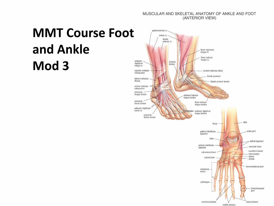
MMT Course Foot and AnkleMod 3

Pes CavusPes Planus
Foot Positions

Tarsal Bones
• Calcaneus- largest and most posterior tarsal bone. It is also known as the heel bone.
The key function of this bone is to form a connection between the leg and the foot so that the body weight may be transferred from the ankle to the leg, enabling a person to walk and maintain balance.
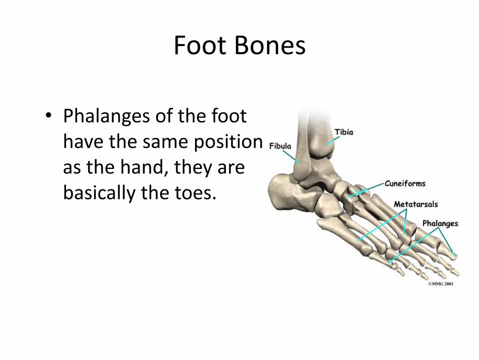
Foot Bones
• Phalanges of the foot have the same position as the hand, they are basically the toes.
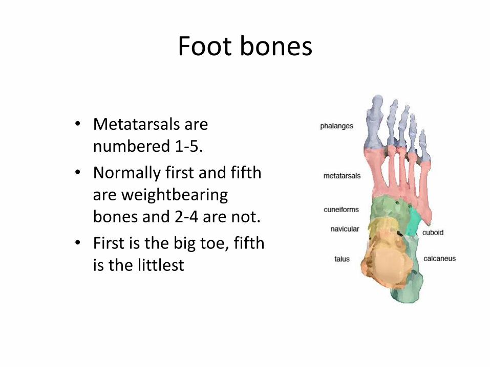
Foot bones
• Metatarsals are numbered 1-5.
• Normally first and fifth are weightbearing bones and 2-4 are not.
• First is the big toe, fifth is the littlest

Joints of the Ankle/Foot
• Talocrural Joint-joint in the ankle found between the tibia, fibula, and talus. Dorsi/plantar flexion
• Subtalar Joint-joint in the ankle found between the talus and calcaneus. Supination and pronation inversion and eversion.
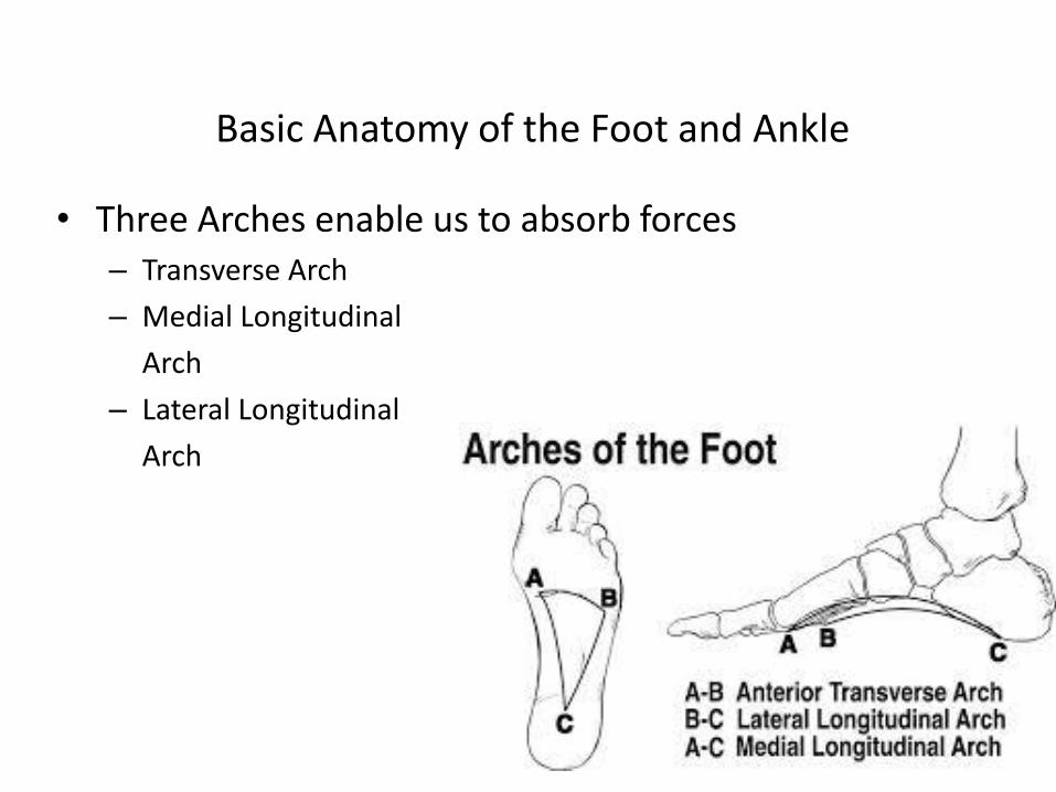
Basic Anatomy of the Foot and Ankle
• Three Arches enable us to absorb forces– Transverse Arch
– Medial Longitudinal
Arch
– Lateral Longitudinal
Arch

• Transverse Arch– Goes across the width of
the foot
– Comprised of the cuneiforms (all three), the cuboid, and the base of the fifth metatarsal.

The Three Arches• Medial longitudinal arch- the highest and
most important arch in the foot.
– Goes the length of the foot on the medial side.
– Comprised of the calcaneus, talus, navicular, cuneiforms and the first three metatarsals.

The Three Arches
• Lateral longitudinal arch- the arch next to the medial one that is flatter and lower.
– Goes the length of the foot on the lateral side.
– Comprised of the calcaneus, talus, cuboid, and the forth and fifth metatarsals.

Gait Cycle and the 3 arches

Pes Planus
Structural or Functional
1. Observe medial arch in both casesa. Pt stands with both feet on the groundb. Pt stands with just toes on the groundc. Pt sits on the table
Functional: if medial arch is restored when either pt is standing on toes or seated is indication of weak muscles or ligaments
Structural : If medial arch remains flat when client is standing on toes and when seated.


Achilles Tendon Rupture
If your Achilles tendon ruptures, you might feel a pop or snap, followed by an immediate sharp pain in the back of your ankle and lower leg that is likely to affect your ability to walk properly.

Ankle Injuries
• The most common direction to sprain the ankle is into inversion with the injury to the lateral ligament, specifically the ATF.

Over use or strained Achilles can lead to chronic issues. This is especially common among the “weekend warriors” groups.


Thompsons Test (Achilles Tendon Rupture)Testing for 3rd Degree strain or Rupture
Pt is prone, feet hanging over edge of table, legs relaxedSqueeze the affected calf muscle
POS sign: Absence of plantarflexion while muscle is squeezed.

Manually Freeing up the Tendon

Youtube Demo
https://youtu.be/p7OAD4zIBos

Image Warning

Achilles Tendon Tear and Repair

After surgery, a cast or walking boot is usually worn for six to 12 weeks. At first, the cast or boot is positioned to keep the foot pointed downward as the tendon heals. The cast or boot is then adjusted gradually to put the foot in a neutral position (not pointing up or down).
Specific gentle exercises (restricted motion) after surgery can shorten the time needed in rehabilitation. Massage is greatly beneficial at this time to increase circulation and help with adhesions and scar tissue build up.
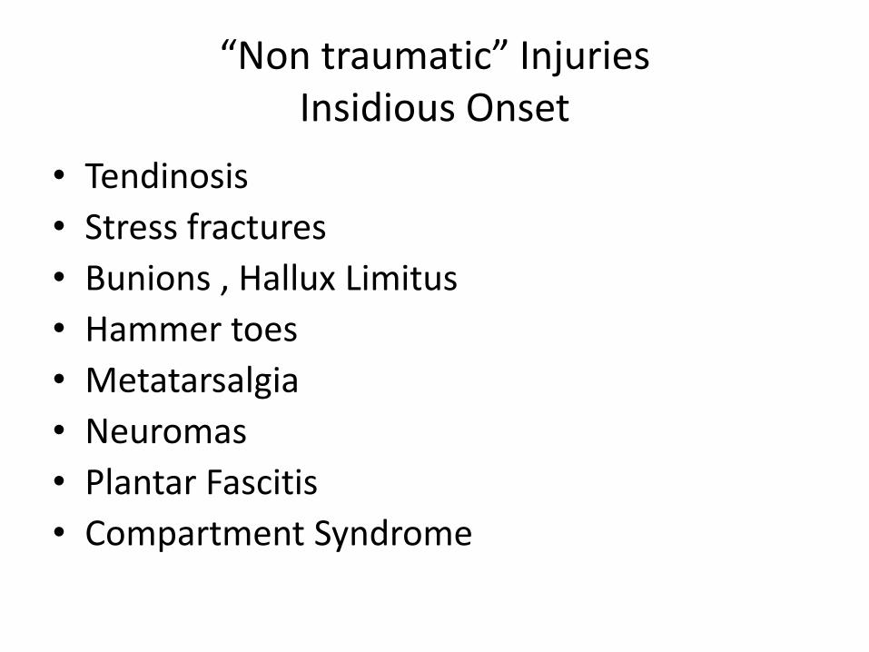
“Non traumatic” InjuriesInsidious Onset
• Tendinosis
• Stress fractures
• Bunions , Hallux Limitus
• Hammer toes
• Metatarsalgia
• Neuromas
• Plantar Fascitis
• Compartment Syndrome
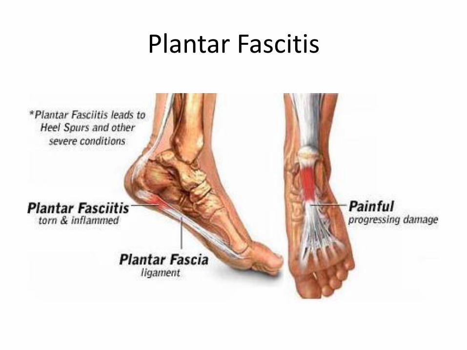
Plantar Fascitis
Causes
• Unlocked midtarsal joint at push off phase of gait causing stretch to fascia
• Variety of foot types
• Tight heelcords for level of function
• Tight great toe flexors or fascia
• Weakness in control of pronation
• Training errors, shoes


Bunions(Hallux Valgus)

Hammer Toe What causes hammer toe?The most common cause of
hammer toe is wearing shoes that do not fit properly. Tight fitting shoes, as well as shoes that have a boxed-in toe area, can lead to the condition. Women who wear high heels on a daily basis may be more prone to hammer toe because the high heels put pressure on the toes while restricting them from moving around freely.

Hammer Toe

Hammer Toe, Mallet Toe, Claw Toe

Bunions
• Both medial (1st MTP) and lateral (5th)
• Over pronated foot with hallux abductus (toe out)
• Tight heel cords
• Forefoot varus
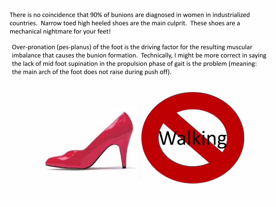
There is no coincidence that 90% of bunions are diagnosed in women in industrialized countries. Narrow toed high heeled shoes are the main culprit. These shoes are a mechanical nightmare for your feet!
Over-pronation (pes-planus) of the foot is the driving factor for the resulting muscular imbalance that causes the bunion formation. Technically, I might be more correct in saying the lack of mid foot supination in the propulsion phase of gait is the problem (meaning: the main arch of the foot does not raise during push off).

On the bottom of your foot, there are two small muscles that can cause much pain and misery. When these muscles develop Myofascial Trigger Points from improper gait, or shoes they combine forces to not only pull your big toe out of alignment but they also produce pain that is virtually indistinguishable from the bunion.
The flexor hallucis brevis, produces pseudo bunion pain. TPs in the muscle identified on the left, adductor hallucis, creates taut bands that pull the big toe in toward the midline, increasing the pain and misery of an already inflamed bunion.

Restoring motion within the Joint capsule

Bunion Stages
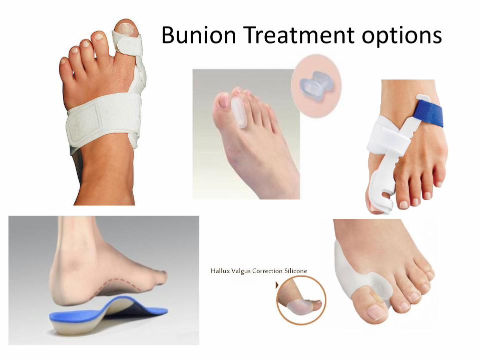
Bunion Treatment options

Image Warning

Bunionectomy
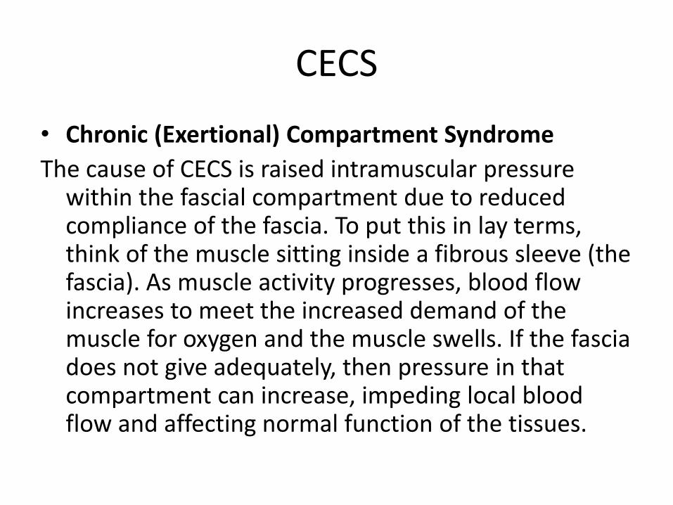
CECS
• Chronic (Exertional) Compartment Syndrome
The cause of CECS is raised intramuscular pressure within the fascial compartment due to reduced compliance of the fascia. To put this in lay terms, think of the muscle sitting inside a fibrous sleeve (the fascia). As muscle activity progresses, blood flow increases to meet the increased demand of the muscle for oxygen and the muscle swells. If the fascia does not give adequately, then pressure in that compartment can increase, impeding local blood flow and affecting normal function of the tissues.

Compartments of the leg
• There are four compartments of the lower leg. If there is inflammation in one it will prevent muscles and nerves from working in that area.
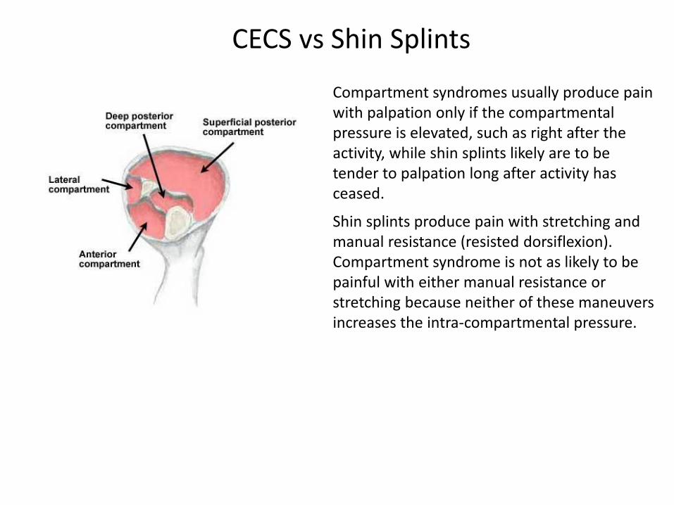
CECS vs Shin Splints
Compartment syndromes usually produce pain with palpation only if the compartmental pressure is elevated, such as right after the activity, while shin splints likely are to be tender to palpation long after activity has ceased.
Shin splints produce pain with stretching and manual resistance (resisted dorsiflexion). Compartment syndrome is not as likely to be painful with either manual resistance or stretching because neither of these maneuvers increases the intra-compartmental pressure.
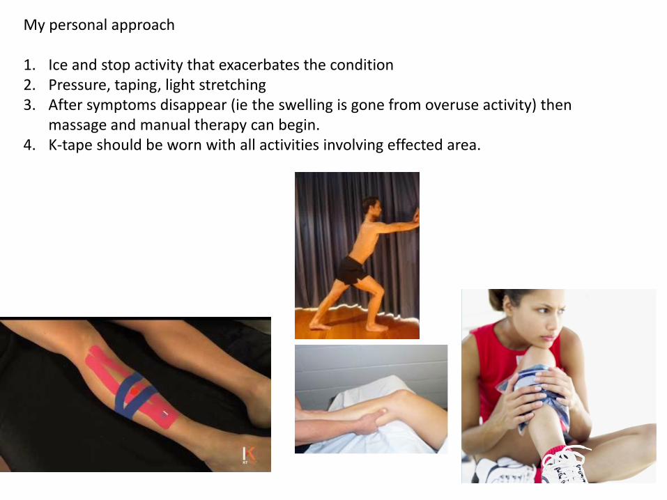
My personal approach
1. Ice and stop activity that exacerbates the condition2. Pressure, taping, light stretching3. After symptoms disappear (ie the swelling is gone from overuse activity) then
massage and manual therapy can begin.4. K-tape should be worn with all activities involving effected area.

Metatarsalgia
• Inflammation of the heads of one or more metatarsal heads (periostitis)
• Caused by uneven loading of forefoot during propulsion
• Caused by forefoot imbalance or deformity
Normally, the small muscles that run between the bones of the feet contract during the final phase of each step (following heel strike) to prevent the forefoot from splaying and the toes from curling. If these small intrinsic muscles don't do their job, the forefoot spreads and the toes curl which causes the metatarsal heads to be forced down and they contact the ground harder and this can lead to injury.

Metatarsalgia

Metatarsalgia

Treatment
Reported by the American Massage Therapy Association, two great methods involve specifically using the thumbs. Several methods seem to work best, including the thumb sweep and walk methods. With the first technique, the thumbs are positioned on the top of the feet and then move in a raking motion up and down. The second technique involves the thumbs being held on the bottom of the feet and are walked upwards while pressing firmly.

Inter Metatarsal (Morton’s) Neuroma
• Enlarged, fibrotic and benign interdigital nerves
• Most commonly between the third and forth metatarsals
• Brought on by shearing between metatarsals
• Aggravated by narrow shoes and forefoot imbalance
• Treatments include special shoes or inserts, NSAIAs and/or cortisone injections, but surgical removal of the growth is sometimes necessary.

Neuromas

MMT Course Foot and AnkleMod 3
Completion