modeling large whale entanglement injuries: an
TRANSCRIPT

The University of MaineDigitalCommons@UMaine
Electronic Theses and Dissertations Fogler Library
8-2006
Modeling Large Whale Entanglement Injuries: AnExperimental Analysis of the Influence of TissueCompliance, Line Tension, and Draw-Length onEpidermal Abrasion ResistanceJeremy Paul Winn
Follow this and additional works at: http://digitalcommons.library.umaine.edu/etd
Part of the Animal Sciences Commons, and the Oceanography Commons
This Open-Access Thesis is brought to you for free and open access by DigitalCommons@UMaine. It has been accepted for inclusion in ElectronicTheses and Dissertations by an authorized administrator of DigitalCommons@UMaine.
Recommended CitationWinn, Jeremy Paul, "Modeling Large Whale Entanglement Injuries: An Experimental Analysis of the Influence of Tissue Compliance,Line Tension, and Draw-Length on Epidermal Abrasion Resistance" (2006). Electronic Theses and Dissertations. 149.http://digitalcommons.library.umaine.edu/etd/149

MODELING LARGE WHALE ENTANGLEMENT INJURIES:
AN EXPERIMENTAL ANALYSIS OF THE INFLUENCE OF
TISSUE COMPLIANCE, LINE TENSION, AND
DRAW-LENGTH ON EPIDERMAL
ABRASION RESISTANCE
BY
Jeremy Paul Winn
B.S. University of Maine, 2003
A THESIS
Submitted in Partial Fulfillment of the
Requirements for the Degree of
Master of Science
(in Marine Bio-Resources)
The Graduate School
The University of Maine
August, 2006
Advisory Committee:
John Riley, Professor of Marine Sciences, Advisor
Michael Peterson, Associate Professor of Mechanical Engineering
Michael Moore, Research Specialist at Woods Hole Oceanographic Institution

MODELING LARGE WHALE ENTANGLEMENT INJURIES:
AN EXPERIMENTAL ANALYSIS OF THE INFLUENCE OF
TISSUE COMPLIANCE, LINE TENSION, AND
DRAW-LENGTH ON EPIDERMAL
ABRASION RESISTANCE
By Jeremy Paul Winn
Thesis Advisor: Dr. John Riley
An Abstract of the Thesis Presented in Partial Fulfillment of the Requirements for the
Degree of Masters of Science (in Marine Bio-Resources)
August, 2006
Two test systems were developed to evaluate the influence of draw-length and
tissue compliance on entanglement-induced epidermal abrasion in humpback (Megaptera
novaeangliae) and right whale (Eubalaena glacialis) tissue samples. Under straight pull
abrasion tests an adult right whale fluke required 3.7 times the load and 15 times the
draw-length of a right whale calf flipper to induce epidermal failure while a humpback
fluke was intermediate between these extremes. Epidermal thickness did not appear to be
the cause of this difference in abrasion resistance. Epidermal thickness averaged 8.0k0.2
mm for the calf flipper, 4.9k0.4 rnrn for the humpback fluke, and 5.1k0.1 mm for the
right whale fluke. It is unknown whether the difference in abrasion resistance is a
function of species (right vs. humpback whale), age (adult vs. calf), or body region (fluke
vs. flipper). Repeated tests with different line materials (new and used float vs. sink) did

not show a significant difference in the rate of epidermal abrasion. However, line
diameter does appear to influence tissue abrasion rates. Thinner (6.4 mm diameter) lines
cut substantially deeper than thicker (9.5 mm diameter) lines under similar loads and
draw-lengths. Tests with the oscillatory abrasion system revealed that draw-lengths that
exceed the tissue compliance limit resulted in substantially increased rates of tissue
abrasion. Draw-lengths below the compliance limit did not penetrate the epidermis while
those exceeding the compliance limit deeply cut into the underlying dermis. In actual
entanglement situations, the relative draw-length to tissue compliance ratio may be the
critical component that determines if the line will cut into the body or benignly press
against the skin. Investigations into the potential for reducing line modulus to allow lines
to stretch with flexure of the body and thus minimize sliding relative to the skin may be
beneficial in reducing the mortality associated with entanglement injuries.

ACKNOWLEDGEMENTS
There are a number of people I would like to thank for their contributions to my
thesis work. I would like to thank my parents for creating their non-traditional junior high
school and firing my interest in whales by setting me loose in the lagoons of Mexico as a
boy. I would also like to thank William Megill of the Coastal Ecosystems Research
Foundation for providing the opportunity to get my feet wet in whale research. I realize
that without the opportunity you provided I would never be where I am now.
I would also like to thank Mick Peterson for his trust and courage in not only
providing a place for me at The University of Maine but also for setting me loose in the
engineering department, allowing me to learn and grow, and continually providing
friendship and support. Thanks to Art Pete for his innumerable lessons and assistance in
helping to solve equipment and manufacturing problems of all sorts. Thanks also to
Michael Moore and the entire Moore family for taking us in and providing a second
home in Massachusetts. Your focus on conserving whales helped to clarify the direction
of this thesis- not to mention your donation of facilities, equipment, and whale bits that
made it a possibility.
I would like to thank John Riley for being willing to take me on as a graduate
student and for supporting me through the process.
I would also like to thank the necropsy team members for obtaining samples from
NEAq Eg 1004, Eg NEFL 0602 and VAQS 2006 1007 Mn with members from the
Virginia Aquarium, UNC Wilmington, and Woods Hole Oceanographic Institution. In
particular, thanks to Sue Barco, Bill McLellan, and Charley Potter for their aid in the

acquisition of tissue specimens. Thanks also to Scott Landry for providing the sketch of
NEAq Eg 2301 and to CT Hany for his help with the necropsy.
But most of all, I need to thank my wife, Becky Woodward, for her continuous
support and assistance in every aspect of this project. Your guidance, endless patience,
and continual assistance and support are really the only things that kept me going. But
more than that, your love and understanding has made me a better person. Without your
help, I would never have come as far as I have. Without it, it is doubtful that I would have
ever completed this degree.
This research was supported by a grant through the National Fish and Wildlife
Foundation (Project # 2005-0327-001). Samples were obtained and are held by Michael
Moore, a coinvestigator, under NMFS permit number 932-1 489-03PRT009526 issued to
Dr. Teri Rowles.

TABLE OF CONTENTS
. . ACKNOWLEDGEMENTS .......................................................................................... 11
LIST OF TABLES ............................................................................................................. vi
. . LIST OF FIGURES ........................................................................................................ v i ~
Chapter
1 . BACKGROUND ............................................................................................................ 1
........................................................................................... The entanglement problem 1
Epidermal structure in cetacea ................................................................................... 2
Experimental modeling of entanglement injuries ......................................................... 6
........................................................................................................... Project objective 8
2 . METHODS .................................................................................................................. 10
........................................................................... Straight pull abrasion system (SPA) 10
..................................................................... Oscillatory pull abrasion system (OPA) 14
....................................................................................... Comparison of line abrasion 18
3 . RESULTS .................................................................................................................... 19
........................................................................... Straight pull abrasion (SPA) testing 19
..................................................................... Oscillatory pull abrasion (OPA) testing 22
............................................................................................................... 4 . DISCUSSION 26
................................................ Comparative abrasion resistance between specimens 26
.......................................................... Differences in epidermal mechanical character 27
.................................................................... Differences between individual samples 32
................................................ The influence of line material on the rate of abrasion 33
Oscillatory abrasion and entanglement injuries .......................................................... 34
Conclusions ................................................................................................................ 36
Recommended future work ................................................................................... 37
BIBLIOGRAPHY ............................................................................................................. 39
BIOGRAPHY OF THE AUTHOR ................................................................................... 44

LIST OF TABLES
TABLE 1. Comparative epidermal thickness ranges reported for baleen
whale species. ........................................................................................... 4
TABLE 2. Comparison of abrasion rates in relation to line material ........................... 22
TABLE 3. Comparison of abrasion rates relating to line diameter and
sample curvature ......................................................................................... 22

LIST OF FIGURES
FIGURE 1 . Diagram of the epidermal layers visible in a cross section .......................... 5
FIGURE 2 . Diagram of the reciprocating load generator used by
Woodward et a1 . (2006) ................................................................................ 7
FIGURE 3 . Comparative differences in h o w appearance and depth
due to line type ............................................................................................. 8
FIGURE 4 . Diagram of the straight pull abrasion (SPA) system .................................. 12
FIGURE 5 . Diagram of the oscillatory pull abrasion (OPA) system ............................ 16
FIGURE 6 . Clamping technique for the OPA system ................................................... 17
FIGURE 7 . Progression of epidermal failure following line abrasion .......................... 20
FIGURE 8 . Tissue flexure during compliance testing of the humpback
fluke specimen ........................................................................................ 23
FIGURE 9 . Comparison of oscillatory abrasion furrows using different
draw-lengths .............................................................................................. 25
FIGURE 10 . Close up image of the stages of epidermal failure ..................................... 28
FIGURE 1 1 . Structural advantages of a vertical fiber orientation within
the epidermis ............................................................................................. 30
FIGURE 12 . Visual comparison of the differences between test specimens .................... 33
FIGURE 13 . The entanglement of NEAq Eg 2301 .......................................................... 35
vii

Chapter 1
BACKGROUND
THE ENTANGLEMENT PROBLEM
Entanglement in commercial fishing gear is one of the leading anthropogenic
causes of serious injury and mortality in baleen whales (Johnson et al. 2005, Knowlton
and Kraus 2001, Robbins and Mattila 200 1). Reduction and the eventual elimination of
these threats is essential to the preservation of five endangered baleen whale species in
the western North Atlantic (blue, fin, sei, humpback, and northern right whales). With
only 300 animals remaining in the population, the North Atlantic right whale is of
particular concern due to its critically low abundance (NMFS 2005). Photographs from
the New England Aquarium's photo-ID catalog indicate that 75.6% of all photographed
right whales show evidence of scarring indicative of rope and net cuts around the tail
stock, while the rate of entanglement appears to be increasing (Knowlton and Kraus
200 1, Knowlton et al. 2005). Additionally, 88% of humpback whales studied in 1999
showed some signs of entanglement-related scarring about the peduncle (Knowlton and
Kraus 2001, Robbins and Mattila 2001). Continued monitoring of the humpback
population suggests that 8-25% of the population receives new scars each year (Johnson
et al. 2005). The extent of entanglements involving other baleen whale species is not
known. Although their more pelagic distribution may decrease their exposure to
commercial fishing gear, the probability of identifying entanglements when they do occur
is also greatly reduced due to a reduction in sightings of pelagic species and the low
probability of animals washing ashore in the event of a mortality.

In the western North Atlantic, right and humpback whales have been found
entangled in all major types of fixed fishing gear (Johnson et al. 2005). Preventative
efforts have included fishing restrictions, mandatory gear removal, deterrent devices such
as pingers, and fishing area closures (Johnson et al. 2005). Fishing gear modifications
such as break-away links, knotless splices, and pop-up buoys are also under development
to help reduce entanglement risks (NMFS 2005). Disentanglement remains a costly and
logistically difficult retroactive defense against entanglement mortalities. However,
model predictions indicate that preventing the deaths of two female right whales per year
would allow population recovery (Fujiwara and Caswell2001). Therefore, minor
improvements may have a significant impact.
To enhance the effectiveness of gear modification efforts, the factors that govern
entanglement injuries must be more fully understood (Johnson et al. 2005). Analysis of
entangling lines and the resulting tissue damage on free swimming whales is extremely
difficult to characterize. Entangling gear on stranded animals is often removed by the
gear owner preventing in-depth analysis during necropsy events (Moore et al. 2005). As a
result, no design criteria presently exist upon which to base future gear modifications or
to verify the effectiveness of existing ones.
EPIDERML STRUCTURE IN CETACEA
In addition to selection for hydrodynamic performance, the cetacean epidermis
has also evolved as a defensive structure. The cetacean epidermis is thick and hairless,
and is often mistakenly referred to as the skin of the whale even though it only composes
the outermost layer of the integument (Haldiman and Tarpley 1993). It has an extremely

smooth, rubbery texture bordering on velvety (Geraci et al. 1986% Haldiman et al. 1985,
Ling 1974). These surface properties reduce frictional forces and provide resistance
against biofouling by marine organisms (Baum et al. 2003, Geraci et al. 1986b). Natural
abrasive stresses on the cetacean epidermis may include but are not limited to
hydrodynamic friction during swimming (Giacometti 1967), attacks by other ceteaceans,
kelp gulls (Rowntree et al. 1998), and other marine organisms, as well as contact with
sediment, ice in the case of northern species, or other marine debris. Physical contact
with other, sometimes barnacle covered, individuals of the same species would also
occur. These interactions would include motherlcalf interactions, mating, and competitive
physical contact during mate selection in some species. Therefore, the evolution of a
hydrodynamicaly smooth and yet durable epidermis would be essential to survival in the
aqueous environment. However, the extent to which these adaptational defenses against
natural stresses are significant as a defense against abrasive impacts from entangling
fishing gear has not been examined.
Only limited data are available on the cetacean integument. The dermal layer is
composed of an extensively cross-linked network of collagen fibers that may function as
an elastic recoil mechanism during swimming activities (Pabst 1996b). A limited number
of studies have looked at the mechanical properties of the cetacean dermis (Hamilton et
al. 2004, Pabst 1996a, Pabst et al. 1995). However, to date the only structural studies on
cetacean epidermis have been measurements of epidermal thickness (Table 1) and
evaluations of surface properties, including surface roughness and resistance to
biofouling (Baum et al. 2002, Baum et al. 2003, Baum et al. 2000). Epidermal thickness
has been reported in 5 species of baleen whales, with thickness ranging from a minimum

of 1.4 mm to a maximum of 25 mm in fin (Balaenoptera physalus) and bowhead whales
(Balaena mysticetus) respectively. There have been no published studies on the
mechanical properties of the cetacean epidermis.
Epidermis Thickness:
<8' a g k S u n o QJ .- P Y U U • . B 9 g 3 3
i
k 0 0 rn 0 3 rn 0 3 g g rn
cJ cJ Balaena - - 25.0 Haldiman et al. 1985 - - 8.6 Haldiman et al. 1985 mysticetus Balaenoptera - - 3.0 Singarajah 1984 - - 3.0 Singarajah 1984 acutorostrata Balaenoptera F 708 3.0 Sokolov 1982 F 708 1.4 Sokolov 1982 acutorostrata Balaenoptera M 156 3.9 Sokolov 1982 F 160 2.5 Giacometti 1967 physalus 5 0 Eubalaena M 107 13.2 Sokolov 1982 F 163 8.6 Sokolov 1982 glacialis 5 0 Megaptera M 857 5.6 Jones and Pfeiffer M 857 4.3 Jones and Pfeiffer novaeangliae 1994 1994
TABLE 1. Comparative epidermal thickness ranges reported for baleen whale species.
The cetacean epidermis is composed of three primary strata as opposed to the five
of terrestrial mammals (Geraci et al. 1986b, Harrison and Thurley 1974, St. Aubin et al.
1990). These include: the stratum extemum, stratum intermedium, and stratum
germinativurn (Figure 1). The stratum extemum is formed of the outermost 12-60 (0.25- 1
rnm) cells of the epidermis. These cells are typically flattened and keratinized forming a
semi-hardened exterior (Haldiman et al. 1985, Pfeiffer and Rowntree 1996, Sokolov
1982, Spearman 1972). Beneath the stratum extemum, the stratum intermedium is by far
the largest portion of the epidermal tissue and may extend as much as 10 mm above the
distal end of the papillae in the bowhead whale (Haldiman et al. 1985). Epidermal
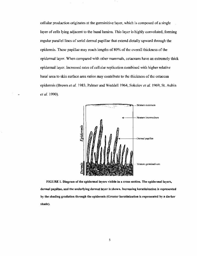
cellular production originates at the germinitive layer, which is composed of a single
layer of cells lying adjacent to the basal lamina. This layer is highly convoluted, forming
regular parallel lines of serial dermal papillae that extend distally upward through the
epidermis. These papillae may reach lengths of 80% of the overall thickness of the
epidermal layer. When compared with other mammals, cetaceans have an extremely thick
epidermal layer. Increased rates of cellular replication combined with higher relative
basal area to skin surface area ratios may contribute to the thickness of the cetacean
epidermis (Brown et al. 1983, Palmer and Weddell 1964, Sokolov et al. 1969, St. Aubin
et al. 1990).
Stratum extemum
Stratum intermedium
-Dermal papillae
Stratum germinativum
FIGURE 1. Diagram of the epidermal layers visible in a cross section. The epidermal layers,
dermal papillae, and the underlying dermal layer is shown. Increasing keratinization is represented
by the shading gradation through the epidermis (Greater keratinization is represented by a darker
shade).

Major arteries in the cetacean skin enter the dermis, branch to an increasing extent
as they approach the epidermis, and turn to run within the dermal bed. The dermal
papillae are well vascularized with capillaries lying against the basal lamina (Haldiman et
al. 1981, Harrison and Thurley 1974, Sokolov 1982). Injuries that result in the
penetration of the protective epidermal layer can easily lead to infection (Oen 1990) and
occasionally death due to bacterial entry into this highly vascularized region. In all cases
in which right whale entanglement mortalities have been examined though necropsy
during the period of 1970-2002, regions of epidermal penetration were noted (Moore et
al. 2005). Microorganisms including diatoms and bacteria that inhabit depressions in the
epidermis have been reported to penetrate deeply into the spinous layer particularly when
necrotic tissue is present as a result of injury (Haldiman et al. 1985, Henk and Mullan
1996). As a result, gear modifications that can help to minimize the penetration of the
epidermis are of high priority.
EXPERIMENTAL MODELING OF ENTANGLEMENT INJURIES
In 2006, Woodward et al. created a reciprocating load generator to model large
whale entanglement injuries. This system was designed to load a line in an oscillatory
manner, periodically causing the line to go taut and then slack. In effect, this simulated an
entanglement scenario in which a line connecting wraps about a flipper and the tail fluke
is loaded and unloaded by the up and down movements of the tail as the whale swims.
The reciprocating load generator used a gear motor and a slider crank mechanism to pull
up and down on an abrading line. An aluminum frame and set of guiding pulleys
positioned the abrading line such that it passed from the slider crank mechanism and
motor, over the leading edge of a fluke tissue sample clamped in a static seawater tank,

and on to a tensioning weight suspended on the far side (Figure 2). The up and down
motion produced by the slider crank mechanism and the return force provided by the
tensioning weight generated an oscillatory load that simulated the abrasion experienced
by a free swimming whale under controlled laboratory conditions.
Testing Apparatus
---/
To laptop compvter
FlGURE 2. Diagram of the reciprocating load generator used by Woodward et al. (2006). The
primary system components are illustrated and a photograph of the sample clamping technique is
shown (lower right).
Abrasion tests were conducted on tissue samples collected from the leading edge
of the fluke of an adult right whale (NEAq Eg 1004). Both new and used float
(polypropylene) and sink (polyester/polypropylene) lines of twisted 3-stranded
construction were tested as well as a hollow braided polypropylene line. All lines were
9.5 mm in diameter. Lines were tested for up to 24 hours using a 9-kg load at an
accelerated loading rate of 60 strokeslminute. Maximum line tension was 267 N. These
tests failed to penetrate the epidermis but succeeded in producing furrows that closely

resembled marks found on entangled whales during necropsy. It was also determined that
the line material, construction, and age affected furrow depth and appearance, suggesting
a potential forensic analysis application of experimental modeling studies (Figure 3).
.ison Between I in
- tine
or 1 Hour
Hollow Braide
Saw
18.3
'2 Y U Y
E 0 8 8 a .* rn
2
-18.3
d Line
FIGURE 3. Comparative differences in furrow appearance and depth due to line type.
Photographs of furrow appearance (left) and a graphical comparison of depth measurements (right)
following 1-hour tests with different line materials are shown (Woodward et aL 2006).
PROJECT OBJECTIVE
Although the Woodward et al. (2006) test system was effective in producing
compression furrows similar to those observed on entangled whales, these marks could
not be compared to entanglement scenarios where the line cut through the epidermis.
Understanding what causes some lines to cut while other do not is essential to preventing
injury and mortality due to entanglement. An evaluation of the physical conditions
leading to epidermal penetration is needed in order to make recommendations for future

gear modifications. During the initial testing, it was noted that the fluke tissue exhibited a
large degree of lateral flexure, or compliance, in response to the pull of the line. This
flexure was thought to absorb a portion of the test system's abrasive impact by helping to
prevent the rope from sliding across the epidermal surface. It is hypothesized that the
tissue compliance is essential to the abrasion resistance capacity of the whale skin.
To further examine this theory, two new test systems were developed based upon
the original design by Woodward eb al. (2006) and used to compare line abrasion on right
and humpback whale skin samples under controlled conditions. Three factors were
examined: 1) the compliance of the skin tissue under load, 2) the relative skin abrasion
following a known length of line pulled under a known tension across the skin, and 3) the
interaction between tissue compliance and line draw-length as they relate to the rate of
tissue abrasion.

Chapter 2
METHODS
Tissue specimens were opportunistically collected at necropsies along the East
Coast of the United States and included samples from the leading edge of a right whale
calf flipper (Eg NEFLO 602), the fluke of an adult right whale (NEAq Eg 1004), and the
fluke of a sub-adult humpback whale (VAQS 2006 1007 Mn). These flipper and fluke
regions are known to be regularly associated with entanglement injuries in large whales
(Johnson et al. 2005, Moore et al. 2005, Woodward et al. 2006). Samples were wrapped
in plastic to avoid hydration change, transported on ice to a storage facility (a process of
several days) and stored in a -20" C freezer until testing was possible. The fluke sample
from Eg1004 had been thawed during the prior Woodward et al. (2006) study and
refrozen following a 4 day period of refrigeration and testing. For the current study,
samples were thawed for 24 h in seawater prior to testing. When tested, tissue specimens
appeared in good condition with the epidermis remaining firmly attached to the
underlying dermal tissue.
Two test systems were developed to examine fishing line related entanglement
injuries in large whales. These systems were designed to independently evaluate straight
pull abrasion and oscillatory abrasion in relation to tissue compliance.
STRAIGHT PULL ABRASION SYSTEM (SPA)
A straight pull abrasion (SPA) system was designed to measure the depth of
epidermal penetration following a unidirectional pull over a standard draw-length. This
system allowed for control over factors such as line material, line diameter, pressure,
contact length, velocity, and pull distance. It has been shown in the tibology literature

that controlling for pressure and velocity allows for the evaluation of the mechanical
interaction between the abrading material and the abraded substance (Amell et al. 1991).
The SPA system utilized a set of two line-positioning pulleys and a tensioning pulley
mounted on an aluminum frame to position and tension an abrading line (Figure 4). The
abrading line was drawn by a sailing winch connected to a %-hp gear motor (Model
6ML5 1, Dayton Electric Manufacturing Company, Lincolnshire, IL). The motor pulled a
sliced loop of line in one direction across a test sample clamped in a static seawater tank.
Tension on the line was maintained using an adjustable weight connected to a tensioning
pulley. The tensioning pulley was free to slide along a rail to absorb any stretch of the test
line and maintain a constant tension in the system. A load cell (Model LC 10 1,
Omegadyne Engineering Inc., Stamford, CT) placed between the tensioning pulley and
the adjustable weight allowed constant evaluation of tension during testing, and a rotating
shaft counter mounted on the tensioning pulley measured the distance the line was pulled
across the test sample (f 1 cm).

Winch >
Positioning pulley
FIGURE 4. Diagram of the straight pull abrasion (SPA) system.
Test samples were submerged and clamped in a static seawater tank. The
clamping system uprights were spaced 7.6 cm apart to minimize lateral flexion in the
sample and maintain the sample's vertical orientation. The abrading line was then pulled
across the test sample. In order to ensure accurate pull distances across the sample
regardless of tissue compliance, tension was first applied to the sawing line by adding
weight to the tensioning pulley. The line was tensioned by manually turning the winch
until the compliance of the tissue was reached and the line began to slide across the
epidermis. At this point the rotary counter was zeroed. A standard draw-length (3.1 m) of
line was then pulled across the fluke sample at a continuous rate of 1.8 cmls.
The line samples used for the abrasion tests were all of 3 stranded twisted
construction. Both new and used polypropylene float and polypropylene/polyester blend
sink lines were tested. Float line diameters included both 6.4-mm and 9.5-mm samples

while only 9.5-mm diameter sink line was tested. The "used rope samples were cut from
the same line used in the Woodward et al. (2006) study. These lines were previously used
for lobstering (Southwest Lobster and Fish Unlimited, Southwest Harbor, ME) and
contained ingrained mud and fraying rope fibers. In order to create a continuous loop of
line, a 3.5-m section was cut and the ends joined using a 15-cm splice. The ends of the
splice were coated with Skotch 667 tape to create a smooth transition across the splice.
Three sets of tests were conducted with the SPA system.
1) Point of epidermal penetration for each test specimen
A standard draw-length of 3.1 m was used with new 9.5-rnm diameter float line to
determine the load and draw length combination necessary to penetrate the
epidermis for each of the tissue specimens: right whale calf flipper, right whale
fluke and humpback whale fluke. Load on the system was increased in 2.3-kg
increments from 2.3 kg to 3 1.8 kg. Epidermal failure was defined as substantial
cracking of the epidermis allowing the underlying dermal material to become
visible. If epidermal failure did not occur, the line was moved to a new test
location and run with an additional load. If the 3 1.8-kg load failed to penetrate the
epidermis, the draw-length was increased in 3.1 -m increments until failure was
achieved.
2 ) Relative line abrasion between line types
Four types of 9.5-mm diameter line were used to examine the relative rate of
abrasion following a standard 3.1 -m draw-length with 3 1.8-kg load. Three tests of
each line type (new and used float and sink) were conducted in random order on
samples from the humpback fluke. The results were compared using a pair- wise
Bonferroni corrected Wilcox-Mann-Whitney test at the a = 0.05 level.
3) Influence of rope diameter and sample curvature on relative abrasion
Both 6.4-rnrn and 9.5-mm diameter new float lines were drawn 3.1 m across
humpback fluke specimens using a 3 1.8-kg load. Tests of each line diameter were
repeated twice: once on the most proximal and once on the most distal portion of
the leading edge of the fluke, representing the widest range in curvatures available
using tissue samples from the same specimen. In this manner, both the relative
influence of line diameter and sample curvature could be compared based on the
rate of epidermal abrasion.
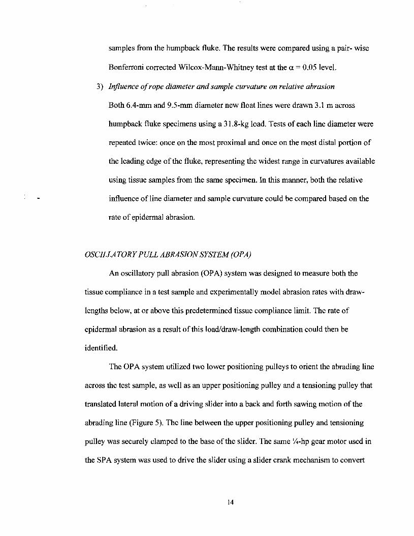
OSCILLATORY PULL ABRASION SYSTEM (OPA)
An oscillatory pull abrasion (OPA) system was designed to measure both the
tissue compliance in a test sample and experimentally model abrasion rates with draw-
lengths below, at or above this predetermined tissue compliance limit. The rate of
epidermal abrasion as a result of this loadldraw-length combination could then be
identified.
The OPA system utilized two lower positioning pulleys to orient the abrading line
across the test sample, as well as an upper positioning pulley and a tensioning pulley that
translated lateral motion of a driving slider into a back and forth sawing motion of the
abrading line (Figure 5). The line between the upper positioning pulley and tensioning
pulley was securely clamped to the base of the slider. The same %-hp gear motor used in
the SPA system was used to drive the slider using a slider crank mechanism to convert

rotary motion of the motor into the linear motion of the driving slider. The slider's
motion in turn drove the lateral sawing motion of the abrading line across the test sample.
The draw-length of the abrading line could be adjusted by changing the throw of the
slider crank mechanism. Tension in the line was maintained using an adjustable weight
connected to the tensioning pulley. Movement of the tensioning pulley absorbed any
stretch in the line while insertion of a cam cleat in the loading line maintained the
position of the tensioning pulley during the return stroke of the slider. A position
transducer (Model PT 10 1, Celesco Transducer Products Inc, Chatsworth, CA) was used
to measure the precise distance of linear travel in the line, while two load cells (Model
LC 10 1, Omegadyne Engineering Inc., Stamford, CT and Model MLP 100, Transducer
Techniques Inc., Temecula, CA) were inserted in either side of the line system to
simultaneously measure load on both sides of the tissue sample.

Powering
7 Slider-crank mechanism
Position transducer
Lip top Iaad s -'d I
Test i : Supports
samplc
FIGURE 5. Diagram of the oscillatory pull abrasion (OPA) system.
In order to simulate the natural compliance of the fluke and flipper samples as
closely as possible, a new clamping technique was developed for use with the OPA
system. In the SPA tests, the tissue sample was securely clamped with bars pressing
along both the sides and upward against the leading edge of the sample, limiting sample
movement as much as possible. In the OPA tests, however, only the base of the sample
was clamped leaving the leading edge free to flex without any form of lateral restraint.
Three 6.4-mrn diameter threaded rods were inserted through the base of the sample and
secured to two pieces of 90" angle irons (3.8 cm wide and 61 cm long). The ends of these
angle irons were then securely bolted to the original clamping frame within the seawater
tank (Figure 6).

FIGURE 6. Clamping technique for the OPA system: (a) angle irons and threaded rods clamping
right whale calf flipper sample, (b) sample clamped in static sea water tank
The OPA system was used to test the influence of tissue compliance on abrasion
using samples from the flipper of the right whale calf and the humpback whale fluke. The
compliance (total lateral flexural due to line pull) in the test sample under a particular
load was determined by applying tension to the line system. A unidirectional load was
applied via the slider until static friction was overcome and the rope started to slide
across the epidermal tissue. The motion of the slider and the force applied to the line
were recorded simultaneously on a laptop computer using the position transducer and
load cells. The linear displacement of the line was calculated at the point when the
maximum load was applied prior to overcoming static friction. The direction of pull was
then reversed until static friction was overcome in the opposite direction. The distance
between these two flexural extremes was recorded as the tissue compliance limit of the
sample. The test was repeated 6 times to determine the average tissue compliance for
each test sample used in the OPA testing.

Prior to oscillatory testing the tissue compliance was determined for each
specimen using a 9.5-mm new float line. Loads were selected to be 2.3 kg above the load
needed to penetrate the epidermis with a 3.1 -m pull in the SPA system. Two 2-hour
oscillatory tests were then conducted: one with a draw-length below the tissue
compliance limit and a second with a draw-length above this limit. The relative abrasion
resulting fiom the different draw-length tests was then compared for each specimen.
COMPARISON OF LINE ABRASION
As in the Woodward et al. (2006) study, individual test sites on the samples were
separated by a minimum of 2 cm to isolate test sites and maintain the integrity of the
tissue. The resulting tissue indentation andlor damage was assessed following each test.
Line h o w patterns were photographically documented and the furrow depth was
measured every 2 cm along its length using a set of digital calipers (f 0.02 mm). The
midline of the leading edge was marked and designated as the zero point for these
measurements. Positive numbers denote proximity to the motor in all measurements.
Relative dermal abrasion was compared in two ways: 1) maximum depth of dermal
penetration and 2) the length of epidermal removal. This length measurement was taken
along the curve of the sample with a flexible ruler (f 1 mrn). It included the region of
exposed dermis but excluded cracked epidermis on either end of the h o w . This
cracking could have represented an additional length of epidermal failure but was not
objectively measurable.

Chapter 3
RESULTS
STRAIGHT PULL ABRASION (SPA) TESTING
Among the three specimens, the right whale calf flipper had the thickest
epidermis (8.0k0.2 mm). Both the right whale and humpback whale flukes had similar
epidermal thicknesses, measuring 5.lf 0.1 mm and 4.9k0.4 mm respectively. Despite
differences in thickness, the mode of epidermal failure was similar in each of the tissue
samples. Prior to abrasion tests the epidermal surface was typically smooth with a slick
rubbery exterior. Once these exterior layers were removed by an initial period of abrasion
a rougher sponge-like tissue was revealed beneath, presumably the stratum intermedium.
As abrasion continued, cracking of the epidermal material began. Initially, crack
formation occurred directly beneath and in the same direction as the abrading line.
Subsequent cracking then appeared at approximately 45' to the direction of line travel,
creating a patchwork of cracked epidermal material reflective of the twisted three
stranded line construction. With continued abrasion this cracking of the epidermis
intensified and was followed by the epidermis being pulled out in chunks, separating
from the underlying dermis at the dermallepidermal interface (Figure 7).

FIGURE 7. Progression of epidermal failure following line abrasion: (a) humpback whale fluke,
(b) adult right whale fluke. Sequence progresses from left to right. Scale bars represent 1 cm.

A substantial difference in epidermal abrasion resistance was found between the 3
test specimens. A 3.1-m draw-length with a 6.8-kg load produced epidermal cracking on
the right whale calf flipper specimen, while an 1 1.3-kg load produced a region of
epidermal removal 15.76-mm long with a maximum depth of 8.44 mm. Thus, the point of
epidermal penetration on the calf flipper occurs somewhere between 6.8 and 1 1.3 kg
using a 3.1 -m draw-length. Using the same draw-length (3.1 m), the humpback whale
fluke withstood a 27.7-kg load prior to epidermal penetration. The right whale fluke
specimen showed an even greater resistance to abrasion. A 3 1 .&kg load and a 45.7-m
draw-length (requiring the splice to pass over the sample 14 times) only produced the
initial stage of epidermal cracking beneath the abrading line.
A second series of tests were conducted on the humpback fluke to determine if
statistical differences in abrasion rates exist when using various line materials. A standard
3 1 .&kg load was applied to the test system using a 3.1 -m pull. Four test materials were
examined, each with a 9.5-mm diameter: new and used polypropylene float and
polypropylene1 polyester sink lines. Each line type was tested 3 times. The resulting
maximum furrow depth and length of epidermal removal are shown in Table 2. No
statistical difference in abrasion rates between line types was found at the 0.05 level
(Wilcox-Ma-Whitney rank sum test). However, the new float line had the smallest
average furrow depth and length of epidermal removal (5.4 mm and 33 mm) while the
new sink line had the largest furrow depth and length (6.8 mrn and 79 mm). Both used
float and new sink were intermediate to these extremes.

Line Material Comparison New-float Used-float New-sink Used-sink
Test1 5.0mm 44mm 6.6mm 64mm 7.6mm 94mm 7.3mm 66mm Test2 5.3mm 26mm 5.5mm 84mm 7.lmm 68mm 6.6mm 73mm Test3 5.Omm 29mm 6.2mm 69mm 5.8mm 76mm 6.6mm 71mm Mean 5.4mm 33mm 6.3mm 72mm 6.8mm 79mm 6.8mm 70mm
SD 0.5 9.6 0.6 10.4 1.0 13.3 0.4 3.6
TABLE 2. Comparison of abrasion rates in relation to line material, The maximum furrow depth
and length of epidermal removal according for each line type are provided.
The influence of line diameter and specimen curvature was examined in two
regions on the humpback fluke. Both 6.4-mm and 9.5-mm diameter lines were tested on
adjacent regions of the most proximal (widest) and most distal (narrowest) portions of the
fluke. The 6.4-mm line caused the greater depth and length of epidermal removal in both
cases (Table 3).
Line Diameter and Sample Curvature Comparison Wide Proximal Sample Narrow Distal Sample
Line Diameter Furrow Depth Length of Removal Furrow Depth Length of Removal 6.4 mm 7.24 mm 147 mm 6.49 mm 117mm 9.5 mm 5.65 mm 44 mm 4.02 mm 61 mm
TABLE 3. Comparison of abrasion rates relating to line diameter and sample curvature. The
maximum furrow depth and length of epidermal removal for each trial are provided.
OSCILLATORY PULL ABRASION (OPA) TESTING
Epidermal abrasion as a result of oscillatory loading of the tissue was examined in
the right whale calf flipper and the humpback whale fluke using the OPA system. With
an 11.3-kg tensioning load applied to a 9.5-mm new float line, the compliance limit in the
calf flipper was determined to be 2.4f 0.08 cm. The compliance limit in the humpback

whale fluke was determined to be 4.45k0.10 cm using a 71.8-kg load on the test system
(Figure 8).
Tissue Flexure During Conipliance Testing
FIGURE 8. Tissue flexure during compliance testing of the humpback fluke specimen. Short line
shows a vertical orientation of the sample under a no-load condition. Longer line shows the midpoint
of the leading edge once the sample has been loaded. Angular difference shows flexure of the tissue as
the load is applied.
Two oscillatory tests were then conducted on each specimen using new 9.5-mm
float line: one with a 2.5-cm and one with a 7.6-cm draw-length. These draw-lengths
were selected to be below or above the compliance limit of the tissue. Test loads were
determined based up on the compliance testing: 1 1.3-kg for the calf flipper and 71.8-kg
for the humpback fluke. In each case, the 7.6-cm draw-length cut deeply into the
underlying dermis while the 2.5-cm draw-length did not break the epidermis (Figure 9).
In the calf flipper, the 7.6-cm draw-length test resulted in a maximum furrow depth 5.9
times as deep as that of the 2.5-cm draw-length (3.78 mrn compared to 22.15 mm). It also
left a 139-mm long region of epidermal removal whereas the 2.5-cm draw-length did not

penetrate the epidermis. The difference in furrow depth was even greater in the
humpback whale fluke. The 7.6-cm draw-length caused a maximum furrow depth 8.5
times as deep as the 2.5-cm draw-length (3.9 mm compared to 33.1 mm), and left a 169-
mm length of dermal tissue exposed. The characteristics of the furrow also differed
between the 2.5-cm and 7.6-cm draw-lengths in each specimen. The shorter draw-length
(2.5 cm) produced a shallow furrow showing extensive streaking and a deeper three-
stranded dimpled pattern representative of the 3-stranded rope construction as noted in
the Woodward et al. (2006) study. The longer draw-length (7.8 cm) left a smooth furrow
without dimpling on either the epidermal or dermal tissue.

Right Whale Calf Flipper 11.3-kg Load
7.6-cm draw 2.5-cm draw
Humpback Whale Fluke 3 1 .%kg Load
7.6-cm draw 2.5-cm draw
FIGURE 9. Comparison of oscillatory abrasion furrows using different draw-lengths: (a) right
whale calf flipper, (b) humpback fluke. The 7.6-cm draw-length cut significantly deeper into the skin
tissue for both tissue specimens. Scale bar represents 1 cm.

Chapter 4
DISCUSSION
COMPARATIVE ABRASION RESISTANCE BETWEEN SPECIMENS
Epidermal abrasion resistance varies according species (humpback vs. right
whale) sample location (flipper vs. fluke) and age (adult vs. calf) of the animal. However,
it does not appear to be related to epidermal thickness. Of the three specimens, the right
whale calf flipper had the thickest epidermis (8.0k0.2 mm) but required the lowest load
(8.6 kg) and draw-length (3.1 m) combination to penetrate the epidermis following tests
with the SPA system. The two fluke specimens had similar skin thicknesses (4.9k0.4 mm
for the humpback and 5.1H.1 mm for the right whale) but required different loads (27.7
kg vs. 3 1.7 kg) with dramatically different draw-lengths (3.1 m vs. 45.7 m) to generate
epidermal failure. At the two extremes, the right whale fluke withstood 15 times the
draw-length (3.1 m vs. 45.7 m) with 3.7 times the load (8.6 kg vs. 3 1.7 kg) of the calf
flipper prior to epidermal failure. Potential factors that may influence differences in the
abrasion resistance among the three specimens include: the age of the animal from which
tissue samples were collected, species specific variations in mechanical properties of the
epidermis, differences in dermal structure between body regions (flippers vs. flukes),
curvature of the tissue and contact length of the line on the sample, and any
freezinglthawing effects on the tissue. The presence of these factors make it difficult to
isolate the influence of individual factors on the observed differences in epidermal
abrasion resistance between samples, but the overwhelming conclusion from this study is
that regardless of age, species, or sample conditions, compliance in the tissue is the
critical factor.

Due to the necessity of opportunistic sample collection at necropsy events,
consistency in age, species and body region could not be maintained across test
specimens. A substantial difference in age existed between the different whale
specimens. Right whale Eg 1004 was a mature adult at least 24 yrs old. Humpback whale
VAQS 2006 1007 Mn was a minimum of 3 yrs. old, and right whale Eg NEFLO 602 was
less than 1 year old (probably 2-3months). The tensile properties of skin in other
vertebrates are known to change with age, typically increasing in strength and decreasing
in elasticity through middle age (Cloete et al. 2004, Cua et al. 1990, Vogel 1994). It is
unknown whether similar age effects may affect skin properties and abrasion resistance
capacity in cetaceans. Oil content in the dermal tissues was also markedly different
between specimens. The adult right whale fluke sample was substantially oilier than
either the humpback fluke or the right whale calf flipper. The oil content may be
indicative of an age-related difference in tissue properties within a species or between
species. In any event, it likely influenced the coefficient of friction between the epidermal
surface and the abrading line, helping to minimize abrasive injuries.
DIFFERENCES IN EPIDERMAL MECHANICAL CHARACTER
Differences in the structure of the epidermis between body regions (flipper vs.
flukes) may exist. Although the stages of epidermal failure appeared consistent across
specimens, the rate at which this failure progressed differed between body regions. In the
calf flipper, epidermal removal followed quickly once the first signs of cracking
appeared. However, in the fluke specimens the progression from initial crack formation
to epidermal removal occurred at a much slower rate with a patchwork of interlocking
cracks spreading across the furrow. This advanced cracking stage could be compared to

a rug-like structure with stalks of epidermal tissue remaining firmly attached to the
dermallepidermal interface beneath, but no longer being attached to adjacent fibers
(Figure 10).
.CI--------- Exposed underlying
Epidermal --+ surface
"k
Region of - stratum externu~n
removal
dermal layer :%'
Final "rug-like" structure apparent prior to epidermal
reinoval 4
4
m C-------- Region of epidermal q , cracking
!
FIGURE 10. Close up image of the stages of epidermal failure. Scale bar represents 1 cm.
There appears to be a vertical orientation in epidermal structure within the
cetacean integument. It has been suggested that the presence of epidermal rods provides
support to regions of thick (>4 mm) epidermis without deep papillae penetration in
bowhead whale skin (Haldiman et al. 1981, Haldiman et al. 1985). In these regions,
epidermal cells encircling the sides and ends of the papillae form solid keratinized

structures separated by epidermal cells with long axes parallel to the epidermal surface.
Henk and Mullan (1 996) observed that superficial lacerations that do not penetrate to the
dermal papillae follow a progressive change in epidermal surface properties during the
healing process. The first stage of this process is keratinization of the epidermal rods
followed by a progression of keratinization within the stratum intermedium beneath the
necrotic tissue, which is then shed. Keratinocyte rosettes have been documented in right
whale calf epidermis, suggesting the presence of similar epidermal features in right
whales (Reeb et al. 2005). However, the structural functions of vertical features within
the epidermis have not been evaluated for any baleen whale species.
Observations from both bowhead and right whales indicate that the primary
strength in the epidermis is oriented in a vertical direction while laterally the structure is
more elastic with lower strength. From a structural standpoint, this type of fiber
integration would have several mechanical advantages. Strong vertically oriented fibers
would both increase the abrasion resistance of the epidermis and may help to decrease the
compressibility of the layer (Figure 11). As a lateral abrading force is applied to the
surface of the epidermis, the strong vertical fibers would start to bend, orienting in the
direction of the applied load. This flexure would allow the load to be applied in line with
the vertical fibers that are anchored through the entire epidermal layer. Retaining elastic
elements between these vertical fibers would maintain the flexibility needed in the
epidermis allowing the epidermis to stretch during normal swimming activities. This
elasticity may be graded through the stratum intermedium as the degree of keratinization
and flattening of the epidermal cells has been noted to increase with distance from the
stratum germinativum (Haldiman et al. 1985).

Resistance Direction of to Indentation Abrading Force z J . 4 4 J . J . P
m
with the stress
4 4
r" - FIGURE 11. Structural advantages of a vertical fiber orientation within the epidermis. Fibers
provide resistance to: (a) indentation and (b) lateral abrasion.
The depth of penetration and character of the dermal papillae may also
substantially influence the strength of the epidermis as a unit. Assuming that a more
multidirectional orientation of collagen fibers in the dermal material enhances the tensile
properties of the dermis, the volume of this dermal material interwoven within the
epidermis would substantially influence the structural characteristics of the layer.
Differences between the right whale flipper and humpback fluke were most
noticeable in the later stages of epidermal failure. The primary failure mechanism
following cracking appeared to be removal of the epidermis in chunks at the dermal
interface. Three potential factors may account for this difference:
1) The epidermalldermal connection could be stronger in the fluke.
2) The depth of penetration of the dermal papillae andlor the shape of the distal
tips of the dermal papillae could be greater in the fluke tissue, increasing
connection surface area and hence increasing attachment strength.

3) Assuming that the dermal papillae are removed along with the epidermis
during epidermal failure, a difference in the degree of collagenization of the
dermal papillae and a subsequent difference in the tensile properties could
exist regionally between the flippers and flukes.
Future studies examining the relative differences between the flipper and fluke
structure from the same animal and multiple individuals from the same species would be
helpful in determining if the observed differences in abrasion resistance are related to the
biomechanical structure of adult versus calf epidermal tissue or if flippers have a lower
abrasion tolerance than flukes. Several types of tests are needed. An abrasion study
examining the comparative durability of the flipper and fluke samples should be
conducted. Second, mechanical evaluation of the tensile properties of the dermis from the
two regions should be made. And finally, an analysis of the 3D structure of the dermal
papillae should be made. Knowlton et al. (2005) noted that a higher percentage of new
entanglement scars are observed on juvenile versus adult animals. Assuming the
difference in abrasion resistance is due to age alone, it may be that benign entanglements
(e.g. non-injury producing and self disentangled) are occurring in adults without scar
formation to a greater extent than has been previously reported. Conversely, if this
difference in observed entanglement scaring is due to variations in epidermal abrasion
resistance between flippers and flukes rather than age, then entanglements involving
flipper wraps may warrant even greater concern than was previously believed.

DIFFERENCES BETWEEN INDIVIDUAL SAMPLES
Variations in sample curvature may also contribute to variations in abrasion
resistance. Straight pull abrasion tests yielded longer but shallower furrows on the right
whale fluke and shorter but deeper furrows on the calf flipper. With the wider curvature
and greater thickness of the leading edge in the right whale fluke (Figure 12), the load
from the abrading line is distributed over a greater surface area, which would tend to
mitigate abrasive impacts. On the other hand, the narrow leading edge of the calf flipper
would tend to concentrate the load at a particular point, accelerating the abrasion process.
It is interesting to note that tests with 6.4-mm and 9.5-mrn diameter float line on the
proximal (widest) and distal (narrowest) regions of the humpback fluke showed a greater
depth and length of abrasion on the wider sample with both line diameters (Table 3).
Although the range in curvatures represented by the proximal and distal ends of the
humpback fluke is not as great as that presented by the right whale fluke and calf flipper,
these results suggest that the mechanical properties of the skin may have a greater
influence on abrasion resistance than the curvature of the sample.
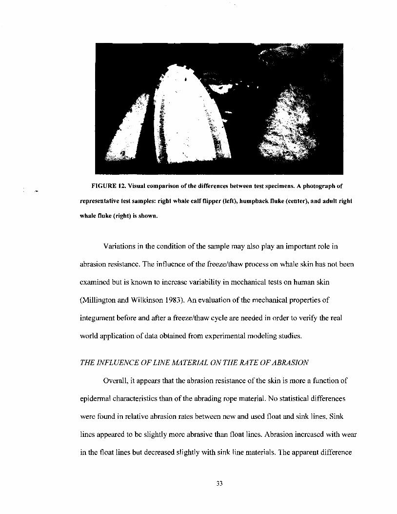
FIGURE 12. Visual comparison of the differences between test specimens. A photograph of
representative test samples: right whale calf flipper (left), humpback fluke (center), and adult right
whale fluke (right) is shown.
Variations in the condition of the sample may also play an important role in
abrasion resistance. The influence of the freezelthaw process on whale skin has not been
examined but is known to increase variability in mechanical tests on human skin
(Millington and Wilkinson 1983). An evaluation of the mechanical properties of
integument before and after a freezelthaw cycle are needed in order to verify the real
world application of data obtained from experimental modeling studies.
THE INFLUENCE OF LINE MATERIAL ON THE RATE OF ABRASION
Overall, it appears that the abrasion resistance of the skin is more a function of
epidermal characteristics than of the abrading rope material. No statistical differences
were found in relative abrasion rates between new and used float and sink lines. Sink
lines appeared to be slightly more abrasive than float lines. Abrasion increased with wear
in the float lines but decreased slightly with sink line materials. The apparent difference

between new and used line was greatest in float lines presumably as a result of fraying of
the polypropylene fibers that increased the abrasiveness of the line. Larger sample sizes
are needed for further statistical comparisons. On the other hand, the diameter of the
abrading line does appear to be an important factor in determining the rate of epidermal
abrasion. Comparisons between 6.4-mm and 9.5-rnm diameter new float lines revealed
that the smaller diameter line consistently cut deeper into the fluke tissue and had a
greater length of epidermal removal in both tests.
OSCILLATORY ABRASION AND ENTANGLEMENT INJURIES
In regards to oscillatory loading, tissue compliance appears to be a primary factor
in mitigating the abrasive impact of rope on whale skin. Tests with the OPA system
revealed that a draw-length above the tissue compliance limit (7.8 cm) cut deeply into the
dermal tissue while draw-lengths below the compliance limit (2.6 cm) did not penetrate
the epidermis in either the right whale calf flipper or the humpback whale fluke (Figure
9). This result suggests that although characteristic indentation furrows such as those
found in the Woodward et al. study are produced as lines are pressed into the skin in an
oscillatory manner, tissue removal does not occur until static friction is overcome and the
line slides across the epidermal surface. Once this sawing and subsequent tissue removal
process begins, the rate of abrasion increases substantially. In actual entanglement
situations, draw-length may be the critical component that determines if the line will cut
into the body or benignly press against the skin.
Differences in wound generation are exemplified by the case of right whale NEAq
Eg 2301. Two entangling lines were woven through and fixed in the baleen, exited the

right side of the animal's mouth, passed over the rostrum and blowhole and terminated in
wraps of line about the animal's left flipper (Figure 13).
FIGURE 13. The entanglement of NEAq Eg 2301. Artist's rendition of the location of entangling
lines as observed from photographs of the animal (left) and photographs of the abrasion marks on
the head (a) and left flipper (b) are shown. Drawing by Scott Landry of the Center for Coastal
Studies.
During necropsy, indentation furrows were visible on either side of the head,
across the rostrum and blowhole of the animal. Although the epidermis was no longer
attached due to an advanced stage of decomposition, marks from the entangling lines
remained imprinted in the underlying dermis. On the head, substantial penetration of the
dermal tissue was not apparent except where one of the lines crossed the left blowhole.
Instead, indentation patterns following the 3-stranded twist of the entangling line were
visible in the dermis (Figure 13a). On the other hand, a severe laceration penetrating to
the bone was observed on the leading edge of the left flipper over the proximal third of

the humerus as a result of the flipper wraps (Figure 13b)(Moore 2005 Necropsy Report).
The oscillatory abrasion results from this study suggest that in the case of Eg 230 1, the
movement of the flipper during normal swimming motions, and presumably the opening
and closing of the blowhole exceeded the tissue compliance and accelerated abrasion
rates in these two locations. In contrast, the movement of the lines passing over the
rostrum and along the side of the animal's head did not exceed the skin compliance and
as a result did not cut into the dermal tissue beneath.
CONCLUSIONS
This study suggests that reducing line movement across the surface of the
epidermis is the most significant factor in preventing serious entanglement injuries such
as the case of Eg 2301. Changing draw-lengths from 2.5 to 7.6 cm could mean the
difference between a deep laceration and a dimpled furrow that does not penetrate the
epidermis. Lines with decreased modulus could be developed to stretch and absorb a
portion of the potential abrasion during entanglement. For a line anchored at both ends, if
the rope flexes with the whale's body, it will help prevent slippage of the line relative to
the skin. Investigations into the potential of decreasing line modulus (or increasing ,the
line "stretchiness") may provide clues that could lead to a reduction in serious
entanglement injuries. Development of such a line, however, would have to incorporate a
reduced recoil speed to retain operational safety for the fisherman. Future studies are
needed in order to resolve these questions and to evaluate the practicality of decreasing
line modulus.

RECOMMENDED FUTURE WORK
0 Mechanical tensile testing of cetacean integument
Mechanical evaluation of the tensile properties of the dermis evaluating both
regional variation and directionality due to tissue fiber orientation
P A method for consistently removing a uniform thickness of dermal tissue
just beneath the epidermis and cutting it into dumbbell shaped test samples
is needed.
P An effective clamping technique must be developed to securely clamp and
align the samples without compromising the test tissue.
Mechanical evaluation of the tensile properties of the epidermis
P Due to the complex 3-D nature of the dermallepidermal junction, an
ultrasonic method may be the best means of determining the tensile
properties of this layer.
0 Analysis of the influence of the freezelthaw process on the mechanical properties
of cetacean integument
0 Additional straight pull abrasion testing
Evaluate the abrasion resistance of the flipper and fluke from the same animal
and a series of animals from the same species.
Evaluate of age related differences in abrasion resistance within a species.
Evaluate of differences between cetacean species.
0 Compressive load analysis
Determine the influence of large (50 to 1,000 kg) loads on the mechanical
behavior of cetacean skin.

P Design and construct a test system to evaluate loading necessary to cause a
laterally static compressive load injury leading to epidermal failure.
P Determine the influence of large loads on the rate of abrasion, tissue
compliance, and draw-length prior to epidermal failure.
0 Development of a surrogate synthetic whale skin
Following tensile testing and mechanical evaluation of cetacean skin, a
synthetic material should be selected with similar properties to that of the
cetacean epidermis.
P This would eliminate the primary problem of obtaining skin tissue samples
and allow statistical evaluation of mechanical tests and proposed gear
modifications.
b It would also provide a means to test other whale research tools such as
tag attachments techniques, drug administering devices, and biopsy
darting practices.
0 Potential gear modification analysis
Evaluate the potential for decreasing line modulus.
P Modify the Oscillatory Abrasion System to allow evaluation of the
influence of line modulus on abrasion resistance in a test sample.
P Estimate the effective line modulus necessary to prevent oscillatory
abrasion in an entangled whale during normal swimming activities.
b Determine if a line modulus change would be practical for the fishing
industry and address any safety concerns associated with this change.

BIBLIOGRAPHY
Arnell, R. D., P. B. Davies, J. Halling and T. L. Whomes. 1991. Tibology: principles and
design applications. Macmillan Education, New York.
Baum, C., W. Meyer, R. Stelzer, L. Fleischer and D. Siebers. 2002. Average nanorough
skin surface of the pilot whale (Globicephala rnelas, Delphinidae): Considerations
of the self-cleaning abilities based on nanoroughness. Marine Biology
140:653-657.
Baum, C., F. Simon, W. Meyer, L. Fleischer, D. Seibers, J. Kacza and J. Seeger. 2003.
Suface properties of the skin of the pilot whale Globicephala rnelas. Biofouling
19 (Supplement): 1 8 1 - 1 86.
Baum, C., R. Stelzer, W. Meyer, D. Siebers and L. Fleischer. 2000. A cryo-scanning
electron microscopic study of the skin surface of the pilot whale Globicephala
melas. Aquatic Mammals 26:7-16.
Brown, W. R., J. R. Geraci, B. D. Hicks, D. J. St. Aubin and J. P. Schroeder. 1983.
Epidermal cell proliferation in the bottlenose dolphin (Tursiops truncatus).
Canadian Journal of Zoology 61 : 1587-1 590.
Cloete, S. W. P., S. J. V. Schalkwyk, L. C. Hoffman and A. Meyer. 2004. Effect of age
on leather and skin traits of slaughter ostriches. South African Journal of Animal
Science 34:80-86.
Cua, A. B., K. P. Wilhelm and H. I. Maibach. 1990. Elastic properties of human skin:
Relation to age, sex, and anatomical region. Archives of Dermatological Research
282:283-288.

Fujiwara, M. and H. Caswell. 2001. Demography of the endangered North Atlantic right
whale. Nature 414:537-541.
Geraci, J. R., D. J. St. Aubin and B. D. Hicks. 1986a. The epidermis of odontocetes: a
view from within. M. M. Bryden, R. Harison, editors. Clarendon Press,
Oxford. England.
Geraci, J. R., D. J. St. Aubin and B. D. Hicks. 1986b. The epidermis of odontocetes: a
view from within. in M. M. Bryden, R. Harison, eds. Research on dolphins.
Clarendon Press, Oxford, UK.
Giacometti, L. 1967. The skin of the whale (Balaenopteraphysalus). Anatomical Record
159:69-76.
Haldiman, J, T., W. G. Henk, F. K. Al-Bagdadi and T. F. Albert. 1981. Observation on
the regional anatomy of bowhead whale skin. American Zoologist 2 1 : 1023.
Haldiman, J. T., W. G. Henk, R. W. Henry, T. F. Albert, Y. Z. Abdelbaki and D. W.
Duffield. 1985. Epidermal and papillary dermal characteristics of the bowhead
whale (Balaena mysticetus). Anatomical Record 2 1 1 :39 1-402.
Haldiman, J. T. and R. J. Tarpley. 1993. Anatomy and physiology. Pages 71-156. in J. J.
Burns, M. J. J, C. J. Cowles, eds. The bowhead whale. The Society for Marine
Mammology, Lawrence, KS.
Hamilton, J. L., R. M. Dillaman, W. Mclellan and D. A. Pabst. 2004. Structural fiber
reinforcement of keel blubber in harbor porpoise (Phocoenaphocoena). Journal
of Morphology 261 : 105- 1 17.

Harrison, R. J. and K. W. Thurley. 1974. Structure of the epidermis in Tursiops,
Delphinus, Orcinus and Phocoena. Pages 45-71. in R. J. Harrison, ed. Functional
anatomy of marine mammals. Academic Press, New York, NY.
Henk, W. G. and D. L. Mullan. 1996. Common epidermal lesions of the bowhead whale,
Balaena mysticetus. Scanning Microscopy 1 0:905-9 16.
Johnson, A., G. Salvador, J. Kenney, J. Robbins, S. D. Kraus, S. Landry and P. J.
Clapham. 2005. Fishing gear involved in entanglements of right and humpback
whales. Marine Mammal Science 2 1 :63 5-645.
Jones, F. M. and C. J. Pfeiffer. 1994. Morphometric comparison of the epidermis in
several cetacean species. Aquatic Mammals 20.1 :29-34.
Knowlton, A. R. and S. D. Kraus. 2001. Mortality and serious injury of northern right
whales (Eubalaena glacialis) in the western North Atlantic Ocean. Journal of
Cetacean Research Management (Special Issue) 2: 193-208.
Knowlton, A. R., M. Marx, H. M. Pettis, P. K. Hamilton and S. D. Kraus. 2005. Analysis
of scarring on North Atlantic right whales (Eubalaena glacialis): Monitoring rates
of entanglement interaction: 1980-2002. Boston: New England Aquarium. Final
Report to the National Marine Fisheries Service Contract #43EANF030107.
Ling, J. K. 1974. The integument of marine mammals. Pages 1-44. in R. J. Harrison, ed.
Functional anantomy of marine mammals. Academic Press, London, UK.
Millington, P. F. and R. Wilkinson. 1983. Skin. Cambridge University Press,
New York, NY.
Moore, M. J., A. R. Knowlton, S. D. Kraus, W. Mclellan and R. K. Bonde. 2005.
Morphometry, gross morphology and available histopathobiology in Northwest

Atlantic right whale (Eubalaena glacialis) mortalities (1 970 to 2002). Journal of
Cetacean Research Management 6: 199-2 14.
Nmfs. 2005. Recovery plan for the North Atlantic right whale (Eubalaena glacialis)
revision. Silver Spring, MD: National Marine Fisheries Service.
Oen, E. 0 . 1990. A review of attachment techniques for radio transmitters to whales.
North Atlantic Studies 2:82-84.
Pabst, D. A. 1996a. Morphology of the subdermal connective tissue sheath of dolphins: A
new fibre-wound, thin-walled, pressurized cylinder model for swimming
vertebrates. Journal of Zoology London 238:35-52.
Pabst, D. A. 1996b. Springs in swimming animals. American Zoologist 36:723-735.
Pabst, D. A., W. Mclellan, J. G. Gosline and P. M. Piermarini. 1995. Morphology and
mechanics of dophin blubber. American Zoologist 35:44A.
Palmer, E. and G. Weddell. 1964. The relationship between structure, innervation and
function of the skin of the bottlenose dolphin. (Tursiops truncatus). Proceedings
of the Zoological Society of London 143:553-568.
Pfeiffer, C. J. and V. J. Rowntree. 1996. Epidermal ultrastructure of the southern right
whale calf (Eubalaena australis). Journal of Submicroscopic Cytology and
Patholology 28:277-286.
Reeb, D., D. W. Dufield and P. B. Best. 2005. Evidence of postnatal ecdysis in southern
right whales, Eubalaena australis. Journal of Mammalogy 86: 13 1-1 38.
Robbins, J. and D. Mattila. 2001. Monitoring entanglements of humpback whales
(Megaptera novaeangliae) in the Gulf of Maine on the basis of caudal peduncle

scarring. Unpublished report to the 53rd Scientific Committee Meeting of the
International Whaling Commission. Hammersmith, London.
Rowntree, V. J., P. Mcguinness, K. Marshall, R. Payne, M. Sironi and J. Seger. 1998.
Increased harassment of right whales (Eubalaena australis) by kelp gulls (Larus
dominicanus) at Peninsula Valdes, Argentina. Marine Mammal Science
Singarajah, K. V. 1984. Observations on the occurrence and behavior of minke whales
off the coast of Brazil. Scientific Reports of the Whales Research Institute, Tokyo
35:17-38.
Sokolov, V. 1982. Mammal skin. University of California Press, London, England.
Sokolov, V., I. Bulina and V. Rodionov. 1969. Interaction of dolphin epidermis with flow
boundary layer. Nature 222:267-268.
Spearman, R. I. C. 1972. The epidermal stratum corneum of the whale. Journal of
Anatomy 113:373-381.
St. Aubin, D. J., T. G. Smith and J. R. Geraci. 1990. Seasonal epidermal molt in beluga
whales, Delphinapterus leucas. Canadian Journal of Zoology 68:3 59-367.
Vogel, H. G. 1994. Age-dependent changes in skin biomechanics, measurements in vitro
and in vivo. Zeitschrift fur Gerontologie 27: 182-1 85.
Woodward, B. L., J. P. Winn, M. J. Moore and M. L. Peterson. 2006. Experimental
modeling of large whale entanglement injuries. Marine Mammal Science 22: 1-1 2.

BIOGRAPHY OF THE AUTHOR
Jeremy Winn was born in San Diego, California on December 1 1, 1977. He spent
his childhood in the Rocky Mountains living predominantly in New Mexico and Montana
and graduating from Darby High School, Darby, Montana in 1996. Jeremy attended the
University of Montana for 3 years in the wildlife biology program but then took two
years off from school to pursue other interests. These included completing a wilderness
emergency medical certification course from the Airie School of Backcountry Medicine,
becoming a certified Baja California, Mexico kayaking guide and eventually becoming a
skipper for the Coastal Ecosystems Research Foundation in British Columbia, Canada,
conducting whale research and ecotourism. He came to The University of Maine in 2001
to pursue his interest in marine biology and received a B.S. in Marine Sciences in
December 2003. Jeremy is a candidate for the Masters of Science degree in Marine Bio-
Resources from The University of Maine in August, 2006.