modeling liquid crystal materials and processes in
TRANSCRIPT

MODELING LIQUID CRYSTAL MATERIALS AND MODELING LIQUID CRYSTAL MATERIALS AND PROCESSES IN BIOLOGICAL SYSTEMSPROCESSES IN BIOLOGICAL SYSTEMS
Alejandro D. Alejandro D. ReyRey
Department of Department of CChemicalhemical Engineering Engineering -- McGill UniversityMcGill University
Center for Scientific Computing and Mathematical Center for Scientific Computing and Mathematical
ModelingModeling
University of Maryland, College ParkUniversity of Maryland, College Park
April 19, 2007April 19, 2007

Liquid Crystalline Phases
nematic cholesteric
homogeneous/achiralisotropic
periodic/chiral
smectic columnar
Sym
met
ry b
reak
ing
crystal
Orientational Order
orientational & 1D/2Dpositional order
disorder
Ani
sotr
opic
vis
co-e
last
ic
T C
T C
T C
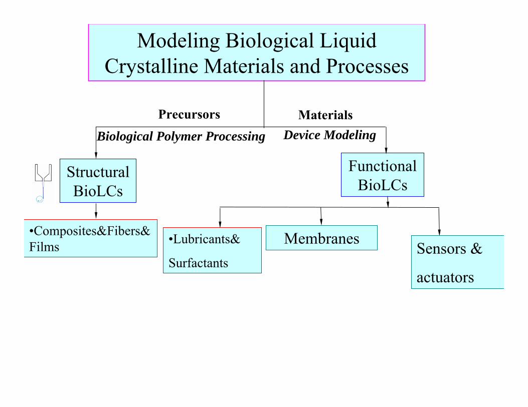
Modeling Biological Liquid Crystalline Materials and Processes
Structural BioLCs
Functional BioLCs
•Composites&Fibers&Films •Lubricants&
SurfactantsSensors &
actuators
Membranes
Precursors
Biological Polymer ProcessingMaterials
Device Modeling
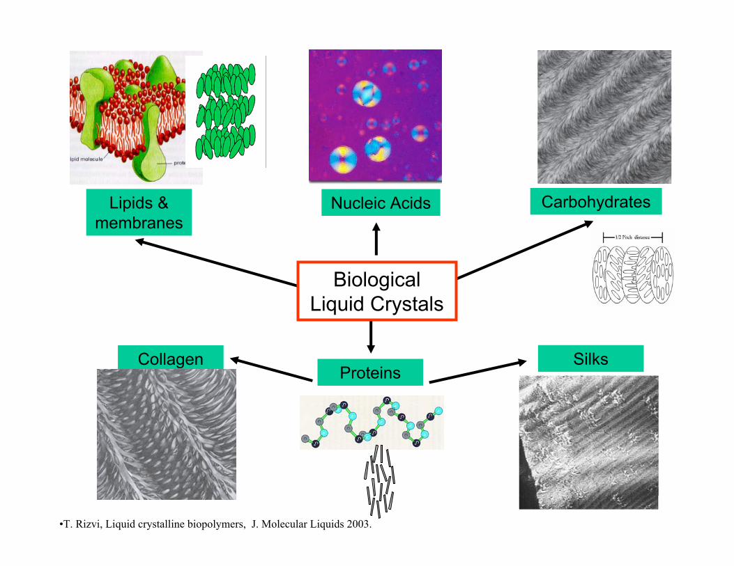
Biological Liquid Crystals
Nucleic Acids
Collagen Silks
CarbohydratesLipids & membranes
Proteins
•T. Rizvi, Liquid crystalline biopolymers, J. Molecular Liquids 2003.

Liquid Crystal Biological Polymer Processing
Spider silk: Biospinning Fiber Spinning
Chitin/collagen/keratin: biological composites Composites
Mussel Byssus: biological reaction injection moldingRIM
systematic technology transfer from nature to engineering
Green engineering, sustainability, efficiency
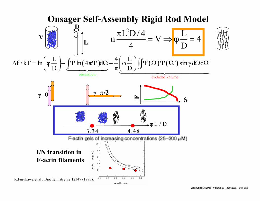
Onsager Self-Assembly Rigid Rod Model
( ) ( ) ( )
excluded volumeorientation
L 4 Lf / kT ln ln 4 d ' sin d d 'D D
⎛ ⎞ ⎛ ⎞Δ = ϕ + Ψ πΨ Ω + ϕ Ψ Ω Ψ Ω γ Ω Ω⎜ ⎟ ⎜ ⎟π⎝ ⎠ ⎝ ⎠∫ ∫∫1442443 144444424444443
2L D / 4 Ln V 44 D
π= ⇒ ϕ =
3.34 4.48
γ=0 γ=π/2SF
L
D
I/N transition in F-actin filaments
L / Dϕ
R.Furukawa et al , Biochemistry,32,12347 (1993).
V

Maier-Saupe-Doi Rigid Rod Model
B/ k Te / Z−ΦΨ =
Doi, 1981, Karlin, Ottinger (2000), R. Kemker et al, EPJ E, (2000)
Maier-Saupe-Doi Potential :
2MS in B Bf U TS 3k TUS / 4 k Tln Z= − = −
/ 2dkTZ e−Φ= ∫ u
u
(partition function)
( ) 1a U : ,U L / DΦ = − = ϕu Q uu
3.11 3.67L / Dϕ
human melanocytes cells

SD
NG
nematicisotropic
coexistence
LC
I

n
Orientation n and Alignment S
Intermediate alignment along n
S = 0.65
No alignmentalong n
S = 0
Perfect alignmentalong n
S = 1
⎛ ⎞⎜ ⎟⎝ ⎠
δQ = S nn -3
Quadrupolar order parameter Q
Orientation & alignment Q
23 1S = cos θ -2 2
molecular domainnanometer
Director field micrometer
n
u
Quadrupolar Order Parameter Q

Cholesteric order=f ( pitch, handedness, helix axisCholesteric order=f ( pitch, handedness, helix axis))
pp00
Macroscopic Macroscopic ChiralityChirality in Liquid Crystalline Phasein Liquid Crystalline Phase
( )⎛ ⎞= +⎜ ⎟⎝ ⎠
oδ BQ(z) Q(z+p ) = S nn- mm- ll3 3
BIAXIAL QUADRUPOLAR ORDER PARAMETER
Periodic/chiral
l
n
m
n
n

Chiral Nematics: Rules and Regulations
L / D 4ϕ > ± ε
1. Molecular asymmetry;optical activity
2. Some are formed by helical molecules
3.
collagenDNAPBG
Geometric packing
pPBG,L 90nm= pHPC,L 12nm=
**
DNA DNAL 50nm,c 100mg / ml= ≈
3.4 nm
μm
.54nm 8nm
A.C. Neville, Cambridge UP, 1993

de Jeu and Longa, Phys.Reports (1980), Lhuiller&Rey JNNFM (2004), de luca and A.D. Rey PRE (2004)
Landau-deGennes Chiral Nematic Model
μm
( ) TB
2B
, 3/ 2k TU
3/ 2k TU :
u u Q : uu
Q Q uu uu
Φ ∇ = −
⎛ ⎞⎛ ⎞ ⎛ ⎞⎛ ⎞ ⎛ ⎞∇ ∇×⎜ ⎟⎜ ⎟ ⎜ ⎟⎜ ⎟ ⎜ ⎟
⎝ ⎠ ⎝ ⎠⎝ ⎠ ⎝ ⎠⎝ ⎠l
0 0
4π 4π× )+ +p p
Q⎛ ⎞⎜ ⎟⎝ ⎠
2 3 2 2h
1 U U Uf /cKT = 1- tr( ) - tr(Q )+ [tr(Q )]2 3 3 4
⎛ ⎞⎛ ⎞∇⎜ ⎟⎜ ⎟⎜ ⎟⎝ ⎠⎝ ⎠
2
2g
0
L 4πf = ( ×Q)+ Q2 p
molecular chirality macroscopic chirality :h,R/L,P
h
nm
22
0
1 3 4 214 4 3 p
SU
⎛ ⎞⎛ ⎞ξ⎜ ⎟= + − + π ⎜ ⎟⎜ ⎟⎝ ⎠⎝ ⎠
22
0
2.7 1 4pICU
⎡ ⎤⎛ ⎞ξ⎢ ⎥= + π ⎜ ⎟⎢ ⎥⎝ ⎠⎣ ⎦
smaller pitch
higher packing
orientation/excluded volume chiralityLandau-deGennesSelf-Assembly Model

Chiral Nematic: A Frustrated Mesophase
I
F
a
b
( )( )⎛ ⎞⎜ ⎟⎝ ⎠
2
0
2πδn(F) = - n(I)× a×bP
P.Oswald and P. Pieranski, Nematics and Cholesteric LC, Taylor and Francis, 2005; C.B. Stanley et al, Biophysical Journal 89 (2005).
Short-fragment (146-bp) DNA , chicken erythrocytes
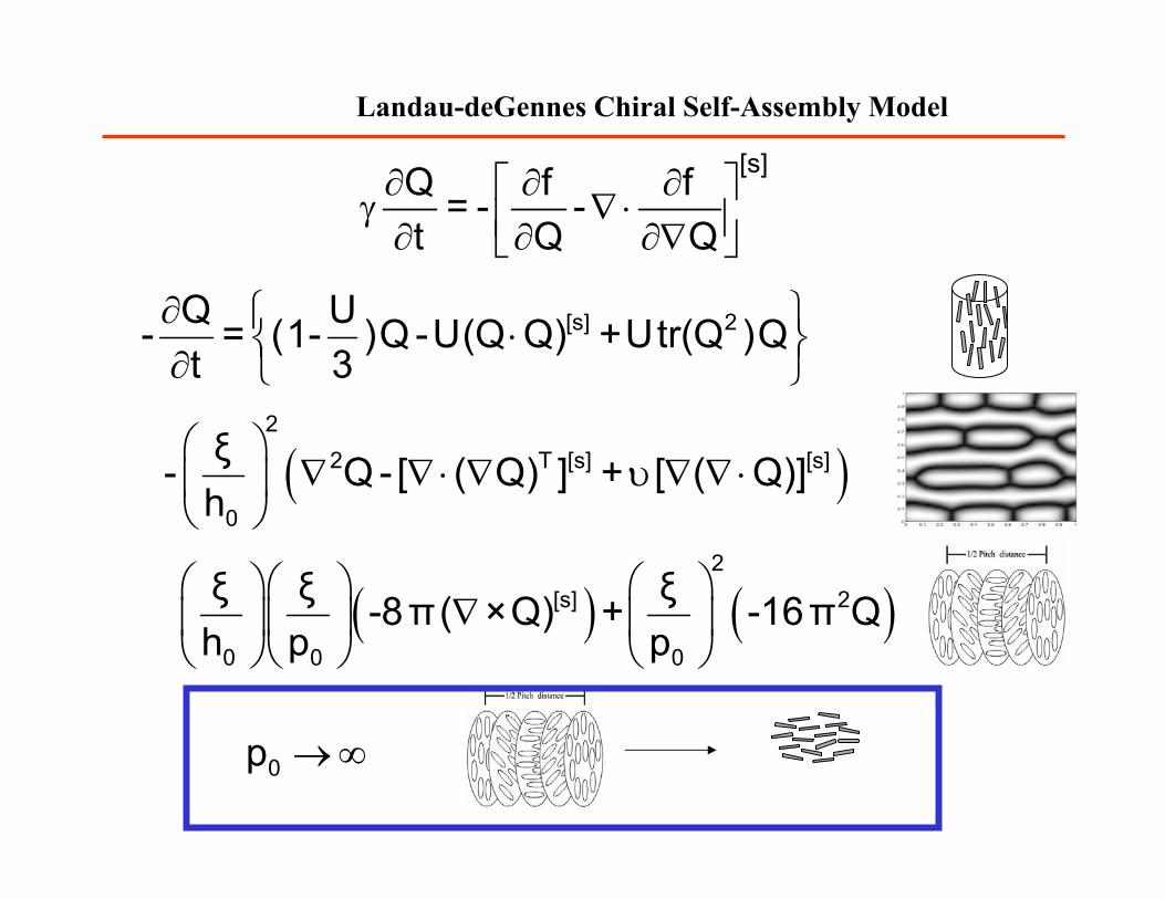
∂ ∂ ∂⎡ ⎤γ ∇ ⋅⎢ ⎥∂ ∂ ∂∇⎣ ⎦
[s]Q f f= - -t Q Q
Landau-deGennes Chiral Self-Assembly Model
∂ ⎧ ⎫⋅⎨ ⎬∂ ⎩ ⎭[s] 2Q U- = (1- )Q -U(Q Q) +Utr(Q )Q
t 3
( )⎛ ⎞∇ ∇ ⋅ ∇ υ ∇ ∇ ⋅⎜ ⎟
⎝ ⎠
22 T [s] [s]
0
ξ- Q - [ ( Q) ] + [ ( Q)]h
→ ∞0p
( ) ( )⎛ ⎞⎛ ⎞ ⎛ ⎞∇⎜ ⎟⎜ ⎟ ⎜ ⎟
⎝ ⎠⎝ ⎠ ⎝ ⎠
2[s] 2
0 0 0
ξ ξ ξ-8π ( ×Q) + -16π Qh p p

Modeling Biological Liquid Crystals
I. DNA solutions: textures and flowsLandau-deGennes, Leslie-Ericksen Nematodynamics
I. Biphasic Equilibrium: TactoidsLiquid Crystal Laplace-Herring Equation
Main Task: Use modeling to recognize and characterizebiological liquid crystal self-assembly

I.Packing helices into small volumes condensed phases of DNA
excluded volume
( ) ( )4L f f ' sin d d 'Dϕ
Ω Ω γ Ω Ωπ ∫∫
chirality
⎛ ⎞⎛ ⎞∇⎜ ⎟⎜ ⎟⎜ ⎟⎝ ⎠⎝ ⎠
2
0
4π( ×Q)+ Qp
140 base pairs, 50nm
Livolant, Leforestier, Prog. Polym. Sci., 1996

Cholesteric Packing of DNA
Livolant, Leforestier, Prog. Polym. Sci., 1996 G.deLuca and A.D.Rey, EJP (2003
Cholesteric DNA in Dinoflagellates
μmsimulations
texture
μm
R.Rill et al, Chromosoma , 1989.
simulationsCryoelectron microscopy of stallion sperm: 80nm sections,2.7nm filaments, 30 filaments
μm
Textured/Polycrystal
MonoCrystal
λ+ τ +
λ− τ −
L
L
L
L
τ− λ+
τ− τ+λ− λ+
λ− τ+
p0/2
p0
p0/2
p0
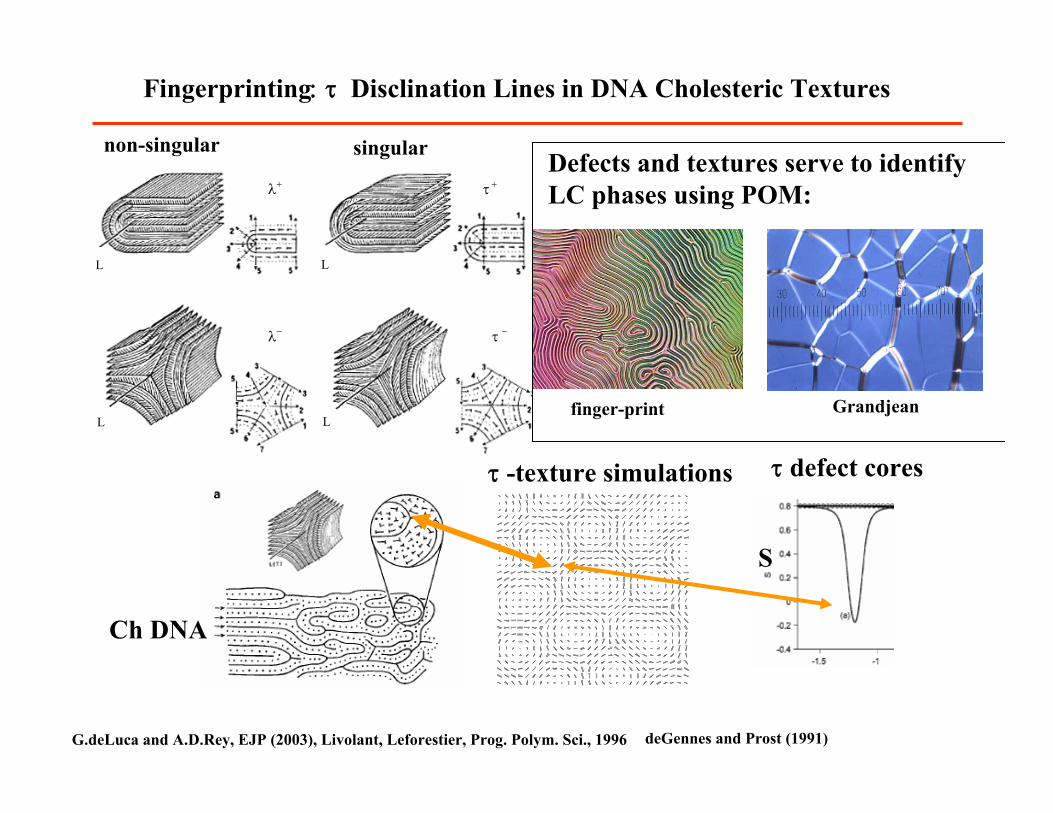
Fingerprinting: τ Disclination Lines in DNA Cholesteric Textures
G.deLuca and A.D.Rey, EJP (2003), Livolant, Leforestier, Prog. Polym. Sci., 1996
non-singular singular
τ -texture simulations
Ch DNA
τ defect cores
Defects and textures serve to identifyLC phases using POM:
finger-print Grandjean
deGennes and Prost (1991)
S

Liquid Crystallinity in Spreading DNA Drops
LeslieLeslie--EricksenEricksen NematodynamicsNematodynamics SimulationsSimulations
A.D. Rey, JOR (1991).N.Morii et al
diluted solution of salmon sperm DNA under drying process
V
silica plate
m=20mg
shear zone
extensional zone
DNS Maxwell Eqns

II. Looking at the I/LC Phase Boundary: Biological Tactoids
( ) ( )( )( )2oΔP γ + W : + s s s
spherical non spherical−
= − ∇ ⋅ −∇ ∇ ⋅ ⋅k k nn k n k14243 1444442444443
Liquid Crystal Laplace Equation
F-Actin Amyloid fibrils
μm
A.D. Rey, JCP (2004), PRE(2004), S.Das and A.D. Rey MTS (06)
Tactoid simulations
Helical FlagellaAt coexistence bioLCs drops should be tactoids!
The shape yields interfacial and bulk elasticity moduli

Modeling Biological Liquid Crystalline Materials and Processes
Structural BioLCs
Functional BioLCs
•Composites: helicoids
•Fibers&Films : silk, cellulosics, collagen, chitin
•Lubricants:
synovial fluids,
mucus
Surfactants: bile
salts
Sensors&
actuators
Membranes
Precursors
Biological Polymer ProcessingMaterials
Device Modeling
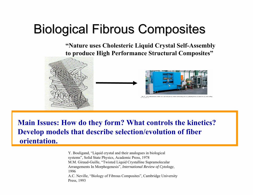
Biological Fibrous CompositesBiological Fibrous Composites
Y. Bouligand, “Liquid crystal and their analogues in biological systems”, Solid State Physics, Academic Press, 1978M.M. Giraud-Guille, “Twisted Liquid Crystalline SupramolecularArrangements In Morphogenesis”, International Review of Cytology, 1996A.C. Neville, “Biology of Fibrous Composites”, Cambridge University Press, 1993
Main Issues: How do they form? What controls the kinetics?Develop models that describe selection/evolution of fiberorientation.
“Nature uses Cholesteric Liquid Crystal Self-Assemblyto produce High Performance Structural Composites”

Universal structural motif of fibrous biological materials
Cross-section of a crab carapace
2 µm 2 µm
Cross-section of a human bone
Cross-section of the stone cell of a pear
2 µm
chitinchitin collagencollagen
cellulosecellulose
Cuticle of dragonflychitin
A.C. Neville, “Biology of Fibrous Composites”, Cambridge University

Helicoids: Plywood architecture with Helicoids: Plywood architecture with chiralchiral nematicnematic orderorder
Arced patternsOblique crossOblique cross--sectionsectionNormal crossNormal cross--sectionsection Fiber
orientation
With a constraining layerWith a constraining layer
Planar Planar monodomainmonodomain plywoodplywood
Without a constraining layerWithout a constraining layer
PolydomainPolydomain plywoodplywoodNeville,TissueNeville,Tissue & Cell 20, 133 (1988).& Cell 20, 133 (1988). A.C. Neville, “Biology of Fibrous Composites”, Cambridge University

Skaife JJ, Abbott NL, (2001)Langmuir, 17, 5595-5604.
Traveling Fronts
λ+ τ +
λ− τ −
L
L
L
L
Static Solitons:Topological Defects
Kinetics of Composite Formation

-0.5 0 0.5 1
-1.5
-1
-0.5
0
0.5
1
1.5
S
P
Pμn
=0 P=3Sμn
=μmUniaxial line
P=Sμm
=0
S μm
=μl
Uniaxial line
P=-Sμl
=0
P=-3Sμn
=μlUniaxial line
SI
SI
SI
SA
SA
SA SO
SI=sinkSA=saddleSO=source
S P(-0.3082, 0.9245) Nodal sink.(-0.3467, -0.9883) Nodal sink.( 0.6675, 0.0259) Nodal sink.( 0.0000, 0.0000) Nodal source.( 0.0582, -0.1745) Saddle point.(-0.1652, 0.0297) Saddle point.( 0.0678, 0.2626) Saddle point.U = 6 , U = 6 , ξξ/p/p00 = 0.02= 0.02
SolitonsSolitons and Traveling Fronts: Sinkand Traveling Fronts: Sink--SourceSource--Saddle ConnectionsSaddle Connections
BB
SS
1. Solitons:SA-SI connections
2.Traveling Fronts:SO-SI connections
G. deLuca and A.D. Rey , PRE (04)
( )⎛ ⎞= +⎜ ⎟⎝ ⎠
δ BQ(z) S nn- mm- ll3 3

∂ ∂ ∂⎡ ⎤γ ∇ ⋅⎢ ⎥∂ ∂ ∂∇⎣ ⎦
[s]Q f f= - -t Q Q
Chiral Self-Assembly Model
∂ ⎧ ⎫⋅⎨ ⎬∂ ⎩ ⎭[s] 2Q U- = (1- )Q -U(Q Q) +Utr(Q )Q
t 3
( ) ( )⎛ ⎞⎛ ⎞ ⎛ ⎞∇⎜ ⎟⎜ ⎟ ⎜ ⎟
⎝ ⎠⎝ ⎠ ⎝ ⎠
2[s] 2
0 0 0
ξ ξ ξ-8π ( ×Q) + -16π Qh p p
( )⎛ ⎞∇ ∇ ⋅ ∇ υ ∇ ∇ ⋅⎜ ⎟
⎝ ⎠
22 T [s] [s]
0
ξ- Q - [ ( Q) ] + [ ( Q)]h
G. deLuca and A.D. Rey , PRE (04)

Nucleation and Growth: Nucleation and Growth: SolitonsSolitons + Traveling Fronts (Hex. Sym)+ Traveling Fronts (Hex. Sym)
defectsdefects
|n|nzz| = | = OrientationOrientation
Red = Red = OutOut--ofof--planeplane
Blue = Blue = In planeIn plane
S = S = alignmentalignment
Red = High Red = High AlignmentAlignment
Blue = Low Blue = Low AlignmentAlignment
λ2+ τ−
Charge balance: -((6)-2)/2+ 2=0
BS
1 2 3+ + =k k k 0

Directed Growth: Unidirectional Traveling FrontsDirected Growth: Unidirectional Traveling Fronts
Strong planar anchoringStrong planar anchoring
SteadySteady--statestate
BS
chitin
simulationo(μm)
{
[s]s e eNsi s
i=3anchoring itensor
entropyenergy adsorption
U W T + μ 0T μ
⎧ ⎫⎪ ⎪∂ ∂ ∂⎪ ⎪+ =⎨ ⎬∂ ∂ ∂⎪ ⎪⎪ ⎪⎩ ⎭
∑n nkkn
%
14243123 14243
MAIN DESIGN TOOL:Interfacial Engineering
n k
G. deLuca and A.D. Rey , PRE (04) A.D. Rey, JCP (2003) A.C. Neville, “Biology of Fibrous Composites”, Cambridge University

( )2 T2 3 2 1
*
L1/ 1 tr( ) tr( ) tr( ) , 2 3 3 4 2
U= 3T /T
Q Q Q Q Q⎛ ⎞ ⎡ ⎤= − − + + ∇ ∇⎜ ⎟ ⎣ ⎦⎝ ⎠M
U U Uf ckT
2 22 3
20
2 2 2 2 4 .t 3 h 3 9 9 9S S U S US US
y⎛ ⎞∂ ξ ∂ ⎛ ⎞− = − + + −⎜ ⎟ ⎜ ⎟∂ ∂ ⎝ ⎠⎝ ⎠
1(y, t) (y t) (y '); /S S v S L ckT= − = ξ =
1 0S = 21 1 2494 4
SU
= − − 31 1 249 .4 4
SU
= + −
2 2
1 2 320
2 4U ( )( )( ) 0,' 3 h ' 9
dS d Sv S S S S S Sdy dy
⎛ ⎞ξ+ − − − − =⎜ ⎟
⎝ ⎠
Moving Homogeneous Flat Phase Ordering Fronts
isotropic nematicmax energy

0
2 1 3 2493 3 h 4 4
UVU
⎡ ⎤⎛ ⎞ξ= − + −⎢ ⎥⎜ ⎟
⎝ ⎠ ⎣ ⎦
1
03 hUK
−⎛ ⎞ξ
= ⎜ ⎟⎝ ⎠
3 3( ) 1 tanh ( )2 2S SS y Vt K y Vt⎧ ⎫⎡ ⎤− = − −⎨ ⎬⎢ ⎥⎣ ⎦⎩ ⎭
V>0: U > Uc=2.7 stable N I
V<0: U < Uc=2.7 stable I N phase
V=0 :U = Uc=2.7 the interface becomes static
Front Velocity
Front Velocity: 0.1m/sec
Popa-Nita and T.J. Sluckin, J. Phys. II(1996).

Process Kinetics: Speed of Process Kinetics: Speed of ChiralChiral FrontsFronts
owhen p v↓⇒ ↓
22
0 0
v1 3 24 969
3 h 4 4 p=
⎡ ⎤⎛ ⎞ ⎛ ⎞ξ ξ⎢ ⎥− + − − π⎜ ⎟ ⎜ ⎟⎢ ⎥⎝ ⎠ ⎝ ⎠⎣ ⎦
UU U
G. deLuca and A.D. Rey , PRE (04), B. Wincure and A.D. Rey, JCP (2006)
7
1rVelocity D
Lξ≈ × ∝
achiral chiralV V>
“The shell can take between a few hours and a few weeksto fully harden, depending on species. They do this by
absorbing calcium carbonate whilst the chitin hardens.”
Moulting Process

Spider Silk SpinningSpider Silk Spinning
““Nature uses Liquid Crystal SelfNature uses Liquid Crystal Self--Assembly to produce SuperAssembly to produce Super--fibersfibers””
Main Issue: use modeling to discover Main Issue: use modeling to discover spider spider biospinningbiospinning principles of valueprinciples of valueto superto super--fiber manufacturing.fiber manufacturing.

Motivation: Super-fiber Manufacturing
Material Science
Kevlar
T = 300 ºC
H2SO4
Spinning of synthetic fibres
Spider’s silk
T = 20 ºC
P = 1 atm
H2O
Spinning of natural silk
Liquid Crystalline Solution
Liquid Crystalline Solution
355Elasticity (%)
1x1094x109Strength (Nm-2)
SilkKevlarMechanical Property
From J.M. Gosline, Endeavor, 1986

Spider Silk Fiber Biospinning Process
R.F. Foelix, Biology of Spiders, Oxford University Press, J.D. van Beek et al , PNS (2002)
Ala
Gly
β-sheetscrystals
1.synthesis 2.self-assembly 3.flow-process 4. solidification

2. Silk Liquid Crystal Self-Assembly
Key Design Principle: LC viscosity is very low!
silk thermodynamics (in progress)
( )31o
nLη = η +
( )21 3o
nD Lη = η +

3. Geometry and Flow-Induced Structural Transformations
Escaped texture
Funnel
Duct
Lyotropic nematic liquid crystal
Sac (Ampulla)
Solid silk
Homeotropic anchoring
Uniaxialorientation parallel to fiber axis
Internal draw-down taper
D.P. Knight & F. Vollrath, Proc. R. Soc. Lond. B (1999) 266, 519-523

Facts on Texture in the S-shaped Duct
“Bidirectional Escaped” texture
Peridoc set of hyperbolic and radial nematic point defects
Polarized Optical Micrograph
Mean repeated period of the pattern ~ 100μm
J.E. Lydon, Liquid Crystals Today, 2004, 13(3), 1–13
Knight and Vollrath, Biomacromolecules, 2(2), 2001
+1 -1 +1 -1 -1
periodic orientation texture

2
HR= , , *
CUC
⎛ ⎞=⎜ ⎟ξ⎝ ⎠
nE = γτ&
6.5 10w
M ≈ ×
( ) ( ) : ) , 0, 0 t
= + ∇ ∇ ⋅ = ∇ ⋅ =2Q R 1C Q A - f(U,Q f Q v TE E
δ βδ
Biospinning Model
1/U(concentration)
E(flow rate)
R(geometry)
phase ordering
FI orientationElasticMode
T. Tsuji and A.D. Rey, MTS (1997)
10 / secrate mm=
20 m<H<100 mμ μ
25%C =
4f
d m= μ
40mm
process pathway?
Circular capillary
DATA
L=40mm

Bi-directional Escape Texture Formation
simulations Spider
Spider Duct 3D Nematodynamics: Elastic Mode
de Luca and A.D. Rey, JCP (2006)
perpendicular anchoring
Knight and Vollrath, Biomacromolecules, 2, 2001 J.E. Lydon, Liquid Crystals Today, 2004, 13
{
[s]s e eNsi s
i=3anchoring itensor
entropyenergy adsorption
U W T + μ 0T μ
⎧ ⎫⎪ ⎪∂ ∂ ∂⎪ ⎪+ =⎨ ⎬∂ ∂ ∂⎪ ⎪⎪ ⎪⎩ ⎭
∑n nkkn
%
14243123 14243
Texture through Interfacial Engineering: Anchoring on Curved Interface

I. Flow-Induced Orientation -Spinneret Nematodynamics
II. Gel-Crystal Transition
pf sf sfsf pf sfsf sf pf
⎛ ⎞⎜ ⎟=⎜ ⎟⎜ ⎟⎝ ⎠
A :ij ijkl lkT Aη=
H20
Flow in hyperbolic die
10 7 2
/.002 / 10 6 10 /
Thread Stress mg Acg x N mπ −
=
= = 7 2
10 /5 10 /
H H Bond kJ mol N Volumex N m− = × ×
=
α-helix β-sheets
S=0.9
Cry=12%
25
7nm
On-going work:
Mw=300,000
E.Iizuka, Adv.Biophys., 24 (1988).

BiomimeticMaterial Science
Optics Wetting Adhesion&Joints
Materials Processing
Friction
Deployable structures Sensors
ConclusionsBiological Polymer Processing
Functional Biological Liquid Crystals
Butterfly photonics
lotus leaf effect
Velcro joints
shark skin

Students and Collaborators
Funding: ERC/Center for Advanced Fibers and Films/Clemson University, Natural Science and Research Council of Canada
1. BiocompositesGino de Luca (McGill), Prof. S. Cowie (CCNY), Prof. D. Passini (McGill)
2. Spider SilkPhD McGill students:Gino de Luca , N. AbukhdeirClemson University Biomimetics Center:Prof. Chris Cox (Math), Prof. Bert Abbot (Genetics), Michael Ellison (Material Science)
3. Liquid Crystal Self-AssemblyProfessor Daniel Lhuiller (P.et M. Curie Institute, Paris)4. BiomimeticsProfessor C. Brebbia, Wessex Institute, UK