modeling sequence-specific polymers using … sequence-specific polymers using anisotropic...
TRANSCRIPT
Modeling Sequence-Specific Polymers Using Anisotropic Coarse-Grained Sites Allows Quantitative Comparison with ExperimentThomas K. Haxton,* Ranjan V. Mannige, Ronald N. Zuckermann, and Stephen Whitelam*
Molecular Foundry, Lawrence Berkeley National Laboratory, Berkeley, California 94720, United States
*S Supporting Information
ABSTRACT: Certain sequences of peptoid polymers (syn-thetic analogs of peptides) assemble into bilayer nanosheetsvia a nonequilibrium assembly pathway of adsorption,compression, and collapse at an air−water interface. As withother large-scale dynamic processes in biology and materialsscience, understanding the details of this supramolecularassembly process requires a modeling approach that capturesbehavior on a wide range of length and time scales, from those on which individual side chains fluctuate to those on whichassemblies of polymers evolve. Here, we demonstrate that a new coarse-grained modeling approach is accurate andcomputationally efficient enough to do so. Our approach uses only a minimal number of coarse-grained sites but retainsindependently fluctuating orientational degrees of freedom for each site. These orientational degrees of freedom allow us toaccurately parametrize both bonded and nonbonded interactions and to generate all-atom configurations with sufficient accuracyto perform atomic scattering calculations and to interface with all-atom simulations. We have used this approach to reproduce allavailable experimental X-ray scattering data (for stacked nanosheets and for peptoids adsorbed at air−water interfaces and insolution), in order to resolve the microscopic, real-space structures responsible for these Fourier-space features. By interfacingwith all-atom simulations, we have also laid the foundation for future multiscale simulations of sequence-specific polymers thatcommunicate in both directions across scales.
Many of the most important processes in molecularbiology, including allostery,1,2 enzyme catalysis,3 molec-
ular recognition,4 protein homeostasis,5 and nucleic acidmetabolism,6 involve the cooperative motion of large, preciselyself-assembled7 biomolecules. Engineering synthetic materialswith similarly sophisticated functionality will require methodsto relate the chemical sequence of large molecules (e.g.,biomolecules or sequence-defined synthetic polymers) to theirself-assembled form and function. These methods must spanmany orders of magnitude in time and space in order todescribe atomically detailed interactions and correlated supra-molecular motions, both of which contribute to materialsassembly and function.One class of nanomaterials that show promise as scaffolds for
molecular recognition and catalysis8 are peptoid nanosheets,solid bilayers that assemble from sequence-defined peptoidpolymers9 (positional isomers of peptides) due to a mechanicalprotocol that acts on many peptoids collectively (Figure1).10−13 Exposed to an air−water interface, amphiphilic peptoidpolymers first adsorb from solution onto the interface, forminga structured monolayer. Subsequently compressing themonolayer past a certain pressure induces irreversible collapseinto bilayer nanosheets 2.9 nm thick and up to 100 μm wide.Such a process is determined by mechanisms operating atmultiple length scales: electrostatic interactions at the angstromscale link side chains on neighboring polymers; amphiphilicpatterning at the 1 nm scale allows for adsorption to the air−water interface; the motion of polymers on scales up to theirfull 10 nm length determines whether in-plane ordering occurs;
and nanosheets can extend to scales of order 100 μm. Theassociated time scales range from picoseconds, for atomic andmolecular fluctuations, to the seconds or minutes on whichnanosheets are produced.Developing a detailed, real-space picture of such a multiscale
process requires a modeling approach able to account formechanisms operating on a broad range of length and timescales. Studies of macromolecules such as nucleic acids andproteins have shown that coarse-grained modeling can, inprinciple, span scales efficiently, by representing explicitly onlythe most important molecular degrees of freedom, andrepresenting implicitly other degrees of freedom via “effective”interactions.14−24 However, reducing the number of degrees offreedom unavoidably discards information, so the resultingcoarse-grained model cannot capture all aspects of theunderlying all-atom system. For example, it has been showngenerally that coarse-grained models parametrized to reproduceall-atom pair distribution functions cannot correctly reproducethermodynamic properties such as energy and pressure, andvice versa.25−27
To mitigate this representability problem,25−27 careful choicesmust be made in the two key aspects of a coarse-grainingscheme: the choice of which degrees of freedom (or “sites”) tobe retained, and how interactions between sites should beparametrized. Most work has focused on the latter, resulting in
Received: September 6, 2014Published: December 10, 2014
Article
pubs.acs.org/JCTC
© 2014 American Chemical Society 303 DOI: 10.1021/ct5010559J. Chem. Theory Comput. 2015, 11, 303−315

the development of rigorous interaction-parametrizationschemes to target particular features (typically distributionfunctions, forces, or energies) of related all-atom simula-tions28−32 or to minimize the relative entropy between coarse-grained and all-atom ensembles.33−36
Fewer authors have investigated the choice of which degreesof freedom to retain. Coarse-grained models based on rigorousparametrization schemes have employed isotropic (sphericallysymmetric) interactions, for which these schemes are mosttractable. However, it has been shown that the accuracy ofisotropic coarse-grained models declines as the underlying all-atom system becomes more anisotropic, due to the fact thatmore information is lost when averaging spherically overanisotropic interactions than over isotropic ones.37,38
One way to improve the accuracy of a coarse-grained modelwith spherically symmetric sites is to increase the number ofsites, a strategy often employed in protein modeling.39−41 Forexample, by including between 3 and 8 sites per amino acidresidue, the PRIMO protein model41 can estimate all-atomconfigurations with such accuracy that all-atom configurationscan be passed between PRIMO and atomistic models with 0.1Å resolution,42 allowing the model to seamlessly interface withall-atom force fields in multiscale simulations.43 While such anapproach clearly represents a protein in great detail, the largenumber of degrees of freedom represented means thatsimulations are only one order of magnitude faster than all-atom simulations.41
Here, we demonstrate an alternative strategy for combiningaccuracy and efficiency within coarse-grained modeling: retainonly a minimal number of coarse-grained sites (two permonomer) but include fluctuating orientational degrees of
freedom for each site. Coarse-grained modelers have recognizedthe importance of anisotropic interactions in biomolecules,having incorporated directional nonbonded interactions intomodels for proteins,44−51 DNA,52−55 and lipids.56 Efforts inprotein modeling have focused on capturing the directionalityof backbone hydrogen bonding (absent in peptoids) byapproximating the dipole−dipole interaction between peptidegroups using only the positions of nearby alpha carbons on thebackbone.45−50 In addition, at least one model has accountedfor the anisotropic shape of protein side chains by usingellipsoidal (but energetically isotropic) side chain sites,44 andone model has accounted for dipole−dipole interactionsbetween polar side chains.51 Coarse-grained DNA modelshave focused more on directional nonbonded interactions,including separate base-pairing, base-stacking, and cross-stacking interactions depending on the relative orientation ofinteracting nucleobases.52−55
Motivated by the success of those strategies, and recognizingthe importance of directional interactions and torsionalconformations in peptoids, we created a model with directionalinteractions depending on independently f luctuating orienta-tions, associating each site with both a position and anindependent symmetry axis. As far as we know, the onlyprevious uses of orientational degrees of freedom in coarse-grained biomolecule models are the protein model of Spiga,Alemani, Degiacomi, Cascella, and Dal Peraro, which includesrotating electric dipoles in polar side chains,51 and the DNAmodel of Morriss-Andrews, Rottler, and Plotkin, which includesone soft orientational degree of freedom per base.53 Relative toan isotropic model, including a symmetry axis increases thenumber of degrees of freedom per site from three to five butimproves the accuracy of our model in two ways. First, it allowsus to parametrize bonded and nonbonded interactions thatincorporate atomic-level details like covalent-bond dihedralangle distributions and electric dipole interactions. Second, itallows us to estimate, or backmap, all-atom configurations withsufficient accuracy to perform detailed scattering calculationsand interface with all-atom simulations.We plan to use our model to investigate the dynamic, large-
scale processes in the nanosheet production cycle (Figure 1):adsorption of solvated peptoid polymers to the air−waterinterface, ordering of the adsorbed monolayer, and collapse intoa free-floating bilayer. In the current study, we establish that ourmodel reproduces all known structural features of theequilibrium states involved in this production cycle, featuresmeasured by solution X-ray scattering of solvated polymers,grazing-incidence X-ray scattering of monolayers, and orientedX-ray scattering of stacks of bilayer nanosheets. In so doing, weprovide a microscopic, real-space interpretation of theseFourier-space features. In addition, we show how our approachcan interface with all-atom simulations with an accuracycomparable to computationally more demanding models thatpossess more degrees of freedom, laying the groundwork forefficient multiscale simulations57−60 of sequence-specificpolymers that communicate in both directions across scales.
■ MODEL AND METHODSIn this paper, we focus on the block-charge peptoid illustratedin Figure 2a−c, although we note that our coarse-grainingscheme is transferable to different peptoid chemistries and isgeneralizable to proteins61. “Block-n” peptoids are (poly)-peptoids of the form ((Nae−Npe)n/4−(Nce−Npe)n/4), builtfrom two equal-length blocks, each consisting of nonpolar N-
Figure 1. (a) Nanosheet production cycle. Exposed to an air−waterinterface, amphiphilic peptoid polymers adsorb from solution onto theair−water interface. Compressing the monolayer induces irreversiblecollapse into free-floating bilayer nanosheets. Decompressing theinterface completes the cycle, allowing additional peptoids to adsorb.(b) Experimental plot of the surface pressure and surface area of themonolayer during one production cycle, illustrating the large hysteresisassociated with the irreversible formation of nanosheets.10−13 (c) Nilered fluorescence micrograph of a solution of free-floating nanosheets.8
Journal of Chemical Theory and Computation Article
DOI: 10.1021/ct5010559J. Chem. Theory Comput. 2015, 11, 303−315
304

(2-phenylethyl)glycine monomers alternating with chargedmonomers. The first block contains positively charged N-(2-aminoethyl)glycine monomers, and the second block containsnegatively charged N-(2-carboxyethyl)glycine monomers. Weinvestigated “block-n” peptoids of several lengths but presentresults primarily for block-28, for which the most extensive setof experimental data exists.As illustrated in Figure 2b, we construct the positions of our
coarse-grained model in the usual way, associating groups ofatoms with coarse-grained sites and linking those sites withvirtual or “coarse-grained” bonds.14−24 We assign two coarse-
grained sites per monomer, one for the backbone and one forthe side chain. The resulting bonded network is a linear chainof n backbone sites, each branched with one side chain site, plus(optionally) an additional backbone site representing therelatively bulky carboxy-terminus. We associate the backbonesites with the backbone atoms of each monomer plus the firstmethylene bridge of the side chain. We associate thephenylethyl, aminoethyl, and carboxyethyl side chain siteswith the remaining atoms of the side chains. With theseassociations, the backbone maps to the molecule N-
Figure 2. (a) Chemical diagram of a 28-monomer “block-28” peptoid polymer (Nae-Npe)7−(Nce-Npe)7. Blue and red parts of the diagram denotepositively and negatively charged polar side chains, yellow parts denote nonpolar side chains, and grey parts denote the backbone. (b) Ball-and-stickrepresentation of our coarse-grained peptoid model, taken from a single strand equilibrated within a monolayer. Blue, red, and yellow balls denotepositions of the positively charged side chain sites, grey balls denote positions of the backbone sites, and arrows denote the symmetry axis of eachsite. (c) All-atom representation backmapped from the positions and (superposed) orientations of the coarse-grained sites. Atoms are coloredaccording to the site with which they are associated. (d-e) Close-up view showing two monomers in the aminoethyl block in (d) coarse-grained and(e) all-atom representations. (f-g) Close-up view showing two monomers in the carboxyethyl bock in (f) coarse-grained and (g) all-atomrepresentations.
Journal of Chemical Theory and Computation Article
DOI: 10.1021/ct5010559J. Chem. Theory Comput. 2015, 11, 303−315
305
methylacetamide and the side chain sites map to the “side chainanalogs”62 toluene, methylammonium, and acetate.Our model distinguishes itself from previous coarse-grained
models by defining each site Xi = {ri,ni} by both a position riand a director n i (arrows in Figure 2b) representing thedominant symmetry axis of the atoms associated with the site.For the backbone sites, defining ri by the backbone N locationand ni by the first side chain (N−Cβ) bond allows us to capturethe softest covalent interactions (bond torsions) with effectiveinteractions that account for the accumulated torsion along thebackbone and side chain branches of the covalent network. Forthe phenylethyl side chain site, defining ri by the center of thearomatic ring and n i by its normal allows us to employanisotropic nonbonded interactions that respect the aromaticring’s approximately axial symmetry. For the aminoethyl andcarboxyethyl side chains, defining ri by the center of mass of theassociated atoms and n i by the direction of the central bond(Cγ−Namino or Cγ−Ccarboxy) aligns the site with the dipolemoment of the associated atoms and leaves only hydrogens andthe carboxy oxygens off the symmetry axis, allowing us toemploy anisotropic nonbonded interactions capturing thedominant (monopolar and dipolar) electrostatic interactionsamong charged groups.The principle of coarse-grained modeling lies in replacing a
potentially exact many-body effective interaction betweencoarse-grained sites with a sum of few-body interactions thatcan be efficiently incorporated into molecular simulations.14−24
We followed this principle by decomposing our effectiveinteraction into bonded and nonbonded terms, as convention-ally done, plus height-dependent solvation terms accounting forthe preference of some molecular groups (such as the aromaticgroups) to adsorb to the air−water interface. Height-dependentsolvation terms are not common in coarse-grained models;instead, coarse-grained models used to study interfaces (such asthe MARTINI lipid model63) typically employ explicit coarse-grained water particles. A height-dependent solvation inter-action has been used previously to model surfactant adsorptiononto a solid interface.64 In future work, we plan to allow theshape of the air−water interface to fluctuate, including a fourthpotential energy term to couple these fluctuations to the surfacetension. We expect that including these fluctuations will benecessary to allow investigation of the mechanism by which themonolayer collapses into a bilayer.We parametrized the potential energy function in two stages.
First, we applied a “bottom-up” parametrization, performing
direct Boltzmann inversion65 to fit a set of effective potentialsto all-atom potentials of mean force,
∫ ∏ δ= − − −α α
α α⎛⎝⎜⎜
⎛⎝⎜
⎞⎠⎟
⎞⎠⎟⎟U R
k Td u
k TR Rr r r
({ })ln exp ( ) ( ( ) )i
ii i
B B
(1)
where {Riα} is an orthogonal set of generalized coordinates
describing a particular bonded, nonbonded, or solvationinteraction Uα; dr are a set of atomic coordinates includingwater atoms; Ri
α(r) is the mapping from atomic coordinates togeneralized coordinates; and u(r) is an all-atom force field. Wecalculated the integral in eq 3 by conducting all-atom moleculardynamics simulations of the smallest molecular systemsadequately constraining Uα.Our use of effective potentials Uα({Ri
α}) depending on morethan one variable is not typical among coarse-grained models.Typically, coarse-grained models built from isotropic sites usesingle-variable effective potentials analogous to those found inall-atom force fields: bonded interactions summed to dependseparately on bond lengths, bond angles, and dihedral angles,and spherically symmetric nonbonded interactions dependingonly on radial separations. Since all-atom distribution functionstypically do not factorize with respect to these variables, directBoltzmann inversion would not be expected to produceaccurate effective potentials. Iterative fitting procedures likeiterative Boltzmann inversion,66 the multiscale coarse-grainingmethod,28−31 and the relative entropy method33−36 aretypically used instead. These methods use a series of coarse-grained simulations to converge to an optimized coarse-grainedmodel. Their success relies on the ability of single-variableeffective potentials to collectively yield accurate many-bodydistribution functions, despite the fact that these distributionfunctions do not factorize into a form commensurate with thesingle-variable terms of the potential energy function.Figure 3 illustrates our approach for the bonded interaction
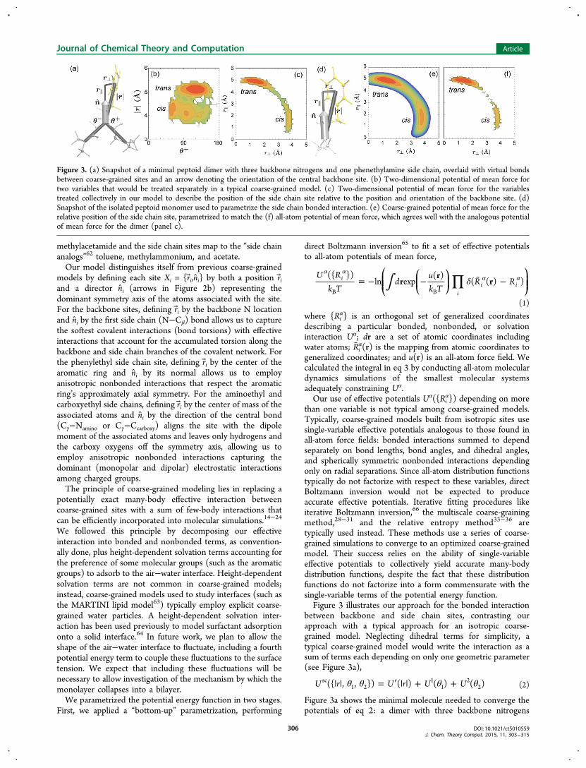
between backbone and side chain sites, contrasting ourapproach with a typical approach for an isotropic coarse-grained model. Neglecting dihedral terms for simplicity, atypical coarse-grained model would write the interaction as asum of terms each depending on only one geometric parameter(see Figure 3a),
θ θ θ θ| | = | | + +U r U r U U({ , , }) ( ) ( ) ( )rsc1 2
11
22 (2)
Figure 3a shows the minimal molecule needed to converge thepotentials of eq 2: a dimer with three backbone nitrogens
Figure 3. (a) Snapshot of a minimal peptoid dimer with three backbone nitrogens and one phenethylamine side chain, overlaid with virtual bondsbetween coarse-grained sites and an arrow denoting the orientation of the central backbone site. (b) Two-dimensional potential of mean force fortwo variables that would be treated separately in a typical coarse-grained model. (c) Two-dimensional potential of mean force for the variablestreated collectively in our model to describe the position of the side chain site relative to the position and orientation of the backbone site. (d)Snapshot of the isolated peptoid monomer used to parametrize the side chain bonded interaction. (e) Coarse-grained potential of mean force for therelative position of the side chain site, parametrized to match the (f) all-atom potential of mean force, which agrees well with the analogous potentialof mean force for the dimer (panel c).
Journal of Chemical Theory and Computation Article
DOI: 10.1021/ct5010559J. Chem. Theory Comput. 2015, 11, 303−315
306
(three backbone sites) and one bulky side chain (one side chainsite) branched from the central nitrogen. As shown in Figure3b, the two-dimensional potential of mean force depending onθ1 and |r| does not factorize into θ1 and |r| terms: the basin ofattraction at large |r| (corresponding to an extended, trans sidechain) is more narrowly distributed in θ than the basinattraction at small |r| (corresponding to a contracted, cis sidechain). Thus, one would not expect direct Boltzmann inversionof single-variable effective interactions to yield an accuratemodel. Instead, one would rely on an iterative procedure toconverge targeted features of the model with those of the all-atom system. These procedures would rely on collective effects
from other interactions, such as a long-range nonbondedinteraction between the side chain site and the terminalbackbone sites, to converge the model.In our model, we write the bonded side chain interaction as a
multiple-variable function that depends on the position andorientation of the bonded side chain and backbone sites butdoes not depend on positions of neighboring backbone sites.Considering only the side chain site’s position for simplicity(ignoring terms introduced to control the side chain site’sorientation), the interaction has the form
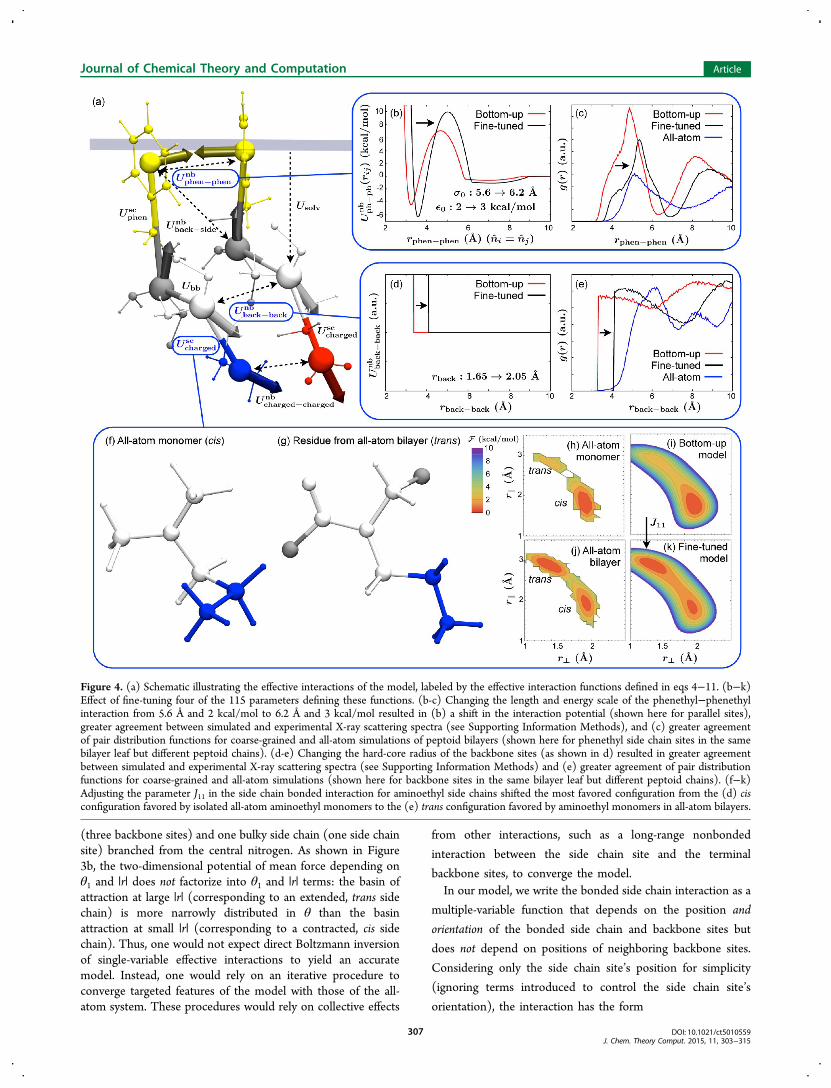
Figure 4. (a) Schematic illustrating the effective interactions of the model, labeled by the effective interaction functions defined in eqs 4−11. (b−k)Effect of fine-tuning four of the 115 parameters defining these functions. (b-c) Changing the length and energy scale of the phenethyl−phenethylinteraction from 5.6 Å and 2 kcal/mol to 6.2 Å and 3 kcal/mol resulted in (b) a shift in the interaction potential (shown here for parallel sites),greater agreement between simulated and experimental X-ray scattering spectra (see Supporting Information Methods), and (c) greater agreementof pair distribution functions for coarse-grained and all-atom simulations of peptoid bilayers (shown here for phenethyl side chain sites in the samebilayer leaf but different peptoid chains). (d-e) Changing the hard-core radius of the backbone sites (as shown in d) resulted in greater agreementbetween simulated and experimental X-ray scattering spectra (see Supporting Information Methods) and (e) greater agreement of pair distributionfunctions for coarse-grained and all-atom simulations (shown here for backbone sites in the same bilayer leaf but different peptoid chains). (f−k)Adjusting the parameter J11 in the side chain bonded interaction for aminoethyl side chains shifted the most favored configuration from the (d) cisconfiguration favored by isolated all-atom aminoethyl monomers to the (e) trans configuration favored by aminoethyl monomers in all-atom bilayers.
Journal of Chemical Theory and Computation Article
DOI: 10.1021/ct5010559J. Chem. Theory Comput. 2015, 11, 303−315
307

∑= − + − −
+
⊥ ⊥ ⊥
=⊥
U r r k r r r r r
J r r
( , ) ((( ) ( ) ) )
(arctan( / ))j
jj
sc1 0
20
2 1/20
2
0
4
1(3)
where r∥ and r⊥ are the components of the side chain separationparallel and perpendicular to the backbone director (Figure 3a)and the other parameters are constants. The first term in eq 3describes an arc traced out by the side chain site as the middleof three side chain hydrocarbon bonds twists, and the secondterm in eq 3 describes the bimodal distribution of dihedralangles with minima corresponding to cis and trans config-urations (Figure 3c). Dihedral rotation of the third hydro-carbon bond does not affect the side chain site’s position(center of mass of the aromatic ring), and we model the firsthydrocarbon bond as freely rotatable, motivated by therelatively weak dihedral interaction in our reference all-atomforce field.67 Thus, a simple pairwise interaction betweenoriented backbone and side chain sites captures the dominantatomic-level energetics of the underlying all-atom system.(Modeling the weak dihedral angle dependence for the firsthydrocarbon bond could be achieved by using fully orientedsites61 rather than sites with only one symmetry axis.)Since the bonded interaction (eq 3) depends only on the
position and orientation of the backbone and side chain sites ina single monomer, we parametrized the interaction by matchingpotentials of mean force (e.g., Figure 3e) to all-atom potentialsof mean force (e.g., Figure 3f) for an isolated monomerdissolved in water (Figure 3d), using a CHARMM-basedpeptoid force field developed in our lab.67,68 Comparing Figure3f to Figure 3c shows that the all-atom potential of mean forcefor the monomer agrees well with the same potential of meanforce for the trimer, justifying our use of the monomer toconstrain the side chain bonded interaction.We parametrized the remaining effective interactions using a
similar Boltzmann inversion procedure. Figure 4a illustrates theeight types of effective interactions, and their functional formsare written below in eqs 4−11. Equation 4 is the backbonebonded interaction Ubb among three adjacent backbone sites,which uses the same coefficients regardless of monomer type.Equation 5 is the side chain bonded interaction betweenbackbone and phenethyl side chain sites and eq 6 is the sidechain bonded interaction between backbone and charged sidechain sites, which uses different coefficients for aminoethyl andcarboxyethyl side chains. Equations 5 and 6 have differentfunctional forms because the symmetry axis of the side chainsites are perpendicular and parallel to the side chain’shydrocarbon chain for phenethyl and charged side chains,respectively. Equation 7 is the nonbonded interaction betweenphenethyl side chain sites; eq 8 is the nonbonded interactionbetween charged side chain sites (with charges and geometricparameters depending on the identities of the two involvedsites); eq 9 is the nonbonded interaction between backboneand any other site (with hard-core radii depending on theidentities of the sites); and eq 10 is the interaction betweennonbonded phenethyl and side chain sites (also with hard-coreradii depending on the identities of the sites). Equation 11 isthe one-body, height-dependent solvation interaction, withcoefficients depending on the monomer type.
∑ ∑
∑ ∑
∑
∑
= | |
+ · − · ·
+ · −| | −
+ · −| | −
=
−
=+
=
−
=− + − +
=−
−
=
−
++
⎛⎝⎜⎜
⎞⎠⎟⎟
⎛⎝⎜⎜
⎞⎠⎟⎟
U K r
K r r r n r n
k r nr r
s
k r nr r
s
( ( )( ))
i
N
jj i i
j
i
N
jj i i i i i i i i i i
j
i
N
i i ii ibb
i
N
i i ii i
bb1
1
0
4
1 , 1bb
2
1
0
4
2 1,bb
, 1bb
1,bb bb
, 1bb bb
2l 1,
bb bb , 1 0l
l
2
1
1
r , 1bb bb , 1
bb0r
r
2
(4)
∑ ∑ ∑
∑ ∑
∑
= − + − −
+ + +
+ + + ·
+ · + · + ·
⊥ ⊥
=⊥
= =
= =
=
U k r r r r r
J r r J r J n r
J n J n J r n n
J r n n J r n n J r n
((( ) ( ) ) )
(arctan( {/} ))
( )
( ) ( ) ( )
jj
j
jj
j
jj
j
jj
j
jj
j
jj
j
phensc
1 02
02 1/2
02
0
4
10
6
200
2
222
2404
0,2,430
1,331
0,232
2331
3340
4
(5)
∑ ∑
∑
= − + − −
+ + −
+ · −
⊥ ⊥
=⊥
=
=
U k r r r r r
J r r k n r r
k r n n n
((( ) ( ) ) )
(arctan( / )) ( ))
( ))
jj
j
jj
j
jj
j
chargedsc
1 02
02 1/2
02
0
4
1 20
2
22
30
2
32
(6)
= + +
+ − +
−
⎜ ⎟⎛⎝
⎞⎠
U X X U X X S r S r U X X
S r S r U X X
( , ) ( , ) 12
( ( ) ( )) ( , )
1 12
( ( ) ( )) ( , )
i j i j iz jz i j
iz jz i j
ph phnb
GB sm
q
(7)
δ δ= | | − +
+ | + − + |−U X X U r R R
U r n r n( , ) ( ( ))
( ( ) ( ) )i j ij i j
j j j i i i
charged chargednb
ex
el (8)
=∞ ≤
>− ⎪
⎪⎧⎨⎩
U rr r
r r( )
,
0,ijij
ijback anynb 0
0 (9)
=∞ ≤
>− ⎪
⎪⎧⎨⎩
U rr r
r r( )
,
0,ijij
ijphen chargednb 0
0 (10)
σ
= − + −+ − −
+ − −
⎛⎝⎜
⎞⎠⎟
⎛⎝⎜⎜
⎛⎝⎜
⎞⎠⎟
⎞⎠⎟⎟
U r U k TU k T
r z w
Ur z
( ) ln 1exp( / ) 1
1 exp( 4( )/ )
exp 12
zz
z
solvation bs B rbs B r
int
intint
int
2
(11)
In eq 7 the expressions entering the nonbonded phenethyl−phenethyl interaction are
Journal of Chemical Theory and Computation Article
DOI: 10.1021/ct5010559J. Chem. Theory Comput. 2015, 11, 303−315
308

ε ε κ ξσσ σ ξσ
ξσσ σ ξσ
= ′ | | − +
− | | − +
⎛
⎝⎜⎜⎛⎝⎜⎜
⎞⎠⎟⎟
⎛⎝⎜⎜
⎞⎠⎟⎟
⎞
⎠⎟⎟
U X X X Xr X X
r X X
( , ) 4 ( , ; )( , )
( , )
i j i jij i j
ij i j
GB 00
0 0
12
0
0 0
6
(12)
ε κ χ κχ κ
χ κ χ κ
′ = − ·
× − ′ ′ · + · + ′ ′ ·
+ · − ·
− ′ ′ ·
⎛⎝⎜⎜
⎛⎝⎜⎜
⎞⎠⎟⎟⎞⎠⎟⎟
X X n nr n r n
n nr n r n
n n
( , ; ) (1 ( ) ( ) )
1 ( )2 1 ( ) 1 ( )
i j i j
ij i ij j
i j
ij i ij j
i j
2 2
(13)
σ χ κχ κ χ κ= −
· + · + ·
+ · − · − ·
−⎛⎝⎜⎜
⎛⎝⎜⎜
⎞⎠⎟⎟⎞⎠⎟⎟X X
r n r nn n
r n r nn n
( , ) 1 ( )2
( )
1 ( )
( )
1 ( )i jij i ij j
i j
ij i ij j
i j
2 2 1/2
(14)
χ κ κκ
= −+
( ) 11
2
2 (15)
χ κ κκ
′ ′ = − ′+ ′
μ
μ( ) 11
1/
1/ (16)
σ= + −S zz z
( ) 11 exp(4( )/ )int int (17)
ε ε κ σ σ σ σ
ε ε κ σ σ σ σ
σ σ
=
− | | − − | | − <
− − | | − − < | | − <
| | − >
⎜ ⎟
⎜ ⎟
⎧
⎨
⎪⎪⎪⎪
⎩
⎪⎪⎪⎪
⎛⎝⎜
⎛⎝
⎞⎠
⎞⎠⎟
⎛⎝⎜
⎛⎝
⎞⎠
⎞⎠⎟
U X X
X Xw
r X X w r X X w
X Xw
r X X w w r X X w
r X X w
( , )
( , ; ) 1 4 ( , )2
, ( , )
( , ; ) 1 4 ( , ) 32
, ( , ) 2
0, ( , ) 2
i j
i j ij i j ij i j
s i j ij i j ij i j
ij i j
sm
r r 2 0
2
0
s ss 2 0
2
0
0 (18)
In eq 8, the expressions entering the nonbonded charged−charged interaction are
Δ =
∞ Δ <
+
− Δ −< Δ <
Δ >
⎧
⎨
⎪⎪⎪⎪
⎩
⎪⎪⎪⎪
⎛⎝⎜⎜
⎛⎝⎜
⎞⎠⎟
⎞⎠⎟⎟
U r
r
f U U
r rr
r r
r r
( )
, 0
( )
1 4/2
,
0
0,
i sj
exsh bs b
sh
sh
2sh
sh
(19)
λε δ δ=
−− − + −U r
Cq q r
r R R r( )
exp( / )
( ( ))i j
i i j jel
D
(20)
εε ε ε λ λε λ
=+ − <
>ε ε
ε
∞
∞⎪
⎪⎧⎨⎩r
r r
r( )
( ) / ,
,0 0
(21)
Sections 1−3 of the Supporting Information Methods discussin detail the Boltzmann inversion procedures we used toparametrize each term. We parametrized bonded side chaininteractions (eqs 5 and 6) by inverting potentials of mean forcefrom all-atom simulations of isolated monomers dissolved inwater using the MF-TOID all-atom peptoid force field,67 asdiscussed above for eq 3 but including the dependence on theside chain sites’ orientations. We used MF-TOID simulations ofa peptoid dimer with three backbone nitrogens to invert thebackbone bonded interaction (eq 4), which includes both two-and three-body terms among neighboring backbone sites.We parametrized the nonbonded interactions by matching
orientation-dependent potentials of mean force to publishedall-atom potentials of mean force for small-molecule analogs ofthe coarse-grained sites.69,70 We used a Gay−Berne potential tomodel the nonbonded interaction between phenethyl sidechain sites,71−74 targeting potentials of mean force fortoluene,69 and we used an anisotropic electrostatic potential
(hard spheres with off-center point charges) with an ad-hocsolvent-mediated term to model the interaction betweenoppositely charged aminoethyl and carboxyethyl side chainsites, targeting potentials of mean force for methylammoniumand acetate.70 We used the same electrostatic parameters tomodel the interactions between equally charged side chain sites.(As far as we know, no one has conducted all-atom simulationsfor equally charged molecules like methylammonium andacetate that are analogous to our charged side chains.) Forsimplicity, we let the remaining, weaker interactions (back-bone−side chain, phenethyl−aminoethyl, and phenethyl−carboxyethyl) be purely repulsive, using hard-core repulsionswith conservatively small radii to prevent erroneous exclusionsof viable all-atom configurations. We let the hard-core radii ofthe charged sites be equal to the hard-core radii used in theinteraction between charged sites, we let the hard-core radius ofthe phenethyl side chain site be equal to the shorter of theprinciple radii for the Gay−Berne potential, and we initially setthe hard-core radii of the backbone site equal to aconservatively small value, the van der Waals radius of thecentral nitrogen atom.Finally, we parametrized a height-dependent solvation free
energy for each type of site, targeting experimental bulksolvation free energies75−77 and height-dependent all-atompotentials of mean force78−80 for the molecular analogs of eachsite. Although the use of molecular analog solvation freeenergies by themselves to predict macromolecule free energieshas been criticized because it neglects solvent exclusion and“self-solvation”,81 we explicitly accounted for those effects byincluding both solvation interactions and solvent-mediatednonbonded interactions.In total, our model has 115 parameters, 114 of which were
parametrized to match experimental free energies or all-atompotentials of mean force for small-molecule analogs, and one ofwhich (the backbone site hard-core radius) was initiallyassigned a conservatively small value.In a second stage of parametrization, we fine-tuned four of
the 115 parameters, including the one that was not para-
Journal of Chemical Theory and Computation Article
DOI: 10.1021/ct5010559J. Chem. Theory Comput. 2015, 11, 303−315
309

metrized from the bottom up, to improve the agreement withexperiments and all-atom simulations82 of peptoid bilayers. Wefine-tuned these parameters to simultaneously improve theagreement with (1) experimental X-ray scattering spectra,which showed a somewhat sharper dominant peak at asomewhat smaller wavenumber, (2) all-atom pair distributionfunctions, some of which showed peaks at larger distances, and(3) distributions for the position and orientation of theaminoethyl side chains, which were shifted more toward thetrans configuration.Figure 4 highlights the effect of fine-tuning the four
parameters, and a detailed discussion appears in Section 7 ofthe Supporting Information Methods. As shown in Figure 4b-c,increasing the size (by 11%) and interaction strength (by 50%)of the phenethyl side chain sites (the dominant nonbondedinteraction in the system) improved the agreement ofphenethyl−phenethyl pair distribution functions between all-atom and course-grained simulations. As shown in Figure 4d-e,increasing the hard-core radius of the backbone sites from theconservatively small nitrogen van der Waals radius improvedthe agreement of backbone−backbone pair distributionfunctions. We chose particular values of the phenethyl sizeand the backbone length scale from a two-dimensional grid tooptimize the agreement with the experimental X-ray scatteringpeak height and location, and we found that our choice was alsoamong the best choices for improving the agreement with all-atom distribution functions. We did not adjust the parameterscontrolling the shape of the phenethyl-phenethyl side chaininteraction, because we found that orientational pair distribu-tion functions (see Supporting Information Methods) agreedwell with the all-atom simulations throughout the range ofphenethyl sizes that we investigated.Finally, as shown in Figure 4f−k, we found that it was
necessary to adjust the side chain bonded interaction for thephenethyl side chains so that the side chains wouldpredominantly adopt a trans configuration, as in all-atomsimulations of peptoid bilayers (Figure 4g), rather than a cisconfiguration, as in all-atom simulations of isolated monomers(Figure 4f). Adjusting the linear coefficient J11 in the fourth-order polynomial describing the bimodal cis−trans transition(see eq 6) resulted in free energies (Figure 4k) that agreed withthe all-atom bilayer (Figure 4j) more closely than the isolatedall-atom monomer (Figure 4h). The bonded interactions forthe carboxyethyl and phenethyl side chains did not need suchan adjustment, because their all-atom free energies are verysimilar for bilayers and isolated monomers.We call our model the Molecular Foundry Coarse-grained
Model for Peptoids (MF-CG-TOID). Source code in C forinitializing, simulating, and analyzing the model are available asSupporting Information.We expect that combining our current model with careful
treatment of fluctuations of the air−water interface will allow usto investigate the large-scale dynamic processes of adsorptionand collapse. To gain confidence that these studies will connectdirectly to experiment, we first establish in the current paperthat our model captures the structural properties of the threemetastable states relevant for the nanosheet production cycle(Figure 1): solvated polymers, adsorbed monolayers, and free-floating bilayers. To calculate these properties, we performedMonte Carlo simulations with periodic boundary conditions inthe appropriate ensembles, as detailed in Sections 4 and 5 ofthe Supporting Information Methods. As discussed in Section 6of the Supporting Information Methods, we compared
simulated and experimental structures by calculating scatteringspectra on all-atom configurations generated from our coarse-grained simulations. Although all-atom scattering spectra havebeen generated from other coarse-grained models,65,85,86
previous approaches have included energy minimization andannealing steps to remove unphysical local configurations. Webelieve that our work, using accurate all-atom configurationsgenerated by anisotropic coarse-grained sites, is the firstexample of a coarse-grained model able to generate accurateall-atom scattering spectra directly.
■ COMPARISON WITH EXPERIMENTWe start our exploration of the nanosheet production cyclewith a study of the equilibrium structure of solvated peptoids.This structure presumably strongly influences the ability ofpeptoids to adsorb to the air−water interface. Experimental X-ray83 and neutron87,88 scattering has indicated that solvatedpeptoids tend to collapse into single-chain globules, except forhighly charged peptoids that form extended conformations in
Figure 5. (a-b) Snapshots of a single solvated block-28 peptoid in (a)coarse-grained and (b) backmapped all-atom representations. (c)Kratky (main) and Guinier (inset) plots of the radially averaged X-rayscattering spectrum I(q) for the solvated block-28 peptoid. The solidcurves are the fit to Guinier’s law, I(q) = I0 exp(−(qRg)
2/3). Wearbitrarily rescale I(q) by I0. (d) Radius of gyration Rg (from the fit toGuinier’s law) vs number of monomers for block-n peptoids. The redline is a fit to the scaling expected for spherical globules, restricting thefit to n ≥ 16. For comparison, the blue points are experimental valuesfor 100-monomer peptoids with either alternating (top point) or“protein-like” (bottom point) patterns of 80% methyl and 20%carboxyethyl side chains,83 and the magenta points are experimentalvalues for globular proteins from ref 84. (e-f) Snapshots of the block-28 peptoid exposed to a horizontal air−water interface. The atoms in band f are shown with twice their van der Waals radii to show theexposed aromatic surfaces (yellow).
Journal of Chemical Theory and Computation Article
DOI: 10.1021/ct5010559J. Chem. Theory Comput. 2015, 11, 303−315
310

low ionic strength solutions.88 Our model block peptoidcollapses into a single-chain globule when equilibrated in waterbecause of the strong attraction between aromatic rings inphenylethyl side chains, as seen in experiments and simulationsof other synthetic polymers with suitably designed amphiphilicpatterns.89−94 Snapshots of the block-28 peptoid at the end ofthe simulation (Figure 5a-b) show its roughly spherical shape,with most of the yellow aromatic side chains sequestered in thecore. The Gaussian peak in the main panel of Figure 5cindicates a roughly spherical shape characterized by a radius ofgyration Rg. The dependence on n for n-monomer blockpeptoids (Figure 5d) indicates that, for large peptoids, theglobule size grows with molecular weight as expected (red line)for spherical globules; peptoids smaller than block-16 do notpack as spherically. Comparing our simulated results withexperimental results for more weakly hydrophobic peptoidsconsisting of 80% methyl and 20% carboxyethyl side chains83
(blue points in Figure 5b) suggests that the negatively chargedmethyl/carboxy peptoids pack less tightly than the blockpeptoids, likely due to repulsions between negative charges.Instead, the agreement of the red scaling with the magenta datapoints for globular proteins84 suggests that our 50% phenyl-ethyl peptoids pack as tightly as globular proteins.When our model peptoid is exposed to an air−water
interface, as in the snapshots of Figure 5e-f, part of thehydrophobic core flattens out along the interface as the peptoidstrikes a balance between maintaining aromatic interactionswithin its core, and exposing nonpolar groups to the air. Thepersistence of the aromatic core may help explain whyexperimental monolayer formation proceeds slower thanwould be expected for a diffusion-limited process.13
The next stage of nanosheet production is the formation atthe air−water interface of structured peptoid monolayers, acrucial intermediate between unassociated peptoids andnanosheets.12,13 Figure 6 demonstrates that our coarse-grainedmodel captures the essential structural features of the
monolayer. We modeled the bulk monolayer by equilibratinga periodic monolayer at fixed values of the surface pressure,13
including the experimental equilibrium surface pressure pS = 31mN/m. We plan to use our model to explore the equilibriumestablished between a peptoid solution and an air−waterinterface; in the current work, we used the surface pressure as acontrol parameter.As shown in Figure 6a, our simulation box equilibrates as a
dense monolayer containing small voids near the terminii ofsome of the peptoids. Nonpolar side chains (yellow) tend tosegregate away from these voids. Inspection of close-up imagessuch as Figure 6b reveals that peptoids tend to align parallelwith their neighbors, separated by a characteristic distance of4.8 Å. The distance 4.8 Å appears as a peak in the radiallyaveraged X-ray spectrum (Figure 6c). Both the location andamplitude of the peak agree well with the experimental peak at4.6 Å obtained from grazing incidence X-ray scattering in ref13. In the simulation, peptoids remain predominantly orientedin the x direction, wrapping around the simulation box, so thesimulation sample is radially anisotropic. This anisotropy allowsus to separate the in-plane spectrum into x and y components,helping to confirm that the dominant peak comes fromseparations (predominantly in the y direction) between parallelpeptoids. The more isotropic halo arises primarily fromcorrelations between nonpolar side chains, which tend toorganize isotropically except for the constraints imposed bytheir bonds to the backbones. Calculating the radial averageonly for atoms within the nonpolar side chains (blue curve inFigure 6c) illustrates that this contribution leads to the secondpeak observed in the simulation, and may explain the peak seenat the right-hand side of the experimental curve (black).Focusing next on the final part of the nanosheet production
cycle, we show in Figure 7 that our model reproduces the keystructural features of the bilayer nanosheets themselves. Wemodeled the interior of a large, free-floating nanosheet bysimulating a periodic bilayer at zero tension (Figure 7a). As
Figure 6. (a) Wide-angle and (b) close-up snapshots of a periodic monolayer of 48 block-28 peptoids simulated at the experimental equilibriumsurface pressures of 31 mN/m. The peptoids are stable at the air−water interface, filling most of the interface and exhibiting small voids near somepeptoid termini. Peptoids remain predominantly parallel to their neighbors, separated by a characteristic distance of 4.8 Å (red arrow in b). (c)Comparison of the experimental grazing-incidence X-ray scattering13 (black) and simulated (red) radially averaged in-plane X-ray spectra show asimilar peak location and amplitude at 4.6 (experiment) and 4.8 (simulation) Å. The spectra are plotted on a log scale and normalized by theamplitude Imax of the dominant peak, in order to allow quantitative comparison without knowing the incident X-ray intensity in experiments (varyingwhich would only shift the log plot up or down). (d) Two-dimensional in-plane spectrum of the radially anisotropic simulated monolayer confirmsthat the dominant peak comes from correlations in the y direction.
Journal of Chemical Theory and Computation Article
DOI: 10.1021/ct5010559J. Chem. Theory Comput. 2015, 11, 303−315
311

shown in Figure 7b-c, the bilayer stabilizes in a rectilinearconfiguration, with parallel peptoids wrapping around the xdirection and seams of terminii running in the y direction. Tocalculate X-ray scattering profiles that could be compared withexperimental ones, we placed two vertically separated bilayersin a periodic box and allowed them to stack together byallowing the attractive forces between the bilayers to drive out
intervening solvent (see Section 6 of the SupportingInformation Methods). As shown in Figure 7e, the bilayersequilibrated at a lamellar spacing of 30 Å. Then, we mimickedthe experiments of ref 7 by performing X-ray scattering in andout of the bilayer plane. (Isolated nanosheets showed very littledifference in their in-plane spectra.)
Figure 7. (a) All-atom representation of one periodic cell of a bilayer nanosheet at zero tension (96 block-28 peptoids). (b-c) Wide-angle snapshotslooking down on the nanosheet, shown in (a) coarse-grained and (b) all-atom representations. The bilayer is stable in a rectilinear configuration,with peptoids running in the x direction and seams of terminii running in the y direction. (d) Close-up snapshots in the coarse-grainedrepresentation illustrate the typical 4.5 Å spacing between parallel peptoid backbones in each leaf of the bilayer. (e) We used a periodic cell of twostacked bilayers to calculate the in-plane and out-of-plane X-ray spectra, in order to compare to those obtained experimentally13 from stackedbilayers. (f) Two-dimensional simulated in-plane X-ray spectrum reveals a dominant peak in the y direction, corresponding to a typical spacing of 4.5Å between parallel peptoids. (g) The radially averaged in-plane spectrum agrees well with the experimental spectrum. (h) Experimental andsimulated transverse X-ray spectra exhibit dominant peaks at 29 and 30 Å, respectively, corresponding to the lamellar spacing between stackednanosheets, as well as peaks at 5.6 and 5.0 Å, respectively, whose origin is more subtle.13 The stacking peaks are larger in simulation due the perfectstacking in the periodic z direction.
Journal of Chemical Theory and Computation Article
DOI: 10.1021/ct5010559J. Chem. Theory Comput. 2015, 11, 303−315
312

As shown in Figure 7f, the in-plane spectrum reveals adominant peak in the y direction corresponding to a separationof 4.5 Å between neighboring peptoids. The distance issomewhat shorter than in the monolayer, probably due to theadditional attractive interactions of the bilayer’s second leaf, andthe peak is more confined to the y direction due to the bilayer’sability to stabilize a rectilinear configuration. The radiallyaveraged in-plane spectrum agrees very well with experiment(Figure 7g). The agreement is better than for the monolayerbecause we fine-tuned four model parameters to match thistarget.The transverse spectrum in Figure 7h also agrees well with
experiment, though the amplitudes of the lamellar peak at 30 Åand its two higher harmonics are larger in simulation due to theperfect stacking in the z direction. As discussed in ref 13, thedominant short-range peak at 5.0 Å (compared to 5.6 Å in theexperiments) arises from a subtle combination of manycontributions, so the agreement in its shape and location is agood indicator of the quality of the coarse-grained model.
■ INTERFACING WITH ALL-ATOM SIMULATIONSThe results described above demonstrate the accuracy affordedby our coarse-grained modeling scheme, which can be achievedin a computationally efficient manner. Our coarse-grainedmodel is at least 104-fold faster than an all-atom calculation: themodel possesses 10 times fewer interaction sites per unitvolume (e.g., 57 coarse-grained sites versus 543 atoms for ablock-28 peptoid, not counting the water molecules necessaryfor the all-atom simulation); it therefore requires 100 timesfewer calculations per unit time per unit volume; and it can bepropagated using a time step roughly 100 times larger than isused for all-atom calculations (10−13 seconds versus 10−15
seconds; see Section 4 of the Supporting InformationMethods).Furthermore, we are able to pass configurations between
coarse-grained and all-atom simulations so as to access theefficiency of the coarse-grained representation and the highresolution of the atomic-scale model. Such multiscalesimulation has been done by embedding small all-atom regionsof interest within coarse-grained simulations,43,95−99 byperforming replica-exchange simulations across scales,100−103
or by initializing all-atom simulations with coordinatesgenerated from coarse-grained simulations.104−106
To establish the feasibility of this approach for the presentmodel, we determined the compatibility of backmapped all-atom configurations of our coarse-grained model with ourpreviously published CHARMM-based68 all-atom peptoid forcefield, MFTOID.67 Following the approach used for the PRIMOprotein coarse-grained model,42 we calculated how far atomsmove when mapped “roundtrip” from an all-atom configuration(AA) to our coarse-grained model (CG) and back to an all-atom configuration (AA). Since the AA → CG mappingreduces the number of degrees of freedom, and the“backmapping” CG → AA is deterministic, atoms must moveduring the AA → CG → AA roundtrip. Because multiscalesimulation schemes are based on either the AA → CGmapping, the CG → AA backmapping, or both, these schemeswork best when the roundtrip distance moved is as small aspossible.Using an all-atom bilayer nanosheet as a test case,82 we found
that the “roundtrip” root-mean-square displacement (RMSD)per heavy atom is 0.291, 0.300, and 0.469 Å for Npe, Nae, andNce monomers, respectively. Table 1 compares these values to
those acquired from reconstructing protein test sets using twocoarse-grained protein models: SICHO/CA, which has twoisotropic sites per monomer and can generate all-atomconfigurations using the Molecular Modeling Tools forStructural Biology toolset,42,107 and PRIMO, an intermediate-scale model designed specifically to interface directly with all-atom simulations.42 Note that the excellent RMSD values forthe PRIMO model, on the order of 0.1 Å, are made possible byusing nearly as many coarse-grained sites as heavy atoms (e.g., 6vs 9 for glutamic acid and 6 vs 11 for phenylalanine). OurRMSD values lie intermediate between the two models,demonstrating that considerable information that can be storedin the orientations of our coarse-grained sites.Ongoing work on a related protein model suggests that
storing the full orientation of each coarse-grained site (ratherthan just the principal symmetry axis) may increase theresolution of a coarse-grained model beyond the intermediate-resolution model: mapping backbone heavy atoms from theProtein Data Bank108 to the protein model and back againyields an RMSD of 0.051 Å.61 Although using a full orientationwould only require increasing the number of degrees offreedom per site from five to six, we chose not to do so for ourpeptoid model because of the added complexity that wouldintroduce to the effective interaction parametrization.The accuracy of our coarse-grained model, reflected both in
its ability to capture atomic-scale interactions and its ability togenerate all-atom configurations, relies on the use of anisotropiccoarse-grained sites. Although such anisotropy makes themodel roughly six times more costly to simulate than anequivalent model comprising only simple isotropic interactionsof a similar range (see Section 4 of the SupportingInformation), it allows us to estimate all-atom configurationswith sufficient accuracy to perform accurate scatteringcalculations and interface directly with all-atom simulations.As discussed above, producing such configurations with all-atom simulations would result in an approximately 104-foldslowdown. Producing them with a high-resolution coarse-grained model such as PRIMO, with three times as many sitesas our coarse-grained model, would result in at least a 9-foldslowdown (due to a 3-fold increase in sites and 3-fold increasein force calculations, plus a shorter time step due to stifferinteractions).
■ CONCLUSIONWe have shown that using a coarse-grained model withanisotropic coarse-grained sites permits efficient and accuratesimulation of sequence-defined polymers. Using a minimalnumber of coarse-grained sites but including an independentlyfluctuating symmetry axis for each, we are able to efficientlysample ensembles of coarse-grained configurations that map to
Table 1. Root Mean Square Displacement Per Heavy Atombetween Initial and Regenerated All-Atom Configurationsfor Coarse-Grained Peptoid and Protein Models
Model Monomer NSites NHeavy atoms RMSD (Å)
SICHO/CA Phe 2 11 1.14942
SICHO/CA Glu 2 9 0.98442
PRIMO Phe 6 11 0.05942
PRIMO Glu 6 9 0.09842
MF-CG-TOID Npe 2 12 0.291MF-CG-TOID Nae 2 7 0.300MF-CG-TOID Nce 2 9 0.469
Journal of Chemical Theory and Computation Article
DOI: 10.1021/ct5010559J. Chem. Theory Comput. 2015, 11, 303−315
313

detailed all-atom configurations with reasonable accuracy.Although we fine-tuned four parameters to improve agreementwith the experimental in-plane X-ray spectrum for the bilayer,other emergent features of the model (the transverse bilayerspectrum and the in-plane monolayer spectrum) matchedexperiment without fine-tuning. We suggest that future effortsto optimize the accuracy of coarse-graining schemes shouldconsider the symmetry of coarse-grained sites as an importantvariable.We plan to use MF-CG-TOID to investigate the large-scale
dynamic processes involved in the peptoid nanosheetproduction cycle, using our coarse-grained model both as astand-alone tool and as a key component of a multiscalesimulation protocol. We expect that MF-CG-TOID (aug-mented by parametrization of the necessary side chains) mayalso be useful in creating a new class of protein-mimeticmaterials based on the precision assembly of sequence-definedpeptoid polymers, building on applications already developed intherapeutics,109 diagnostics,110−112 transfection,113,114 and anti-biotics.115
■ ASSOCIATED CONTENT*S Supporting Information(1) Supporting Information Methods describing the modelparametrization, Monte Carlo simulations, initialization, gen-eration of all-atom configurations and scattering spectra, andmodel fine-tuning. (2) Source code in C for initializing,simulating, and analyzing the model. This material is availablefree of charge via the Internet at http://pubs.acs.org/.
■ AUTHOR INFORMATIONCorresponding Authors*E-mail: [email protected].*E-mail: [email protected] authors declare no competing financial interest.
■ ACKNOWLEDGMENTSThis project was funded by the Defense Threat ReductionAgency under Contract No. IACRO-B1144571. Work at theMolecular Foundry and the National Energy Research ScientificComputing Center was supported by the Office of Science,Office of Basic Energy Sciences, of the U.S. Department ofEnergy under Contract No. DE-AC02-05CH11231.
■ REFERENCES(1) Collier, G.; Ortiz, V. Arch. Biochem. Biophys. 2013, 538, 6.(2) Cui, Q.; Karplus, M. Protein Sci. 2008, 17, 1295.(3) McGeagh, J. D.; Ranaghan, K. E.; Mulholland, A. J. Biochim.Biophys. Acta 2011, 1814, 1077.(4) Tuffery, P.; Derreumaux, P. J. R. Soc. Interface 2012, 9, 20.(5) England, J.; Lucent, D.; Pande, V. Curr. Opin. Struct. Biol. 2008,18, 163.(6) Wang, H.; Cui, J.; Hong, W.; Paterson, I. C.; Laughton, C. A. J.Mol. Model 2013, 19, 4997.(7) Bowman, G. R.; Voelz, V. A.; Pande, V. S. Curr. Opin. Struct. Biol.2011, 21, 4.(8) Olivier, G. K.; Cho, A.; Sanii, B.; Connolly, M. D.; Tran, H.;Zuckermann, R. ACS Nano 2013, 7, 9276.(9) Sun, J.; Zuckermann, R. N. ACS Nano 2013, 7, 4715−4732.(10) Nam, K. T.; Shelby, S. A.; Choi, P. H.; Marciel, A. B.; Chen, R.;Tan, L.; Chu, T. K.; Mesch, T. A.; Lee, B.-C.; Connolly, M. D.;Kisielowski, C.; Zuckermann, R. N. Nat. Mater. 2010, 9, 454.
(11) Kudirka, R.; Tran, H.; Sanii, B.; Nam, K. T.; Choi, P. H.;Venkateswaran, N.; Chen, R.; Whitelam, S.; Zuckermann, R. N. PeptideSci. 2011, 96, 586.(12) Sanii, B.; Kudirka, R.; Cho, A.; Venkateswaran, N.; Olivier, G.K.; Olson, A. M.; Tran, H.; Harada, R. M.; Tan, L.; Zuckermann, R. N.J. Am. Chem. Soc. 2011, 133, 20808.(13) Sanii, B.; Haxton, T. K.; Olivier, G. K.; Barton, B.; Proulx, C.;Whitelam, S.; Zuckermann, R. N. ACS Nano 2014, 8, 11674.(14) Nielson, S. O.; Lopez, C. F.; Srinivas, G.; Klein, M. L. J. Phys.:Condens. Matter 2004, 16, R481.(15) Clementi, C. Curr. Opin. Struct. Biol. 2008, 18, 10.(16) Murtola, T.; Bunker, A.; Vattulainen, I.; Deserno, M.; Karttunen,M. Phys. Chem. Chem. Phys. 2009, 11, 1869.(17) Tozzini, V. Q. Rev. Biophys. 2010, 43, 3.(18) Trylska, J. J. Phys.: Condens. Matter 2010, 22, 453101.(19) Kamerlin, S. C. L.; Vicatos, S.; Dryga, A.; Warshel, A. Annu. Rev.Phys. Chem. 2011, 62, 41.(20) Hyeon, C.; Thirumalai, D. Nat. Commun. 2011, 2, 487.(21) Takada, S. Curr. Opin. Struct. Biol. 2012, 22, 130.(22) Shinoda, W.; DeVane, R.; Klein, M. L. Curr. Opin. Struct. Biol.2012, 22, 175.(23) Saunders, M. G.; Voth, G. A. Annu. Rev. Biophys. 2013, 42, 73.(24) Noid, W. G. J. Chem. Phys. 2013, 139, 090901.(25) Louis, A. A. J. Phys.: Condens. Matter 2002, 14, 9187.(26) Stillinger, F. H.; Sakai, H.; Torquato, S. J. Chem. Phys. 2002, 117,288.(27) Johnson, M. E.; Head-Gordon, T.; Louis, A. A. J. Chem. Phys.2007, 126, 144509.(28) Izvekov, S.; Voth, G. A. J. Phys. Chem. B 2005, 109, 2469.(29) Noid, W. G.; Chu, J.-W.; Ayton, G. S.; Krishna, V.; Izkekov, S.;Voth, G. A.; Das, A.; Andersen, H. C. J. Chem. Phys. 2008, 128,244114.(30) Noid, W. G.; Liu, P.; Wang, Y.; Chu, J.-W.; Ayton, G. S.;Izvekov, S.; Andersen, H. C.; Voth, G. A. J. Chem. Phys. 2008, 128,244115.(31) Izvekov, S.; Chung, P. W.; Rice, B. M. J. Chem. Phys. 2010, 133,064109.(32) Muller, M. J. Stat. Phys. 2011, 145, 967.(33) Shell, M. S. J. Chem. Phys. 2008, 129, 144108.(34) Chaimovich, A.; Shell, M. S. Phys. Rev. E 2010, 81, 060104.(35) Chaimovich, A.; Shell, M. S. J. Chem. Phys. 2011, 134, 094112.(36) Carmichael, S. P.; Shell, M. S. J. Phys. Chem. B 2012, 116, 8383.(37) Kowalczyk, P.; Gauden, P. A.; Ciach, A. J. Phys. Chem. B 2009,113, 12988.(38) Kowalczyk, P.; Gauden, P. A.; Ciach, A. J. Phys. Chem. B 2011,115, 6985.(39) Monticelli, L.; Kandasamy, S. K.; Periole, X.; Larson, R. G.;Tieleman, D. P.; Marrink, S.-J. J. Comput. Chem. 2008, 4, 819.(40) Hills, R. D.; Lu, L.; Voth, G. A. PLoS Comput. Biol. 2010, 6,e1000827.(41) Kar, P.; Gopal, S. M.; Cheng, Y.-M.; Predeus, A.; Feig, M. J.Chem. Theory Comput. 2013, 9, 3769.(42) Gopal, S. M.; Mukherjee, S.; Cheng, Y.-M.; Feig, M. Proteins2010, 78, 1266.(43) Predeus, A. V.; Gul, S.; Gopal, S. M.; Feig, M. J. Phys. Chem. B2012, 116, 8610.(44) Liwo, A.; Oldziej, S.; Pincus, M. R.; Wawak, R. J.; Rackovsky, S.;Scheraga, H. A. J. Comput. Chem. 1997, 18, 849.(45) Liwo, A.; Oldziej, S.; Czaplewski, C.; Kozlowska, U.; Scheraga,H. A. J. Phys. Chem. B 2004, 108, 9421.(46) Yap, E.-H.; Fawzi, N. L.; Head-Gordon, T. Proteins 2008, 70,626.(47) Majek, P.; Elber, R. Proteins 2009, 76, 822.(48) Alemani, D.; Collu, F.; Cascella, M.; Dal Peraro, M. J. Chem.Theory Comput. 2010, 6, 315.(49) Enciso, M.; Rey, A. J. Chem. Phys. 2010, 132, 235102.(50) Enciso, M.; Rey, A. J. Chem. Phys. 2012, 136, 215103.(51) Spiga, E.; Alemani, D.; Degiacomi, M. T.; Cascella, M.; DalPeraro, M. J. Chem. Theory Comput. 2013, 9, 3515.
Journal of Chemical Theory and Computation Article
DOI: 10.1021/ct5010559J. Chem. Theory Comput. 2015, 11, 303−315
314

(52) Ouldridge, T. E.; Louis, A. A.; Doye, J. P. K. Phys. Rev. Lett.2010, 104, 178101.(53) Morriss-Andrews, A.; Rottler, J.; Plotkin, S. S. J. Chem. Phys.2010, 132, 035105.(54) Linak, M. C.; Tourdot, R.; Dorfman, K. D. J. Chem. Phys. 2011,135, 205102.(55) Sulc, P.; Romano, F.; Ouldridge, T. E.; Rovigatti, L.; Doye, J. P.K.; Louis, A. A. J. Chem. Phys. 2012, 137, 135101.(56) Orsi, M.; Essex, J. W. PLoS One 2011, 6, e28637.(57) Ayton, G. S.; Noid, W. G.; Voth, G. A. Curr. Opin. Struct. Biol.2007, 17, 192.(58) Praprotnik, M.; Delle Site, L.; Kremer, K. Annu. Rev. Phys. Chem.2008, 59, 545.(59) Peter, C.; Kremer, K. Soft Matter 2009, 5, 4347.(60) Meier, K.; Choutko, A.; Dolenc, J.; Eichenberger, A. P.; Riniker,S.; van Gunsteren, W. F. Angew. Chem., Int. Ed. 2013, 52, 2820.(61) Haxton, T. K. High-resolution coarse-grained modeling usingoriented coarse-grained sites. Submitted; available online at arxiv.org/abs/1409.8658.(62) Wolfenden, R.; Andersson, L.; Cullis, P. M.; Southgate, C. C. B.Biochemistry 1981, 20, 849.(63) Marrink, S. J.; Risselada, H. J.; Yefimov, S.; Tieleman, D. P.; deVries, A. H. J. Phys. Chem. B 2007, 111, 7812.(64) Nielson, S. O.; Srinivas, G.; Lopez, C. F.; Klein, M. L. Phys. Rev.Lett. 2005, 94, 228301.(65) Tschop, W.; Kremer, K.; Hahn, O.; Batoulis, J.; Burger, T. ActaPolym. 1998, 49, 75.(66) Reith, D.; Putz, M.; Muller-Plathe, F. J. Comput. Chem. 2003, 24,1624.(67) Mirijanian, D. T.; Mannige, R. V.; Zuckermann, R. N.;Whitelam, S. J. Comput. Chem. 2014, 35, 360.(68) Mackerell, A. D.; Feig, M.; Brooks, C. L. J. Comput. Chem. 2004,25, 1400.(69) Chipot, C.; Jaffe, R.; Maigret, B.; Pearlman, D. A.; Kollman, P.A. J. Am. Chem. Soc. 1996, 118, 11217.(70) Zhu, S.; Elcock, A. H. J. Chem. Theory Comput. 2010, 6, 1293.(71) Gay, J. G.; Berne, B. J. J. Chem. Phys. 1981, 74, 3316.(72) Gupta, S.; Sediawan, W. B.; McLaughlin, E. Mol. Phys. 1988, 65,961.(73) Walsh, T. R. Mol. Phys. 2002, 100, 2867.(74) Cacelli, I.; Cinacchi, G.; Prampolini, G.; Tani, A. J. Chem. Phys.2004, 120, 3648.(75) Wolfenden, R. Biochemistry 1978, 17, 201.(76) Radzicka, A.; Wolfenden, R. Biochemistry 1988, 27, 1664.(77) Kang, Y. K.; Nemethy, G.; Scheraga, H. A. J. Phys. Chem. 1987,91, 4118.(78) Shaytan, A. K.; Ivanov, V. A.; Shaitan, K. V.; Khokhlov, A. R. J.Comput. Chem. 2010, 31, 204.(79) Minofar, B.; Vacha, R.; Wahab, A.; Mahiuddin, S.; Kunz, W.;Jungwirth, P. J. Phys. Chem. B 2006, 110, 15939.(80) Jungwirth, P. private communication.(81) K onig, G.; Bruckner, S.; Boresch, S. Biophys. J. 2013, 104, 453.(82) Mannige, R. V.; Haxton, T. K.; Proulx, C.; Butterfoss, G. L.;Zuckermann, R. N.; Whitelam, S. submitted.(83) Murnen, H. K.; Khokhlov, A. R.; Khalatur, P. G.; Segalman, R.A.; Zuckermann, R. N. Macromolecules 2012, 45, 5229.(84) Mylonas, E.; Svergun, D. I. J. Appl. Crystallogr. 2007, 40, s245.(85) Shih, A. Y.; Freddolino, P. L.; Sligar, S. G.; Schulten, K. NanoLett. 2007, 7, 1692.(86) Perlmutter, J. D.; Sachs, J. N. Biochim. Biophys. Acta 2009, 1788,2284.(87) Rosales, A. R.; Murnen, H. K.; Kline, S. R.; Zuckermann, R. N.;Segalman, R. A. Soft Matter 2012, 8, 3673.(88) Murnen, H. K.; Rosales, A. M.; Dobrynin, A. V.; Zuckermann,R. N.; Segalman, R. A. Soft Matter 2013, 9, 90.(89) Lozinsky, V. I. Adv. Polym. Sci. 2006, 196, 87.(90) Altintas, O.; Barner-Kowollik, C. Macromol. Rapid Commun.2012, 33, 958.(91) Akagi, T.; Piyapakorn, P.; Akashi, M. Langmuir 2012, 28, 5249.
(92) Moreno, A. J.; Lo Verso, F.; Sanchez-Sanchez, A.; Arbe, A.;Colmenero, J.; Pomposo, J. A. Macromolecules 2013, 46, 9748.(93) Lo Verso, F.; Pomposo, J. A.; Colmenero, J.; Moreno, A. J. SoftMatter 2014, 10, 4813.(94) Terashima, T.; Sugita, T.; Fukae, K.; Sawamotot, M.Macromolecules 2014, 47, 589.(95) Neri, M.; Anselmi, C.; Cascella, M.; Maritan, A.; Carloni, P.Phys. Rev. Lett. 2005, 95, 218102.(96) Shi, Q.; Izvekov, S.; Voth, G. A. J. Phys. Chem. B 2006, 110,15045.(97) Machado, M. R.; Dans, P. D.; Pantano, S. Phys. Chem. Chem.Phys. 2011, 13, 18134.(98) Mamonov, A. B.; Lettieri, S.; Ding, Y.; Sarver, J. L.; Palli, R.;Cunningham, T. F.; Saxena, S.; Zuckerman, D. M. J. Chem. TheoryComput. 2012, 8, 2921.(99) di Pasquale, N.; Marchisio, D.; Carbone, P. J. Chem. Phys. 2012,137, 164111.(100) Lyman, E.; Ytreberg, F. M.; Zuckerman, D. M. Phys. Rev. Lett.2006, 96, 028105.(101) Lyman, E.; Zuckerman, D. M. J. Chem. Theory Comput. 2006,2, 656.(102) Christen, M.; van Gunsteren, W. F. J. Chem. Phys. 2006, 124,154106.(103) Moritsugu, K.; Terada, T.; Kidera, A. J. Chem. Phys. 2010, 133,244105.(104) Thogersen, L.; Schiott, B.; Vosegaard, T.; Nielsen, N. C.;Tajkhorshid, E. Biophys. J. 2008, 95, 4337.(105) Stansfeld, P. J.; Sansom, M. S. P. J. Chem. Theory Comput.2011, 7, 1157.(106) Permutter, J. D.; Drasler, W. J.; Xie, W.; Gao, L.; Popot, J.-L.;Sachs, J. N. Langmuir 2011, 27, 10523.(107) Feig, M.; Karanicolas, J.; Brooks, C. L. J. Mol. Graph. Model.2004, 22, 377.(108) Berman, H. M.; Westbrook, J.; Feng, Z.; Gilliland, G.; Bhat, T.N.; Weissig, H.; Shindyalov, I. N.; Bourne, P. E. Nucleic Acids Res.2000, 28, 235.(109) Zuckermann, R. N.; Kodadek, T. Curr. Opin. Mol. Ther. 2009,11, 299.(110) Gao, C. M.; Yam, A. Y.; Wang, X.; Magdangal, E.; Salisbury, C.;Peretz, D.; Zuckermann, R. N.; Connolly, M. D.; Hansson, O.;Minthon, L. PLoS One 2010, e15725.(111) Yam, A. Y.; Wang, X.; Gao, C. M.; Connolly, M. D.;Zuckermann, R. N.; Bleu, T.; Hall, J.; Fedynyshyn, J. P.; Allauzen, S.;Peretz, D. Biochemistry 2011, 50, 4322.(112) Reddy, M. M.; Wilson, R.; Wilson, J.; Connell, S.; Gocke, A.;Hynan, L.; German, D.; Kodadek, T. Cell 2011, 144, 132.(113) Utku, Y.; Dehan, E.; Oeurfelli, O.; Piano, F.; Zuckermann, R.N.; Pagano, M.; Kirshenbaum, K. A. Mol. BioSyst. 2006, 2, 312.(114) Lobo, B. A.; Vetro, J. A.; Suich, D. M.; Zuckermann, R. N.;Middaugh, C. R. J. Pharm. Sci. 2003, 92, 1905.(115) Chongsiriwatana, N. P.; Patch, J. A.; Czyzewksi, A. M.; Dohm,M. T.; Ivankin, A.; Gidalevitz, D.; Zuckermann, R. N.; Barron, A. E.Proc. Natl. Acad. Sci. U.S.A. 2008, 105, 2794.
Journal of Chemical Theory and Computation Article
DOI: 10.1021/ct5010559J. Chem. Theory Comput. 2015, 11, 303−315
315