modeling the colchicum autumnale tubulin and a comparison
TRANSCRIPT

International Journal of
Molecular Sciences
Article
Modeling the Colchicum autumnale Tubulin and aComparison of Its Interaction with Colchicine toHuman Tubulin
Ivana Spasevska 1 ID , Ahmed T. Ayoub 2, Philip Winter 3, Jordane Preto 3, Gane K.-S. Wong 4 ID ,Charles Dumontet 1 and Jack A. Tuszynski 3,*
1 INSERM 1052/CNRS 5286/UCBL, Cancer Center Research of Lyon, 69008 Lyon, France;[email protected] (I.S.); [email protected] (C.D.)
2 Medicinal Chemistry Department, Heliopolis University, Cairo-Belbeis Desert Rd, El-Nahda, El Salam,Cairo Governorate 11777, Egypt; [email protected]
3 Department of Oncology, University of Alberta, Edmonton, AB T6G 1Z2, Canada;[email protected] (P.W.); [email protected] (J.P.)
4 Department of Biological Sciences and Department of Medicine, University of Alberta,Edmonton, AB T6G 2E9, Canada; [email protected]
* Correspondence: [email protected]; Tel.: +1-780-432-8906
Received: 31 May 2017; Accepted: 28 July 2017; Published: 2 August 2017
Abstract: Tubulin is the target for many small-molecule natural compounds, which alter microtubulesdynamics, and lead to cell cycle arrest and apoptosis. One of these compounds is colchicine, a plantalkaloid produced by Colchicum autumnale. While C. autumnale produces a potent cytotoxin, colchicine,and expresses its target protein, it is immune to colchicine’s cytotoxic action and the mechanism ofthis resistance is hitherto unknown. In the present paper, the molecular mechanisms responsible forcolchicine resistance in C. autumnale are investigated and compared to human tubulin. To this end,homology models for C. autumnale α-β tubulin heterodimer are created and molecular dynamics(MD) simulations together with molecular mechanics Poisson–Boltzmann calculations (MM/PBSA)are performed to determine colchicine’s binding affinity for tubulin. Using our molecular approach,it is shown that the colchicine-binding site in C. autumnale tubulin contains a small number of aminoacid substitutions compared to human tubulin. However, these substitutions induce significantreduction in the binding affinity for tubulin, and subsequently fewer conformational changes in itsstructure result. It is suggested that such small conformational changes are insufficient to profoundlydisrupt microtubule dynamics, which explains the high resistance to colchicine by C. autumnale.
Keywords: tubulin; colchicine; C. autumnale; binding site; cytotoxicity; molecular modeling
1. Introduction
Microtubules (MT), the main components of the cytoskeleton, are formed of polymers of α andβ tubulin expressed as stable heterodimers. They organize to create a stable interphase microtubulenetwork, and the highly dynamic mitotic spindle, during cell division. In eukaryotic cells, microtubulesplay crucial roles in a number of cellular functions, including cell signaling, cellular transport,morphogenesis, cell motility, and cell division [1]. Any alteration in tubulin polymerization disruptsthe cell homeostasis and leads to mitotic arrest, eventually resulting in cell death [2]. Several naturalcompounds of various structures, such as colchicine, vinca alkaloids and taxanes have been found tobind to tubulin and alter MT dynamics. Hence, these compounds have demonstrated an immensepotential in clinic as anti-mitotic drugs [3]. Whereas vinca alkaloids and taxanes turned out to beeffective chemotherapeutic agents and have found their way into anti-cancer treatments, colchicine
Int. J. Mol. Sci. 2017, 18, 1676; doi:10.3390/ijms18081676 www.mdpi.com/journal/ijms

Int. J. Mol. Sci. 2017, 18, 1676 2 of 14
and its analogues and derivatives are still in preclinical stages of development due to their high toxicityand severe side effects.
Colchicine is a plant alkaloid that was first isolated in 1820 from the leaves of Colchicum autumnale(autumn crocus, also known as meadow saffron). Since then, colchicine has been used—and continuesto be used—for the treatment of gout, familial Mediterranean fever, and pericarditis [4]. Although it hasbeen used as a therapeutic agent for many years, colchicine’s biological action remained unknown until1968, when Taylor et al. recognized tubulin as its biological target [5]. In 2004, Ravelli et al. [6] identifieda colchicine-binding site at the interface of the α-β tubulin heterodimer (Figure 1a). While the bindingsite for colchicine is located between the α and β tubulin monomers, the principal interaction zone islocated on the β subunit. The colchicine-binding pocket was identified within the intermediate domainof β tubulin. Three distinct regions of interaction, which include several amino acids, can be identifiedwithin a 6 Å cutoff range of the bound colchicine [7,8], namely, residues 235–260, residues 310–320,and residues 350–360. The binding of colchicine to human tubulin induces important structuralchanges within the tubulin subunits, shifting from a straight to a curved conformation that renders thetubulin dimer assembly incompetent for microtubules. Taking the N-terminal domain of β tubulin asa reference, in going from a straight to a curved conformation, the following conformational changesoccur: (I) the H7 helix of the β subunit translates, along with an important rotation of the intermediatedomain; (II) the H6–H7 loop and the H6 helix protrude at the longitudinal interface between tubulinsubunits [9] (Figure 1). The presence of tubulin dimers with curved conformations leads to the blockingof microtubules polymerization, which induces their depolymerization. The MT depolymerizationtriggered by the tubulin–colchicine complex is the main cause for its cytotoxicity.
In a previous paper [10], some of the present authors analyzed the structure of tubulin expressedin the yew tree, and compared it to that expressed in Homo Sapiens. It was concluded that the yew treetubulin has an overall structural similarity compared to human tubulin, but there is a very significantdifference in the paclitaxel-binding site. Consequently, the toxin produced by the yew tree, paclitaxel,has a much lower binding affinity for yew tree tubulin than human tubulin. In humans, paclitaxelis a cytotoxic agent used in cancer chemotherapy, but the yew tree is insensitive to it. In the presentpaper, we have attempted to analyze a similar situation for colchicine and the plants that produce it.Although colchicine has a high toxic effect on human cells, it does not affect its source plant. Producedas a secondary metabolite of C. autumnale and Gloriosa superba, colchicine is involved in these plants’self-defense mechanisms against pathogens. Interestingly, both plants and humans express the targetprotein for the action of the toxin. However, only the human cells are susceptible to it. As tubulin is ahighly conserved protein in eukaryotic cells, we explored the reasons for the resistance of C. autumnaleto its own toxin given the fact that, at the same time, human cells are extremely sensitive to colchicine.It was suggested that colchicine binding to C. autumnale tubulin either does not induce conformationalchanges, or induces less of a conformational change. This is unlike the binding of colchicine to humantubulin, which results in an important conformational shift in the tubulin structure, that is, a shift froma straight conformation to a curved one. In order to test out this hypothesis, molecular models forboth human and C. autumnale tubulins were created. For each model, colchicine-binding affinity wasestimated, and possible conformational modifications due to the binding were investigated.
This study summarizes the interaction of the β tubulin of C. autumnale with colchicine, as assessedby comparative genomics, computational studies, and molecular modeling. Comparison of themolecular model for the C. autumnale tubulin dimer with the human model showed that the resistanceto colchicine is related to amino acid substitutions, leading to a weaker interaction with colchicine.Contrary to human tubulin, it is suggested that conformational changes associated with the binding ofcolchicine to C. autumnale tubulin do not significantly disrupt MT dynamics, thus explaining the highresistance of colchicine for C. autumnale.

Int. J. Mol. Sci. 2017, 18, 1676 3 of 14
Int. J. Mol. Sci. 2017, 18, 1676 3 of 14
Figure 1. Structure of human tubulin heterodimer bound to colchicine, guanosine triphosphate (GTP) and guanosine diphosphate (GDP). (a) Location of the colchicine-binding site: α and β tubulins are presented in red and green ribbon structures, respectively, with the intermediate domains in darker colors. GTP and GDP are shown in yellow and purple, respectively. Colchicine (CLN) is represented in blue, with a zoom on its chemical structure on the picture below (the image was prepared with MOE2012.10 [11], adapted from the Protein Data Bank (PDB) ID:1SA0). (b) Schematic representation of the conformational changes in tubulin, undergoing from straight to curved structures. The α subunit is bound to GTP (yellow ball), and the β subunit to GDP (purple ball). The tubulin dimer representation was redrawn based on the information obtained from Ravelli et al.’s 2004 study [6].
2. Results
2.1. Colchicum autumnale and Human Tubulin Sequences
Plant tubulin protein sequences were assembled from the 1000 Plants Initiative (1KP) transcriptome database [12]. Two different species of colchicine-producing plants were available: C. autumnale and G. superba. After quality trimming of the sequences, only one α tubulin sequence was available: sample SFCT from C. autumnale. We decided to use a sequence for β tubulin from the same plant. Instead of using consensus sequences assembled from all available sequences, we chose the NHIX sample sequence, since it is the only one obtained from the plant bulb, and it is known that plant bulbs produce colchicine. Moreover, no significant differences between the available β sequences were observed (more then 94% identity and 97% similarity).
As for the human tubulin sequences, we selected the tubulin α chain (TUBA1C) (UniProt AC: Q9BQE3), since it has the highest expression signals averaged over all samples; and the tubulin βI
Figure 1. Structure of human tubulin heterodimer bound to colchicine, guanosine triphosphate (GTP)and guanosine diphosphate (GDP). (a) Location of the colchicine-binding site: α and β tubulins arepresented in red and green ribbon structures, respectively, with the intermediate domains in darkercolors. GTP and GDP are shown in yellow and purple, respectively. Colchicine (CLN) is representedin blue, with a zoom on its chemical structure on the picture below (the image was prepared withMOE2012.10 [11], adapted from the Protein Data Bank (PDB) ID:1SA0). (b) Schematic representation ofthe conformational changes in tubulin, undergoing from straight to curved structures. The α subunit isbound to GTP (yellow ball), and the β subunit to GDP (purple ball). The tubulin dimer representationwas redrawn based on the information obtained from Ravelli et al.’s 2004 study [6].
2. Results
2.1. Colchicum autumnale and Human Tubulin Sequences
Plant tubulin protein sequences were assembled from the 1000 Plants Initiative (1KP)transcriptome database [12]. Two different species of colchicine-producing plants were available:C. autumnale and G. superba. After quality trimming of the sequences, only one α tubulin sequencewas available: sample SFCT from C. autumnale. We decided to use a sequence for β tubulin from thesame plant. Instead of using consensus sequences assembled from all available sequences, we chosethe NHIX sample sequence, since it is the only one obtained from the plant bulb, and it is known thatplant bulbs produce colchicine. Moreover, no significant differences between the available β sequenceswere observed (more then 94% identity and 97% similarity).
Int. J. Mol. Sci. 2017, 18, 1676 4 of 14
As for the human tubulin sequences, we selected the tubulin α chain (TUBA1C) (UniProt AC:Q9BQE3), since it has the highest expression signals averaged over all samples; and the tubulin βIisoform (TUBB) (UniProt AC: P07437), since it is an isoform ubiquitously expressed, and it is the mostabundant isotype in most tumors.
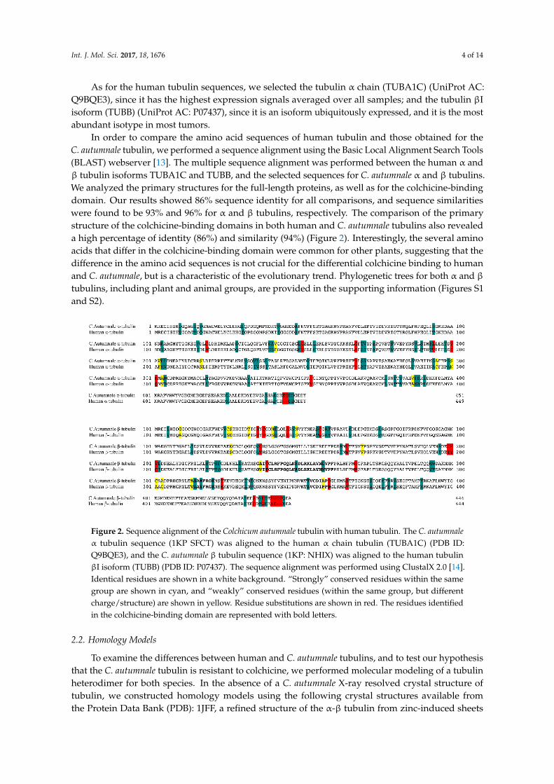
In order to compare the amino acid sequences of human tubulin and those obtained for theC. autumnale tubulin, we performed a sequence alignment using the Basic Local Alignment Search Tools(BLAST) webserver [13]. The multiple sequence alignment was performed between the human α andβ tubulin isoforms TUBA1C and TUBB, and the selected sequences for C. autumnale α and β tubulins.We analyzed the primary structures for the full-length proteins, as well as for the colchicine-bindingdomain. Our results showed 86% sequence identity for all comparisons, and sequence similaritieswere found to be 93% and 96% for α and β tubulins, respectively. The comparison of the primarystructure of the colchicine-binding domains in both human and C. autumnale tubulins also revealeda high percentage of identity (86%) and similarity (94%) (Figure 2). Interestingly, the several aminoacids that differ in the colchicine-binding domain were common for other plants, suggesting that thedifference in the amino acid sequences is not crucial for the differential colchicine binding to humanand C. autumnale, but is a characteristic of the evolutionary trend. Phylogenetic trees for both α and βtubulins, including plant and animal groups, are provided in the supporting information (Figures S1and S2).
Int. J. Mol. Sci. 2017, 18, 1676 4 of 14
isoform (TUBB) (UniProt AC: P07437), since it is an isoform ubiquitously expressed, and it is the most abundant isotype in most tumors.
In order to compare the amino acid sequences of human tubulin and those obtained for the C. autumnale tubulin, we performed a sequence alignment using the Basic Local Alignment Search Tools (BLAST) webserver [13]. The multiple sequence alignment was performed between the human α and β tubulin isoforms TUBA1C and TUBB, and the selected sequences for C. autumnale α and β tubulins. We analyzed the primary structures for the full-length proteins, as well as for the colchicine-binding domain. Our results showed 86% sequence identity for all comparisons, and sequence similarities were found to be 93% and 96% for α and β tubulins, respectively. The comparison of the primary structure of the colchicine-binding domains in both human and C. autumnale tubulins also revealed a high percentage of identity (86%) and similarity (94%) (Figure 2). Interestingly, the several amino acids that differ in the colchicine-binding domain were common for other plants, suggesting that the difference in the amino acid sequences is not crucial for the differential colchicine binding to human and C. autumnale, but is a characteristic of the evolutionary trend. Phylogenetic trees for both α and β tubulins, including plant and animal groups, are provided in the supporting information (Figures S1 and S2).
Figure 2. Sequence alignment of the Colchicum autumnale tubulin with human tubulin. The C. autumnale α tubulin sequence (1KP SFCT) was aligned to the human α chain tubulin (TUBA1C) (PDB ID: Q9BQE3), and the C. autumnale β tubulin sequence (1KP: NHIX) was aligned to the human tubulin βI isoform (TUBB) (PDB ID: P07437). The sequence alignment was performed using ClustalX 2.0 [14]. Identical residues are shown in a white background. “Strongly” conserved residues within the same group are shown in cyan, and “weakly” conserved residues (within the same group, but different charge/structure) are shown in yellow. Residue substitutions are shown in red. The residues identified in the colchicine-binding domain are represented with bold letters.
2.2. Homology Models
To examine the differences between human and C. autumnale tubulins, and to test our hypothesis that the C. autumnale tubulin is resistant to colchicine, we performed molecular modeling of a tubulin heterodimer for both species. In the absence of a C. autumnale X-ray resolved crystal structure of tubulin, we constructed homology models using the following crystal structures available from the Protein Data Bank (PDB): 1JFF, a refined structure of the α-β tubulin from zinc-induced sheets stabilized with paclitaxel (the “straight” conformation of tubulin) and obtained from bovine [15], and 1SA0, a tubulin–colchicine and stathmin-like domain complex (the “curved” conformation of tubulin) obtained from bovine and rat [6]. Both templates were used for threading of the C. autumnale genes and human genes for the α and β tubulins. The generated homology models were expected to have a similar resolution to the original structures i.e., around 3.5 Å (3.50 Å for 1JFF, and 3.58 Å for
Figure 2. Sequence alignment of the Colchicum autumnale tubulin with human tubulin. The C. autumnaleα tubulin sequence (1KP SFCT) was aligned to the human α chain tubulin (TUBA1C) (PDB ID:Q9BQE3), and the C. autumnale β tubulin sequence (1KP: NHIX) was aligned to the human tubulinβI isoform (TUBB) (PDB ID: P07437). The sequence alignment was performed using ClustalX 2.0 [14].Identical residues are shown in a white background. “Strongly” conserved residues within the samegroup are shown in cyan, and “weakly” conserved residues (within the same group, but differentcharge/structure) are shown in yellow. Residue substitutions are shown in red. The residues identifiedin the colchicine-binding domain are represented with bold letters.
2.2. Homology Models
To examine the differences between human and C. autumnale tubulins, and to test our hypothesisthat the C. autumnale tubulin is resistant to colchicine, we performed molecular modeling of a tubulinheterodimer for both species. In the absence of a C. autumnale X-ray resolved crystal structure oftubulin, we constructed homology models using the following crystal structures available fromthe Protein Data Bank (PDB): 1JFF, a refined structure of the α-β tubulin from zinc-induced sheets

Int. J. Mol. Sci. 2017, 18, 1676 5 of 14
stabilized with paclitaxel (the “straight” conformation of tubulin) and obtained from bovine [15], and1SA0, a tubulin–colchicine and stathmin-like domain complex (the “curved” conformation of tubulin)obtained from bovine and rat [6]. Both templates were used for threading of the C. autumnale genesand human genes for the α and β tubulins. The generated homology models were expected to have asimilar resolution to the original structures i.e., around 3.5 Å (3.50 Å for 1JFF, and 3.58 Å for 1SA0).The sequence alignment of the C. autumnale and human tubulin genes to the 1JFF and 1SA0 templatesindicated high sequence homology: 81% sequence identity for C. autumnale, and 97% for the humansequences. In order to calculate the colchicine-binding energies to human and C. autumnale tubulins,respectively, as well as to visualize any conformational changes, molecular modeling was conducted onthe holo (ligand bound) and apo (no ligand bound) forms of the protein. At the same time, molecularmodeling was performed using paclitaxel as a ligand (Figure 3). Structures of colchicine, paclitaxeland other colchicine-related compounds are provided in the supporting information (Figure S3).The binding of paclitaxel to human tubulin has already been studied, and it is known that paclitaxelbinding to β-tubulin stabilizes the tubulin dimer in the straight conformation. In this study, weused paclitaxel as a positive control for the straight conformation and for the binding free energycalculations. Each homology model was energy minimized to refine the geometry of each system, andto relieve any bad contacts among the added hydrogen atoms. The geometry-optimized systems weresimulated in physiological conditions until they reached equilibrium. The root-mean-square deviation(RMSD) of each complex backbone with respect to the initial protein structure was plotted for thefull-length protein and the ligand-binding domain (Figure 4). Although the RMSDs of the holo formare higher than those of the apo form, all of the systems reached equilibrium after approximately 50 ns,indicating that the 65 ns simulations are adequate for performing the binding free energy analysis.
Int. J. Mol. Sci. 2017, 18, 1676 5 of 14
1SA0). The sequence alignment of the C. autumnale and human tubulin genes to the 1JFF and 1SA0 templates indicated high sequence homology: 81% sequence identity for C. autumnale, and 97% for the human sequences. In order to calculate the colchicine-binding energies to human and C. autumnale tubulins, respectively, as well as to visualize any conformational changes, molecular modeling was conducted on the holo (ligand bound) and apo (no ligand bound) forms of the protein. At the same time, molecular modeling was performed using paclitaxel as a ligand (Figure 3). Structures of colchicine, paclitaxel and other colchicine-related compounds are provided in the supporting information (Figure S3). The binding of paclitaxel to human tubulin has already been studied, and it is known that paclitaxel binding to β-tubulin stabilizes the tubulin dimer in the straight conformation. In this study, we used paclitaxel as a positive control for the straight conformation and for the binding free energy calculations. Each homology model was energy minimized to refine the geometry of each system, and to relieve any bad contacts among the added hydrogen atoms. The geometry-optimized systems were simulated in physiological conditions until they reached equilibrium. The root-mean-square deviation (RMSD) of each complex backbone with respect to the initial protein structure was plotted for the full-length protein and the ligand-binding domain (Figure 4). Although the RMSDs of the holo form are higher than those of the apo form, all of the systems reached equilibrium after approximately 50 ns, indicating that the 65 ns simulations are adequate for performing the binding free energy analysis.
Figure 3. Schematic representation of each of the eight homology models constructed for this study. The α tubulin is represented in blue, and the β tubulin in green. Each subunit is bound to a nucleotide. The α subunit is bound to GTP (green ball), and the β subunit to GDP (blue ball). Colchicine is represented in orange, and paclitaxel in purple. The graphical representation of the tubulin dimers was generated based on the information obtained from Krebs et al.’s 2005 study [16].
Figure 3. Schematic representation of each of the eight homology models constructed for this study.The α tubulin is represented in blue, and the β tubulin in green. Each subunit is bound to a nucleotide.The α subunit is bound to GTP (green ball), and the β subunit to GDP (blue ball). Colchicine isrepresented in orange, and paclitaxel in purple. The graphical representation of the tubulin dimers wasgenerated based on the information obtained from Krebs et al.’s 2005 study [16].

Int. J. Mol. Sci. 2017, 18, 1676 6 of 14
Int. J. Mol. Sci. 2017, 18, 1676 6 of 14
Figure 4. The root-mean-square deviation (RMSD) from the initial minimized structures for each of the studied systems. Plot (a) is human tubulin in straight conformation without ligand, plot (b) is human tubulin in straight conformation bound to paclitaxel, plot (c) is human tubulin in curved conformation without ligand, plot (d) is human tubulin in curved conformation bound to colchicine. Plot (e) is C. autumnale tubulin in straight conformation without ligand, plot (f) is C. autumnale tubulin in straight conformation bound to paclitaxel, plot (g) is C. autumnale tubulin in curved conformation without ligand, plot (h) is C. autumnale tubulin in curved conformation bound to colchicine. RMSD analyses are shown for both full-length protein (black curved) and the ligand-binding domain when the ligand is present (red curve).
2.3. Free Energy of Binding Calculations
In order to investigate whether colchicine has a different affinity between human and C. autumnale tubulins, we calculated the binding free energies using the Molecular Mechanics Poisson–Boltzmann Surface Area (MM/PBSA) technique [17]. The MM/PBSA method combines molecular
Figure 4. The root-mean-square deviation (RMSD) from the initial minimized structures for each of thestudied systems. Plot (a) is human tubulin in straight conformation without ligand, plot (b) is humantubulin in straight conformation bound to paclitaxel, plot (c) is human tubulin in curved conformationwithout ligand, plot (d) is human tubulin in curved conformation bound to colchicine. Plot (e) isC. autumnale tubulin in straight conformation without ligand, plot (f) is C. autumnale tubulin in straightconformation bound to paclitaxel, plot (g) is C. autumnale tubulin in curved conformation withoutligand, plot (h) is C. autumnale tubulin in curved conformation bound to colchicine. RMSD analyses areshown for both full-length protein (black curved) and the ligand-binding domain when the ligand ispresent (red curve).
2.3. Free Energy of Binding Calculations
In order to investigate whether colchicine has a different affinity between human and C. autumnaletubulins, we calculated the binding free energies using the Molecular Mechanics Poisson–Boltzmann

Int. J. Mol. Sci. 2017, 18, 1676 7 of 14
Surface Area (MM/PBSA) technique [17]. The MM/PBSA method combines molecular mechanicsand solvation energies on the molecular dynamics (MD) trajectory. Further estimates of the bindingenergy performed from another popular continuum solvation model, i.e., the Molecular MechanicsGeneralized-Born Surface Area (MM/GBSA) technique were provided (Table 1). The main differencebetween the MM/PBSA and MM/GBSA techniques lies in the estimation of the polar contribution ofthe solvation free energy (see Equation (1), Section 4.4). For MM/PBSA, this contribution is typicallyobtained by solving the Poisson–Boltzmann equation while the generalized born (GB) model is usedin the MM/GBSA approach [18].
Table 1. The binging free energy ∆G in kcal/mol of colchicine and paclitaxel with human and Colchicumautumnale tubulin.
Species Ligand Generalized Born ∆G (kcal/mol) Poisson–Boltzman ∆G (kcal/mol)
Homo Sapiens Paclitaxel −23.25 −43.30C. autumnale Paclitaxel −20.78 −44.81
Homo Sapiens Colchicine −51.79 −76.68C .autumnale Colchicine −43.44 −64.71
All binding energy calculations were performed on the MD trajectories during which thecomplexes were equilibrated, in this case, from 50 to 65 ns.
Our calculations of the binding free energies for paclitaxel have confirmed its binding to humantubulin, with the same affinity found in experimental tests. In addition, our calculations suggested thatpaclitaxel binds to C. autumnale tubulin with almost the same affinity as to human tubulin (Table 1).Although the free energy estimated from the MM/GBSA technique showed a fairly substantialfree energy difference, i.e., 3 kcal/mol of paclitaxel had more affinity for human tubulin than forC. autumnale, the difference obtained from the MM/PBSA technique is only 1 kcal/mol with anopposite tendency, i.e., paclitaxel binding was slightly more preferential for the C. autumnale tubulin.Energy decomposition per residue within 8.0 Å of the bound paclitaxel showed key residues known tointeract with paclitaxel (Table 2) [10].
On the other hand, colchicine was clearly found to have high affinity and a preferred binding modeto human tubulin (∆G0 = −51.79 kcal/mol with the GBSA model and ∆G0 = −76.68 kcal/mol with thePBSA model), but significantly lower affinity to the C. autumnale tubulin (∆G0 = −43.44 kcal/mol withthe GBSA model and ∆G0 = −64.71 kcal/mol with the PBSA model) (Table 1). To obtain a better insightinto the differences in the binding free energy of colchicine, we performed energy decomposition.We looked into the residues located within an 8.0 Å cut-off range of the bound colchicine (Table 2).In human tubulin, we found eight residues that significantly contribute to colchicine binding. Tworesidues are located in the α subunit, whereas the other six are located in the colchicine-binding domainin the β subunit. In the case of C. autumnale, we found six amino acids that significantly contributeto colchicine binding. Interestingly, five out of the six residues involved in colchicine binding toC. autumnale tubulin are also key residues in human tubulin. The exception is βLeu257 in C. autumnale,which contributes to colchicine binding, but is not significantly involved in human tubulin binding,although this residue is conserved between the two species. In addition to the common residues inhuman tubulin, we found that βCys239, βAla248 and βAla314, with βAla248 are the residues withhighest energy contributions. Moreover, the contribution of colchicine is different for the two species,with ∆G0 predicted to be −28.22 kcal/mol for human tubulin, and −23.55 kcal/mol for C. autumnale.

Int. J. Mol. Sci. 2017, 18, 1676 8 of 14
Table 2. Binding free energy (kcal/mol) decomposition into key residues mediating thetubulin–ligand interaction.
Ligand Species Residue ∆E vdw ∆E elec ∆E Polar ∆E Non p ∆G Total
Paclitaxel Homo Sapiens
βLeu215 −2.90 0.48 0.43 −0.36 −2.36βThr274 −0.48 −2.91 2.07 −0.05 −1.37βSer275 −1.69 −2.00 1.77 −0.14 −2.07βArg276 −2.69 −4.00 4.75 −0.43 −2.28
PTX −18.28 −15.35 22.19 0.19 −14.40
Paclitaxel C. autumnale
βLeu272 −2.10 0.19 0.72 −0.28 −1.84βThr274 −1.02 0.53 0.11 −0.11 −0.49βIle358 −2.28 −1.22 2.22 −0.50 −1.78βLeu360 −1.65 −0.24 0.46 −0.29 −1.72
PTX −18.23 −15.11 22.93 −3.36 −12.77
Colchicine Homo Sapiens
αAla180 −1.53 −2.19 0.74 −0.14 −3.13αVal181 −0.89 −2.52 1.00 −0.055 −2.46βCys239 −0.80 −0.76 0.52 −0.10 −1.12βLeu246 −2.71 −1.08 1.00 −0.28 −3.07βAla248 −1.77 −0.26 0.48 −0.13 −1.67βLeu253 −2.68 −0.30 −0.089 −0.28 −3.36βAla314 −1.24 −0.32 −0.080 −0.18 −1.82βLys350 −2.24 −0.25 0.89 0.17 −1.77
CLN −32.12 −14.78 23.65 −4.96 −28.22
Colchicine C. autumnale
αSer180 −1.43 −1.53 0.43 −0.19 −2.72αVal181 −1.19 −2.35 0.93 −0.058 −2.66βLeu246 −2.58 −0.55 0.75 −0.52 −2.90βLeu253 −2.25 −0.57 0.50 −0.22 −2.57βLeu257 −2.13 −0.21 0.20 −0.19 −2.33βLys350 −2.35 −2.20 1.46 −0.30 −3.38
CLN −27.14 −12.85 20.86 −4.42 −23.55
2.4. Atomic Fluctuations
In order to analyze the behavior of the residues and their flexibility, we calculated the atomicfluctuations for each residue in the presence and absence of the ligand, respectively. In human tubulin,the presence of colchicine did not affect the fluctuations of the residues in the α subunit. However,more fluctuations are induced for residues at the interface between α and βmonomers, which suggestsa conformational change of the dimer (Figure 5a). Generally, residues in the β subunit show largerfluctuations, except for the residues involved in colchicine binding. These residues are stabilizedby the presence of colchicine, confirming their implication in the binding. On the other hand, inC. autumnale tubulin, the bound colchicine did not cause any major modifications in the atomicfluctuations (Figure 5b). We observed an increase of atomic fluctuations in the N-terminal tail in the αsubunit, but not as large as in human tubulin. In the β subunit, residues are in general more stabilized.This includes residues involved in colchicine binding, especially βLys350. Taken together, these resultssuggest an important conformational change in human tubulin into a curved shape. This changecreated a gap between the two subunits, followed by an increasing size of atomic fluctuations at theα–β interface. However, no such significant increase was observed in plant tubulin, which suggestsless important conformational changes in that case.
2.5. Conformational Changes
In order to examine how the presence of colchicine influences tubulin conformation, we extractedthe lowest energy structures obtained from MD simulations and superimposed the structures byaligning the N-terminals of the β tubulin. To investigate the conformational changes, we lookedinto the β subunit intermediate domain, which is known to be affected by the colchicine binding.By superimposing the human tubulin bound to colchicine with the human tubulin in straightconformation that is unbound to colchicine, we observed a significant dislocation in the intermediatedomain, especially in regions involving the helices H7, H9 and H10 (Figure 5c). In C. autumnale tubulin,

Int. J. Mol. Sci. 2017, 18, 1676 9 of 14
we also observed a shift in the intermediate domain, again, in regions including the H7, H9 and H10helices (Figure 5d). However, this shift was less important than in human tubulin, confirming thatcolchicine bound to C. autumnale induces less notable conformational changes.
Figure 5. Comparison between the holo and apo forms of tubulin. (a,b): average atomic fluctuationsper residue in human (a) and in C. autumnale (b) tubulins. The tubulin heterodimer is representedin red in the presence of colchicine, and in black in the absence of colchicine. Key amino acidsinvolved in colchicine binding are depicted on the graphs. (c,d): overlay of the conformations of thecolchicine-binding domain of β tubulin in the presence and absence of colchicine. The conformationsof the intermediate domain of the human β-tubulin are shown in (c) and the ones of the C. autumnale βtubulin are depicted in (d). The holo form of tubulin bound to colchicine is colored in blue, and the apoform in red.
3. Discussion
Colchicine, a plant alkaloid, has an enormous pharmacological importance due to its ability tobind to tubulin and inhibit microtubule polymerization, which leads to mitotic arrest. Yet, the immensepharmacological potential of colchicine remains unfulfilled due to its severe side effects. Interestingly,while colchicine is extremely toxic for human cells, the plants that endogenously express colchicine,such as C. autumnale and G. superba, seem to be unaffected by its cytotoxicity even though they expresstubulin, the biological target of colchicine. This observation suggests that there could be a method

Int. J. Mol. Sci. 2017, 18, 1676 10 of 14
of redesigning colchicine analogues to enable differential binding between tubulin isotypes in thehuman body. This might allow for a reduction of side effects, while maintaining efficacy. In thispaper, we performed a computational study in order to explore what might be the cause for theresistance of C. autumnale to colchicine. For this purpose, molecular modeling of C. autumnale tubulinwas performed based on the transcriptomic data obtained from the Alberta 1000 Plants Initiative(1KP) database.
Tubulin is a highly conserved protein in all eukaryotic cells, although multiple tubulin isotypes arewidespread among eukaryotes. In order to investigate whether the differential colchicine binding tohuman and C. autumnale tubulin is caused by sequence variations, we performed a sequence alignment.The results revealed a high conservation of the amino acid residues with 86% sequence identitybetween the C. Autumnale and the human tubulins. In addition, we compared the colchicine-bindingdomains in both species. Again, high sequence homology was found. The five residues differing inthe colchicine-binding site between human and C. autumnale were T238C, A248S, M257L, V258I, andR359I of the β subunit. Interestingly, all of these residues were found to be common among otherplants such as Taxus baccata, Podophyllum peltatum, Larrea tridentate and Catharanthus roseus. The highconservation of these residues in other plant species suggests that the differential colchicine binding tohuman and C. autumnale tubulin cannot be explained only by sequence variations and substitutions inthe colchicine-binding site; it also requires an analysis of evolutionary trends in general.
Since sequence variation is not sufficient to explain colchicine resistance within C. autumnale,we investigated whether any difference in the colchicine-binding affinity could be observed betweenhuman and C. autumnale tubulins. For that purpose, we performed MM/PBSA and compared thebinding free energies of both human and C. autumnale tubulins. It was found that colchicine has asignificantly higher affinity for human tubulin than C. autumnale tubulin, even though our resultsindicated that colchicine is still able to bind to C. autumnale. The energy decomposition per residuerevealed eight residues that are critical for colchicine binding to human tubulin, with αAla180, βLeu246,and βLeu253 having the highest energy contribution. αAla180 and αVal181 are both hydrophobicamino acids, which establish van der Waals contacts with the methoxy tropone ring (C ring) ofcolchicine. βCys239, βLeu246, βAla248, βLeu253, βAla314, βLys350 delimit a hydrophobic pocket intubulin, and interact with the trimethoxy benzene ring (A ring). The thiol group of βCys239 is involvedin a hydrogen bond with the methoxy group of the A ring. On the other hand, in C. autumnale, fewerresidues were involved in the binding of colchicine, with a major contribution of βLeu246 and βLys350.Moreover, βLeu257 was found to be involved in the colchicine binding to C. autumnale, but not tohuman tubulin. Moreover, position 180 in C. autumnale is associated with serine, while it is associatedwith alanine in human tubulin. However, this substitution does not seem to modify colchicine binding,since both amino acids establish van der Waals contacts with the same strength. Another variationoccurs at position 248. In human tubulin, βAla248 turns out to be a key residue for colchicine binding,whereas βSer248 in C. autumnale tubulin does not seem to be involved. These amino acids are locatedin the hydrophobic pocket of β tubulin. Alanine, being a hydrophobic amino acid, contributes tohydrophobic interactions with the A ring of colchicine. On the contrary, serine, as a polar aminoacid with a hydroxyl group, causes local destabilization in the hydrophobic pocket. Surprisingly,βCys239 was not involved in C. autumnale, which suggests a less stable interaction between the A ringof colchicine and β tubulin.
In order to estimate the flexibility of tubulin residues in the presence or absence of colchicine, welooked into the atomic fluctuations of each residue. When analyzing the human tubulin fluctuations,the presence of colchicine induces a higher flexibility of the residues of the β subunit, which are locatedat its interface within the α subunit. This result confirms the conformational change in human tubulincaused by the binding of colchicine. On the contrary, no significant changes in the residues’ flexibilitywere observed in C. autumnale, both in the presence and absence of colchicine. This indicates thatC. autumnale tubulin is more rigid than human tubulin when bound to colchicine, and also undergoesfewer conformational changes. These results were also confirmed by the superposition of the structures

Int. J. Mol. Sci. 2017, 18, 1676 11 of 14
bound and unbound to colchicine. The minor shift in the intermediate domain of C. autumnale β tubulinindicates slight changes in tubulin conformation. Taken together, these findings indicate that colchicineis able to bind to tubulin in C. autumnale in the identified colchicine-binding domain, but with aweaker affinity when compared to human tubulin. This low affinity is mostly due to weak interactionsinvolving the A ring of colchicine, which is known to be a critical pharmacophore in the binding totubulin. Analogues and derivatives having the same A ring as colchicine, such as podophyllotoxinand cornigerine, have proven the crucial role of the A ring in this interaction [19]. Weak interactionsof the A ring in C. autumnale contribute to a decrease in the total binding energy of colchicine, thusresulting in fewer conformational changes in the tubulin structure. These slight conformationalchanges can be viewed as the result of forming reversible pre-equilibrium complexes without evolvingto a stable tubulin–colchicine complex [20]. Moreover, these conformational changes seem not to alterthe microtubules polymerization and their dynamics. In order to further investigate the differentconformational changes in tubulin induced by colchicine binding, principal component analysis (PCA)can be performed in the future [21]. However, this is outside the scope of the present study.
An experimental assay on binding affinity would be necessary to confirm the binding freeenergy calculations. Since βSer248 seems to cause destabilization in the hydrophobic binding pocket,experiments with βAla248 mutated to βSer248 in human tubulin might give a better understanding ofthe binding site and its interaction with the A ring. It may also be interesting to investigate whetherβSer248 in C. autumnale tubulin includes any post-translational modifications, such as phosphorylation.
In conclusion, this study reported on the features of the C. autumnale tubulin protein and itscolchicine-binding site. It provides a better understanding of the interaction of colchicine and itstarget tubulin in plants endogenously expressing colchicine, and broadens our understanding ofthis pharmacologically important drug–protein interaction. Since colchicine-binding site agentsare examples of tubulin-binding agents, which have not yet reached clinical applications in cancertreatment, it is crucial to understand the biological relevance of the colchicine-binding site. Colchicinehas an immense but as yet unrealized potential as an antimitotic drug. The dozens of colchicinederivatives and analogues under development should soon result in clinical trials leading to novelanti-cancer therapeutics. Understanding what types of differences provide resistance against this toxincan aid in the understanding of drug resistance in human cancers as well as contribute to the creationof more specific and more efficacious drugs that bind to the same binding site as colchicine. In a futurepublication, we intend to examine a similar relation between the tubulin structures expressed by allremaining plants that produce toxins, and the binding affinity for these toxins. We intend this futurestudy to validate the generalization of the hypothesis posed in the present study, that the binding sitesare suitably modified to lower the affinity for its own toxic agents. We have seen this in the case ofcolchicine and C. autumnale and G. superba, as well as in the case of paclitaxel and the yew tree.
4. Materials and Methods
4.1. Sequence Assembly
Plant tubulin protein sequences were assembled from the 1000 Plants Initiative (1KP) transcriptomedatabase [12] using the following method. Sequence read data were downloaded in FASTQ formatfrom the 1KP data server for the two plant species producing colchicine: C. autumnale and G. superba.There were three samples available for C. autumnale, and only one sample available for G. superba.The FASTQ files were trimmed to remove low-quality reads, using a quality threshold of Q20 with theFASTX-Toolkit (The Apache Software Foundation, Forest Hill, MD, USA).
Human tubulin protein sequences were obtained from UniProt [22]. Tubulin α chain (TUBA1C)(Q9BQE3) was chosen as reference sequence for human α tubulin, and βI isoform (TUBB) (P07437)was chosen for human β tubulin. Sequence analysis was performed using BLAST [13], and multiplesequence alignment was performed with ClustalX 2.0 software (UCD Conway Institute, Dublin,Ireland) [14].

Int. J. Mol. Sci. 2017, 18, 1676 12 of 14
4.2. Homology Modeling
As mentioned before, homology models were constructed using 1JFF and 1SA0 templates forthe straight and the curved conformations of tubulin, respectively. Coordinates of missing residueswithin the PDB crystal structure 1JFF were obtained from the 1TUB structure of tubulin after RMSDalignment of the two structures [23].
C. autumnale α tubulin (1KP sample code SFCT) and β tubulin (1KP sample code NHIX) sequences,as well as human α tubulin (Q9BQE3) and β tubulin (P07437) sequences, were aligned to both 1JFFand 1SA0 structures using the MOE 2012.10 software (Montreal, QC, Canada) [11]. For each targetsequence, the maximum number of independent models constructed, available via the “maximum ofmainchain models” option in MOE, was set to five. Final models were chosen based on GA431, DOPEand molpdf scores, as well as the lowest RMSD alignment with the template structure. Eight homologymodels were created in total, four for the plant tubulin dimer, and four for the human tubulin dimer.
The missing hydrogen atoms were added using the tLEAP module of AMBER 12 (San Francisco,CA, USA) [24] with the AMBER12SB force field. Each protein model was solvated in 12 Å box ofTIP3P water and neutralized with 30 atoms of Na+. In order to bring the salt concentration to thephysiological value of 0.1 M, 64 atoms of Na+ and Cl− were added.
Protonation states of all ionizable residues were calculated at pH 7 using the program PROPKA3.1 (GitHub, San Francisco, CA, USA) [25].
The homology models were then energy minimized with periodic boundaries using the AMBER12 software (San Francisco, CA, USA) [24]. A first minimization run was performed for 2000 cycleswith a cutoff of 8.0 Å and heavy restraints on all backbone atoms. A second minimization run wasperformed for 4500 cycles with a cutoff of 8.0 Å and no restraints.
4.3. Molecular Dynamics Simulations
The fully minimized structures were used as starting configurations for the subsequent MDsimulations. The MD simulations were carried out in three steps: heating, density, and production.First, each solvated system was heated to 300 K for 50 ps, with weak restraints on all backbone atoms.Next, density equilibration was carried out for 50 ps of constant pressure equilibration at 300 K, withweak restraints. Finally, MD production runs were performed on all systems for 65 ns, during whichatomic coordinates were saved every 10 ps. The runs were conducted on 8 Central Processing Units(CPUs) in each case, which enabled the simulation of the protein dynamics at a rate of approximately0.3–0.4 ns per day with the SANDER module of Amber 12 [24]. Each trajectory produced after a MDsimulation was examined. The MD trajectories were analyzed using the ptraj module of AMBER12 utilities to obtain the RMSD and the total energy. The structures with the lowest energy wereextracted using the ptraj module of AMBER 13 tools [24], and superposed with the MOE 2012.10software (Chemical Computing Group, Montreal, QC, Canada) [11].
4.4. Binding Free Energy Calculations
The Molecular Mechanics Poisson–Boltzmann Surface Area (MM/PBSA) technique was used tocalculate the free energy associated with ligand binding [17]. This method combines molecularmechanics with continuum solvation models. The total free energy is estimated as the sum ofaverage molecular mechanical gas phase energies (EMM) and solvation free energies (Gsolv) of thebinding reaction:
G = EMM + Gsolv (1)
The total molecular mechanical (EMM) energy can be further decomposed into contributionsfrom electrostatic (Eele), van der Waals (Evdw), and internal energies (Eint). The molecular mechanical(EMM) energy of each snapshot during the last 15 ns of the MD simulation was calculated using theSANDER module of Amber 12 [24], with all pairwise interactions assuming a dielectric constant(ε) of 1.0. The solvation free energy (Gsolv) was estimated as the sum of electrostatic solvation

Int. J. Mol. Sci. 2017, 18, 1676 13 of 14
free energy, calculated by the finite-difference solution of the Poisson–Boltzmann equation in theAdaptive Poisson–Boltzmann Solver (APBS) and non-polar solvation free energy, calculated fromthe solvent-accessible surface area (SASA) algorithm. Applying the thermodynamic cycle for eachtubulin-colchicine and tubulin-paclitaxel complex, the binding free energy was approximated by:
∆G0 = ∆GLig−Protgas + ∆GLig−Prot
solv −{
∆GProtsolv + ∆GLig
solv
}(2)
Here, ∆GLig−Protgas represents the free energy per mole for the non-covalent association of the
ligand–protein complex in vacuum (gas phase) at a representative temperature, while (−∆Gsolv)
stands for the work required to transfer a molecule from its solution conformation to the sameconformation in vacuum (assuming that the binding conformation of the colchicine protein complex isthe same in solution and in vacuum). The decomposition of the binding free energy per residue wasalso calculated using the MM/PBSA technique [17]. Only the residues within an 8 Å cutoff range ofthe ligand were taken into account for the calculations. Less than an hour was typically needed toestimate binding energies on 8 CPUs, each using the MMPBSA/MMGBSA technique.
4.5. Atomic Fluctuation
The atomic fluctuations were calculated for every atom and averaged for each residue, during thelast 15 ns of MD simulations, using the cpptraj module available within the AMBER 12 package [24].
Supplementary Materials: Supplementary materials can be found at www.mdpi.com/1422-0067/18/8/1676/s1.
Acknowledgments: This research was supported by funds from the Allard Foundation.
Author Contributions: Ivana Spasevska performed the computational studies and produced a first version of themanuscript; Ahmed T. Ayoub and Philip Winter provided valuable assistance during computational studies andfor the writing of the manuscript; Jordane Preto reexamined computational data and produced the final versionof the manuscript; Gane K.-S. Wong provided access to the 1KP database from which tubulin sequences wereobtained; Charles Dumontet and Jack A. Tuszynski. supervised the study and edited the final manuscript.
Conflicts of Interest: The authors declare no conflict of interest.
Abbreviations
APBS Adaptive Poisson–Boltzmann SolverBLAST Basic Local Alignment Search ToolsCLN ColchicineGDP Guanosine DiphosphateGTP Guanosine TriphosphateMD Molecular DynamicsMM/PBSA Molecular Mechanics Poisson–Boltzmann Surface AreaMM/GBSA Molecular Mechanics Generalized-Born Surface AreaMT MicrotubuleRMSD Root-Mean-Square DeviationSASA Solvent Accessible Surface Area
References
1. Hyams, J.S.; Lloyd, C.W. Microtubules; Wiley-Liss: New York, NY, USA, 1994.2. Mitchison, T.; Kirschner, M. Dynamic instability of microtubule growth. Nature 1984, 312, 237–242. [CrossRef]3. Jordan, M.A.; Wilson, L. Microtubules as a target for anticancer drugs. Nat. Rev. Cancer 2004, 4, 253–265.
[CrossRef] [PubMed]4. Wallace, S.L. Colchicine. Colchicine 1974, 3, 369–381. [CrossRef]5. Weisenberg, R.C.; Borisy, G.G.; Taylor, E.W. The colchicine-binding protein of mammalian brain and its
relation to microtubules. Biochemistry 1968, 7, 4466–4479. [CrossRef] [PubMed]6. Ravelli, R.B.; Gigant, B.; Curmi, P.A.; Jourdain, I.; Lachkar, S.; Sobel, A.; Knossow, M. Insight into tubulin
regulation from a complex with colchicine and a stathmin-like domain. Nature 2004, 428, 198–202. [CrossRef][PubMed]

Int. J. Mol. Sci. 2017, 18, 1676 14 of 14
7. Luis, L.; Serrano, M.L.; Hidalgo, M.; Mendoza-Leon, A. Comparative analyses of the β-tubulin gene andmolecular modeling reveal molecular insight into the colchicine resistance in kinetoplastids organisms.BioMed Res. Int. 2013, 2013, 843748. [CrossRef] [PubMed]
8. Mane, J.Y.; Klobukowski, M.; Huzil, J.T.; Tuszynski, J. Free energy calculations on the binding of colchicine andits derivatives with the α/β-tubulin isoforms. J. Chem. Inf. Model. 2008, 48, 1824–1832. [CrossRef] [PubMed]
9. Gigant, B.; Cormier, A.; Dorleans, A.; Ravelli, R.B.; Knossow, M. Microtubule-destabilizing agents: Structuraland mechanistic insights from the interaction of colchicine and vinblastine with tubulin. Top. Curr. Chem.2009, 286, 259–278. [PubMed]
10. Tuszynski, J.A.; Craddock, T.J.; Mane, J.Y.; Barakat, K.; Tseng, C.Y.; Gajewski, M.; Winter, P.; Alisaraie, L.;Patterson, J.; Carpenter, E.; et al. Modeling the yew tree tubulin and a comparison of its interaction withpaclitaxel to human tubulin. Pharm. Res. 2012, 29, 3007–3021. [CrossRef] [PubMed]
11. MOE-Molecular_Operating_Environment. Available online: https://www.chemcomp.com/announcements/2013-10-23-MOE2013.08.pdf (accessed on 23 August 2013).
12. Matasci, N.; Hung, L.H.; Yan, Z.; Carpenter, E.J.; Wickett, N.J.; Mirarab, S.; Nguyen, N.; Warnow, T.;Ayyampalayam, S.; Barker, M.; et al. Data access for the 1000 plants (1kp) project. GigaScience 2014, 3, 17.[CrossRef] [PubMed]
13. Altschul, S.F.; Madden, T.L.; Schaffer, A.A.; Zhang, J.; Zhang, Z.; Miller, W.; Lipman, D.J. Gapped blast andpsi-blast: A new generation of protein database search programs. Nucleic Acids Res. 1997, 25, 3389–3402.[CrossRef] [PubMed]
14. Larkin, M.A.; Blackshields, G.; Brown, N.P.; Chenna, R.; McGettigan, P.A.; McWilliam, H.; Valentin, F.;Wallace, I.M.; Wilm, A.; Lopez, R.; et al. Clustal w and clustal x version 2.0. Bioinformatics 2007, 23, 2947–2948.[CrossRef] [PubMed]
15. Lowe, J.; Li, H.; Downing, K.H.; Nogales, E. Refined structure of α β-tubulin at 3.5 a resolution. J. Mol. Biol.2001, 313, 1045–1057. [CrossRef] [PubMed]
16. Krebs, A.; Goldie, K.N.; Hoenger, A. Structural rearrangements in tubulin following microtubule formation.EMBO Rep. 2005, 6, 227–232. [CrossRef] [PubMed]
17. Kollman, P.A.; Massova, I.; Reyes, C.; Kuhn, B.; Huo, S.; Chong, L.; Lee, M.; Lee, T.; Duan, Y.; Wang, W.;et al. Calculating structures and free energies of complex molecules: Combining molecular mechanics andcontinuum models. Acc. Chem. Res. 2000, 33, 889–897. [CrossRef] [PubMed]
18. Genheden, S.; Ryde, U. The MM/PBSA and MM/GBSA methods to estimate ligand-binding affinities.Expert Opin. Drug Discov. 2015, 10, 449–461. [CrossRef] [PubMed]
19. Bhattacharyya, B.; Panda, D.; Gupta, S.; Banerjee, M. Anti-mitotic activity of colchicine and the structuralbasis for its interaction with tubulin. Med. Res. Rev. 2008, 28, 155–183. [CrossRef] [PubMed]
20. Hastie, S.B. Interactions of colchicine with tubulin. Pharmacol. Ther. 1991, 51, 377–401. [CrossRef]21. Jolliffe, I.T.; Cadima, J. Principal component analysis: A review and recent developments. Philos. Trans. A
Math. Phys. Eng. Sci. 2016, 374, 20150202. [CrossRef] [PubMed]22. UniProt, C. Update on activities at the universal protein resource (uniprot) in 2013. Nucleic Acids Res. 2013,
41, D43–D47.23. Nogales, E.; Wolf, S.G.; Downing, K.H. Structure of the α β tubulin dimer by electron crystallography. Nature
1998, 391, 199–203. [CrossRef] [PubMed]24. Pearlman, D.A.; Case, D.A.; Caldwell, J.W.; Ross, W.S.; Cheatham, T.E.; DeBolt, S.; Ferguson, D.; Seibel, G.;
Kollman, P. Amber, a package of computer programs for applying molecular mechanics, normal modeanalysis, molecular dynamics and free energy calculations to simulate the structural and energetic propertiesof molecules. Comput. Phys. Commun. 1995, 91, 1–41. [CrossRef]
25. Sondergaard, C.R.; Olsson, M.H.; Rostkowski, M.; Jensen, J.H. Improved treatment of ligands and couplingeffects in empirical calculation and rationalization of pka values. J. Chem. Theory Comput. 2011, 7, 2284–2295.[CrossRef] [PubMed]
© 2017 by the authors. Licensee MDPI, Basel, Switzerland. This article is an open accessarticle distributed under the terms and conditions of the Creative Commons Attribution(CC BY) license (http://creativecommons.org/licenses/by/4.0/).