modern technologies in endodontics - core · modern technologies in endodontics ... the root canal...
TRANSCRIPT

LITERATURE REVIEW/REVISIONE DELLA LETTERATURA
Modern technologies in Endodontics
Moderne tecnologie in Endodonzia
Alfredo Iandolo a,*, Giuseppe Iandolo b, Mariano Malvano c,Giuseppe Pantaleo a, Michele Simeone a
aDepartment of Neurosciences, Reproductive and Odontostomatological Sciences, University of NaplesFederico II, Naples, ItalybMedical Director, ASL Avellino 2, Avellino, ItalycPrivate Practice in Naples, Italy
Received 23 September 2015; accepted 11 December 2015Available online 15 January 2016
Giornale Italiano di Endodonzia (2016) 30, 2—9
KEYWORDSIrrigation solutions;Activation;Microscope;Ultrasonic tips.
Abstract
Aim: In Endodontics, a complete chemo-mechanical cleansing of the root canal system isessential to achieving success, which is gained through adequate tridimensional obturation ofthe endodontic space.Materials and methods: Today, thanks to modern technologies as Operative Microscope, ultra-sonic tips, M-Wire Files, devices to activate irrigation and tridimensional obturation performedwith thermo plasticized gutta-percha, satisfactory results can be obtained.Results: This study shows all the technologies that are available today to increase the chemo-mechanical cleansing and obturation of the entire and complicated endodontic system.Conclusions: The positive results highlighted by these clinical cases demonstrate how the use ofmodern technologies are essential to avoid iatrogenic injury, and guarantee, on the other hand,safe and reproducible results.� 2015 Societa Italiana di Endodonzia. Production and hosting by Elsevier B.V. All rights reserved.
Peer review under responsibility of Societa Italiana di Endodonzia.
* Corresponding author at: DDS, University of Naples Federico II, via S. Pansini 5, 80131 Naples, Italy.E-mail: [email protected] (A. Iandolo).
Available online at www.sciencedirect.com
ScienceDirect
j ou rn al home pag e: www. el sev ie r. com/l oca te/ g i e
http://dx.doi.org/10.1016/j.gien.2015.12.0011121-4171/� 2015 Societa Italiana di Endodonzia. Production and hosting by Elsevier B.V. All rights reserved.

Introduction
The long-term success of endodontic treatment is closelylinked to adequate cleansing, shaping and then to a completetridimensional obturation of the complex root canal sys-tem.1—3 Probably, a significant percentage of failures iscaused by the presence of residual pulp tissue and to aninsufficient cleansing of the roots canals.4 The endodonticsystem is composed by spaces easily accessible to hand androtary files (main canals) and, as demonstrated by manyclinical and histological studies,5,6 by not easily accessibleor inaccessible spaces (isthmus, delta, loop, lateral andaccessory canals and dentinal tubules) (Figs. 1 and 2).7
Root canal shaping is not able to reach all areas of the rootcanal system, regardless of the technique used; so not allsections of canal are treated.8 For this reason it is necessarythe endodontic biochemistry cleansing (accessible and not
PAROLE CHIAVESoluzioni irriganti;Attivazione;Microscopio;Punte ultrasoniche.
Riassunto
Obiettivo: In Endodonzia una completa detersione chemio-meccanica del complesso sistema deicanali radicolari e fondamentale per il raggiungimento del successo, il quale viene mantenutoattraverso un’adeguata otturazione tridimensionale dello spazio endodontico.Materiali e metodi: Oggi, grazie alle moderne tecnologie, Microscopio Operatorio, Punte ultra-soniche, Files in lega M-Wire, dispositivi per l’attivazione degli irriganti e all’otturazione tridi-mensionale eseguita con guttaperca termoplasticizzata si possono ottenere risultati ben piu chesoddisfacenti.Risultati: L’articolo che segue mette in risalto tutte le tecnologie che oggi abbiamo a disposizioneper aumentare il grado di detersione chemio-meccanica e otturazione tridimensionale delcomplicato e intero sistema endodontico.Conclusioni: Gli esiti positivi, evidenziati da questi casi clinici, dimostrano come l’utilizzo dellemoderne tecnologie siano indispensabili nell’evitare danni iatrogeni e garantire, invece, risultatisicuri e riproducibili.� 2015 Societa Italiana di Endodonzia. Production and hosting by Elsevier B.V. Tutti i dirittiriservati.
Figure 2 Diaphanization of a lower central incisor: an isthmusbetween the two root canals is shown.
Modern technologies in Endodontics 3
accessible spaces); once cleaned, it can be filled and obturedwith gutta percha and cement during obturation.9
Figure 1 Root apex of the mesial root of a lower first molar atSEM: a lot of exits are shown.
The outcomes of current endodontic treatments are basedon old working methods (operators without experience,treatments performed without the aid of the operatingmicroscope, chemo-mechanical preparation performed withnormal Ni-Ti files, use of irrigants without activation)10,11;
In the endodontic treatment we can distinguish differentphases:(a) The pulp chamber opening, the most difficult phase in
accordance with literature, because an error during thisphase could compromise the treatment. The opening ofthe pulp chamber should be performed under constantmagnification and lighting4,12—16;
(b) The shaping phase with the new modified NiTi instru-ments17—19;
(c) The cleansing phase, where irrigants are activated andenhanced20—22;
(d) The obturation phase, where in addition to modern sys-tems using thermoplastic gutta percha, new root canalsfilling materials are proposed.23—27
Of course, the treatment has to be concluded with anappropriate post-endodontic restoration.
After a careful analysis of the case that has to be treated,by X-ray and clinical examinations, it is possible to proceedwith the endodontic treatment.

Figure 4 Endodontic ultrasonic tips.
4 A. Iandolo et al.
Materials and methods
Modern technologies
The pulp chamber openingFirst step that has to be performed is the isolation of theoperative field with a dental dam, than under constantmagnification and lighting we have to proceed with theopening of the pulp chamber with rotary instruments andultrasonic tips.
The main aid of the operating microscope (Fig. 3) is theincrease of the PDR, or power of resolution, namely theability to see distant two points that are very closetogether. The human eye, in fact, it’s not able to distin-guish between two points separated by a minimum dis-tance of 0.1 mm (PDR: 0.1 mm),28,29 it will sum them as asingle image. By using the operating microscope, thepower of resolution increases from 0.1 mm to 0.005 mmequal to 5 micron, thus making the human eye able toobserve more details.
The ultrasonic instruments which are now available indentistry include various types of tips that have differentkind of shapes, length and construction materials (Fig. 4).Furthermore, with the introduction of new advancedsources of ultrasound, it was possible to optimize theuse of each type of tip with the option to control thefrequency and the amplitude of vibration. The ultrasonictips guarantee a great cutting accuracy thanks to theirreduced dimensions that allow greater view of the operat-ing field than the rotary instruments, greater view thatincreases the use of magnification systems as the operatingmicroscope.4,30
So, only after root canal entrances identification (Fig. 5),it is possible to proceed with the phases of shaping, cleansingand tridimensional obturation.12—14
Figure 3 Operating microscope.
Figure 5 Opening the pulp chamber in a lower first molarperformed under constant magnification and illumination, usingultrasonic tips.
The shaping phase with the new modified NiTiinstrumentsThe use of Ni-Ti represented a turning point in the history ofEndodontics, in fact it allowed the construction and produc-tion of new manual and rotary endodontic instruments withcharacteristics that were superior to stainless steel instru-ments, obtaining more effective and reproducible thera-pies.31,32 The Ni-Ti alloys used in dentistry have an equalatomic composition of Ni and Ti, corresponding to 55% bymass of Ni and 45% by mass of Ti.33
The main properties of Ni-Ti are the shape memory andsuperelasticity (or pseudoelasticity), although in Endodonticsthe first characteristic is not used.
The superelasticity or pseudoelasticity on the other hand,is particularly useful because it lends to the alloy the abilityto bend and adapt to the shape of the canal, allowing toshape the canal with a movement of rotation, keeping acentered position even in the presence of accentuated cur-vatures, in that way the restoring force and its negative
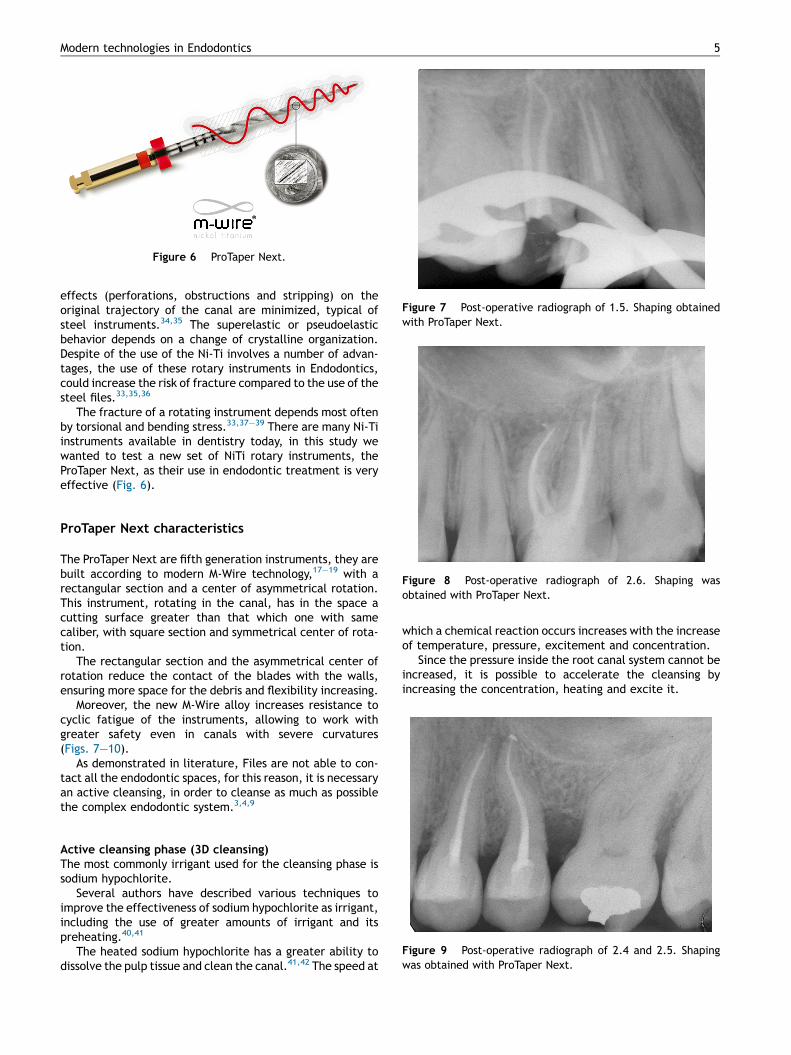
Figure 6 ProTaper Next.
Figure 9 Post-operative radiograph of 2.4 and 2.5. Shapingwas obtained with ProTaper Next.
Figure 7 Post-operative radiograph of 1.5. Shaping obtainedwith ProTaper Next.
Figure 8 Post-operative radiograph of 2.6. Shaping wasobtained with ProTaper Next.
Modern technologies in Endodontics 5
effects (perforations, obstructions and stripping) on theoriginal trajectory of the canal are minimized, typical ofsteel instruments.34,35 The superelastic or pseudoelasticbehavior depends on a change of crystalline organization.Despite of the use of the Ni-Ti involves a number of advan-tages, the use of these rotary instruments in Endodontics,could increase the risk of fracture compared to the use of thesteel files.33,35,36
The fracture of a rotating instrument depends most oftenby torsional and bending stress.33,37—39 There are many Ni-Tiinstruments available in dentistry today, in this study wewanted to test a new set of NiTi rotary instruments, theProTaper Next, as their use in endodontic treatment is veryeffective (Fig. 6).
ProTaper Next characteristics
The ProTaper Next are fifth generation instruments, they arebuilt according to modern M-Wire technology,17—19 with arectangular section and a center of asymmetrical rotation.This instrument, rotating in the canal, has in the space acutting surface greater than that which one with samecaliber, with square section and symmetrical center of rota-tion.
The rectangular section and the asymmetrical center ofrotation reduce the contact of the blades with the walls,ensuring more space for the debris and flexibility increasing.
Moreover, the new M-Wire alloy increases resistance tocyclic fatigue of the instruments, allowing to work withgreater safety even in canals with severe curvatures(Figs. 7—10).
As demonstrated in literature, Files are not able to con-tact all the endodontic spaces, for this reason, it is necessaryan active cleansing, in order to cleanse as much as possiblethe complex endodontic system.3,4,9
Active cleansing phase (3D cleansing)The most commonly irrigant used for the cleansing phase issodium hypochlorite.
Several authors have described various techniques toimprove the effectiveness of sodium hypochlorite as irrigant,including the use of greater amounts of irrigant and itspreheating.40,41
The heated sodium hypochlorite has a greater ability todissolve the pulp tissue and clean the canal.41,42 The speed at
which a chemical reaction occurs increases with the increaseof temperature, pressure, excitement and concentration.
Since the pressure inside the root canal system cannot beincreased, it is possible to accelerate the cleansing byincreasing the concentration, heating and excite it.

Figure 10 Post-operative radiograph of 1.6. Shaping wasobtained with Files in M-Wire alloy.
Figure 11 IG-File (Iandolo Gauging File), Ni-Ti instrumentdesigned exclusively for the measurement of apical diameterand for the sonic activation of the irrigants.
6 A. Iandolo et al.
The excitement is easily achieved by sonic or ultrasonicsources43 (Figs. 11 and 12). The solutions concentrationavailable today on the market, to prevent possible irritantreactions, does not exceed 6%. Finally we can consider theheating.
Normally the solution is pre-heated outside of the tooth toa temperature of 508.44
The preheated solutions have limited utility, since theystabilize rapidly at a temperature included between bodyand ambient temperature.
New technique for the sodium hypochloriteheating: operating protocol
Sodium hypochlorite has a boiling temperature includedbetween 968 and 1208.
We use a heat carrier (System-B or similar). The tempera-ture of the heat carrier is set to 1508.
Figure 12 MicroCT photo shows how, thanks to the sonic activationin the middle third of a root canal.
The heat carrier used will be the 30/04, so that the incomefrom 3 mm working length can be easily achieved withoutexcessive preparations.
The root canal is filled with sodium hypochlorite throughthe endodontic needle. The heat carrier is used cold approxi-mately 3 mm from the working length, and subsequentlyactivated.
Each cycle of activation of the heat-carrier lasts 5 s with afurther 5 s intervals.
During activation, the heat carrier makes brief excursionsup and down of few millimeters to shake the irrigant.
The most important aspect is not to take contact with thecanal walls during the activation of the heat carrier.
, we had a good penetration of the irrigant in a small lateral canal

Figure 13 Preoperative radiograph of 4.6, there is a bigperiapical lesion of the distal and mesial root.
Figure 14 Post-operative radiograph of 4.6. Obturation withthermoplasticized guttapercha after activation and heating ofirrigants.
Figure 15 X-ray follow up at 6 months of 4.6. We can noticethe successful healing with the disappearance of periapicallesions.
Modern technologies in Endodontics 7
After each cycle the irrigant is replaced with fresh solutionso as to have increasingly greater amount of hypochloritewith active chlorine.
The activation cycle is repeated 5 times. During eachactivation of the irrigant, the vapors are sucked by a cannula.
A parameter used was the heating of the outer surface ofthe root, at the third coronal level, middle, apical andforamen level. During activation of the irrigant with aninfrared thermometer (resolution: 0.18) the temperatureson the outer surface of the root were measured. Using thevalues exposed in the operating protocol, there were notdetected external temperature higher than 42.58. So tem-peratures close to 478 were avoided, dangerous for thecells of the periodontal ligament, avoiding medico-legalconsequences.45—47 After chemo-mechanical cleansing(Figs. 13—15), fundamental for the achievement of clinicalsuccess, we proceed to tridimensional obturation withthermo-plasticized guttapercha.
Root canal obturation phase
As obturation technique, if continuous wave of condensationis used, it is important to underline that the heat carrier,must be brought to about 3 mm from the working length, toobtain adequate thermoplasticization of apical gutta-percha.48
For the obturation techniques spread by carrier availableon the market today we find Guttacore Pink obturators, witha gutta percha crosslinked core.24
In addition, recent studies, are proposing new materialsfor the filling of root canals, with remarkable results.49
Conclusions
The positive results highlighted by these clinical casesdemonstrate how the use of modern technologies, operatingmicroscope, ultrasonic tips, rotary files of new generation,systems enhancing cleansing and methods used to obtain avalid tridimensional seal, are essential to avoid iatrogenicdamage and ensure, however, safe and reproducible results.
Of course, to confirm what we have described, furtherresearch and scientific studies are needed, however, theclinical cases performed with these technologies and tech-niques have proved very satisfactory results, especially in thetreatment of endodontic teeth that have large osteolyticlesions and canals with accentuated curvatures.
Conflict of interest
The authors have no conflict of interest.
References
1. Paduano S, Uomo R, Amato M, Riccitiello F, Simeone M, VallettaR. Cyst-like periapical lesion healing in an orthodontic patient: acase report with five-year follow-up. G It Endod 2013;27(2):95—104.
2. Silvani M, Brambilla E, Cerutti A, Amato M, Gagliani M. Root canaltreatment quality in undergraduate program: a preliminarreport on NiTi reciprocating files. G It Endod 2013;27:33—7.

8 A. Iandolo et al.
3. Ametrano G, Riccitiello F, Amato M, Formisano A, Muto M, GrassiR, et al. mCT analysis of mandibular molars before and afterinstrumentation by Reciproc files. Recenti Progressi Medicina2013;104:420—4.
4. Iandolo A. Reperimento del canale mesiale mediano nei primimolari inferiori. Il Dentista moderno 2011;18:269.
5. Carratu P, Amato M, Riccitiello F, Rengo S. Evaluation of leakageof bacteria and endotoxins in teeth treated endodontically bytwo different techniques. J Endod 2002;28(4):272—5.
6. Spagnuolo G, Ametrano G, D’Anto V, Formisano A, Simeone M,Riccitiello F, et al. Microcomputed tomography analysis of mesio-buccal orifices and major apical foramen in first maxillarymolars. Open Dent J 2012;6:118—25.
7. Riccitiello F, Di Caprio MP, D’Amora M, Pizza NL, Vallone G,D’Ambrosio C, et al. Repair of a root perforation by usingMTA: a case report. Recenti Progressi Medicina2013;104:453—8.
8. Spagnuolo G, Ametrano G, D’Anto V, Rengo C, Simeone M,Riccitiello F, et al. Effect of autoclaving on the surfaces ofTiN-coated and conventional nickel-titanium rotary instruments.Int Endod J 2012;45(12):1148—55.
9. Santarcangelo F, Castellucci A. L’irrigazione canalarenell’Endodonzia moderna: casi semplici. Blog in odontoiatria:www.zerodonto.com, sezione Endodonzia.
10. Friedman S, Abitbol S, Lawrence HP. Treatment outcome inendodontics: the Toronto Study. Phase 1: initial treatment. JEndod 2003;29(December (12)):787—93.
11. Ng YL, Mann V, Gulabivala K. A prospective study of the factorsaffecting outcomes of nonsurgical root canal treatment: part 1:periapical health. Int Endod J 2011;44(July (7)):583—609.
12. Das S, Warhadpande MM, Redij SA, Jibhkate NG, Sabir H. Fre-quency of second mesiobuccal canal in permanent maxillary firstmolars using the operating microscope and selective dentinremoval: a clinical study. Contemp Clin Dent 2015;6(January—March (1)):74—8.
13. de Toubes KM, Cortes MI, Valadares MA, Fonseca LC, Nunes E,Silveira FF. Comparative analysis of accessory mesial canal iden-tification in mandibular first molars by using four differentdiagnostic methods. J Endod 2012;38(April (4)):436—41.
14. Rampado ME, Tjaderhane L, Friedman S, Hamstra SJ. The benefitof the operating microscope for access cavity preparationby undergraduate students. J Endod 2004;30(December (12)):863—7.
15. Wu D, Shi W, Wu J, Wu Y, Liu W, Zhu Q. The clinical treatment ofcomplicated root canal therapy with the aid of a dental operat-ing microscope. Int Dent J 2011;61(October (5)):261—6.
16. Lo Giudice G, Lo Giudice R, Matarese G, Isola G, Cicciu M,Terranova A, et al. Evaluation of magnification systems inrestorative dentistry. An in-vitro study. Dent Cadm 2015;83(5):296—305.
17. Ferrara G, Taschieri S, Corbella S, Ceci C, Del Fabbro M, MachtouP. Comparative evaluation of the shaping ability of two differentnickel-titanium rotary files in curved root canals of extractedhuman molar teeth. J Investig Clin Dent )2015;(September).
18. Berutti E, Alovisi M, Pastorelli MA, Chiandussi G, Scotti N,Pasqualini D. Energy consumption of ProTaper Next X1 afterglide path with PathFiles and ProGlider. J Endod 2014;40(Decem-ber (12)):2015—8.
19. Capar ID, Arslan H, Akcay M, Ertas H. An in vitro comparison ofapically extruded debris and instrumentation times with ProTa-per Universal, ProTaper Next, Twisted File Adaptive, and HyFlexinstruments. J Endod 2014;40(October (10)):1638—41.
20. Schmidt TF, Teixeira CS, Felippe MC, Felippe WT, Pashley DH,Bortoluzzi EA. Effect of ultrasonic activation of irrigants onsmear layer removal. J Endod 2015;41(August (8)):1359—63.
21. Sahar-Helft S, Sarp AS, Stabholtz A, Gutkin V, Redenski I, Stein-berg D. Comparison of positive-pressure, passive ultrasonic, andlaser-activated irrigations on smear-layer removal from the
root canal surface. Photomed Laser Surg 2015;33(March (3)):129—35.
22. Rodrıguez-Figueroa C, McClanahan SB, Bowles WR. Spectropho-tometric determination of irrigant extrusion using passive ultra-sonic irrigation, EndoActivator, or syringe irrigation. J Endod2014;40(October (10)):1622—6.
23. Buchanan LS. Endodontic obturation techniques. The state of theart in 2015. Dent Today 2015;34(3):94—5. 90, 92.
24. Li GH, Niu LN, Selem LC, Eid AA, Bergeron BE, Chen JH, et al.Quality of obturation achieved by an endodontic core-carriersystem with crosslinked gutta-percha carrier in single-rootedcanals. J Dent 2014;42(September (9)):1124—34.
25. Keles A, Alcin H, Kamalak A, Versiani MA. Micro-CT evaluation ofroot filling quality in oval-shaped canals. Int Endod J2014;47(December (12)):1177—84.
26. Paul ML, Mazumdar D, Vyavahare NK, Baranwal AK. Healing ofthe periapical lesion in posterior teeth with mineral trioxideaggregate using orthograde technique — two case reports. Con-temp Clin Dent )2012;(September).
27. Yoo JS, Chang SW, Oh SR, Perinpanayagam H, Lim SM, Yoo YJ,et al. Bacterial entombment by intratubular mineralizationfollowing orthograde mineral trioxide aggregate obturation: ascanning electron microscopy study. Int J Oral Sci2014;6(December (4)):227—32.
28. Amato M, Scaravilli MS, Farella M, Riccitiello F. Bleaching teethtreated endodontically: long-term evaluation of a case series. JEndod 2006;32(4):376—8.
29. Riccitiello F, Maddaloni G, D’Ambrosio C, Amato M, Rengo S,Simeone M. Operating microscope: diffusion and limits. G ItEndod 2012;26(2):67—72.
30. Lambertini G, editor. Anatomia umana- Piccin, Padova. 1978.31. Iandolo A. L’utilizzo degli ultrasuoni per una maggiore predici-
bilita nella rimozione delle calcificazioni. G It Endod 2009;23:3.32. Pettiette MT, Metzger Z, Phillips C, Trope M. Endodontic com-
plications of root canal therapy performed by dental studentswith stainless-steel K-files and nickel-titanium hand files. JEndod 1999;25:230—4.
33. Sonntag D, Guntermann A, Kim SK, Stachniss V. Root canalshaping with manual stainless steel files and rotary NiTi filesperformed by students. Int Endod J 2003;36:248—55.
34. Bonaccorso A, Tripi RT. Il Nichel Titanio in Endodonzia. EdizioneMartina Bologna 2006;4:10—9.
35. Spagnuolo G, Desiderio C, Rivieccio V, Amato M, Rossetti DV,D’Anto V, et al. In vitro cellular detoxification of triethyleneglycol dimethacrylate by adduct formation with N-acetylcys-teine. G Dent Mater 2013;29(8):e153—60.
36. Cantatore G, Ceci A. L’ Endodonzia verso il duemila, Prepar-azione canalare con strumenti Ni-Ti Evoluzione delle tecniche.Dental Cadmos 1996;2:21—8.
37. Suter B, Lussi A, Sequeira P. Probability of removing fracturedinstruments from root canals. Int Endod J 2005;38:114—23.
38. Sattapan B, Nervo GJ, Palamara JE, Messer HH. Defects in rotarynickel-titanium file after clinical use. J Endod 2000;26(3):161—5.
39. D’Anto V, Valletta R, Amato M, Schweikl H, Simeone M, PaduanoS, et al. Effect of nickel chloride on cell proliferation. Open DentJ 2012;6:177—81.
40. Abou Rass M, Oglesby SW. The effects of temperature, concen-tration and tissue type on the solvent ability of sodium hypo-chlorite. J Endod 1981;7:376—7.
41. Cunningham WT, Joseph SW. Effect of temperature on thebactericidal action of sodium hypochlorite endodontic irrigant.Oral Surg Oral Med Oral Pathol 1980;50:569—71.
42. Cunnigham WT, Balekjian AY. Effect of temperature on collagen-dissolving ability of sodium hypochlorite endodontic irrigant.Oral Surg Oral Med Oral Pathol 1980;49:175—7.
43. Iandolo A, Ametrano G, Amato M, Rengo S, Simeone M. IG-File:un nuovo strumento per l’ottimizzazione della detersione

Modern technologies in Endodontics 9
canalare e per la misurazione del diametro apicale. G It Endod2011.
44. Berutti E, Marini R. A scanning electron microscopic evaluationof the debridement capability of sodium hypochlorite at differ-ent temperatures. J Endod 1996;22:467—70.
45. Simeone M, Valletta A, Giudice A, Di Lorenzo P, Iandolo A. Theactivation of irrigation solutions in endodontics: a perfectedtechnique. G It Endod 2015 [in press].
46. Di Lorenzo P, Niola M, Pantaleo G, Buccelli C, Amato M. On thecomparison of age determination methods based on dentaldevelopment radiographic studies in a sample of Italian popula-tion. Dent Cadm 2015;83:38—45.
47. Di Lorenzo P, Niola M, Buccelli C, Re D, Cortese A, Pantaleo G,et al. Professional responsibility in dentistry: analysisof an interdepartmental case study. Dent Cadm 2015;83:324—40.
48. Simeone M, De Santis R, Panico G, Riccitiello F, Rengo S. Valu-tazione del profilo termico dell’ otturazione canalare acaldo mediante l utilizzo del system-B. G It Endo 2004;18(4):179—83.
49. Vitti RP, Prati C, Silva EJ, Sinhoreti MA, Zanchi CH, de Souza eSilva MG, et al. Physical properties of MTA Fillapex sealer. JEndod 2013;39(July (7)):915—8.