modes of transmission of loma salmonae (microsporidia) · loma species are unknown, for...
TRANSCRIPT

Vol. 33: 151-156. 1998 DISEASES OF AQUATIC ORGANISMS
Dis Aquat Org ~ Published June 19
Modes of transmission of Loma salmonae (Microsporidia)
'Department of Zoology, 6270 University Boulevard. University of British Columbia. Vancouver, British Columbia V6T 124, Canada 2Departrnent of Fisheries and Oceans. Pacific Biological Station, Nanaimo, British Columbia V9R 5K6, Canada
ABSTRACT: Loma salmonae (Putz, Hoffman and Dunbar, 1965) Morrison and Sprague, 1981 (Micro- sporidia) causes prominent gill disease in pen-reared chinook salmon Oncorhynchus tshawytscha in the Pacific Northwest. Transmission of the parasite was examined by exposing Pacific salmon Oncorhynchus spp. to infectious spores by various routes: per OS, intraperitoneal, intramuscular, and intravascular injection, by cohabitation with infected fish, and by placement of spores directly on the gill. All exposure methods led to infections except placement of spores on the gill. Putative sporoplasms were visible in epithelial cells of the alimentary canal within 24 h of per os exposure. L. salmonae may initially infect alimentary epithelia1 cells and then migrate into the lamina propria to access the blood stream. Positive results obtained by intravascular injection suggest that autoinfec.tion from spores of ruptured xeno~nas in the endothelium may also occur. The cohab~tation cxpc!r~ment demonstr~ttes that flsh may become infected by spores released from live fish.
KEY WORDS: Loma salmonae Microspondia Transmission
INTRODUCTION
Loma salmonae (Putz, Hoffman and Dunbar, 1965) Morrison and Sprague, 1981 is a microsporidian para- site infecting endothelial cells of salmonids. Fish infected w ~ t h L. salnlonae d t e n exhibit pale gills with petechial hemorrhages, inflammation, hyperplasia, and white cysts termed xenomas (Wales & Wolf 1955, Hauck 1984, Kent 1992). Outbreaks of L. salmonae in Pacific salmon Oncorhynchus spp. and rainbow trout 0 . mykiss have occurred in the Pacific Northwest (Hauck 1984, Kent et al. 1989), eastern United States (Markey et al. 1994), and Scotland (Bruno et al. 1995).
Kent et al. (1995) demonstrated that fish can be infected by Loma salmonae when ingesting infected tissues. Whereas details on the early development of Loma species are unknown, for microsporidia in general the spore extrudes a polar filament which pierces a gut epithelial cell and injects the parasite's sporoplasm. For genera such as Loma and Glugea, the
sporoplasm eventually divides, forming numerous spores within a hypertrophied cell (i.e. a xenoma) (Canning & Lom 1986).
Loma salmonae has been transmitted experimentally both in fresh and sea water by feeding fish macerated gills containing spores (Kent et al. 1995) Hauck (1984) suggested that gills may be infected directly by phago- cytic uptake of pillar cells. However, the method by which Lonla spp, spread within the host is unknown. It may be spread by infected macrophages, or by utilis- ing body fluids (Hauck 1984). In the present study, experimental transmission of L. salmonae to chinook Oncorhynchus tshawytscha and coho 0. kisutch was attempted by various routes: per OS, intraperitoneal (IP), intran~uscular (IM), and intravascular (IV) injec- tion, cohabitation of infected fish with naive fish, and placement of L. salmonae spores directly on the gills.
MATERIALS AND METHODS
Fish husbandry. Fish were obtained from Rosewall Creek Hatchery, Fisheries and Oceans Canada, Van-
0 Inter-Research 1998 Resale of full article not permitted
152 Dis Aquat Org 33: 151-156. 1998
couver Island, Canada. This hatchery receives only well water and has no history of the disease. Unless noted elsewhere, all fish were held in flow-through tanks receiving 14 to 16OC dechlorinated tresh water. Before handling, fish were anesthetised with tricaine methanesulfonate (MS-222). During sampling fish were killed with an overdose of MS-222; the first left gill arch of each fish was examined by wet mount for Loma salmonae. The rest of the gills were placed in Davidson's solution (Humason 1979) along with the heart, stomach, intestine, kidney, liver, and spleen, and a section of dorsal muscle for histology. All these tissues were processed using standard histological techniques. Experiments were terminated at 56 d , un- less otherwise noted, to allow sufficient time for devel- opmer?t c?! visih!~ xeccmas (Kent et al. 199.5)
Preparation of parasite. Spores of Loma salmonae were purified from infected chinook gills obtained from a seawater netpen site on the west coast of Van- couver Island. Gill tissue suspended in dechlorinated fresh water was ground using a Polytron tissue homogeniser (Luzer, Switzerland) to create a slurry. This slurry was centrifuged at 2000 X g for 10 min. The pellet was resuspended in water and filtered through a wire mesh and then through a 50 pm nylon screen. The filtrate was then centrifuged at 800 X g for 45 min on a layered 34 %/51% Percol gradient. Pure spores were collected from the pellet and the 34 W 5 1 % layer.
Exposure protocol. Loma salmonae spores were introduced directly into the stomach using a 16 g needle tipped with 2.0 mm inner diameter tubing as either a gill tissue slurry (0.25 ml) or as a suspension of pure spores (0.1 ml). For anal gavage, a 22 g needle tipped with 0.58 mm inner diameter tubing was used. Various amounts of pure spores (Tables 1 & 2) in 0.1 m1 innocula were used for anal gavage, IP, IM, and IV injections. TV injections were given using the ventral gill sinus or dorsal aorta.
Various trials were conducted using the above expo- sure methods as outlined in Tables 1 & 2. Controls had their left ventral fin clipped and were kept in the same tank as exposed fish. In trial 6, controls were injected with Earle's Buffered Saline Solution (EBSS) and blood smears were collected at all sampling times from fish receiving IV injections. For trial 3, blood smears from 3 fish were made within 30 min of injection. Smears were air dried, heat and methanol fixed, and an indi- rect fluorescent antibody test (IFAT) was conducted to locate spores. Monoclonal antibody (MAB) for Loma salmonae developed using standard techniques (Schots et al. 1992) was provided by F. Markham, Department of Pathology and Microbiology, Atlantic Veterinary School, Charlottetown, Prince Edward Island, Canada. The MAB (225 p1) was applied directly to blood smears and allowed to incubate at room temperature for
30 min. The smears were washed 3 times and incu- bated in phosphate buffered saline (PBS) at pH 7.2 for 10 min. Fluorescein isothiocynate (FITC) conjugated goat anti-mouse IgG antibody diluted in PBS (1:128) was counterstained with l % Evan's blue (1:156) and applied to the smears. Smears were incubated for 30 min, washed 3 times and incubated for 10 min in PBS. IFAT slides were examined with a fluorescent microscope at x1000 using pH 9 mounting fluid (Difco).
In trial 4, pure spores (0.1 ml) .were placed directly on the left gill arch of 10 chinook held out of water for 1 min. After 1 min, each chinook was dipped for 5 s in a rinse bucket before being placed in the holding tank. Controls were passed through the rinse bucket before
Table 1 Loma salmonae in Oncorhynchus tshawytscha and 0, kisutch. Infection of L salmonae in chinook and coho salmon exposed by various routes. Fish were examined a t 56 d post-exposure and considered positive upon detection of
the parasite in wet mounts or histological sections
Trial No. of fish No. positive/ exposed No examined
Trial 1 Chinook (avg. 16.6 cm; 38.4 g) Spore dosage (except controls) = 2.8 X 10"
Per OS 10 Anal gavage 10 Control 4
Trial 2 Coho (avg. 18.8 cm; 60.1 g) Spore dosage = 1.2 X 10'
Per OS
lntraperitoneal Intravascular
Trial 3 Chinook (avg. 23 2 cm; 119.2 g) Spore dosage (except controls) = 5.9 X 10⁵
Per os 4 Intravascular"' 13 Control 4
Trial 4 Chinook (avg. 11.2 cm; 12.9 g) Spore dosage (except controls) = 2.8 X 10h
Gill arch 10 Control 2
Trial 5 Cohabitation exposure Donor chinook (avg. 19.8 cm; 66.4 g) Recipient chinook (avg. 12.4 cm; 17 4 g)
Donor fish (spore dosage = 1.25 X 10" 10 Recipient fish, 24 h exposured 45 Recipient fish, 3 d exposured 35 Recipient fish, 5 d exposured 25 Recipient fish, 7 d exposure" 15
"Injection by ventral gill sinus bInjection by dorsal aorta 'Spores detected in blood by an indirect fluorescent anti- body test (IFAT)
dFish removed at time indicated and raised separately for 56 d before examination

Shaw et al.: Modes of Loma salmonae transmission
being placed in the same tank. The same innoculum Table 2. Lo~na salrnonae in Oncorhynchus k i s ~ ~ t c h . Infection
was used here as in trial 1. of L. salmonae in coho salmon from trial 6 exposed by various routes to 1.2 X 10h spores per fish (except controls). Fish were
For 5' donor were infected per and cons~dered positive upon detection of the parasite in wet held for 72 d at 12 to 14°C. Ten donor fish with clinical mounts or histoloaicdl sections signs of Loina salnionae (anemia and petechial hemor- rhages on gills) were placed in a 204 1 tank with 45 nalve chinook (differentiated by size from infected fish). After exposure periods, sets of 10 recipient fish were removed (Table 1) . Following 7 d cohabitation (i.e. 79 d post-exposure of donor fish), all donor fish were examined for L. salmonae. Intensity of infection in donors was measured from the average of 3 counts of the number of xenomas per xlOO field of view (1.5 mm diameter).
RESULTS
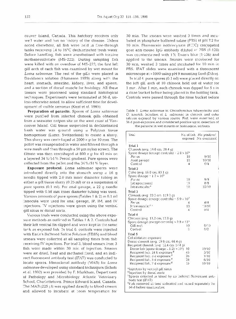
All exposure methods, except placement of Loma salmonae spores directly on the gill, resulted in infec- tion (Tables 1 & 2). When administered per OS, spores were found within 6.5 h in histological sections of stomach in free material and in association with epi- thelial cells (Figs. 1 & 2). Between 2.5 and 24 h possible sporoplasms were found in epithelial cells of the stom- ach and pyloric caeca. These stages contained dark- staining, probably nuclear, material and a lighter-stain- ing outer area. At 24 h epithelial cells of the anterior intestine contained these sporoplasms, and some sporoplasms were also associated with the lamlna pro- pria (Figs. 3-5). These structures were not found in controls or in fish sampled at 35 or 56 d.
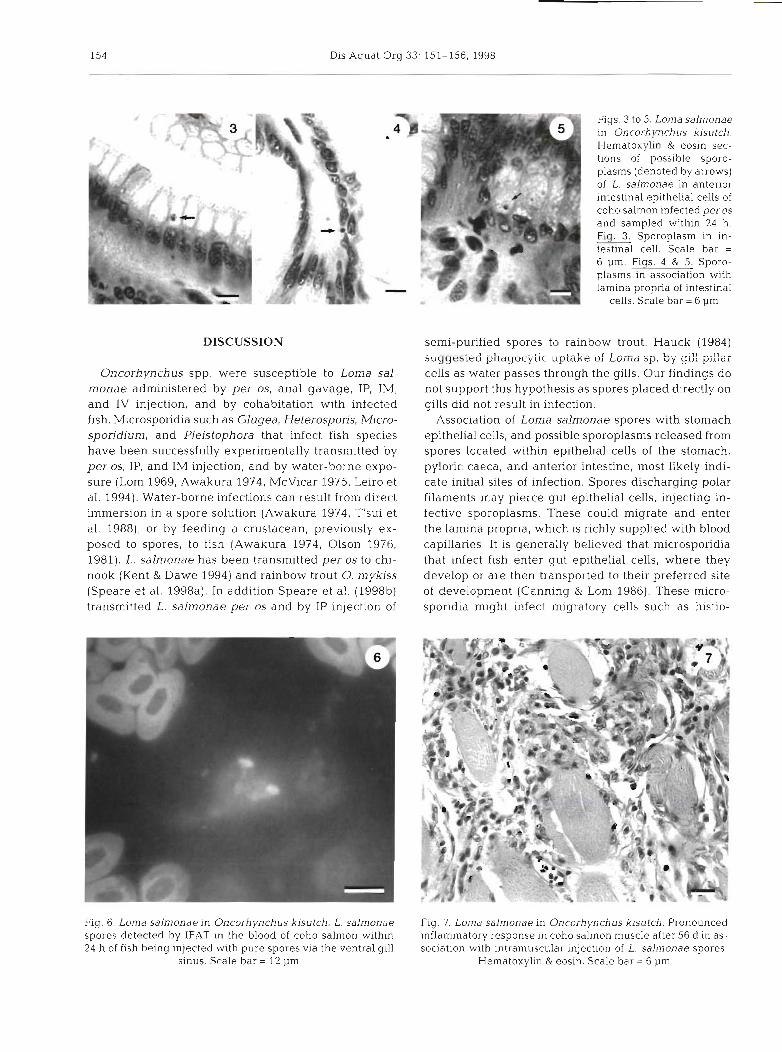
Lonla salmonae spores were detected in the blood by IFAT after fish received IV injections via the dorsal aorta and up to 24 h after injections via the ventral gill
Mode of No. of Time No. positive/ exposure fish examined No. examined
Per os 16 2.5 h" 0/2 6.5 h'," 0/2 24 h" 0/2 35 d 1/2 56 d 7/8
Control 8 2.5 h-56 d 0/8
lntraperitoneal 16 2.5 h 0/2 6.5 h 0/2 24 h 0/2 35 d 0/2 56 d 3/4
Control 2.5 h-56 d 0/4
1ntravascularC 2.5 hd 0/2 6.5 h d 0/2 24 h" 0/2 35 d 1/2 56 d 616
Control 2.5 h-56 d 018
Intramuscular 2.5 h 0/2 6.5 h 0/2 24 h 0/2 35 d 112 56 d 6/7
Control 8 2.5 h-56 d 0/7
4Possible sporoplaslns detected in alimentary canal "spores detected in histological sections of stomach 'Injection b), ventral gill sinus "Spores detected in blood by IFAT
Figs. 1 & 2. Loma salmonae in Oncorhynchus kisutch. Hematoxylin & eosin sections of L. salmonae spores i.n coho salmon infected per OS and sampled within 6.5 h. Fig. Spores in stomach material denoted by arrows. Scale bar = 12 p m Single spore in association with stomach epithelia1 cells.
Scale bar = 12 pm
sinus (Fig. 6). By 56 d all fish given IP injec- tions had swollen kidneys and spleens, petechial hemorrhaging, and bloody ascites. This was not seen in fish infected per OS or in controls. One fish given an IV injection showed edema and hemorrhag- ing. Fish receiving IM injections of spores exhibited edema and a pronounced in- flammatory response in muscle tissue at the site of injection by 35 d (infection shown after 56 d in Fig. 7). No inflamma- tory changes were seen in controls. In trial 6 (Table 2) xenomas were first detected 35 d post-infection in the gills, heart and spleen. Xenomas were found most often in the gills followed by heart and spleen (Table 3). Xenomas were not found in intestine, liver, muscle, or stomach.
Xenomas in donor fish (Table 1; trial 5) were completely opaque and filled with spores. Intensity in donor fish was 2.32 xeno- mas (range 1 to 6.3) per X l00 field of view.
Dis Aquat Org 33: 151-156, 1998
DISCUSSION
Oncorhynchus spp. were susceptible to Lorna sal- rnonae administered by per OS, anal gavage, IP, IM, and IV injection, and by cohabitation with infected fish. M~crosporida such as Glugea, Heterospofis. Micro- sporidium, and Pleistophora that infect fish species have been successfully experimentally transmitted by per OS, IP, and IM injection, and by water-borne expo- sure (Lom 1969, Awakura 1974, McVicar 1975, Leiro et al. 1994). Water-borne infections can result from direct immersion in a spore solution (Awakura 1974, T'sui et al. 1988), or by feeding a crustacean, previously ex- posed to spores, to fish (Awakura 1974, Olson 1976, 1981). L. salmonae has been transmitted per os to chi- nook (Kent & Dawe 1994) and rainbow trout 0. mykiss (Speare et al. 1998a). In addition Speare et al. (1998b) transmitted L. salmonae per os and by IP injection of
Fig. 6. Loma salmonae in Oncorhynchus k~sutch. L. salmonae spores detected by IFAT in the blood of coho salmon within 24 h of fish being injected with pure spores via the ventral gill
sinus. Scale bar = 12 pm
Figs. 3 to 5. Loma salmonae in Oncorhynchus kisutch. Hematoxylin & eosin sec- tions of possible sporo- plasms (denoted by arrows) of L. salmonae in anterior intestinal epithelial cells of coho salmon infected per OS
and sampled within 24 h. Fig. 3 . Sporoplasm in In-
testinal cell. Scale bar = 6 pm. Figs. 4 & 5. Sporo- plasms in association with lamina propria of intest~nal
cells. Scale bar = 6 pm
semi-purified spores to rainbow trout. Hauck (1984) suggested phagocyiic uptake of Loma sp. by gi:: pillar cells as water passes through the gills. Our findings do not support this hypothesis as spores placed directly on gills did not result in infection.
Association of Loma salmonae spores with stomach epithelial cells, and possible sporoplasms released from spores located within epithelial cells of the stomach, pyloric caeca, and anterior intestine, most likely indi- cate initial sites of infection. Spores discharging polar filaments may pierce gut epithelial cells, injecting in- fective sporoplasms. These could migrate and enter the lamina propria, which is richly supplied with blood capillaries It is generally believed that microsporidia that infect fish enter gut epithelial cells, where they develop or are then transported to their preferred site of development (Canning PL Lom 1986). These micro- sporidia might infect migratory cells such as histio-
Fig. 7. Loma salmonae in Oncorhynchus kisutch. Pronounced inflammatory response in coho salmon muscle after 56 d in as- sociation wlth intramuscular injection of L. salmonae spores.
Hematoxylin & eosin. Scale bar = 6 pm
Shaw et al.. Modes ot L o ~ ~ r a aalr~~onde transmission 155
Table 3. Loma salrnonae in Oncorhynchus kisulch. Distribu- tion of L. sa ln~onae in organs of coho salmon from trials 2 and 6 combined, at 56 d after exposure by various routes. Values for tissue are percentage of fish showing infection in
organ indicated
Infection method No. of Tissue fish G111 Heart Spleen K~dney
Per os 17 100.0 22.2 17.6 0.0 Intraperitoneal 1 2 9 2 2 41.7 41.7 25.0 Intravascular 16 100.0 43.8 41.2 6.3 Intramuscular 7 85.7 14.3 0.0 0.0
cytes, neutrophils or macrophages (Weissenberg 1968, McVicar 1975, Canning & Lom 1986). Matthews &
Matthews (1980) proposed that macrophages act as a transport mechanism for Tetrarnicra brevifilum Matthews & Matthew, 1980 through the endothelium of turbot Scophthalmus maximus, or become infected themselves during phagocytosis of the parasite in the lamina propria of the intestine. However, to our knowl- edge none of these hypotheses have been previously experimentally demonstrated. Further studies using in situ hybridisation will probably elucidate the site of initial infection and how subsequent spread of L. sal- rnonae occurs within the fish host. Docker et al. (1997) developed a polymerase chain reaction (PCR) test for L. salrnonae which would be useful for this purpose.
Autoinfection within fish-infecting' microsporidia has also been proposed but not verified (Lom & Dykova 1992). The systemic distribution of Loma sp. xenomas and free spores suggests that, as xenomas rupture, infective stages are liberated and move throughout the fish via the blood (Hauck 1984, Markey et al. 1994). Our experiments, in which free spores injected into the blood resulted in xenoma formation in the gills sup- port the hypothesis that autoinfection of L. salmonae may occur, i.e. in natural infections, xenomas may rup- ture in blood vessels and free spores may circulate and establish new xenomas. The high concentration of xenomas in the gills compared to other organs, regard- less of the route of exposure, suggests that the gill endothelium is the preferred site of development of L. salmonae, rather than a coincidental site of infection due to route of exposure or circulatory patterns.
Transmission of Loma salmonae from infected to naive fish within 24 h of cohabitation in a flow-through system indicates how readily the parasite may be transmitted. Kano et al. (1982) noted that Heterosporis anguillarum Hoshina, 195 1 (syn. Pleistophora anguil- larum) spread to healthy eels from infected eels in the same aquarium. During cohabitation in our study, naive fish may have ingested spores from ruptured or whole xenomas released from the gills (i.e. the sec- ondary lamellae) of infected fish. Spores could also be
liberated in fish urine when fish have kidney infections (Hauck 1984).
Our results demonstrate that Lorna salmonae is transmissible by experimental (e.g IP, IM) and natural (e.g. per OS, cohabitation) exposure routes. Infection by IV injection and distribution of xenomas throughout vascular tissu.e suggest that once a fish is infected, L. salmonae inay spread within the host by autoinfection. Kent et al. (1995) proposed that L. salnlonae spreads within a seawater netpen site via spores released from decomposing fish or by other fish feeding on the remains of mortalities. Salmonid farmers should be aware of this and the possibility of live infected fish transmitting the parasite to naive fish when formulat- ing management strategies to control the infection.
Acknowledgements. Funding for this research was provided by NSERC strategic grant no. 582073. We thank the fish farm companies involved in thls project, S. St-Hilaire for help with injecting fish, S Dawe for assistance with ln~tlal photo- graphics preparations, and F. Markham for providing the Loma-specific monoclonal antibody.
LITERATURE CITED
Awakura T (1974) Studies on the microsporidian infection in salmonid fishes. Sci Rep Hokkaido Fish Hatchery 29:l-96
Bruno DW, Collins RO, hlorrison CM (1995) The occurrence of Loma salrnonae (Protozoa: Microspora) in farmed rain- bow trout, Oncorhynchrrs rnykiss Walbaum, In Scotland. Aquacullure 133:341-344
Canning EU, Lom J (1986) The Microsporidia of vertebrates. Academic Press, New York, p 1-39
Docker MF, Devlin RH. Richard J, Khattra J. Kent ML (1997) Sensitive and specific polymerase chain reaction assay for detection of Loma salrnonae (Microsporea). Dis Aquat Org 29:41-48
Hauck AK (1984) A mortality and associated tissue reactions of chinook salmon. Oncorhynchus tshawytscha (Wal- baum), caused by the microsporidian Lorna sp. J Fish Dis 7:217-229
Humason GL (1979) Animal tissue techniques, 4th edn. WH Freeman, San Francisco
Kano T, Okauchi T, Fukui 1-1 (3.982) Studies on Pleisfopl~ora infection in eel, Anguilla japonica-11. Preliminary tests for application of furnagillin. Fish Path01 1?:107-114
Kent ML (1992) Diseases of seawater netpen-reared salmonid fishes in the Pacific Northwest. Can Spec Pub1 Fish Aquat Sci 116:39-42
Kent ML, Dawe SC (1994) Efficacy of Fumagillin DCH against experimentally induced Loma salmonae (Microsporea) in- fections in chinook salmon Oncorhynchus tsha wytscha. Dis Aquat Org 20:231-233
Kent ML, Dawe SC. Speare DJ (1995) Tra .nsn~~ss~on of Loma salmonae (Microsporea) to chinook salmon In sea water Can Vet J 36:98-101
Kent ML, Elliott DG, Groff JM, Hedrick RP (1989) Loma salmonae (Protozoa: Microspora) infections in seawater reared coho salmon Oncorhynchus kisutch. Aquaculture 80:211-222
Leiro J, Estevez J, Ubeira FM, Santamarina MT. Sanmartin ML (1994) Seriological relationships between two micro- sporidian parasites of fish. Aquaculture 125:l-9

156 Dis Aquat Org 33: 151-156. 1998
Lom J (1969) Experimental transmission of a microsporidian Plistophora hyphessobryconis, by intramuscular trans- plantation. J Protozool 16:17
Lom J, Dykovd I (1992) Developments in aquaculture and fisheries science, Vol 26. Protozoan parasites of fishes. Elsevier Science, Amsterdam, p 125-157
Markey PT, Blazer VS, Ewiny MS, Kocan KM (1994) Loma sp, in salmonids from the Eastern United States: asso- ciated lesions in rainbow trout. J Aquat Anim Health 6: 318-328
Matthews RA, Matthews BF (1980) Cell and tissue reactions of turbot Scophthalmus maximus ( L ) to Tetramicra brevi- filum gen. N., sp. n. (Microspora). J Fish DIS 3:495-515
McVicar AH (1975) Infection of plaice Pleuronectes platessa L , with Glugea (Nosema) stephani (Hagenmiiller 1899) (Protozoa: Microsporidia) in a fish farm and under expen- mental conditions. J Fish Biol7:611-619
Olson RE (1976) Laboratory and field studles on Glugea stephani [Har~anmiillar), a micrnspnridan parasite nf pleuronectid flatfishes. J Protozool 23:158-164
Olson RE (1981) The effect of low temperature on the devel- opment of a microsporidan Glugea stephani in English sole (Parophrya vetulus). J Wild1 Dis 17:559-562
Editorial responsibility: Wolfgang Korting, Hannover, Germany
Schots A , Pomp R , Muiswinkel WB (19921 Production of monoclonal antibod~es. In: Stolen SA, Fletcher TC. Ander- son DP, Kaattari SL, Rowley AF (eds) Techniques in fish immunoloyy, Vol 2 SOS Publicat~uns, Fair Haven, NJ, p 1-18
Speare DJ, Arsenault GJ, Boute MA (1998a) Evaluation of rainbow trout as a model species for studyi'ng the patho- genesis of the branchial microsporidian Loma sahonae . Contemp Top Lab Anim Sci 37:55-58
Speare DJ, Reaman HJ, Jones SRM, Markham RJF, Arsenault GJ (1998b) Induced resistance of rainbow trout to gill disease associated with the microsporidian gill parasite Loma salmonae. J Flsh Dis 21:93-100
T'sui WN, Wang CH, Lo CF (1988) On the Plistophora infec- tion in eel I1 The development of Plistophora anguillarum in experimentally infected elvers. Anguilla japon~ca. Bull Inst Zoo1 Acad Sin 27:249-258
Wales JM, Wolf H (1955) Three protozoan diseases of trout in Cs!ifornia. Ca!if Fish Game 41: 183-187
Weissenberg R (1968) lntracellular development of the micro- sporidian Glugea anomala Moniez in hypertophying migra- tory cells of the fish Gasterosteus aculeatus L., an example of the formation of 'xenoma tumors'. J Protozool 1544-57
Submitted: December 15, 1997; Accepted: March 10, 1998 Proofs received from author@): May 25, 1998