module 4 shoulder. normal anatomy positioning slides discuss three views versus four view series ap...
TRANSCRIPT

MODULE 4
SHOULDER

NORMAL ANATOMY
• Positioning slides
• Discuss three views versus four view series
• AP - internal rotation
• AP - external rotation
• Abduction - baby arm, 90o/90o view– at least one view should demonstrate lung
apices, usually baby arm

LINES OF MENSURATION
• Axial angle of humerus in AP with external rotation
• Humeral shaft line
• Humeral head line– greater tubercle– medial/inferior articular surface
LINES OF MENSURATION
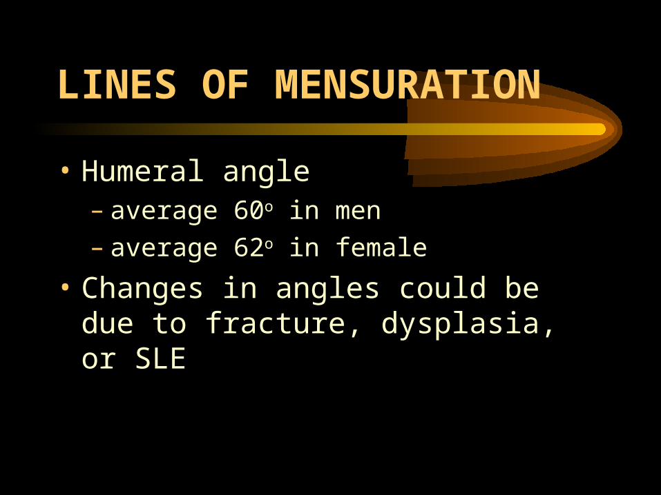
• Humeral angle– average 60o in men– average 62o in female
• Changes in angles could be due to fracture, dysplasia, or SLE

Humeral angle=HAFig 2.68
Humeral shaft line=AHumeral head line=B

LINES OF MENSURATION
• Glenohumeral joint in AP with external rotation
• Measure superior, middle, and inferior
• Add and divide by three and report average (mobility versus stability)
• 4-5 mm essential normal

Glenohumeral joint in AP with external rotationFig 2.69

LINES OF MENSURATION
• Increased measure indicative of infection, inflammatory arthritide, acromegaly, posterior dislocation
• Decreased measure indicative of degenerative joint disease, calcium pyrophosphate dihydrate (CPPD), inflammatory arthritide, septic arthritis

LINES OF MENSURATION
• Acromiohumeral joint space
• AP shoulder (or external rotation)
• Measure inferior surface of acromion to humeral head
• Average 9.0mm (range 7-11mm)

Acromiohumeral joint spaceFig 2.70

LINES OF MENSURATION
• Decrease (<7.0mm) indicative of rotator cuff tear, degenerative tendonitis
• Increase (>11.0mm) indicative of post-traumatic subluxation, dislocation, joint effusion, stroke, brachial plexus lesion (drooping shoulder)

LINES OF MENSURATION
• Acromioclavicular joint space
• AP, PA, external view
• Measure superior and inferior surfaces and average the measurements
• Males average 3.3 with a range of 2.5-4.1
• Females average 2.9 with a range of 2.1-3.7
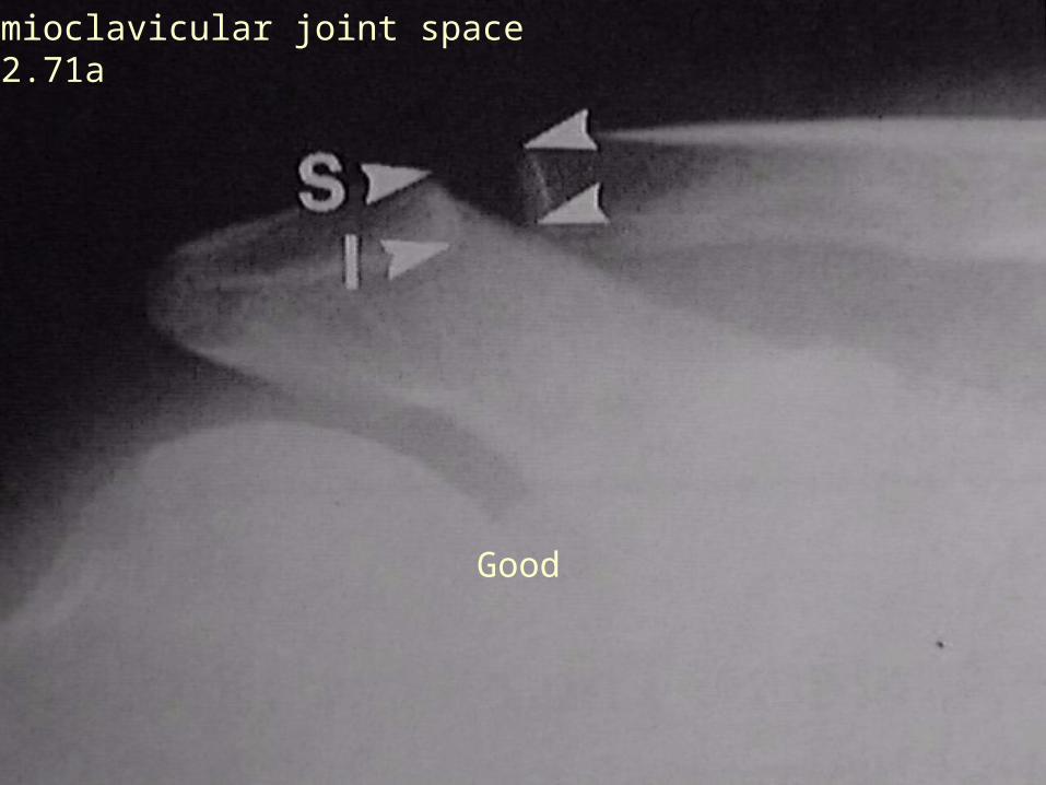
Acromioclavicular joint spaceFig 2.71a
Good

Bad DJD Bad dog!!!
Acromioclavicular joint spaceFig 2.71b

LINES OF MENSURATION
• Decrease measure (<2.5/2.1) indicative of degenerative joint disease
• Increase measure (>4.1/3.7) indicative of traumatic separation, resorption (osteolysis), HPT, rheumatoid arthritis, backpacking syndrome
• May require weighted and non-weighted comparison study

COMMON CONDITIONS
• Degenerative joint disease
• Rotator cuff tear
• Calcific tendonitis
• Dislocation and humerus fracture
• Clavicle fracture

CLINICAL DIAGNOSIS
• Impingement
• Adhesive capsulitis
• Tendonitis/bursitis

SLIDES
• Normal positioning• Text measure• Gross specifications• Degenerative joint
disease• Calcified tendonitis• Rotator cuff tear
• Acromioclavicular separation Osteolysis
• Scapula fracture• Clavicular fracture• Surgical sign of
clavicle removal• Ischemic necrosis• Impingement

MENTION
• Adhesive capsulitis
• Tendonitis/bursitis