module two: the neurological...
TRANSCRIPT
MODULE TWO: THE NEUROLOGICAL EXAMINATION The neurological examination is a systematic survey of the functional status of the nervous system, which when abnormal allows the physician to state with confidence WHERE a lesion resides. A neurological exam should always be interpreted within the context of a more general assessment that includes a patient history, general physical exam (which includes the neurologic exam as one part), and other diagnostic tests such as radiological studies and blood tests. While the neurological exam can tell you WHERE the lesion is in the nervous system, other aspects of a complete assessment, such as history and radiological tests, are typically required to ascertain WHAT the lesion is (i.e., correct diagnosis of the disease). This information is being presented in Module Two of this course because knowledge of the clinical relevance of the neurological examination will be important in interpretation of clinical cases presented in subsequent Modules. A neurologic exam consists of the following six subdivisions:
1) Mental status 2) Cranial nerves 3) Motor exam 4) Reflexes 5) Coordination and gait 6) Sensory exam Each of these subdivisions will be briefly summarized below with an
emphasis on those parts of the exam that all neuropsychologists should be familiar with. Students are urged to read this section, Chapter Three in the Blumenfeld text, and then to review the important portions of the neurological examination via video demonstrations at the text’s companion website, www.neuroexam.com. Since the information presented in this module may be difficult to fully absorb upon first presentation, students are encouraged to frequently refer back to the neurological examination procedures when future clinical cases are presented to become more thoroughly knowledgeable. As with most of the material to be learned in this course, after describing the core content to be learned, discussions about the neuroanatomical underpinnings and clinical significance will follow. Not every patient will require an exhaustive neurological examination. The core elements of a minimal screening neurologic exam are as follows:
1) MENTAL STATUS EXAM a. Level of alertness and orientation b. Assess attention using months forwards/backwards c. Immediate registration and delayed recall of 3 objects for four
minutes (timed). d. Naming of parts of watch e. Note behavior, language, affect, etc., while history taking
2) CRANIAL NERVES
2
a. Pupil light reflexes b. Ophthalmoscopic exam c. Visual fields, including extinction testing d. Horizontal and vertical smooth pursuit eye movements e. Facial sensation to light touch including extinction testing f. Facial asymmetry during emotional smile g. Hearing of finger rub bilaterally h. Palate elevation i. Note quality of voice during remainder of exam j. Head turning and shoulder shrug against resistance k. Tongue protrusion
3) MOTOR EXAMINATION a. Drift b. Rapid hand and foot tapping c. Upper and lower extremity tone d. Strength in several proximal and distal muscles in the upper and
lower extremities bilaterally i. Finger extensors ii. Finger abductors iii. Wrist extensors iv. Biceps v. Triceps vi. Deltoids vii. Iliopsoas viii. Quadriceps ix. Foot and toe dorsiflexors x. Knee flexors
4) REFLEXES
a. Bilateral biceps b. Brachioradialis c. Patellar d. Achilles tendon e. Plantar reflexes
5) COORDINATION AND GAIT
a. Finger-to-nose bilaterally b. Heel-to-shin bilaterally c. Gait d. Tandem walking
6) SENSORY EXAMINATION
a. Light touch in hands and feet, including extinction testing b. Pin prick or temperature testing in feet bilaterally c. Vibration and joint position sense in feet bilaterally
Each section of this core neurological examination will be discussed below.
3
I. MENTAL STATUS EXAMINATION
Level of Alertness, Attention, and Cooperation. Levels of alertness are generally described on a continuum from mildly inattentive to lethargic to stupor us to a full coma. Neurologists typically assess attention by spelling a short word forward and backward (such as WORLD or DLROW), digit span forward and backward, or naming the months of the year forward and then backward (see www.neuroexam.com Video 4). Degree of cooperation should also be noted since this may influence the results of many aspects of the exam. Neuroanatomic Correlations. Level of consciousness is severely impaired by damage to the brainstem reticular formation, bilateral lesions of the thalamus, and in extensive bilateral lesions of the cerebral hemispheres (as in toxic or metabolic disturbances). Clinical Correlations. Generalized impaired attention is a nonspecific abnormality that can occur in many different focal brain lesions as well as in diffuse abnormalities such as some dementias or encephalitis. Level of cooperation is most often encountered in behavioral or mood disorders or in cases where litigation is involved (e.g., worker’s compensation evaluations). Orientation and Memory. Many non-neurological physicians are notoriously liberal in interpretation of patient’s orientation to person, place, and time (“A&OX3”). Ask the patient’s full name, location (name of the hospital or clinic), and full date including day of week and time of day. Memory is typically assessed by asking the patient to recall three or four items or a brief story after a 3 to 4 minute delay (see www.neuroexam.com Video 6). Neuroanatomical and Clinical Correlations. Memory can be impaired on different timescales. Inability to register and recall something within a few seconds after it was said is an abnormality of attention (or “immediate memory”) which was discussed above. If immediate recall is intact, then difficulty with recall after 5 minutes often signifies damage to the limbic memory structures located in the medial temporal lobes or medial diencephalon. Such memory difficulties may also occur at times with frontal lobe lesions and in psychogenic amnesia. Speech and Language. Spontaneous Speech. The neurologist attends to the patient’s fluency, including phrase length, rate, and abundance of spontaneous speech (see www.neuroexam.com Video 8) and also notes the tonal modulation (prosody) and whether paraphasic errors (inappropriately substituted words or syllables) or neologisms (nonexistent words), or errors of grammar are present. Comprehension. This is usually assessed by whether the patient can understand simple questions and commands (see www.neuroexam.com Video 9).
4
Naming. Both easy (e.g., pen, watch, tie, jacket) and harder (e.g., fingernail, belt buckle, stethoscope) objects should be named (see www.neuroexam.com Video 10). Repetition. Can the patient repeat single words and sentences (a standard sentence is, “no ifs, ands, or buts”)? Other more difficult sentences may also be given, such as “the spy fled to Greece” or “the phantom soared across the foggy heath” (see www.neuroexam.com Video 11). Reading and Writing. The patient may be asked to read a newspaper headline, a brief passage, or single words and tested for comprehension. Handwriting samples often consist of asking the patient to write his or her name and a dictated sentence or two (see www.neuroexam.com Video 12). Neuroanatomical and Clinical Correlations. Different types of speech and language abnormalities may be caused by lesions in the dominant (usually left) frontal lobe including Broca’s area; the left temporal and parietal lobes, including Wernicke’s area; subcortical white matter and gray matter structures, including thalamus and caudate nucleus. Apraxia. This term typically refers to the inability to follow a motor command that is not due to a primary motor deficit or language impairment. It is thought to be caused by a higher-order planning or conceptualization of the motor task. Patient’s are often giving commands such as, “pretend to comb your hair” or “pretend to strike a match and blow it out” (see www.neuroexam.com Video 15). This type of apraxia is referring to as “ideomotor apraxia.” Unfortunately, there are several different forms of apraxia, and the term is even applied to other abnormalities that are not true apraxias such as “constructional apraxia,” “dressing apraxia,” or “ocular apraxia.”
Neuroanatomical and Clinical Correlations. The ideomotor type of apraxia described above indicates cortical brain dysfunction usually accompanying aphasia although it may occur in isolation. Ideomotor apraxia is commonly present in lesions affecting the inferior parietal region of the dominant (usually left) cerebral hemisphere. Neglect and Constructions. Hemineglect is an abnormality of inattention to one side of space or the body that is not due to a primary motor or sensory deficit. In sensory neglect, patients ignore visual, somatosensory, or auditory stimuli on side opposite the lesion despite intact primary sensation. This defect is most often observed when testing for extinction upon double simultaneous stimulation. In addition, some drawing tasks, such as line bisection or clock drawing, can demonstrate neglect. Constructional tasks involve drawing complex figures or making patterns by manipulating colored blocks may show impairments due to neglect or to a visual-spatial deficit. These are shown at www.neuroexam.com in Videos 16 and 17).
Neuroanatomical and Clinical Correlations. Hemineglect is most often caused by lesions of the right (nondominant) parietal lobe, causing patients to
5
neglect the left side. Abnormal constructions also frequently occur following damage to the nondominant (usually right) parietal lobe. Sequencing Tasks and Frontal Release Signs. Patients with damage to the frontal lobes will often get stuck and repeat an action over and over again (called perseveration) or may have difficulties changing from one action to the next when asked to perform a repeated sequence of actions. These deficits may be evaluated by alternating sequential motor actions, such as tapping the table with a fist, open palm, and side of open hand as quickly as possible repetitively. Motor impersistence is another common form of distractibility seen in frontal lobe disease in which patients will have difficulty sustaining a motor action such as keeping the arms raised or maintain eye gaze to the right in the face of distraction (such as the examiner’s fingers snapping). Additional support for frontal lobe pathology comes from frontal release signs, such as the grasp reflex which is discussed later in this module under Cranial Nerves. This portion of the mental status examination may be viewed at www.neuroexam.com in Videos 18-20). Psychiatric Symptoms. The mental status portion of the neurologic exam also looks for signs of mood changes typical of depression, anxiety or mania as well as unusual false beliefs constituting delusions or visual or auditory hallucinations (see www.neuroexam.com Video 23). Neuroanatomical and Clinical Correlations. Delusions and hallucinations may be seen in toxic or metabolic derangement and in primary psychiatric disorders. In addition, abnormal sensory phenomena can be caused by focal lesions or seizures in primary sensory cortices.
II. CRANIAL NERVES
Olfaction (CN I). Is often assessed by asking the patient to identify by smell coffee, soap, or peanut butter (see www.neuroexam.com Video 24). Neuroanatomical and Clinical Correlations. Impairment can be due to nasal obstruction, damage to olfactory nerves in nasal mucosa, damage to the nerves as the cross the cribiform plate, or intracranial lesions affecting the olfactory bulbs. Damage to CN I may occur with significant closed head / traumatic brain injuries. Ophthalmoscopic Exam (CN II). Both retinas are examined carefully with an ophthalmoscope (see www.neuroexam.com Video 25).
Neuroanatomical and Clinical Correlations. This allows direct visualization of damage to the retina or retinal vessels, optic nerve atrophic changes, optic neuritis such as that seen early in multiple sclerosis, or papilledema signaling increased intracranial pressure. Vision (CN II). Visual acuity is tested using an eye chart or pocket screening card by covering one eye at a time. Visual fields are assessed by asking the
6
patient to fixate straight ahead and to report when a finger can be seen moving into each quadrant (see www.neuroexam.com Video 27). Visual extinction on double simultaneous stimulation is assessed by asking the patients how many fingers they see when fingers are presented to both sides at the same time (see www.neuroexam.com Video 27). In visual extinction, patients do not report seeing the fingers on the affected side (usually left) of the visual field, but can see the fingers when they’re presented to that side alone. Neuroanatomical and Clinical Correlations. Damage anywhere along the visual pathway from the eye to the visual cortex can cause defects in the visual fields of one or both eyes. Lesions in front of the optic chiasm (eye, optic nerve) cause visual defects in one eye only. Lesions behind the optic chiasm (optic tract, thalamus, white matter, visual cortex) will result in visual field deficits that are similar in both eyes. Visual hemineglect or extinction is usually caused by contralateral parietal lobe lesions, and less often by frontal or thalamic lesions. Pupillary Responses (CN II, III). The size and shape of pupils are first noted at rest. Next, the direct response, meaning constriction of the illuminated pupil and the consensual response, meaning construction of the opposite pupil are observed (see www.neuroexam.com Video 29). Finally, the pupillary response to accommodation (response to looking at something moving toward the eye) is tested. The pupils will normally constrict while fixating on an object moving toward the eye (see www.neuroexam.com Video 31).
In a patient’s medical chart, normal results of this part of the neurologic exam are often reported as, “PERRLA,” to mean “pupils are equal, responsive, and reactive to light and accommodation.”
Neuroanatomical and Clinical Correlations. The direct response (pupil
illuminated) is impaired in lesions of the ipsilateral optic nerve, pretectal area, ipsilateral parasympathetics traveling in CN III, or the pupillary constrictor muscle of the iris.
The consensual response (contralateral pupil illuminated) is impaired in lesions of the contralateral optic nerve, pretectal area, ipsilateral parasympathetics traveling in CN III, or the contralateral pupillary constrictor muscle.
Accommodation is impaired in lesions of the ipsilateral optic nerve, ipsilateral parasympathetics traveling in CN III, the pupillary constrictor muscle, or in bilateral lesions of pathways from the optic tracts to the visual cortex. Extraocular Movements (CN III, IV, VI). The muscles controlling movement of the eyes is first checked by having the patient move their eyes in all directions. Smooth pursuit is tested by having the patient follow an object moved across their full range of horizontal and vertical eye movements (see www.neuroexam.com Video 32). Convergent movements are tested by having the patient fixate on an object as it is moved slowly toward a point right between the patient’s eyes.
7
The eyes are also observed at rest to see if there are any abnormalities, such as nystagmus or dysconjugate gaze (eyes not both fixated on the same point), resulting in diplopia (double vision). Neuroanatomical Correlations. Testing can identify abnormalities in individual muscles or in specific cranial nerves (oculomotor, trochlear, or abducens) in their course from the brainstem to the orbit, in the brainstem nuclei, or finally, in the higher-order centers and pathways in the cortex and brainstem that control eye movements. Facial Sensation and Muscles of Mastication (CN V). Facial sensation is typically tested with a cotton swab (Q-tip) and a sharp object (safety pin). Assessment for tactile extinction is also routinely accomplished via double simultaneous stimulation. See www.neuroexam.com Video 37. Neuroanatomical and Clinical Correlations. Facial sensation can be impaired by lesions of the trigeminal nerve (CN V), trigeminal sensory nuclei in the brainstem, or ascending sensory pathways to the thalamus and somatosensory cortex in the postcentral gyrus. Tactile extinction in the presence of intact primary sensation is usually caused by right parietal lobe lesions. Muscles of Facial Expression and Taste (CN VII). Weakness of the facial muscles may be seen in asymmetries in facial shape or in the depth of furrows such as the nasolabial fold with spontaneous facial expressions and blinking. Patients also may be asked to smile, puff out their cheeks, clench their eyes tight, or wrinkle their brow to bring out facial asymmetry. See www.neuroexam.com Video 40. Taste is checked with sugar, salt, or lemon juice on cotton swabs applied to the lateral aspect of each side of the tongue.
Neuroanatomical and Clinical Correlations. Facial weakness can be caused by lesions of upper motor neurons in contralateral motor cortex, descending motor pathways, lower motor neurons in the ipsilateral facial nerve nucleus (CN VII), or exciting nerve fibers, the neuromuscular junction, or the facial muscles. Upper motor neuron lesions, such as stroke, cause contralateral facial weakness only involving the lower portion of the face (sparing the forehead) because the lower part of the facial nerve nucleus does not receive bilateral cortical input (but the upper part of the nucleus does). Hearing and Vestibular Sense (CN VIII). Hearing is tested by gently rubbing the fingers together just outside the external auditory canal (see www.neuroexam.com Video 42). A tuning fork can be used to distinguish neural from mechanical conductive hearing problems. Vestibular sense is generally not tested except in patient with vertigo, limitations of horizontal or vertical gaze, or coma.
Neuroanatomical and Clinical Correlations. Hearing loss can be caused by lesions in the acoustic and mechanical elements of the ear, the neural
8
elements of the cochlea, or the acoustic nerve (CN VIII). After the hearing pathways enter the brainstem, they cross over at multiple levels and ascend bilaterally to the thalamus and auditory cortex. Therefore, clinically significant unilateral hearing loss is invariably caused by peripheral neural or mechanical lesions.
Abnormalities in vestibular testing can be associated with lesions in the vestibular apparatus of the inner ear, the vestibular portion of CN VIII, vestibular nuclei in the brainstem, the cerebellum, or pathways in the brainstem (such as the medial longitudinal fasciculus) that connect vestibular and oculomotor systems. Palate Elevation and Gag Reflex (CN IX, X). Does the palate elevate symmetrically when the patient says, “Aah” (see www.neuroexam.com Video 44)? Does the patient gag when the posterior pharynx is brushed? The gag reflex only needs to be tested in patients with suspected brainstem pathology, impaired consciousness, or impaired swallowing. Neuroanatomical and Clinical Correlations. Palate elevation and gag reflex are impaired in lesions involving CN IX, CN X, the neuromuscular junction, or the pharyngeal muscles. Muscles of Articulation (CN V, VII, IX, X, XII). This is examined simple by listening to the patient speak and obtaining some history. Have there changes in the patient’s speech, such that it has become more hoarse, slurred, quiet, breathy, nasal, low or high pitched, or other changes? See www.neuroexam.com Video 45).
Neuroanatomical and Clinical Correlations. Abnormal speech articulation can occur in lesions involving the muscles of articulation, the neuromuscular junction, or the peripheral or central portions of CN V, VII, IX, X or XII. Dysarthria (speech misarticulation) can also be abnormal as a result of lesions in the motor cortex, cerebellum, basal ganglia, or descending pathways to the brainstem. Sternocleidomastoid and Trapezius Muscles (CN XI). These muscles of the neck and shoulders are assessed by asking the patient to shrug their shoulders, turn their head in both directions, and raise their head from the bed flexing forward against the force of the examiner’s hands (see www.neuroexam.com Video 46). Neuroanatomical and Clinical Correlations. Weakness in these muscles may be caused by lesions in the muscles, neuromuscular junction, or lower motor neurons of the spinal accessory nerve (CN XI). Upper motor neuron lesions in cortex or descending pathways may cause contralateral weakness of the trapezius, with relative sparing of sternocleidomastoid strength (similar to how upper motor neuron facial lesions spare the upper part of the face). When sternocleidomastoid weakness is present with upper motor neuron lesions, there is weakness of head turning away from the side of the lesion.
9
Tongue Muscles (CN XII). The examiner first looks to see if there are any fasciculations (spontaneous quivering movements) of the tongue while it is resting in the mouth. The patient is then asked to stick their tongue straight out, and the examiner looks to see if the tongue curves to one side or the other. Patients are also asked to move their tongue from side to side and to push it forcefully against the inside of each check (see www.neuroexam.com Video 47). Neuroanatomical and Clinical Correlations. Fasciculations and atrophy are signs of lower motor neuron damage (see the table below). Unilateral tongue weakness causes the tongue to deviate toward the weak side. Tongue weakness may result from lesions of the tongue muscles, the neuromuscular junction, lower motor neurons of the hypoglossal nerve (CN XII), or upper motor neurons originating in the motor cortex. Lesions of motor cortex cause contralateral tongue weakness.
III. MOTOR EXAMINATION The motor exam consists of several steps including:
1) observation 2) inspection 3) palpation 4) muscle tone testing 5) functional testing, and 6) strength testing of individual muscle groups
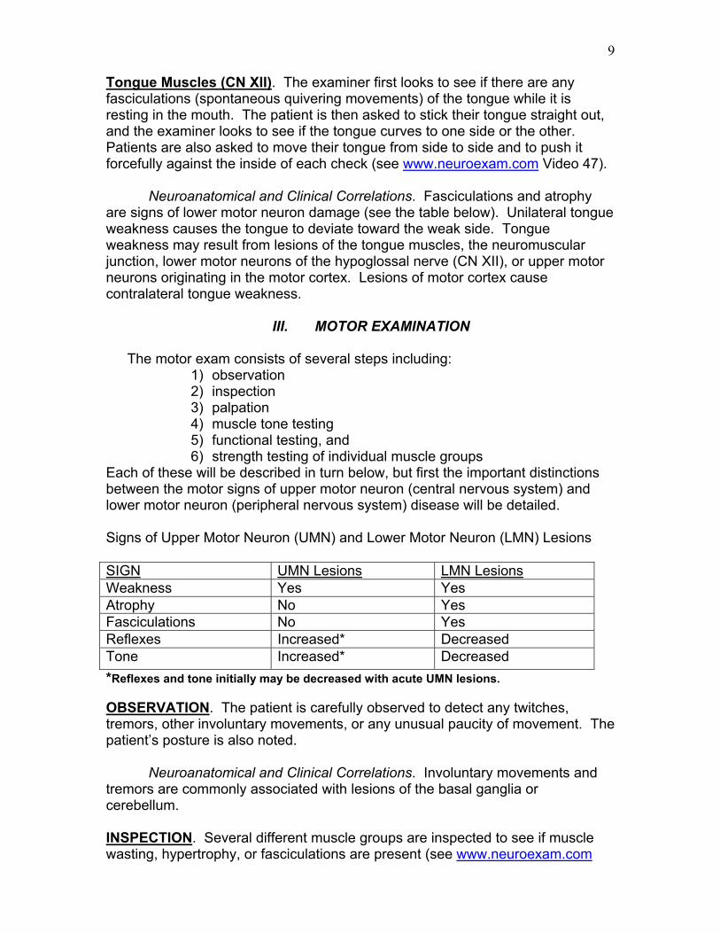
Each of these will be described in turn below, but first the important distinctions between the motor signs of upper motor neuron (central nervous system) and lower motor neuron (peripheral nervous system) disease will be detailed. Signs of Upper Motor Neuron (UMN) and Lower Motor Neuron (LMN) Lesions SIGN UMN Lesions LMN Lesions Weakness Yes Yes Atrophy No Yes Fasciculations No Yes Reflexes Increased* Decreased Tone Increased* Decreased *Reflexes and tone initially may be decreased with acute UMN lesions. OBSERVATION. The patient is carefully observed to detect any twitches, tremors, other involuntary movements, or any unusual paucity of movement. The patient’s posture is also noted. Neuroanatomical and Clinical Correlations. Involuntary movements and tremors are commonly associated with lesions of the basal ganglia or cerebellum. INSPECTION. Several different muscle groups are inspected to see if muscle wasting, hypertrophy, or fasciculations are present (see www.neuroexam.com
10
Video 48). Fasciculations are most easily seen in the hands, shoulders, and thighs. PALPATION. Muscles are palpated or probed to see if there is any muscle tenderness in cases of suspected myositis (muscle inflammation). MUSCLE TONE TESTING. The patient is asked to relax, and then each limb is passively moved at several joints to get a feeling for any resistance or rigidity (see www.neuroexam.com Videos 49 and 50). Neuroanatomical and Clinical Correlations. Parts of the motor exam can help distinguish between upper motor neuron and lower motor neuron lesions (see Table above). Recall UMNs project via the corticospinal tract to lower motor neurons in the anterior horn of the spinal cord. Signs of lower motor neuron damage include weakness, atrophy, fasciculations, and hyporeflexia. Signs of upper motor neuron damage include weakness, hyperreflexia (increased reflexes), and increased tone. NOTE with acute UMN lesions there is often flaccid paralysis with decreased tone and decreased reflexes. With time (hours to weeks), hypertonicity (increased tone) and hyperreflexia (increased reflexes) usually develop. Increased tone may also be seen in diseases of the basal ganglia. Slow or awkward fine finger movements or toe tapping in the absence of weakness can also signify mild corticospinal tract damage or lesions of the cerebellum or basal ganglia. FUNCTIONAL TESTING. Before testing each individual muscle group, a gross motor examination may alert the examiner to certain abnormalities. Drift is checked by having the patient hold up both arms or both legs and then close their eyes (see www.neuroexam.com Video 51). Fine movements are tested by rapid finger tapping, rapid hand tapping, and rapid foot tapping (against the floor). STRENGTH OF INDIVIDUAL MUSCLE GROUPS. Patterns of weakness help localize a lesion to a specific region on the neuraxis whether it is in cortex, white matter, spinal cord level, nerve root, peripheral nerve, or muscle. Each major muscle group is systematically assessed and typically rated on a scale ranging from 0/5 to 5/5 strength as follows: 0/5 = No contraction 1/5 = Muscle flicker, but no movement 2/5 = Movement possible, but not against gravity 3/5 = Movement possible against gravity, but not against resistance 4/5 = Movement possible against mild resistance by examiner 5/5 = Normal strength
11
IV. REFLEXES
Deep tendon reflexes and plantar response should be checked in all
patients. Deep tendon reflexes are examined using impulses from a reflex hammer to stretch the muscle and tendon in a relaxed limb. When reflexes are very brisk, clonus (repetitive vibratory contraction of the muscle in response to muscle and tendon stretch) may be observed (see www.neuroexam.com Video 58). Deep tendon reflexes (DTRs) are often rated according to the following scale:
0 = Absent reflexes 1+ = Trace 2+ = Normal 3+ = Brisk 4+ = Nonsustained clonus 5+ = Sustained clonus
Deep tendon reflexes are normal if they are rated as 1+, 2+, or 3+ unless they are significantly asymmetrical with respect to side of body or in the upper versus lower extremities.
Neuroanatomical and Clinical Correlations. DTRs may be diminished by
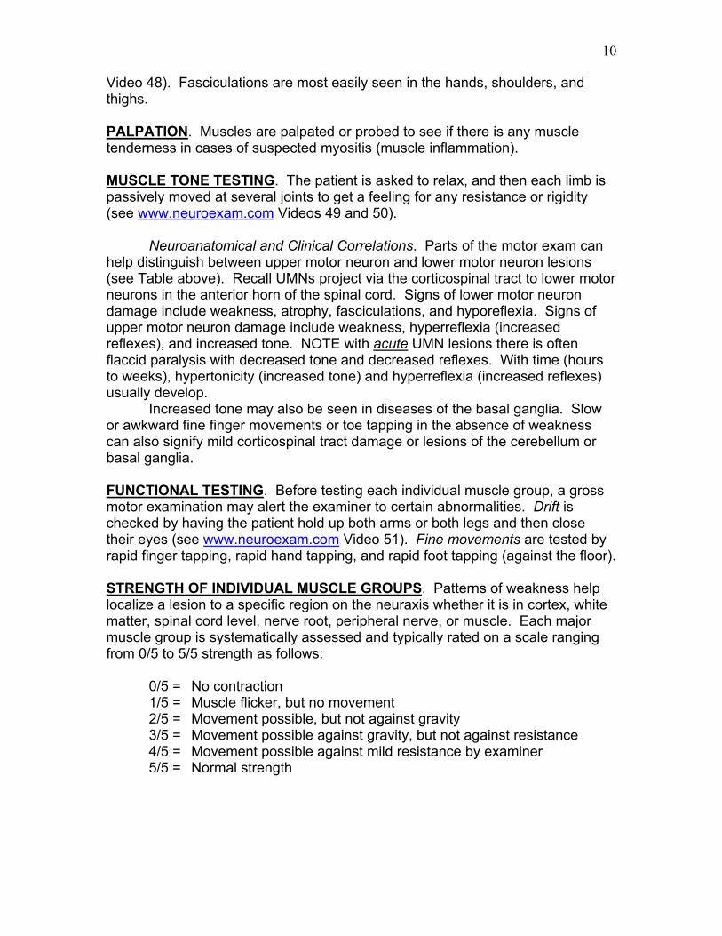
abnormalities in muscles, sensory neurons, lower motor neurons, and the neuromuscular junction; acute UMN lesions; and in joint disease. Hyperreflexia (abnormally increased reflexes) is associated with UMN damage. PLANTAR RESPONSE. The plantar response is tested by scraping an object (e.g., reflex hammer handle) across the sole of the foot beginning from the heel, moving forward toward the small toe, and then arching medially toward the big toe (see www.neuroexam.com Video 59). The normal response is downward contraction of the toes. The abnormal response, called Babinski’s sign, is indicated by an upgoing big toe and fanning out of the other toes. Neuroanatomical and Clinical Correlations. Babinski’s sign is associated with upper motor neuron lesions anywhere along the corticospinal tract.
12
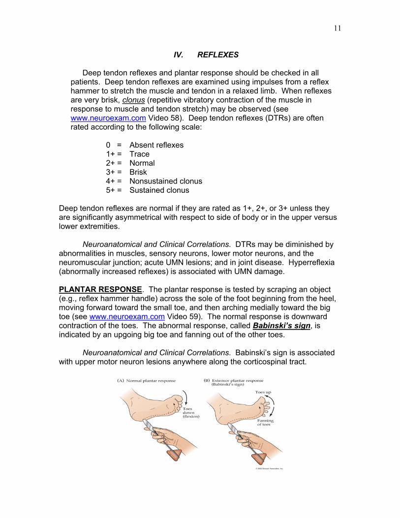
FRONTAL RELEASE SIGNS. Frontal lobe lesions can cause reemergence of some primitive reflexes that are normally present in infants but are abnormal in adults. These so-called frontal release signs include the grasp, snout, root, and suck reflexes (see www.neuroexam.com Video 18 for demonstration of the grasp reflex). REFLEX EXAM STICK FIGURE. The deep tendon reflex examination is typically summarized in the medical record or chart using a stick figure (shown below) for each of the five major reflex sites (also shown below and in the book in Table 3.6). The up- or down-going arrows at the feet represent the direction of the plantar response (e.g., upgoing arrows for a positive Babinski sign).
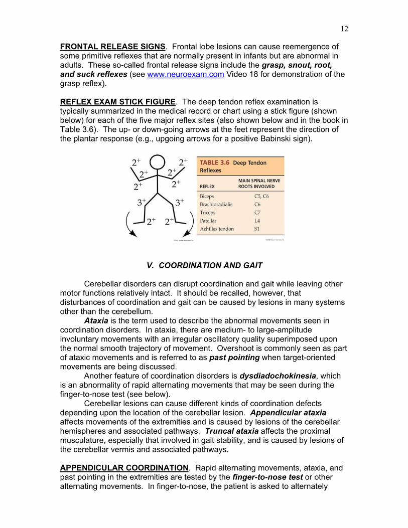
V. COORDINATION AND GAIT Cerebellar disorders can disrupt coordination and gait while leaving other motor functions relatively intact. It should be recalled, however, that disturbances of coordination and gait can be caused by lesions in many systems other than the cerebellum. Ataxia is the term used to describe the abnormal movements seen in coordination disorders. In ataxia, there are medium- to large-amplitude involuntary movements with an irregular oscillatory quality superimposed upon the normal smooth trajectory of movement. Overshoot is commonly seen as part of ataxic movements and is referred to as past pointing when target-oriented movements are being discussed. Another feature of coordination disorders is dysdiadochokinesia, which is an abnormality of rapid alternating movements that may be seen during the finger-to-nose test (see below). Cerebellar lesions can cause different kinds of coordination defects depending upon the location of the cerebellar lesion. Appendicular ataxia affects movements of the extremities and is caused by lesions of the cerebellar hemispheres and associated pathways. Truncal ataxia affects the proximal musculature, especially that involved in gait stability, and is caused by lesions of the cerebellar vermis and associated pathways. APPENDICULAR COORDINATION. Rapid alternating movements, ataxia, and past pointing in the extremities are tested by the finger-to-nose test or other alternating movements. In finger-to-nose, the patient is asked to alternately
13
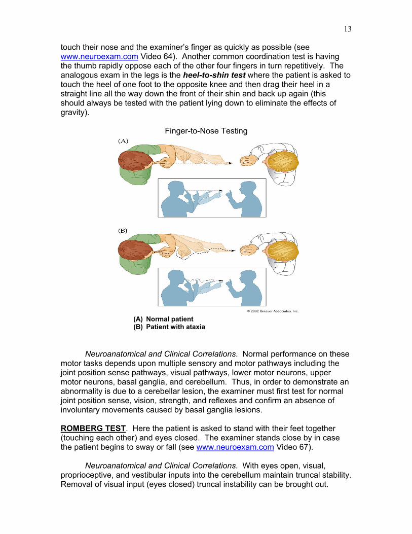
touch their nose and the examiner’s finger as quickly as possible (see www.neuroexam.com Video 64). Another common coordination test is having the thumb rapidly oppose each of the other four fingers in turn repetitively. The analogous exam in the legs is the heel-to-shin test where the patient is asked to touch the heel of one foot to the opposite knee and then drag their heel in a straight line all the way down the front of their shin and back up again (this should always be tested with the patient lying down to eliminate the effects of gravity).
Finger-to-Nose Testing
(A) Normal patient (B) Patient with ataxia
Neuroanatomical and Clinical Correlations. Normal performance on these motor tasks depends upon multiple sensory and motor pathways including the joint position sense pathways, visual pathways, lower motor neurons, upper motor neurons, basal ganglia, and cerebellum. Thus, in order to demonstrate an abnormality is due to a cerebellar lesion, the examiner must first test for normal joint position sense, vision, strength, and reflexes and confirm an absence of involuntary movements caused by basal ganglia lesions. ROMBERG TEST. Here the patient is asked to stand with their feet together (touching each other) and eyes closed. The examiner stands close by in case the patient begins to sway or fall (see www.neuroexam.com Video 67). Neuroanatomical and Clinical Correlations. With eyes open, visual, proprioceptive, and vestibular inputs into the cerebellum maintain truncal stability. Removal of visual input (eyes closed) truncal instability can be brought out.
14
Severe damage to the proprioceptive or vestibular pathways or to the midline cerebellum (vermis) will cause the patient to be unable to maintain the Romberg position. GAIT. To bring out abnormalities in gait and balance, the patient’s tandem gait may be tested by asking them to walk a straight line while touching the heel of one foot to the toe of the other with each step (see www.neuroexam.com Video 68). Patients with truncal ataxia due to damage of the cerebellar vermis will have difficulty with this task since they tend to have a wide-based, unsteady gait, and become more unsteady when attempting to keep their feet together.
V. SENSORY EXAMINATION
The sensory exam should be performed in all extremities, as well as on the face and trunk, with the patient’s eyes closed to improve objectivity. PRIMARY SENSATION. Light touch is typically tested using a cotton-tipped swab. Pain sensation is usually examined by randomly alternating stimuli with the sharp or dull end of a safety pin (see www.neuroexam.com Video 70). Temperature sensation is often tested with a cool piece of metal such as a tuning fork while vibratory sense is assessed by placing a vibrating tuning fork on the ball of the large toe or fingers and asking them to report when the vibration stops (see www.neuroexam.com Video 72). Joint position sense can be tested by moving one of the patient’s fingers or toes up or down. The digit should only be moved very slightly since normal patients can detect very subtle movements. CORTICAL SENSATION. Higher-order aspects of sensation, such as graphesthesia, stereognosis, and tactile extinction, are routinely examined as part of the neurologic examination. To test graphesthesia, patients are asked to close their eyes and identify numbers or letters that are being drawn onto their palm or tips of their fingers (see www.neuroexam.com Video 75). To test stereognosis, patients are asked to close their eyes and identify various objects by touch using one hand at a time (see www.neuroexam.com Video 76). Tactile extinction is tested for by randomly alternating single and double simultaneous stimulation (see www.neuroexam.com Video 77).
Neuroanatomical and Clinical Correlations. Somatosensory deficits can be caused by lesions in the peripheral nerves, nerve roots, posterior columns or anterolateral (a.k.a., ventrolateral) sensory pathways in the spinal cord or brainstem, the thalamus, or sensory cortex. Remember that position and vibration sense ascend in the dorsal (posterior) column pathway and cross over in the medulla, while pain and temperature cross over in the spinal cord shortly after entering the cord and then ascend in the anterolateral pathway.
Intact primary sensation with deficits in cortical sensation such as graphesthesia or stereognosis suggests a lesion in the contralateral sensory cortex.