molecular basis for the specification of floral organs by ... · molecular basis for the...
TRANSCRIPT

Molecular basis for the specification of floral organs byAPETALA3 and PISTILLATASamuel E. Wuesta,1,2, Diarmuid S. O’Maoileidigha,2, Liina Raea, Kamila Kwasniewskaa, Andrea Raganellia,Katarzyna Hanczaryka, Amanda J. Lohanb, Brendan Loftusb, Emmanuelle Gracieta, and Frank Wellmera,3
aSmurfit Institute of Genetics, Trinity College Dublin, Dublin 2, Ireland; and bConway Institute, University College Dublin, Dublin 4, Ireland
Edited by Martin F. Yanofsky, University of California at San Diego, La Jolla, CA, and approved July 10, 2012 (received for review April 27, 2012)
How different organs are formed from small sets of undifferenti-ated precursor cells is a key question in developmental biology. Tounderstand the molecular mechanisms underlying organ specifica-tion in plants, we studied the function of the homeotic selectorgenes APETALA3 (AP3) and PISTILLATA (PI), which control theformation of petals and stamens during Arabidopsis flower devel-opment. To this end, we characterized the activities of the trans-cription factors that AP3 and PI encode throughout flowerdevelopment by using perturbation assays aswell as transcript pro-filing and genomewide localization studies, in combination witha floral induction system that allows a stage-specific analysis offlower development by genomic technologies. We discovered con-siderable spatial and temporal differences in the requirement forAP3/PI activity during flower formation and show that they controldifferent sets of genes at distinct phases of flower development.The genomewide identification of target genes revealed thatAP3/PI act as bifunctional transcription factors: they activate genesinvolved in the control of numerous developmental processes re-quired for organogenesis and repress key regulators of carpel for-mation. Our results imply considerable changes in the compositionand topology of the gene network controlled by AP3/PI during thecourse of flower development. We discuss our results in light ofa model for the mechanism underlying sex-determination in seedplants, in which AP3/PI orthologues might act as a switch betweenthe activation of male and the repression of female development.
Flowers are typically composed of four organ types, which aredisposed in four floral whorls. From the outside of the flower to
the center, they are sepals, petals, stamens, and carpels (the sub-units of the gynoecium). The developmental fate of these differenttypes of organs is specified by a small number of floral organidentity genes. The pivotal role of these genes was uncoveredthrough the analysis of mutants that form flowers with homeotictransformations, i.e., the replacement of one type of organ withanother (1–4). Based on the morphological defects of the in-dividual mutants and their genetic interactions, it was proposedthat the floral organ identity genes act in a combinatorial mannerand have distinct functions during flower development, with theso-called A function genes being required for the formation ofsepals and petals, B function genes for petal and stamen de-velopment, and C function genes for the formation of stamens andcarpels. This well-established ABC model of floral organ identityspecification (5) has provided, since its introduction more than20 y ago, an invaluable framework for the analysis of the geneticmechanisms underlying the formation and evolution of flowers.Molecular characterization of the floral organ identity genes in
different species revealed that they encode transcription factorsand belong, with few exceptions, to the family of MADS domainproteins (1–3). The floral organ identity factors were shown toform higher-order complexes together with flower-specific cofac-tors, which are also MADS domain transcription factors (6, 7).Further insights into the molecular functions of these regulatorshave come from the recent identification of some of their targetgenes and interacting proteins (7–11). Despite this progress, ourunderstanding of the developmental mechanisms that mediate thespecification and formation of floral organs is still vastly incomplete.
To gain detailed insights into the processes controlled by floralorgan identity factors during morphogenesis, we analyzed the ac-tivities of the B function regulators APETALA3 (AP3) and PIS-TILLATA (PI) from Arabidopsis throughout flower development.AP3 and PI are closely related MADS domain proteins that arethought to act as obligate heterodimers (12–14). Only few directtarget genes of AP3/PI have been described, which include the Afunction gene APETALA1 (AP1) (15), NAP (NAC-like, activatedby AP3/PI) (16), two GATA transcription factors (17), and threerelated BANQUO genes (18), which also encode transcriptionalregulators. To unravel the gene network underlying B function ona global scale, we used gene perturbation assays, transcript pro-filing, and genomewide localization studies, in combination witha floral induction system, which allows the analysis of regulatoryprocesses during early flower development by genomic technolo-gies. We supplemented these molecular data with a morphologicalanalysis of the spatial and temporal requirement for AP3/PIactivity during flower development. Taken together, our resultsprovide a molecular framework for the control of organ specifi-cation by B function.
ResultsGenes and Processes Controlled by AP3/PI. To unravel the tran-scriptional program underlying B function, we identified geneswhose expression depends on AP3/PI activity during early flowerdevelopment. To this end, we used a floral induction system, whichallows the collection of a large number of flowers that are at ap-proximately the same developmental stage (19). Morphologicaland molecular analyses have shown that flowers produced by thissystem can serve as a model for the early phase of flower de-velopment (19). To perturb B function activity, we introduced thenull mutant alleles ap3-3 and pi-1, respectively, into the floral in-duction system. Activation of flower development in these geneticbackgrounds led to the formation of flowers with petal-to-sepaland stamen-to-carpel transformations (Fig. 1 A–E), as expectedfor B function mutants. We collected mutant flowers at differentstages of early flower development (stages according to ref. 20)and compared their gene expression profiles, by whole-genomemicroarray analysis, with those of corresponding flowers in whichB function was not affected. As expected, the number of differ-entially expressed genes identified in these experiments increased
Author contributions: S.E.W., D.S.O., E.G., and F.W. designed research; S.E.W., D.S.O., L.R.,K.K., A.R., K.H., A.J.L., B.L., E.G., and F.W. performed research; S.E.W., D.S.O., E.G., andF.W. analyzed data; and S.E.W., D.S.O., E.G., and F.W. wrote the paper.
The authors declare no conflict of interest.
This article is a PNAS Direct Submission.
Data deposition: The microarray and ChIP-Seq data reported in this paper have beendeposited in the Gene Expression Omnibus (GEO) database, www.ncbi.nlm.nih.gov/geo(accession no. GSE38363).1Present address: Institute of Evolutionary Biology and Environmental Studies, Universityof Zurich, CH-8057 Zurich, Switzerland.
2S.E.W. and D.S.O. contributed equally to this work.3To whom correspondence should be addressed. E-mail: [email protected].
This article contains supporting information online at www.pnas.org/lookup/suppl/doi:10.1073/pnas.1207075109/-/DCSupplemental.
13452–13457 | PNAS | August 14, 2012 | vol. 109 | no. 33 www.pnas.org/cgi/doi/10.1073/pnas.1207075109
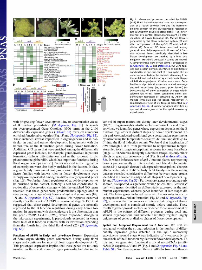
with progressing flower development due to accumulative effectsof B function perturbation (SI Appendix, Fig. S1). A searchfor overrepresented Gene Ontology (GO) terms in the 2,100differentially expressed genes (Dataset S1) revealed numerousenriched functional categories (Fig. 1F and SI Appendix, Fig. S2).These included several implicated in organogenesis and in par-ticular, in petal and stamen development, in agreement with theknown role of the B function genes during flower formation.Additional GO terms that were enriched among the differentiallyexpressed genes included, for example, genes involved in patternformation, cellular differentiation, and in the response to thephytohormone gibberellin, which has important functions duringfloral organ development (21). Genes involved in the regulationof transcription were also highly enriched in the dataset. In fact,a gene family enrichment analysis showed that transcriptionfactor families with known roles in flower development werestrongly overrepresented among the differentially expressed genes(Fig. 1G). We further found regulators of carpel development tobe enriched in the dataset. Notably, a test for coordinated di-rectionality of expression changes within the enriched GO termsrevealed that these genes were predominantly up-regulated invery young (i.e., stage ∼3–4) flowers of ap3 and pi mutants (Fig.1H and SI Appendix, Fig. S2). Because this response occurredshortly after the onset of AP3/PI expression at stage 3 (13, 14), itsuggested that these carpel developmental genes are normallyrepressed by the B function regulators during early flower for-mation. In agreement with this conjecture is the observation thatthe gene CRABS CLAW (CRC), which responded strongly inthe microarray experiments, is precociously expressed in youngfloral buds of B function mutants, with its expression expandingfrom the fourth into the third floral whorl (22) (SI Appendix,Fig. S3).
Functions of AP3/PI in Early- and Late-Stage Flowers. Expressionof floral organ identity genes commences during early floralstages and continues for most of floral organ development (3).This prolonged expression implies that these genes are not onlyinvolved in the specification of organ primordia but also in the
control of organ maturation during later developmental stages(10, 23). To gain insights into themolecular basis of these differentactivities, we identified genes whose expression depends on the Bfunction regulators at distinct stages of flower development. Tothis end, we conducted conditional gene perturbation experimentsby introducing the temperature-sensitive AP3 mutant allele ap3-1(4) into the floral induction system. We found that inactivation ofAP3 through a shift from permissive to nonpermissive temper-atures led to a strong transcriptional response in young floral buds(stage∼3–4), whereas, in slightly older buds (stage∼6), onlyminoreffects on gene expression occurred (Fig. 1I and SI Appendix, Fig.S2). In whole inflorescences of ap3-1 mutant plants, representingflowers predominantly of intermediate and late developmentalstages (24), we again detected widespread transcriptional changesafter a perturbation ofAP3 activity. A GO analysis of the resultingdatasets revealed considerable differences between gene groupsidentified as enriched at early and late stages of development (Fig.1F and SI Appendix, Fig. S2). Furthermore, genes responding earlyshowed, as expected, a significant overlap (P < 0.0001, Pearson χ2test) with genes identified as differentially expressed in the nullmutant experiments, whereas genes identified at late stages didnot. The latter genes included many with known roles in micro-sporogenesis (i.e., pollen formation; Fig. 1F and SI Appendix, Fig.S2), a process that commences at intermediate stages of flowerdevelopment and is completed shortly before anthesis. Theseresults together provide molecular evidence for an involvement ofAP3/PI in the control of different processes during petal andstamen organogenesis and indicate that they regulate largelyunique sets of genes at distinct phases of flower development.
Spatial and Temporal Requirement for B Function. We next in-vestigated whether the strong reduction in the number of differ-entially expressed genes detected in the ap3-1 microarrayexperiments around stage 6 was indicative of a temporarily re-duced role of the B function regulators in flower development. Tothis end, we generated functional artificial microRNAs (amiR-NAs) (25) against AP3 and PI (Fig. 2 and SI Appendix, Fig. S4 andTable S1). We then expressed these amiRNAs from an ethanol-
H
A B C D E
updo
wn
I40
020
00
200
400
600
num
ber
of g
enes
3-4 6 infl566
30149
385
29
335
GF
Stages
IM
regulation of transcriptioncarpel developmentflower developmentplant−type cell wall organizationpollen developmentclathrin coat assemblyprotein phosphorylationcell differentiationpollen wall assemblycellular process involved in reproductionstamen developmentphotosynthesisstomatal complex developmentresponse to light stimulusshoot developmentregionalizationadaxial/abaxial pattern specificationresponse to gibberellin stimuluspetal developmentovule developmentfloral organ morphogenesis
3-4 6 StagesIM 3-4 6 3-4 6 infl
ap3-3 pi-1 ap3-10 10 20
-log10(adjust. p-value) IM
B3 TF familyB3 domainREM TF familyHelix-loop-helix domainBasic helix-loop-helix TF familyHomeoboxHomeobox TF familyNPH3 domainC2H2 TF familyCytoplasmic ribosomal proteins familyDUF617GRAS TF domainGRAS TF familyBTB/POZ domainZinc finger, GATA-typeC2C2-GATA TF familyDUF623 (ovate family proteins)DUF584F−box, cyclin-likeC2H2 zinc finger
3-4 6 IM 3-4 6
ap3-3 pi-1
Stages
0 4 8-log10(adjust. p-value)
12
petal developmentstamen developmentfloral organ morphogenesisovule developmentadaxial/abaxial pattern specificationcarpel developmentpollen wall assembly
4 0 2 4
down up
2
Stages
ap3-3 pi-1
ap3-1
3-4 6 3-4 6 3-4 3-4 6 3-4 6 3-4
ap3-3 pi-1
ap3-1
Repression Activation
-log10(p-value)
Fig. 1. Genes and processes controlled by AP3/PI.(A–E) Floral induction system based on the expres-sion of a fusion between AP1 and the hormone-binding domain of the glucocorticoid receptor inap1 cauliflower double-mutant plants (19). Inflor-escences of a control plant (A) and a plant 6 d afterinduction of flower formation (B). Mature flowersgenerated by the floral induction system (C) andafter introgression of the ap3-3 (D) and pi-1 (E)alleles. (F) Selected GO terms enriched amonggenes differentially expressed in flowers of B func-tion mutants. Terms specifically identified in lateflower development are marked by a blue box.Benjamini–Hochberg-adjusted P values are shown.A comprehensive view of GO terms is presented inSI Appendix, Fig. S2 and Dataset S3. (G) Gene fam-ilies and protein domains identified as significantlyenriched (or in one case, that of F-box, cyclin-like, asunder-represented) in the datasets stemming fromthe ap3-3 and pi-1 microarray experiments. Benja-mini–Hochberg-adjusted P values are shown. Genefamilies and protein domains are labeled in orangeand red, respectively. (TF, transcription factor.) (H)Directionality of gene expression changes withinselected GO terms. Terms containing genes pre-dominantly repressed or activated by AP3/PI aremarked with blue and red boxes, respectively. Acomprehensive view of GO terms is presented in SIAppendix, Fig. S2. (I) Number of genes identified asup- and down-regulated in the ap3-1 microarrayexperiments.
Wuest et al. PNAS | August 14, 2012 | vol. 109 | no. 33 | 13453
PLANTBIOLO
GY
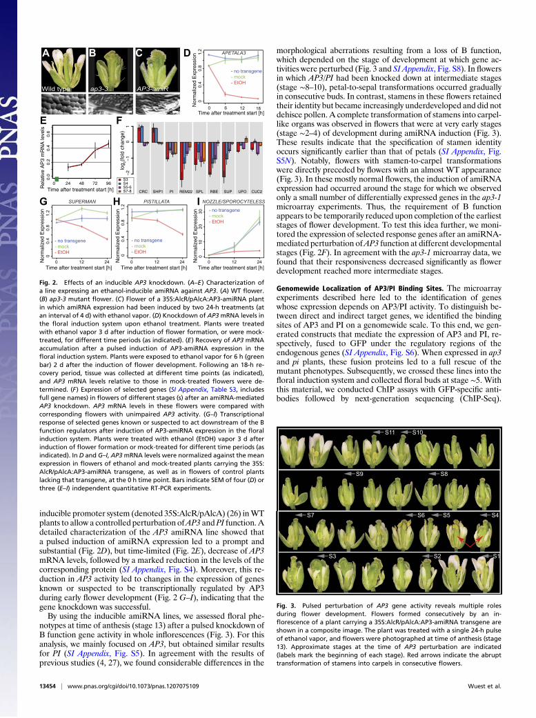
inducible promoter system (denoted 35S:AlcR/pAlcA) (26) inWTplants to allow a controlled perturbation ofAP3 and PI function. Adetailed characterization of the AP3 amiRNA line showed thata pulsed induction of amiRNA expression led to a prompt andsubstantial (Fig. 2D), but time-limited (Fig. 2E), decrease of AP3mRNA levels, followed by a marked reduction in the levels of thecorresponding protein (SI Appendix, Fig. S4). Moreover, this re-duction in AP3 activity led to changes in the expression of genesknown or suspected to be transcriptionally regulated by AP3during early flower development (Fig. 2 G–I), indicating that thegene knockdown was successful.By using the inducible amiRNA lines, we assessed floral phe-
notypes at time of anthesis (stage 13) after a pulsed knockdown ofB function gene activity in whole inflorescences (Fig. 3). For thisanalysis, we mainly focused on AP3, but obtained similar resultsfor PI (SI Appendix, Fig. S5). In agreement with the results ofprevious studies (4, 27), we found considerable differences in the
morphological aberrations resulting from a loss of B function,which depended on the stage of development at which gene ac-tivities were perturbed (Fig. 3 and SI Appendix, Fig. S8). In flowersin which AP3/PI had been knocked down at intermediate stages(stage ∼8–10), petal-to-sepal transformations occurred graduallyin consecutive buds. In contrast, stamens in these flowers retainedtheir identity but became increasingly underdeveloped and did notdehisce pollen. A complete transformation of stamens into carpel-like organs was observed in flowers that were at very early stages(stage ∼2–4) of development during amiRNA induction (Fig. 3).These results indicate that the specification of stamen identityoccurs significantly earlier than that of petals (SI Appendix, Fig.S5N). Notably, flowers with stamen-to-carpel transformationswere directly preceded by flowers with an almost WT appearance(Fig. 3). In these mostly normal flowers, the induction of amiRNAexpression had occurred around the stage for which we observedonly a small number of differentially expressed genes in the ap3-1microarray experiments. Thus, the requirement of B functionappears to be temporarily reduced upon completion of the earlieststages of flower development. To test this idea further, we moni-tored the expression of selected response genes after an amiRNA-mediated perturbation ofAP3 function at different developmentalstages (Fig. 2F). In agreement with the ap3-1 microarray data, wefound that their responsiveness decreased significantly as flowerdevelopment reached more intermediate stages.
Genomewide Localization of AP3/PI Binding Sites. The microarrayexperiments described here led to the identification of geneswhose expression depends on AP3/PI activity. To distinguish be-tween direct and indirect target genes, we identified the bindingsites of AP3 and PI on a genomewide scale. To this end, we gen-erated constructs that mediate the expression of AP3 and PI, re-spectively, fused to GFP under the regulatory regions of theendogenous genes (SI Appendix, Fig. S6). When expressed in ap3and pi plants, these fusion proteins led to a full rescue of themutant phenotypes. Subsequently, we crossed these lines into thefloral induction system and collected floral buds at stage ∼5. Withthis material, we conducted ChIP assays with GFP-specific anti-bodies followed by next-generation sequencing (ChIP-Seq).
Time after treatment start [h]
Nor
mal
ized
Exp
ress
ion
PISTILLATA
Nor
mal
ized
Exp
ress
ion
Time after treatment start [h]
- no transgene- mock- EtOH
NOZZLE/SPOROCYTELESS
Nor
mal
ized
Exp
ress
ion
Time after treatment start [h]
- no transgene- mock- EtOH
Time after treatment start [h]
Nor
mal
ized
Exp
ress
ion
Wild type
A
ap3-3
B
AP3-amiR
C D
G H ISUPERMAN
- no transgene- mock- EtOH
0 12 24
0.0.
41.
20.
8APETALA3
- no transgene- mock- EtOH
0 6
00.
40.
81.
2
12 18
0.0.
41.
20.
8
0 12 24 0 12 24
010
2030
S3S4S5-6S7-8
log 2(
fold
cha
nge)
−2
−1
01
CRC SHP1 PI REM22 SPL RBE SUP UFO CUC2
0.0
0.2
0.4
0.6
Time after treatment start [h]Rel
ativ
e A
P3
mR
NA
leve
ls
0 24 48 72 96
E F
Fig. 2. Effects of an inducible AP3 knockdown. (A–E) Characterization ofa line expressing an ethanol-inducible amiRNA against AP3. (A) WT flower.(B) ap3-3 mutant flower. (C) Flower of a 35S:AlcR/pAlcA:AP3-amiRNA plantin which amiRNA expression had been induced by two 24-h treatments (atan interval of 4 d) with ethanol vapor. (D) Knockdown of AP3mRNA levels inthe floral induction system upon ethanol treatment. Plants were treatedwith ethanol vapor 3 d after induction of flower formation, or were mock-treated, for different time periods (as indicated). (E) Recovery of AP3 mRNAaccumulation after a pulsed induction of AP3-amiRNA expression in thefloral induction system. Plants were exposed to ethanol vapor for 6 h (greenbar) 2 d after the induction of flower development. Following an 18-h re-covery period, tissue was collected at different time points (as indicated),and AP3 mRNA levels relative to those in mock-treated flowers were de-termined. (F) Expression of selected genes (SI Appendix, Table S3, includesfull gene names) in flowers of different stages (s) after an amiRNA-mediatedAP3 knockdown. AP3 mRNA levels in these flowers were compared withcorresponding flowers with unimpaired AP3 activity. (G–I) Transcriptionalresponse of selected genes known or suspected to act downstream of the Bfunction regulators after induction of AP3-amiRNA expression in the floralinduction system. Plants were treated with ethanol (EtOH) vapor 3 d afterinduction of flower formation or mock-treated for different time periods (asindicated). In D and G–I, AP3mRNA levels were normalized against the meanexpression in flowers of ethanol and mock-treated plants carrying the 35S:AlcR/pAlcA:AP3-amiRNA transgene, as well as in flowers of control plantslacking that transgene, at the 0 h time point. Bars indicate SEM of four (D) orthree (E–I) independent quantitative RT-PCR experiments.
S11 S10
S9 S8
S7 S6 S5 S4
S3 S2 S1
Fig. 3. Pulsed perturbation of AP3 gene activity reveals multiple rolesduring flower development. Flowers formed consecutively by an in-florescence of a plant carrying a 35S:AlcR/pAlcA:AP3-amiRNA transgene areshown in a composite image. The plant was treated with a single 24-h pulseof ethanol vapor, and flowers were photographed at time of anthesis (stage13). Approximate stages at the time of AP3 perturbation are indicated(labels mark the beginning of each stage). Red arrows indicate the abrupttransformation of stamens into carpels in consecutive flowers.
13454 | www.pnas.org/cgi/doi/10.1073/pnas.1207075109 Wuest et al.
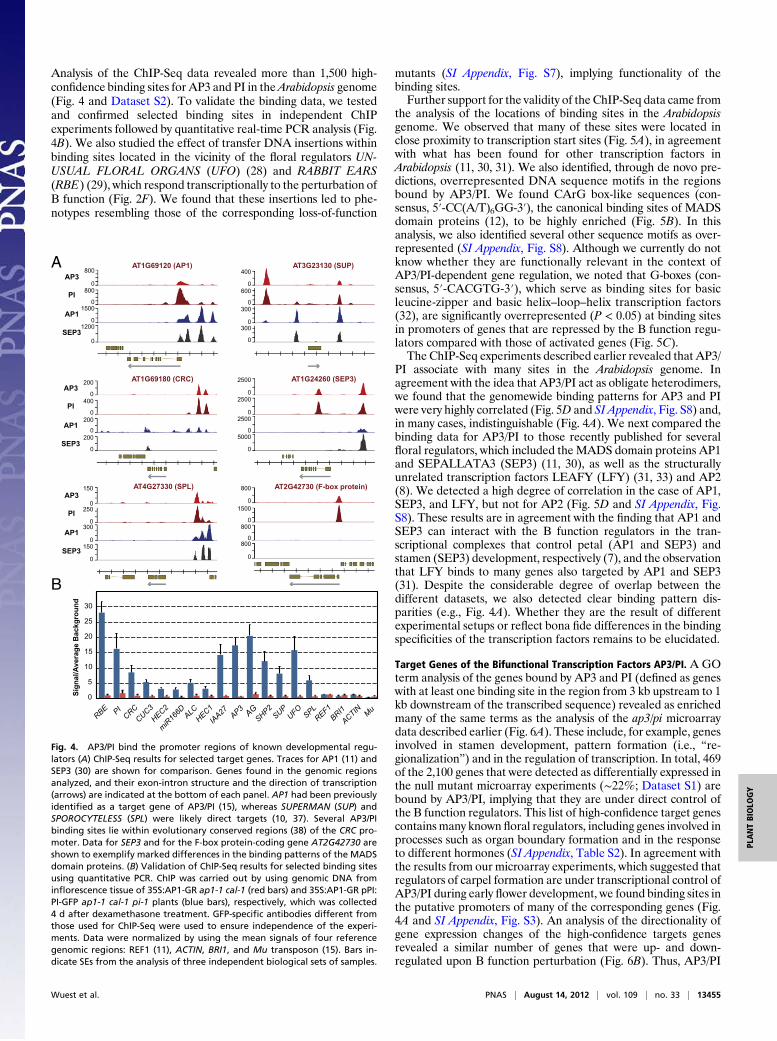
Analysis of the ChIP-Seq data revealed more than 1,500 high-confidence binding sites for AP3 and PI in theArabidopsis genome(Fig. 4 and Dataset S2). To validate the binding data, we testedand confirmed selected binding sites in independent ChIPexperiments followed by quantitative real-time PCR analysis (Fig.4B). We also studied the effect of transfer DNA insertions withinbinding sites located in the vicinity of the floral regulators UN-USUAL FLORAL ORGANS (UFO) (28) and RABBIT EARS(RBE) (29), which respond transcriptionally to the perturbation ofB function (Fig. 2F). We found that these insertions led to phe-notypes resembling those of the corresponding loss-of-function
mutants (SI Appendix, Fig. S7), implying functionality of thebinding sites.Further support for the validity of the ChIP-Seq data came from
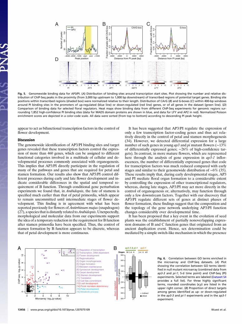
the analysis of the locations of binding sites in the Arabidopsisgenome. We observed that many of these sites were located inclose proximity to transcription start sites (Fig. 5A), in agreementwith what has been found for other transcription factors inArabidopsis (11, 30, 31). We also identified, through de novo pre-dictions, overrepresented DNA sequence motifs in the regionsbound by AP3/PI. We found CArG box-like sequences (con-sensus, 5′-CC(A/T)6GG-3′), the canonical binding sites of MADSdomain proteins (12), to be highly enriched (Fig. 5B). In thisanalysis, we also identified several other sequence motifs as over-represented (SI Appendix, Fig. S8). Although we currently do notknow whether they are functionally relevant in the context ofAP3/PI-dependent gene regulation, we noted that G-boxes (con-sensus, 5′-CACGTG-3′), which serve as binding sites for basicleucine-zipper and basic helix–loop–helix transcription factors(32), are significantly overrepresented (P < 0.05) at binding sitesin promoters of genes that are repressed by the B function regu-lators compared with those of activated genes (Fig. 5C).The ChIP-Seq experiments described earlier revealed that AP3/
PI associate with many sites in the Arabidopsis genome. Inagreement with the idea that AP3/PI act as obligate heterodimers,we found that the genomewide binding patterns for AP3 and PIwere very highly correlated (Fig. 5D and SI Appendix, Fig. S8) and,in many cases, indistinguishable (Fig. 4A). We next compared thebinding data for AP3/PI to those recently published for severalfloral regulators, which included theMADS domain proteins AP1and SEPALLATA3 (SEP3) (11, 30), as well as the structurallyunrelated transcription factors LEAFY (LFY) (31, 33) and AP2(8). We detected a high degree of correlation in the case of AP1,SEP3, and LFY, but not for AP2 (Fig. 5D and SI Appendix, Fig.S8). These results are in agreement with the finding that AP1 andSEP3 can interact with the B function regulators in the tran-scriptional complexes that control petal (AP1 and SEP3) andstamen (SEP3) development, respectively (7), and the observationthat LFY binds to many genes also targeted by AP1 and SEP3(31). Despite the considerable degree of overlap between thedifferent datasets, we also detected clear binding pattern dis-parities (e.g., Fig. 4A). Whether they are the result of differentexperimental setups or reflect bona fide differences in the bindingspecificities of the transcription factors remains to be elucidated.
Target Genes of the Bifunctional Transcription Factors AP3/PI. A GOterm analysis of the genes bound by AP3 and PI (defined as geneswith at least one binding site in the region from 3 kb upstream to 1kb downstream of the transcribed sequence) revealed as enrichedmany of the same terms as the analysis of the ap3/pi microarraydata described earlier (Fig. 6A). These include, for example, genesinvolved in stamen development, pattern formation (i.e., “re-gionalization”) and in the regulation of transcription. In total, 469of the 2,100 genes that were detected as differentially expressed inthe null mutant microarray experiments (∼22%; Dataset S1) arebound by AP3/PI, implying that they are under direct control ofthe B function regulators. This list of high-confidence target genescontains many known floral regulators, including genes involved inprocesses such as organ boundary formation and in the responseto different hormones (SI Appendix, Table S2). In agreement withthe results from our microarray experiments, which suggested thatregulators of carpel formation are under transcriptional control ofAP3/PI during early flower development, we found binding sites inthe putative promoters of many of the corresponding genes (Fig.4A and SI Appendix, Fig. S3). An analysis of the directionality ofgene expression changes of the high-confidence targets genesrevealed a similar number of genes that were up- and down-regulated upon B function perturbation (Fig. 6B). Thus, AP3/PI
AT1G69120 (AP1)
0
800AT3G23130 (SUP)
0
400
0
600
0
300
0
300
AP3
PI
AP1
SEP3
0
800
0
1500
0
1200
AP3
PI
AP1
SEP3
A
AT4G27330 (SPL)
0
150
0
250
0
300
0
150
AT1G69180 (CRC)
0
200
0
400
0
200
0
200
AT1G24260 (SEP3)
0
2500
0
2500
0
2500
0
5000
AT2G42730 (F-box protein)
0
800
0
1500
0
800
0
800
RBE
PI
CRC
CUC3
HEC2
miR
166D
ALC
HEC1
IAA27
AP3
AG
SHP2
SUP
UFO
SPL
REF1
BRI1
ACTIN
Mu
Sig
nal/A
verag
e B
ackg
ro
un
d
0
5
10
15
20
25
30
B
AP3
PI
AP1
SEP3
Fig. 4. AP3/PI bind the promoter regions of known developmental regu-lators (A) ChIP-Seq results for selected target genes. Traces for AP1 (11) andSEP3 (30) are shown for comparison. Genes found in the genomic regionsanalyzed, and their exon-intron structure and the direction of transcription(arrows) are indicated at the bottom of each panel. AP1 had been previouslyidentified as a target gene of AP3/PI (15), whereas SUPERMAN (SUP) andSPOROCYTELESS (SPL) were likely direct targets (10, 37). Several AP3/PIbinding sites lie within evolutionary conserved regions (38) of the CRC pro-moter. Data for SEP3 and for the F-box protein-coding gene AT2G42730 areshown to exemplify marked differences in the binding patterns of the MADSdomain proteins. (B) Validation of ChIP-Seq results for selected binding sitesusing quantitative PCR. ChIP was carried out by using genomic DNA frominflorescence tissue of 35S:AP1-GR ap1-1 cal-1 (red bars) and 35S:AP1-GR pPI:PI-GFP ap1-1 cal-1 pi-1 plants (blue bars), respectively, which was collected4 d after dexamethasone treatment. GFP-specific antibodies different fromthose used for ChIP-Seq were used to ensure independence of the experi-ments. Data were normalized by using the mean signals of four referencegenomic regions: REF1 (11), ACTIN, BRI1, and Mu transposon (15). Bars in-dicate SEs from the analysis of three independent biological sets of samples.
Wuest et al. PNAS | August 14, 2012 | vol. 109 | no. 33 | 13455
PLANTBIOLO
GY
appear to act as bifunctional transcription factors in the control offlower development.
DiscussionThe genomewide identification of AP3/PI binding sites and targetgenes revealed that these transcription factors control the expres-sion of more than 460 genes, which can be assigned to differentfunctional categories involved in a multitude of cellular and de-velopmental processes commonly associated with organogenesis.This implies that AP3/PI directly participate in the regulation ofmany of the pathways and genes that are required for petal andstamen formation. Our results also show that AP3/PI control dif-ferent processes during early and late flower development and in-dicate considerable differences in the spatial and temporal re-quirement of B function. Through conditional gene perturbationexperiments we found that, in Arabidopsis, the fate of stamens isspecified much earlier than that of petal primordia, which appearto remain uncommitted until intermediate stages of flower de-velopment. This finding is in agreement with what has beenreported previously for flowers of Antirrhinum majus (snapdragon)(27), a species that is distantly related toArabidopsis. Unexpectedly,morphological and molecular data from our experiments supportthe idea of a temporary reduction in the requirement for B functionafter stamen primordia have been specified. Thus, the control ofstamen formation by B function appears to be discrete, whereasthat of petal development is more continuous.
It has been suggested that AP3/PI regulate the expression ofonly a few transcription factor-coding genes and thus act rela-tively directly in the control of petal and stamen morphogenesis(34). However, we detected differential expression for a largenumber of such genes in young ap3 and pi mutant flowers (∼13%of differentially expressed genes; ∼26% of high-confidence tar-gets). In contrast, in more mature flowers, which are representedhere through the analysis of gene expression in ap3-1 inflor-escences, the number of differentially expressed genes that codefor transcription factors was much reduced compared with earlystages and similar to their genomewide distribution of ∼6% (35).These results imply that, during early developmental stages, AP3and PI mediate floral organ formation to a considerable extentby controlling the expression of other transcriptional regulators,whereas, during late stages, AP3/PI may act more directly in thecontrol of organogenesis or, alternatively, may function throughonly a few downstream factors. Together with our discovery thatAP3/PI regulate different sets of genes at distinct phases offlower formation, these findings suggest that the composition andthe topology of the gene network underlying AP3/PI functionchanges considerably over developmental time.It has been proposed that a key event in the evolution of seed
plants was the establishment of partially nonoverlapping expres-sion domains of B and C function genes that originated from anancient duplication event. Hence, sex determination could bemediated by a simple switch-likemechanism in which the presence
Peak position
5010
015
0
Num
ber o
f pea
ks
−3000 −2000 −1000 start end +1000
PIAP3
PI
peak-5kb 5kb
0 1 2 3 4 >5AP3
peak-5kb 5kb
0 1 2 3 4 >5AP1
peak-5kb 5kb
0 1 2 3 4 >5SEP3
peak-5kb 5kb
0 1 2 3 4 >5LFY
peak-5kb 5kb
0 1 2 3 4 >5AP2
peak-5kb 5kb
0 1 2 3 4 >5
CBA
D
0.2
0.3
0.4
0.5
0.6
Distance from PI peak [bp]Frac
tion
of p
eaks
with
CA
rG-b
ox
-4000 -2000 0 2000 4000
CArG-box
0.05
0.1
0.15
0.2
Distance from PI peak [bp]
Frac
tion
of p
eaks
with
G-b
ox
-4000 -2000 0 2000 4000
G-box
Fig. 5. Genomewide binding data for AP3/PI. (A) Distribution of binding sites around transcription start sites. Plot showing the number and relative dis-tribution of ChIP-Seq peaks in the proximity (from 3,000 bp upstream to 1,000 bp downstream) of transcribed regions of potential target genes. Binding sitepositions within transcribed regions (shaded box) were normalized relative to their length. Distribution of CArG (B) and G-boxes (C) within 400-bp windowsaround PI binding sites in the promoters of up-regulated (blue line) or down-regulated (red line) genes, or of all genes in the dataset (green line). (D)Comparison of binding data for selected floral regulators. Heat maps show binding data from different ChIP-Seq experiments for genomic regions sur-rounding 1,852 high-confidence PI binding sites (data for MADS domain proteins are shown in blue, and data for LFY and AP2 in red). Normalized Poissonenrichment scores are depicted in a color-code scale. All data were sorted (from top to bottom) according to descending PI peak height.
BA ap3-3 & pi-1 ap3-1
Stages IM 3-4 6 3-4 6 infl
up-r
egul
ated
dow
n-re
gula
ted
0 2 4 6 8 10
02
46
810
Microarray -log10(p-value)
ChI
P-s
eq -
log 1
0(p-v
alue
)
[13] Leaf development
[16] Metabolic process
[1] Regulation of transcription
800
600
400
200
0
200
400
600
800
7
29
21
19
1627
15
25
27
15
7
5
indirectdirect
num
ber o
f gen
es
[2] Transcription, DNA-dependent[3] Flower development[4] Regionalization[5] Organ morphogenesis[6] Organ formation[7] Meristem maintenance[8] Gynoecium development[9] Carpel development[10] Stamen development[11] Translation[12] Cell fate commitment
[14] Floral meristem determinacy[15] Petal development
[3][1][2]
(33, 27)
(32, 25)(13, 22)
[4]
[5]
[6]
[7]
[8]
[9][10]
[11][12]
[13]
[14]
[15] [16]
Fig. 6. Correlation between GO terms enriched inthe microarray and ChIP-Seq datasets. (A) Plotshowing the correlation between GO terms identi-fied in null mutant microarray (combined data fromap3-3 and pi-1; 5-d time point) and ChIP-Seq (PI)experiments. Selected terms are labeled (Dataset S4provides a full list). For three highly significantterms, rounded coordinates (x;y) are listed in theupper right corner. (B) Proportion of direct targetsamong genes identified as up- or down-regulatedin the ap3-3 and pi-1 experiments and in the ap3-1experiment.
13456 | www.pnas.org/cgi/doi/10.1073/pnas.1207075109 Wuest et al.
or absence of B function results in the formation of male or femalestructures, respectively (36). Our results show that, in Arabidopsis,the B function regulators AP3/PI are bifunctional transcriptionfactors that activate or repress downstream targets, thus acting asa context-dependent transcriptional switch. In some cases, thisappears to happen in concert with C function, for example, in theactivation of the key regulator of microsporogenesis, SPL (Figs. 2Iand 4A) (10). On the contrary, we found that AP3/PI suppressgenes, such as those involved in carpel development (e.g., CRC),which are otherwise activated by C function (9), implying opposingregulatory roles. Based on these results, we propose that themolecular mechanism for sex-determination in seed plantsdepends on the interplay of synergistic and antagonistic inter-actions between B and C function regulators.
Materials and MethodsPreviously published Arabidopsis strains used in this study included the fol-lowing: 35S:AP1-GR ap1-1 cal-1 (19), ap3-1 (4), ap3-3 (14), and pi-1 (4). Plantswere grown on a soil:vermiculite:perlite (3:1:1) mixture at 20 °C under
constant illumination with cool white fluorescent light unless indicatedotherwise. Cloning strategies and methods used for PCR-based genotypingof mutant alleles and transgenes are described in SI Appendix. Plant trans-formations, quantitative RT-PCR assays, and in situ hybridizations werecarried out as outlined in SI Appendix. Transcript profiling experiments weredone by using custom microarrays (Agilent Technologies) and a previouslydescribed protocol (11). ChIP-Seq analysis was performed, with minor mod-ifications, as described previously (11). Analysis of microarray and ChIP-Seqdata were done using custom R scripts and functions provided by the Bio-conductor project. Detailed experimental and data analysis procedures areprovided in SI Appendix.
ACKNOWLEDGMENTS. The authors thank J. L. Riechmann for sharing thedesign of the Agilent microarrays, T. Kavanagh and M. Ramaswami forsharing equipment, J. Muiño for advice on data analysis, and J. L. Riechmannand J. Goodrich for comments on the manuscript. This study was supportedby Science Foundation Ireland Grants 09/SIRG/B1600 (to E.G.), 07/IN.1/B851(to F.W.), and 10/IN.1/B2971 (to F.W.). A.R. was supported by a scholarshipfrom the Irish Research Council for Science, Engineering and Technology.E.G. was supported by a long-term fellowship from the European MolecularBiology Organization.
1. Lohmann JU, Weigel D (2002) Building beauty: The genetic control of floral pat-terning. Dev Cell 2:135–142.
2. Jack T (2004) Molecular and genetic mechanisms of floral control. Plant Cell 16(Suppl):S1–S17.
3. Krizek BA, Fletcher JC (2005) Molecular mechanisms of flower development: Anarmchair guide. Nat Rev Genet 6:688–698.
4. Bowman JL, Smyth DR, Meyerowitz EM (1989) Genes directing flower development inArabidopsis. Plant Cell 1:37–52.
5. Coen ES, Meyerowitz EM (1991) The war of the whorls: Genetic interactions con-trolling flower development. Nature 353:31–37.
6. Goto K, Kyozuka J, Bowman JL (2001) Turning floral organs into leaves, leaves intofloral organs. Curr Opin Genet Dev 11:449–456.
7. Smaczniak C, et al. (2012) Characterization of MADS-domain transcription factorcomplexes in Arabidopsis flower development. Proc Natl Acad Sci USA 109:1560–1565.
8. Yant L, et al. (2010) Orchestration of the floral transition and floral development inArabidopsis by the bifunctional transcription factor APETALA2. Plant Cell 22:2156–2170.
9. Gómez-Mena C, de Folter S, Costa MM, Angenent GC, Sablowski R (2005) Transcrip-tional program controlled by the floral homeotic gene AGAMOUS during early or-ganogenesis. Development 132:429–438.
10. Ito T, et al. (2004) The homeotic protein AGAMOUS controls microsporogenesis byregulation of SPOROCYTELESS. Nature 430:356–360.
11. Kaufmann K, et al. (2010) Orchestration of floral initiation by APETALA1. Science 328:85–89.
12. Riechmann JL, Krizek BA, Meyerowitz EM (1996) Dimerization specificity of Arabi-dopsis MADS domain homeotic proteins APETALA1, APETALA3, PISTILLATA, andAGAMOUS. Proc Natl Acad Sci USA 93:4793–4798.
13. Goto K, Meyerowitz EM (1994) Function and regulation of the Arabidopsis floralhomeotic gene PISTILLATA. Genes Dev 8:1548–1560.
14. Jack T, Brockman LL, Meyerowitz EM (1992) The homeotic gene APETALA3 of Ara-bidopsis thaliana encodes a MADS box and is expressed in petals and stamens. Cell 68:683–697.
15. Sundström JF, Nakayama N, Glimelius K, Irish VF (2006) Direct regulation of the floralhomeotic APETALA1 gene by APETALA3 and PISTILLATA in Arabidopsis. Plant J 46:593–600.
16. Sablowski RW, Meyerowitz EM (1998) A homolog of NO APICAL MERISTEM is animmediate target of the floral homeotic genes APETALA3/PISTILLATA. Cell 92:93–103.
17. Mara CD, Irish VF (2008) Two GATA transcription factors are downstream effectors offloral homeotic gene action in Arabidopsis. Plant Physiol 147:707–718.
18. Mara CD, Huang T, Irish VF (2010) The Arabidopsis floral homeotic proteins APE-TALA3 and PISTILLATA negatively regulate the BANQUO genes implicated in lightsignaling. Plant Cell 22:690–702.
19. Wellmer F, Alves-Ferreira M, Dubois A, Riechmann JL, Meyerowitz EM (2006) Ge-nome-wide analysis of gene expression during early Arabidopsis flower development.PLoS Genet 2:e117.
20. Smyth DR, Bowman JL, Meyerowitz EM (1990) Early flower development in Arabi-dopsis. Plant Cell 2:755–767.
21. Plackett AR, Thomas SG, Wilson ZA, Hedden P (2011) Gibberellin control of stamendevelopment: A fertile field. Trends Plant Sci 16:568–578.
22. Bowman JL, Smyth DR (1999) CRABS CLAW, a gene that regulates carpel and nectarydevelopment in Arabidopsis, encodes a novel protein with zinc finger and helix-loop-helix domains. Development 126:2387–2396.
23. Ito T, Ng KH, Lim TS, Yu H, Meyerowitz EM (2007) The homeotic protein AGAMOUScontrols late stamen development by regulating a jasmonate biosynthetic gene inArabidopsis. Plant Cell 19:3516–3529.
24. Wellmer F, Riechmann JL, Alves-Ferreira M, Meyerowitz EM (2004) Genome-wideanalysis of spatial gene expression in Arabidopsis flowers. Plant Cell 16:1314–1326.
25. Schwab R, Ossowski S, Riester M, Warthmann N, Weigel D (2006) Highly specific genesilencing by artificial microRNAs in Arabidopsis. Plant Cell 18:1121–1133.
26. Deveaux Y, et al. (2003) The ethanol switch: A tool for tissue-specific gene inductionduring plant development. Plant J 36:918–930.
27. Zachgo S, et al. (1995) Functional analysis of the Antirrhinum floral homeotic DEFI-CIENS gene in vivo and in vitro by using a temperature-sensitive mutant. De-velopment 121:2861–2875.
28. Lee I, Wolfe DS, Nilsson O, Weigel D (1997) A LEAFY co-regulator encoded by UN-USUAL FLORAL ORGANS. Curr Biol 7:95–104.
29. Takeda S, Matsumoto N, Okada K (2004) RABBIT EARS, encoding a SUPERMAN-likezinc finger protein, regulates petal development in Arabidopsis thaliana. De-velopment 131:425–434.
30. Kaufmann K, et al. (2009) Target genes of the MADS transcription factor SE-PALLATA3: Integration of developmental and hormonal pathways in the Arabidopsisflower. PLoS Biol 7:e1000090.
31. Winter CM, et al. (2011) LEAFY target genes reveal floral regulatory logic, cis motifs,and a link to biotic stimulus response. Dev Cell 20:430–443.
32. Sibéril Y, Doireau P, Gantet P (2001) Plant bZIP G-box binding factors. Modularstructure and activation mechanisms. Eur J Biochem 268:5655–5666.
33. Moyroud E, et al. (2011) Prediction of regulatory interactions from genome sequencesusing a biophysical model for the Arabidopsis LEAFY transcription factor. Plant Cell23:1293–1306.
34. Zik M, Irish VF (2003) Global identification of target genes regulated by APETALA3and PISTILLATA floral homeotic gene action. Plant Cell 15:207–222.
35. Riechmann JL, et al. (2000) Arabidopsis transcription factors: Genome-wide compar-ative analysis among eukaryotes. Science 290:2105–2110.
36. Theissen G, Melzer R (2007) Molecular mechanisms underlying origin and di-versification of the angiosperm flower. Ann Bot (Lond) 100:603–619.
37. Sakai H, Krizek BA, Jacobsen SE, Meyerowitz EM (2000) Regulation of SUP expressionidentifies multiple regulators involved in Arabidopsis floral meristem development.Plant Cell 12:1607–1618.
38. Lee JY, et al. (2005) Activation of CRABS CLAW in the Nectaries and Carpels ofArabidopsis. Plant Cell 17:25–36.
Wuest et al. PNAS | August 14, 2012 | vol. 109 | no. 33 | 13457
PLANTBIOLO
GY