molecular characterization of colletotrichum ... filemolecular characterization of colletotrichum...
TRANSCRIPT
International Society for Tropical Root Crops (ISTRC) 157
Molecular characterization of Colletotrichum gloeosporioides responsible for anthracnose of yam and cassava in Nigeria, and development a diagnostic PCR assay
Kamal Sharma, M. Ayodele, R. Bandyopadhyay, R. Asiedu and P. Lava Kumar*
International Institute of Tropical Agriculture (IITA), Oyo Road, PMB 5320, Ibadan, Nigeria Corresponding author: [email protected]
Anthracnose, caused by Colletotrichum gloeosporioides Penz, is one the major diseases of yam and cassava in West Africa. Thirteen isolates, ten from yam and three from cassava, isolated in Nigeria were classified based on their symptoms and morphological characters into two groups: spot (S) and blight (B). Both S and B isolates infect yam, but only B isolates infect cassava. All these isolates were analyzed by nucleotide sequencing of nuclear ribosomal internal transcribed spacer (ITS) sequence (ITS-1, 5.8S and ITS-2), actin and histone genes. Phylogenetic analysis of the three gene sequence data grouped these 13 isolates into two major clades. Both S and B isolates distributed between the two clades, with all the three cassava infecting isolates formed a separate sub-clade. Further studies are underway to assess the implication of S and B isolates on host resistance.
A set of primers was designed from the nucleotide sequences of rDNA ITS region for specific detection of yam and cassava infecting C. gloeosporioides isolates. A simple method for extracting DNA from mycelia, leaf, stem and roots was established for C. gloeosporioides diagnostic PCR, which was validated by testing 36 C. gloeosporioides isolates from yam and 85 isolates from cassava. Specific product (500 bp) was obtained from all the yam and cassava isolates but not in C. gloeosporioides isolate from soybean indicating the high-specificity of the primers. This assay can is useful in diagnosis of C. gloeosporioides of yam and cassava.
Introduction
Nigeria rank first for production of yam in Africa and for cassava in World (FAO, 2007). These serve as staple food for millions of people in Nigeria. These crops are infected by several pathogens i.e. virus, fungus and bacteria. Among fungal Colletotrichum gloeosporioides are most important. This pathogen may once it appears in the field can destroy all susceptible variety and gave a blighted appearance to the field in few days.
A combination of techniques (culture on agar plates, colony morphology, growth on selective media, and various biochemical reactions) is used for the identification of fungi and are used to detect specific fungi or its strains (Jyan et al., 2002). The accuracy and reliability of the conventional methods depend largely on the experience and skill of the person making the diagnosis. This approach is time consuming (several days to weeks) and impractical to analyze large number of samples. Further, such methods are unsuitable for reliable identification of new strains or species that may not show distinctive phenotypic or physiological features. With the advent of biotechnology, molecular methods, such as polymerase chain reaction (PCR), are fast establishing as a choice method for routine identification of fungal pathogens (Henson and French, 1993). These so called modern methods are rapid, highly specific and can be used to detect minute quantities of fungal DNA from environmental samples before symptoms occur, therefore, allowing implementation of early control methods. PCR technology can also provide very accurate quantitative data supplying the necessary additional information required for control and quarantine decisions and for assessing how effective fungal agents are in the case of biological control (Atkins and Clark, 2004). These methods exploit genes such as rDNA, tubulin, the most and know to be conserved in a wide array of fungal species.
We design the primer based on rDNA region for gloeosporioides specific for diagnostic purposes and for Phylogenetic analysis. The method was tested for 121 strains of yam and cassava and 21 strains of other fungi to show the specificity of primer. The primer only amplified a specific band with yam and cassava strain.
Materials and methods
The C. gloeosporioides isolates are of dioscorea and cassava were received from Pathology and Germplasm Health Unit. The fungi cultured on potatao dextrose agar (PDA) at 28 °C. The 5 mm diameter for each isolate
158 15th Triennial ISTRC Symposium
incubated in rotary shaker at 100 rpm at 28°C. The mycelial mass of each isolate was harvested by filteration through sterile filter paper and used immediately for DNA extractions.
Genomic DNA extractions
Total genomic DNA extraction protocol developed in our lab and this was used in all tests performed. The 100 mg mycelia was grinded with mortar and pestle and 1.5 ml of extraction buffer {(100 mM Tris (pH 8), 10 mM EDTA (pH 8), SDS 1%, PVP 2% and 1 M NaCl. Autoclave and add 1% β-mercaptoethanol just before use} added containing proteinase K incubated to at 65 °C for 30 minutes in water bath. Bring the tubes out and cool it to room temperature for 3-5 minutes and centrifuge for 13,000 rpm for 10 minutes. Transfer the supernatant to new tubes and add 200 μl of PEG. It was observed that incubating the tubes at 4°C for 10 minutes improve the yield. Centrifuge at 13,000 rpm in microcentrifuge and transfer the supernatant to new fresh tubes. Add 2/3 volume isopropanol and incubate the tube at -20°C for 1 hour to precipitate DNA. Centrifuge at 13,000 rpm for 10 min. at 4 °C, remove supernatant, and wash the DNA pellet with 70% Ethanol, air dry, and resuspend in 50-100 μl of TE (10 mM Tris (pH 8) and 1 mM EDTA (pH 8) buffer. (It was observed that centrifugation at room temperature doesn’t effect the quality of DNA). Remove RNA by adding 1 μl of RNase (10 mg/ml) incubated at 37°C for 30 minutes, and stored at -20°C until use.
PCR amplification and sequencing of ITS, Actin and Histone region
The 10 type isolate of yam and 3 type isolate of cassava were characterized by nucleotide sequence analysis. For this, the 18S ribosomal RNA gene partial, ITS1 1, 5.8S ribosomal RNA gene, ITS 2 complete and 28S ribosomal RNA partial gene sequence, regions were amplified with the universal primers ITS1 and ITS4 (White et al., 1990). Histone H3 is used for phylogenetic analysis with primers CylH3F and CylH3R (Crous et al., 2004) and actin region was amplified using primers Act512 and Act 783R (Carbone and Kohn, 1999). The PCR reaction (25 μL final volume) reaction mixture consisted of 5.0 μL 5x PCR buffer (Promega, Madison, WI, USA), 25 mM MgCl2, 10 mM dNTPs, 10 pmol of each primer, 0.5 units of Taq polymerase (Promega) and 4.0 to 10 ng of template DNA. PCR conditions for the ITS were, after the initial denaturation at 94 °C for 5 min, 35 PCR cycles were performed with 30 sec at 94 °C, 1min at 55 °C and 1.5 min at 72 °C. After the last cycle, the temperature was maintained at 72 °C for 8 min and for actin and Histone annealing temperature used 58 °C for 30 sec and extension for 1 minute. DNA amplification was performed in a MJ Research Thermal Cycler. PCR products were visualized by electrophoresis in 1.5% (wt/vol) agarose gels run 1 X Tris-acetate-EDTA (TAE) buffer stained with ethidium bromide and visualized under UV light. DNA sequencing was performed with two primers (ITS1 and ITS4) in both directions to ensure that there was no misreading. PCR products were purified and sequenced by (BangloGenei, India). Alignment and edition were carried out with the BioEdit Program v 7.0.5 (Hall, 2005) and visually corrected. Sequences were then compared against those available in the GenBank database.
Diagnostics PCR with Strain-Specific primer
The primer YA(I)R (5’-GTTACTACGCAAAGGAGG-3’) specific for gloeosporioides for yam and cassava was used in conjunction with the conserved primer ITS1 (White et al., 1990) for rDNA amplification. The PCR reaction and condition are same as explained above PCR products were visualized by electrophoresis in 1.5% (wt/vol) agarose gels run 1 X Tris-acetate-EDTA (TAE) buffer stained with ethidium bromide and visualized under UV light.
Results
PCR amplification and sequencing of ITS region
The DNA of fungal isolates 13 from yam and 3 from cassava were used in PCR with the Universal primers ITS1 and ITS4 for the amplification of the rDNA region comprising the two non-coding internal transcribed spacers ITS1 and ITS2 and the 5.8S rRNA gene, partial 18S and 28S. All isolates amplified a PCR product of approximately 550 bp. Sequences were determined of each isolate, and blast analyses were carried out for 13 isolates, previously identified by morphological and biochemical study. The results showed a 99-100% homology with DNA sequences from other C. gloeosporioides strains deposited in the GeneBank.
International Society for Tropical Root Crops (ISTRC) 159
PCR amplification and sequencing of Actin and Histone region
The DNA of fungal isolates 13 from yam and 3 from cassava were used in PCR with the primers Act512 and Act783 for the amplification of the actin region and primers CylH3F and CylH3R for the amplification of Histone3 region. All isolates amplified a PCR product of approximately 270 bp with actin and 390 bp with histone. Sequences were determined of each isolate, and blast analyses were carried out for 13 isolates. Since there are no more sequences deposited in NCBI for C. gloeosporioides based on Histone and Actin there was no homology determined.
Diagnostics PCR with Strain-Specific primer
With PCR testing of gloeosporioides from fungal strains yam and cassava with primer pairs YA(I)R and ITS1, 500 bp band was successfully detected in all strains. Among the samples tested. To show the specificity of primer other fungal and bacterial strains were tested and no band was detected.
Discussions
The purpose of this research was to perform phylogenetic analysis of the C. gloeosporioides and develop data on the number of nucleotide differences between blight and spot causing strains to yam and cassava and to determine whether spot and blight represent different species. The another aim of this research was to use PCR-based methods to identify rapidly and accurately the species of gloeosporioides responsible for anthracnose in yam and cassava in Nigeria.
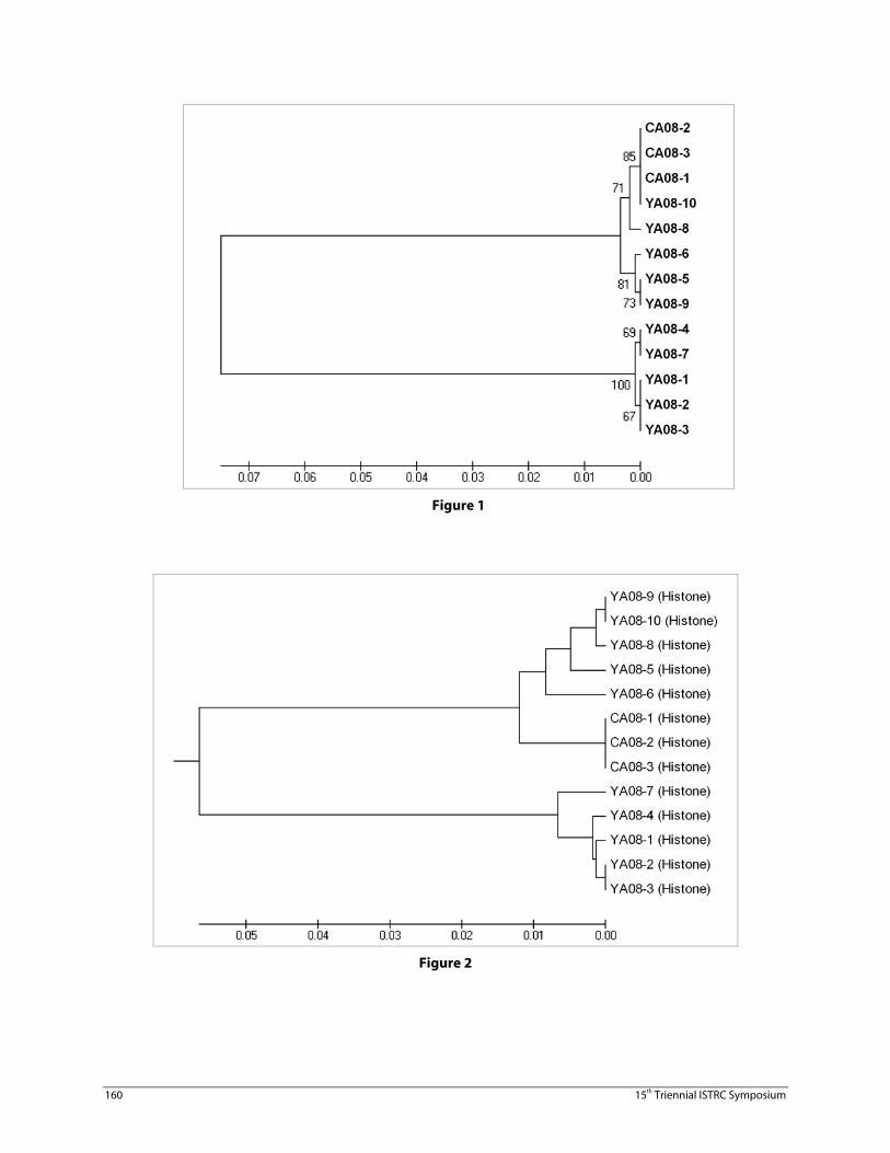
The size of the amplification product 500 bp confirmed with specific designed primer. PCR with ITS universal primers amplification of the 18S-5.8S-ITS-28S region of DNA, subsequent sequence analysis of the rDNA product revealed clearly the existence of two strains causing anthracnose lesions on yam. PCR amplification of the three target genes, rDNA ITS resulted in DNA bands of expected size from all the isolates ~550bp, actin 270 bp and histone 390 bp. The amplified fragments were sequenced and data was analyzed using bioinformatics packages, BioEdit and MEGA. The 13 isolates clustered into two major groups in a phylogenetic tree inferred from rDNA sequence information, with the isolates from cassava forming a sub-group in one of the major clades along with one of Yam strain (YA08-10) (Figure 1) suggesting that yam and cassava strain are genetically very close. Phylogenetic analysis revealed the composition of subclades by blight and spot isolates from Nigeria. This may be a consequence of intermatings between isolates introduced from distant regions and the resident isolates of the respective regions.
160 15th Triennial ISTRC Symposium
Figure 1
Figure 2
International Society for Tropical Root Crops (ISTRC) 161
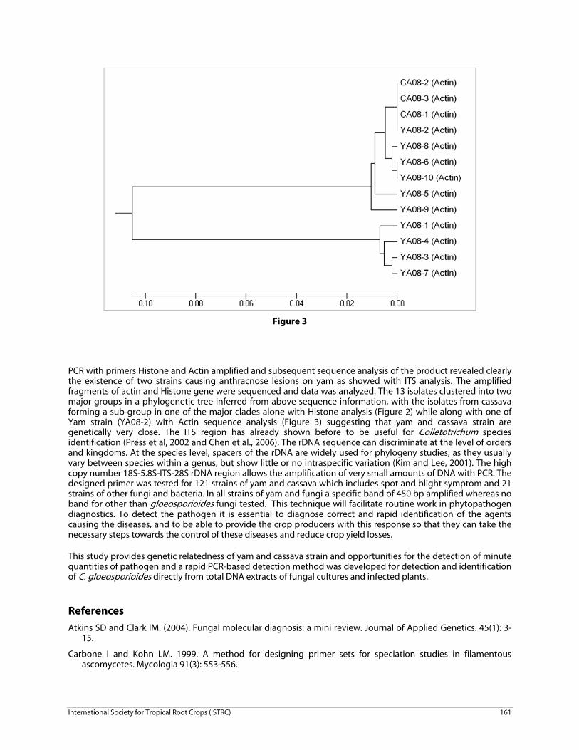
Figure 3
PCR with primers Histone and Actin amplified and subsequent sequence analysis of the product revealed clearly the existence of two strains causing anthracnose lesions on yam as showed with ITS analysis. The amplified fragments of actin and Histone gene were sequenced and data was analyzed. The 13 isolates clustered into two major groups in a phylogenetic tree inferred from above sequence information, with the isolates from cassava forming a sub-group in one of the major clades alone with Histone analysis (Figure 2) while along with one of Yam strain (YA08-2) with Actin sequence analysis (Figure 3) suggesting that yam and cassava strain are genetically very close. The ITS region has already shown before to be useful for Colletotrichum species identification (Press et al, 2002 and Chen et al., 2006). The rDNA sequence can discriminate at the level of orders and kingdoms. At the species level, spacers of the rDNA are widely used for phylogeny studies, as they usually vary between species within a genus, but show little or no intraspecific variation (Kim and Lee, 2001). The high copy number 18S-5.8S-ITS-28S rDNA region allows the amplification of very small amounts of DNA with PCR. The designed primer was tested for 121 strains of yam and cassava which includes spot and blight symptom and 21 strains of other fungi and bacteria. In all strains of yam and fungi a specific band of 450 bp amplified whereas no band for other than gloeosporioides fungi tested. This technique will facilitate routine work in phytopathogen diagnostics. To detect the pathogen it is essential to diagnose correct and rapid identification of the agents causing the diseases, and to be able to provide the crop producers with this response so that they can take the necessary steps towards the control of these diseases and reduce crop yield losses.
This study provides genetic relatedness of yam and cassava strain and opportunities for the detection of minute quantities of pathogen and a rapid PCR-based detection method was developed for detection and identification of C. gloeosporioides directly from total DNA extracts of fungal cultures and infected plants.
References
Atkins SD and Clark IM. (2004). Fungal molecular diagnosis: a mini review. Journal of Applied Genetics. 45(1): 3-15.
Carbone I and Kohn LM. 1999. A method for designing primer sets for speciation studies in filamentous ascomycetes. Mycologia 91(3): 553-556.
162 15th Triennial ISTRC Symposium
Chen, L. S., Chu, C., Liu, C. D., Chen, R. S., & Tsay, J. G. (2006). PCR-based detection and differentiation of anthracnose pathogens, Colletotrichum gloeosporioides and C. truncatum, from vegetable soybean in Taiwan. Journal of Phytopathology, 154, 654–662.
Crous PW, Groenewald JZ, Risède JM, Simoneau P
and Hywel-Jones NL. 2004.
Calonectria species and their Cylindrocladium anamorphs: species with sphaeropedunculate vesicles. Studies in Mycology 50: 415–430.
Hall, T. (2005).BioEdit v 7.0.5. http://www.mbio.ncsu.edu/BioEdit/.
Henson JM. And French R. (1993). The polymerase chain reaction and plant disease diagnosis. Annual review of phytopathology. 31: 81-109.
Jyan M.H., Huang LC., Ann PJ and Liou RF. (2002). Rapid detection of Phytophthora infestans by PCR. Plant Pathology Bulletin. 11: 45-56.
Kang HW, Choyg, Yoon UH and Eun MY (1998). A Rapid DNA Extraction Method for RFLP and PCR Analysis from a Single Dry Seed. Plant Mol Biol Rep. 16:1–9.
Kim HJ and Lee YS (2001). Development of an In Planta Molecular Marker for the Detection of Chinese Cabbage (Brassica campestris ssp. pekinensis) Club Root Pathogen Plasmodiophora brassicae. The Journal of Microbiology, 39(1): 56-61.
Peres, N. A. R., Kuramae, E. E., Dias, M. S. C., & de Sousa, N. L. (2002). Identification and characterization of Colletotrichum spp affecting fruit after harvest in Brazil. Journal of Phytopathology, 150, 128–134
White TJ, Bruns T, Lee S, and Taylor J. (1990). Amplification and direct sequencing of fungal ribosomal RNA genes for phylogenetics. In Innis M, Gelfand D, Sninsky J, and White T. (Eds.), PCR protocols, a guide to methods and applications (pp. 315–322). San Diego CA USA: Academic Press.
Wulff EG, Torres S and Vigil EG (2002). Protocol for DNA Extraction from potato tubers. Plant Mol Biol Rep. 20:187a–187e.