molecular diagnostic methods recombinant dna technology recombinant dna technology
TRANSCRIPT
Molecular Diagnostic Molecular Diagnostic MethodsMethods
Molecular Diagnostic Molecular Diagnostic MethodsMethods
Recombinant DNA TechnologyRecombinant DNA Technology
Nucleic Acid Test for Infectious Disease
• Accurate to ID MO– Specific – DNA, RNA– Sensitive - detect low number
• Timely ID MO causing disease• Rapid start proper clinical treatment
in patient• Guide Public Health to prevent
spread of infection
Utility Nucleic Acid Test• ID nonculturable agents:
– Human papilloma virus– Hepatitis B virus
• ID fastidious, slow-growing agents:– Mycobacterium tuberculosis– Legionella pneumophilia
• ID highly infectious agents dangerous to culture:– Francisella tularensis– Brucella melitensis– Coccidioidis immitis
Utility Nucleic Acid Test• In situ detection of MO:
– Helicobacter pylori (stomach)– Toxoplasma gondii (intracellular protozoan)
• MO present low number:– Human immunodeficiency virus (HIV) in antibody
negative patient– Cytomegalovirus (CMV) in transplant organ
• Differentiate antigenically MO• Detect specific virus genotypes associated
with human cancers (Papilloma virus 16, 18)• MO in small volume specimens:
– Intra-ocular fluid– Forensic sample
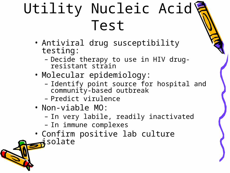
Utility Nucleic Acid Test• Antiviral drug susceptibility testing:
– Decide therapy to use in HIV drug-resistant strain
• Molecular epidemiology:– Identify point source for hospital and
community-based outbreak– Predict virulence
• Non-viable MO:– In very labile, readily inactivated– In immune complexes
• Confirm positive lab culture isolate
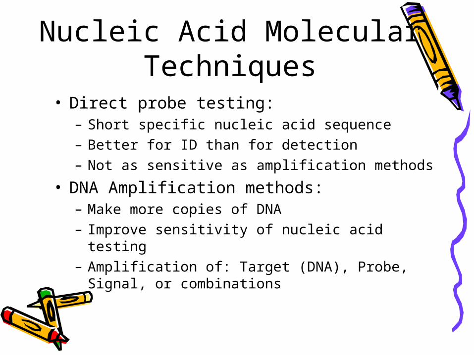
Nucleic Acid Molecular Techniques
• Direct probe testing:– Short specific nucleic acid sequence– Better for ID than for detection– Not as sensitive as amplification methods
• DNA Amplification methods:– Make more copies of DNA– Improve sensitivity of nucleic acid testing– Amplification of: Target (DNA), Probe,
Signal, or combinations
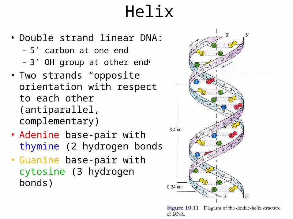
DNA Structure: Double Helix
• Double strand linear DNA:– 5’ carbon at one end– 3’ OH group at other end
• Two strands “opposite” orientation with respect to each other (antiparallel, complementary)
• Adenine base-pair with thymine (2 hydrogen bonds)
• Guanine base-pair with cytosine (3 hydrogen bonds)
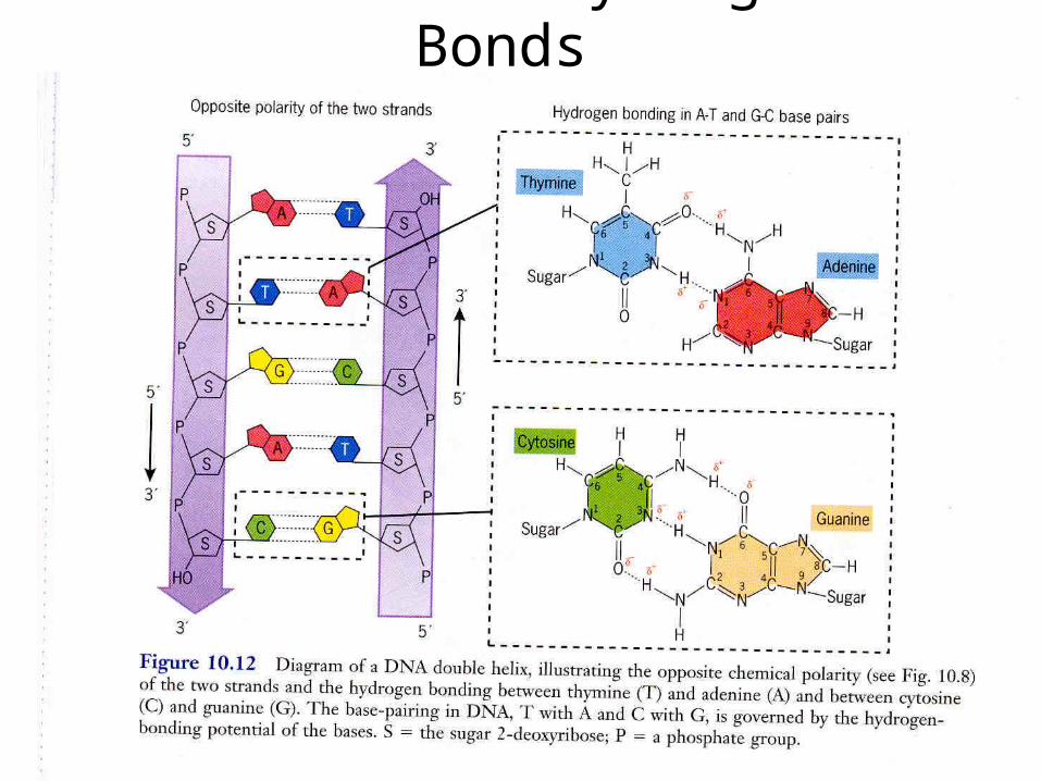
DNA Helix: Hydrogen Bonds
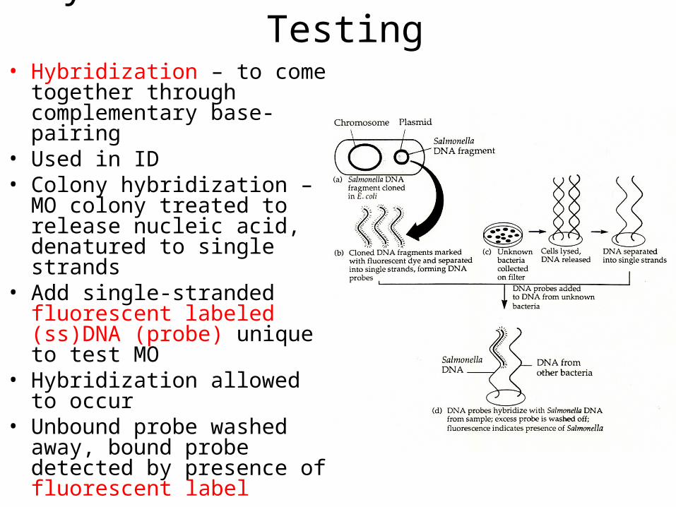
Hybridization: Direct Probe Testing
• Hybridization – to come together through complementary base-pairing
• Used in ID • Colony hybridization – MO
colony treated to release nucleic acid, denatured to single strands
• Add single-stranded fluorescent labeled (ss)DNA (probe) unique to test MO
• Hybridization allowed to occur
• Unbound probe washed away, bound probe detected by presence of fluorescent label
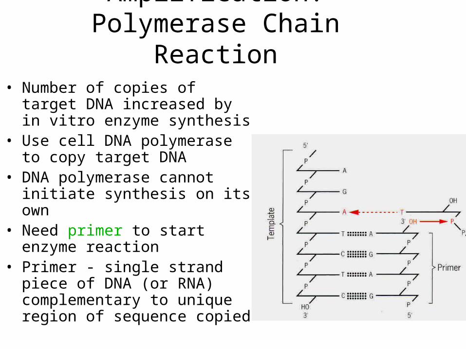
Target DNA Amplification: Polymerase Chain
Reaction• Number of copies of target
DNA increased by in vitro enzyme synthesis
• Use cell DNA polymerase to copy target DNA
• DNA polymerase cannot initiate synthesis on its own
• Need primer to start enzyme reaction
• Primer - single strand piece of DNA (or RNA) complementary to unique region of sequence copied
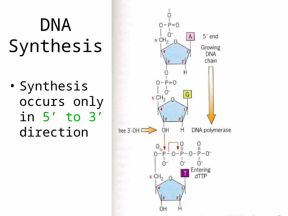
DNA Synthesis
• Synthesis occurs only in 5’ to 3’ direction
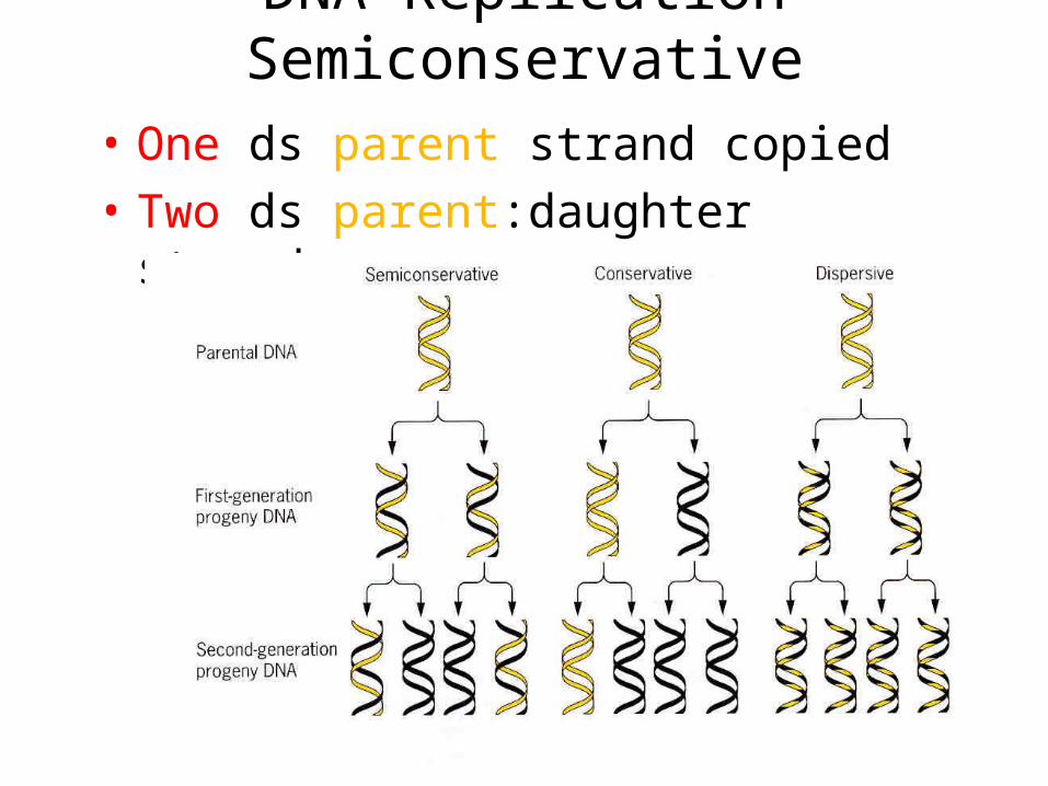
DNA Replication Semiconservative
• One ds parent strand copied • Two ds parent:daughter strands
Polymerase Chain Reaction (PCR)
• Enzyme reaction to amplify DNA: – Two primers - one binds one strand of
dsDNA, other binds other strand– Four nucleotide DNA precursors (dNTPs)– Thermostable DNA polymerase– Buffer (ions, salts) for enzyme reaction
• Primers (~15-20 bases) unique sequence to DNA being amplified
• PCR reaction has three basic steps:– 1) Denaturation– 2) Primer Annealing– 3) Primer Extension
PCR Assay: 1) Denaturation
• Separate dsDNA into ssDNA• Heating 950 C, 15 seconds to 1
minute• The two ssDNA strands serve as
templates for DNA synthesis
PCR Assay: 2) Primer Annealing
• DNA comes together through complementary base-pairing (hybridization)
• Primers base-pair with their complementary sequences on ssDNA template
• Primer concentration in excess of DNA template concentration
• Excess primer ensures base-pairing with complementary sequence on template DNA instead of ssDNA templates base-pairing back together

Primer Annealing• Temperature used should ensure annealing
will occur only with DNA sequences that are completely complementary. WHY?
• The annealing temperature depends upon length and sequence of primers
• The longer the primer and the more Gs and Cs in the sequence, the higher the annealing temperature. WHY?
• Annealing temperature range from 45-65ºC• Annealing time usually 15 seconds to 1 minute
PCR Assay: 3) Primer Extension
• DNA polymerase use dNTPs to synthesize DNA complementary to template DNA
• Thermostable Taq DNA polymerase (isolated from Thermus aquaticus) extends annealed primers
• Temperature used is 720 C, optimum reaction temperature for Taq DNA pol
• The extension time is usually 15 seconds to 1 minute
• Why is this high temperature used?• Why is a thermostable polymerase used?
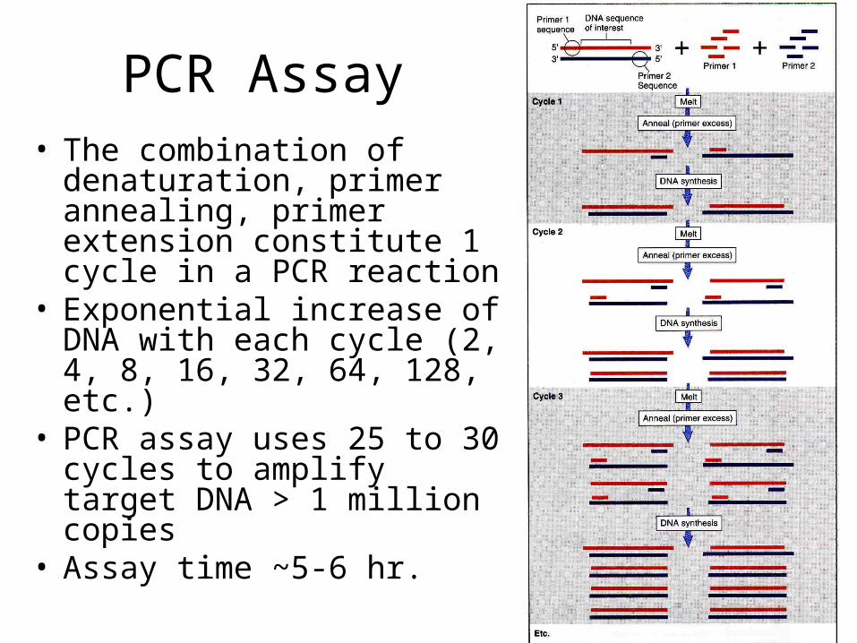
PCR Assay• The combination of
denaturation, primer annealing, primer extension constitute 1 cycle in a PCR reaction
• Exponential increase of DNA with each cycle (2, 4, 8, 16, 32, 64, 128, etc.)
• PCR assay uses 25 to 30 cycles to amplify target DNA > 1 million copies
• Assay time ~5-6 hr.
PCR Product DNA Analysis• DNA agarose gel electrophoresis
– ID DNA molecular weight by migration in gel– Estimate size using DNA markers (100 bp
ladder; 100-1500 bp)– Assay time ~1-2 hr.
• Real-Time PCR– ID DNA during PCR assay in closed tube– Use fluorescent reporter dye released
during enzyme reaction– Thermal Cylcler with UV light to excite
reporter dye and camera for detection– Assay time ~1 hr.
MICR 302 Lab PCR Exercise• First PCR assay will amplify DNA
sequence in bacterial 16S ribosomal RNA (rRNA) gene
• Use primers for DNA consensus sequence found in all bacteria
• Primers not unique to a specific bacteria, but unique to a conserved region of 16S rRNA gene of all bacteria
Bacterial 16S rRNA Gene: ID Unknown Bacteria
• Amplify portion of 16S rRNA gene of unknown bacteria
• DNA of all bacteria are amplified and yield DNA product using the consensus primers
• Sequence of the amplified DNA will be determined by Automated DNA Sequencer
• Identity of unknown found by searching and matching your result in the National Institutes of Health (NIH) DNA sequence database using “BLAST” program
MICR 302 Lab PCR Exercise• Second PCR assay will amplify DNA sequence
in Shiga Toxin gene• Use two primer sets unique for the two
different Shiga toxin genes of EHEC• Only E. coli that carry one or both of these
toxin genes yield DNA product using these primers
• Positive DNA product thus identifies bacteria isolate as EHEC strain
• For lab diagnosis, only this second type of PCR assay is of practical use for MO ID
Advantages of MolecularDNA Test
• High sensitivity - theoretically detect single MO
• High specificity – one MO species – Detect specific genotypes– Determine drug resistance– Predict virulence
• Speed - more rapid than traditional MO culture
• Simplicity - assays are automated; Real-time PCR
Disadvantages of Molecular DNA Test
• Expensive• Very specific, must have good clinical data to
support infection by MO before testing initiated
• Contaminating DNA give false positive• Miss new MO unless sequencing is done (as
for your lab molecular unknown); not practical in clinical lab
• Problem with mixed cultures – assay for all MOs possibly causing infection
• Too sensitive? Are results clinically relevant?• No isolated MO grown in culture
DNA Sequencing• Determine actual DNA sequence of MO• Using computer, based on nucleic acid
data:– Identify MO – Predict protein sequence and function
• The most commonly used sequencing method is the dideoxy method
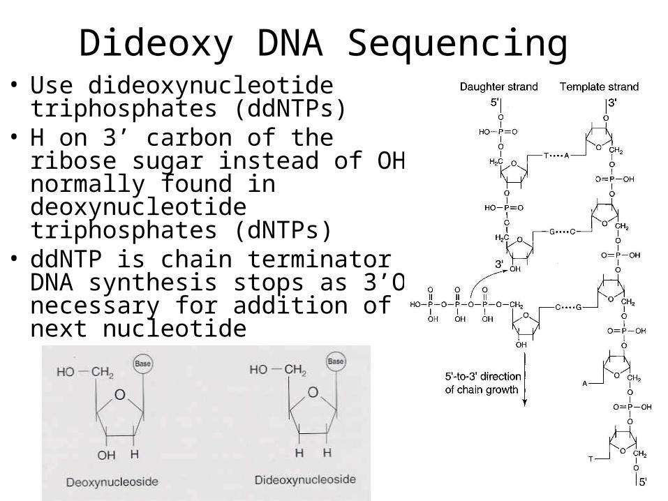
Dideoxy DNA Sequencing• Use dideoxynucleotide
triphosphates (ddNTPs)• H on 3’ carbon of the ribose
sugar instead of OH normally found in deoxynucleotide triphosphates (dNTPs)
• ddNTP is chain terminator - DNA synthesis stops as 3’OH necessary for addition of next nucleotide
Dideoxy DNA Sequencing
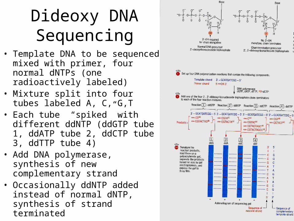
• Template DNA to be sequenced mixed with primer, four normal dNTPs (one radioactively labeled)
• Mixture split into four tubes labeled A, C, G,T
• Each tube “spiked” with different ddNTP (ddGTP tube 1, ddATP tube 2, ddCTP tube 3, ddTTP tube 4)
• Add DNA polymerase, synthesis of new complementary strand
• Occasionally ddNTP added instead of normal dNTP, synthesis of strand terminated
Dideoxy DNA Sequencing
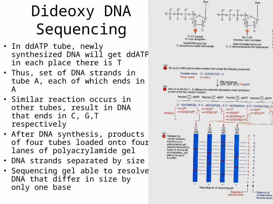
• In ddATP tube, newly synthesized DNA will get ddATP in each place there is T
• Thus, set of DNA strands in tube A, each of which ends in A
• Similar reaction occurs in other tubes, result in DNA that ends in C, G,T respectively
• After DNA synthesis, products of four tubes loaded onto four lanes of polyacrylamide gel
• DNA strands separated by size• Sequencing gel able to resolve
DNA that differ in size by only one base
Dideoxy DNA Sequencing
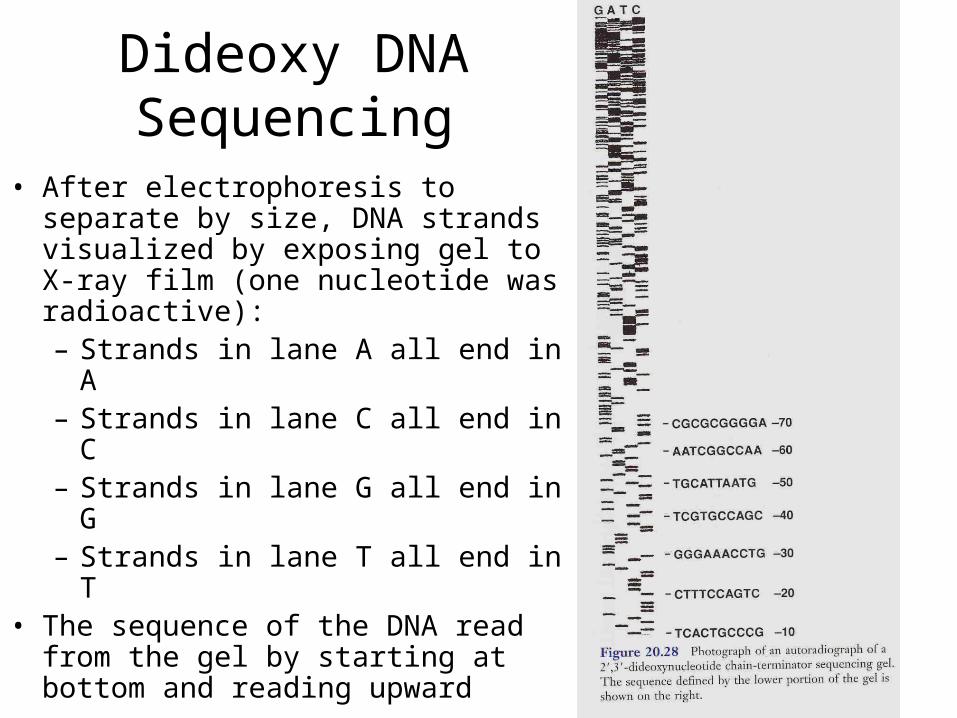
• After electrophoresis to separate by size, DNA strands visualized by exposing gel to X-ray film (one nucleotide was radioactive):– Strands in lane A all end in A– Strands in lane C all end in C– Strands in lane G all end in G– Strands in lane T all end in T
• The sequence of the DNA read from the gel by starting at bottom and reading upward
Automated DNA Sequencing• All four ddNTP reactions done in single tube• Each ddNTP labeled with a different
fluorescent dye• Dye present in each synthesized strand
corresponds to ddNTP that terminate DNA synthesis
• DNA strands in reaction tube analyzed by gel electrophoresis
• The Sequencing reaction depends on quality and purity of the amplified DNA product
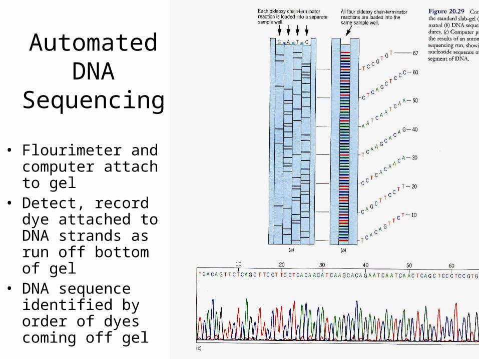
Automated DNA
Sequencing
• Flourimeter and computer attach to gel
• Detect, record dye attached to DNA strands as run off bottom of gel
• DNA sequence identified by order of dyes coming off gel
MICR 302 Lab Molecular Unknown
• Culture of an isolated Unknown bacteria• Isolate and purify DNA from Unknown bacteria• Use PCR to amplify rRNA gene sequence of
Unknown bacteria DNA• Do Automated Sequencing reaction on
amplified rRNA DNA product using fluorescent dyes
• Submit DNA to CSULA Sequencing Lab• Use NIH DNA databank to search for matching
DNA sequence to ID Unknown
Class Assignment• Textbook Reading: Chapter 11
Applications of Molecular Diagnostics– Introduction to Nucleic Acid Amplification
Procedures– Polymerase Chain reaction– Omit: Other Nucleic Acid Amplification
Reactions• Key Terms – only from assigned reading• Omit: Learning Assessment Questions
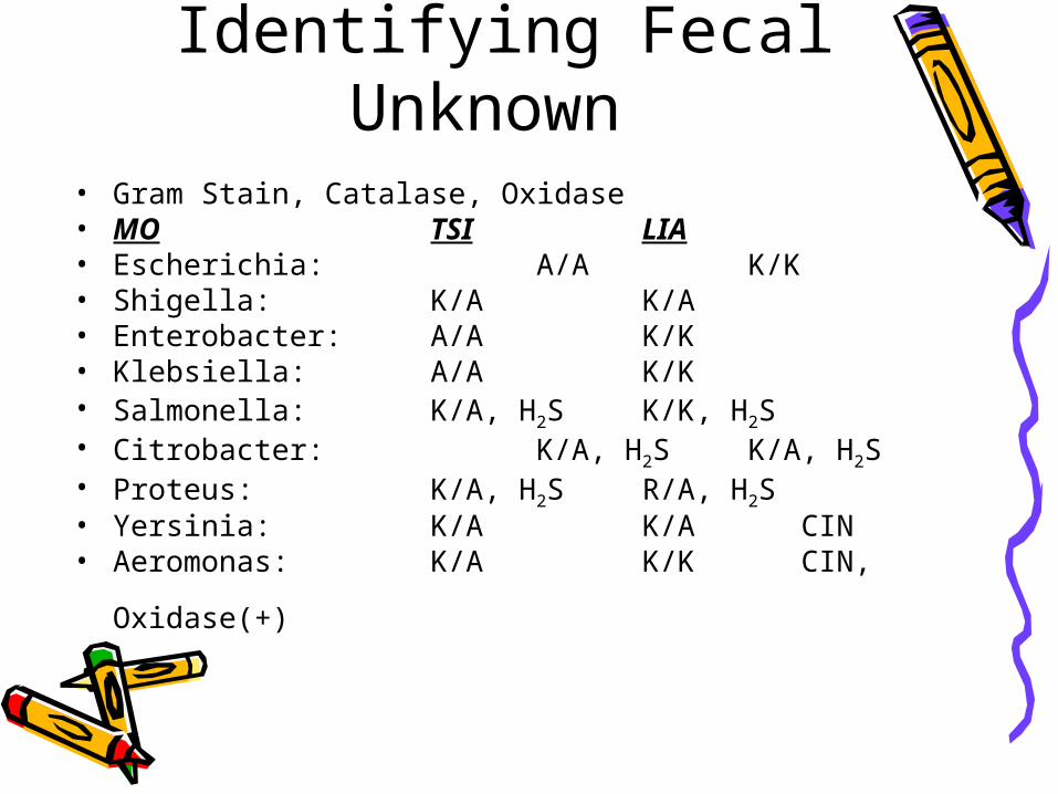
Identifying Fecal Unknown • Gram Stain, Catalase, Oxidase• MO TSI LIA• Escherichia: A/A K/K• Shigella: K/A K/A• Enterobacter: A/A K/K• Klebsiella: A/A K/K• Salmonella: K/A, H2S K/K, H2S• Citrobacter: K/A, H2S K/A, H2S• Proteus: K/A, H2S R/A, H2S• Yersinia: K/A K/A CIN• Aeromonas: K/A K/K CIN,
Oxidase(+)

Lecture Exam IITue., Feb. 28, 2012
• Vibrio thru New Molecular Methods • Lecture, Reading, Key Terms,
Learning Assessment Questions• Case Study 4, 5, 6 (Pseudomonas,
Francisella, Bacillus)• Review, Review, Review!• Repetition is the key to retention• Repetition is the key to retention• Repetition is the key to retention
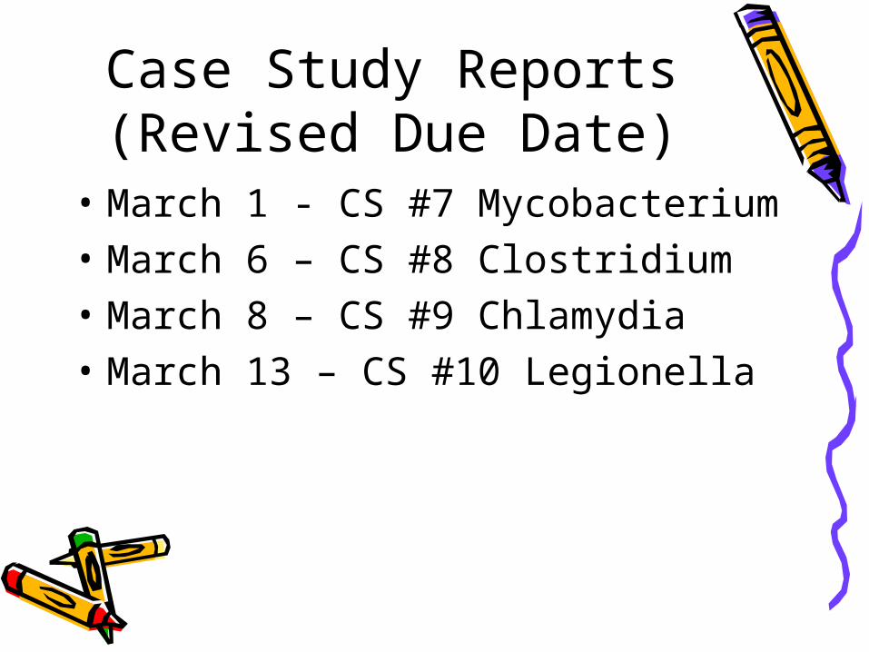
Case Study Reports (Revised Due Date)
• March 1 - CS #7 Mycobacterium• March 6 – CS #8 Clostridium• March 8 – CS #9 Chlamydia• March 13 – CS #10 Legionella