molecular epidemiology, genome characterization, and ... · ily, picornaviridae. the virus has...
TRANSCRIPT
Virology 421 (2011) 159–166
Contents lists available at SciVerse ScienceDirect
Virology
j ourna l homepage: www.e lsev ie r .com/ locate /yv i ro
Molecular epidemiology, genome characterization, and recombination event ofhuman parechovirus
Thaweesak Chieochansin, Preeyaporn Vichiwattana, Sumeth Korkong,Apiradee Theamboonlers, Yong Poovorawan ⁎Center of Excellence in Clinical Virology, Department of Pediatrics, Faculty of Medicine, Chulalongkorng University, Bangkok, Thailand
⁎ Corresponding author at: Center of Excellence in ClPediatrics, Faculty of Medicine, Chulalongkorn UniversFax: +66 2256 4929.
E-mail address: [email protected] (Y. Poovorawan
0042-6822/$ – see front matter © 2011 Elsevier Inc. Alldoi:10.1016/j.virol.2011.09.021
a b s t r a c t
a r t i c l e i n f oArticle history:Received 1 July 2011Returned to author for revision22 September 2011Accepted 24 September 2011Available online 21 October 2011
Keywords:Human parechovirusHPeVRecombinationGenotype
Human Parechovirus (HPeV), a member of the Picornaviridae family, is an infectious agent mostly affectingchildren. There are 16 recognized genotypes which have globally spread. This study incorporated a total of2957 nasopharyngeal (NP) swab and 759 fecal samples that were collected from different parts of Thailand.The NP of HPeV was detected in 0.4% of NP swab and 6.1% of fecal samples. The majority of HPeV infectionsoccur in infants below the age of 2 years, while infections were detected in children above the age of 10 yearsas well. Various genotypes comprising 1A, 1B, 2, 3, 4, 5, 6, 10 and 14 have been characterized. This studyrevealed recombination events in 16 samples in which HPeV1B was shown as the highest frequency. Inconclusion, HPeV can be detected in both the respiratory and GI tract. Moreover, HPeV which circulates inThailand is highly diverse and subject to recombination.
inical Virology, Department ofity, Bangkok 10330, Thailand.
).
rights reserved.
© 2011 Elsevier Inc. All rights reserved.
Introduction
Human parechovirus (HPeV) is a small non-enveloped singlestranded RNA virus of positive polarity which belongs to the vast fam-ily, Picornaviridae. The virus has first been discovered from an out-break of diarrhea among children in 1961. Based on their serologyand clinical presentation, the virus was defined as echovirus 22 and23 of the genus Enterovirus (Wigand and Sabin, 1961). However,studies on genetics, protein translation and biological properties ofthe virus have shown that it is different from other members of thegenus Enterovirus. Hence, it has been reclassified into a new genus,Parechovirus and those viruses previously defined as echovirus 22and 23 were re-named HPeV1 and HPeV2, respectively (Stanwayand Hyypia, 1999; Stanway et al., 1994). Recently, additional types ofHPeV have been reported, whichwere associated with different clinicalmanifestations. For example, HPeV3 was isolated from nasopharyngealaspirates (NPA) and related to sepsis in neonates (Ito et al., 2004).HPeV4 was isolated from fecal samples and related to fever in neonates(Benschop et al., 2006a). HPeV5 had previously been defined as HPeV2based on serology of children presenting with high fever but upongenome analysis was re-classified as type five (Oberste et al., 1998).HPeV6 was isolated from one child suffering from Reye's syndrome(Watanabe et al., 2007), HPeV7was identified bymetagenomicmethod
in fecal samples of a healthy child (Li et al., 2009), HPeV8 was isolatedfrom fecal samples during an outbreak of acute diarrhea in Brazil(Drexler et al., 2009), and HPeV9 to HPeV16 which unpublished buthad already been assigned by the following website (http://www.picornastudygroup.com/types/parechovirus/hpev.html).
Since the first report of HPeV infection, the epidemics of virus havebeen continuously reported. The most common genotype of the virusthat could be isolated worldwide was HPeV1B followed by HPeV3. Incontrast, other genotypes such as HPeV1A, HPeV2, and HPeV4-6 wereless common among infected patients than those two dominant geno-types (Benschop et al., 2008b; Ito et al., 2010; Pham et al., 2010; Tapiaet al., 2008). It should be noted that HPeV7 to 16 are a new genotypesthat have been recently discovered. Thus, epidemiology and prevalenceof these genotypes have not been fully established. The virus wasmostlydetected in children especially in infants below the age of 3 years(Benschop et al., 2006b; Tauriainen et al., 2007; Verboon-Macioleket al., 2005). However, patients above the age of 10 years have alsobeen noted (Abed and Boivin, 2006; Figueroa et al., 1989; Tapia et al.,2008; Watanabe et al., 2007). The longitudinal surveillance in USA be-tween 1983 and 2005 has shown 3% of HPeV1 and 68% of HPeV2 hadbeen isolated from infants less than 1 year old (Khetsuriani et al., 2006).In contrast, a study conducted in the Netherlands in 2000 has proposedthat children below the age of 3 years were infected with HPeV1 andHPeV3 (Benschop et al., 2006b). The clinical presentations of HPeV infec-tionwere associatedwithmild disease of the respiratory and gastrointes-tinal (GI) tract (Harvala et al., 2008; Zhang et al., 2010; Zhong et al., 2011).According to a recent study, HPeV, especially HPeV3, was strongly associ-ated with sepsis in neonates (Boivin et al., 2005; Harvala et al., 2009).
160 T. Chieochansin et al. / Virology 421 (2011) 159–166
The HPeV genome comprises approximately 7300 nucleotidesflanked by an un-translated region (UTR) at both the 5′ and 3′ end.The virus translates a polyprotein with one single open readingframe (ORF) which is subsequently cleaved during the post-transla-tional process into 3 structural proteins, VP0, VP3 and VP1, and 7non-structural proteins, 2A–2C and 3A–3D (Harvala and Simmonds,2009; Harvala et al., 2010).
Based on differences in antigenicity along with vast diversities inthe virus genome, 16 types of HPeV have been classified (http://www.picornastudygroup.com/types/parechovirus/hpev.html). Rapidchanges in their genomes normally occurring in the course of the
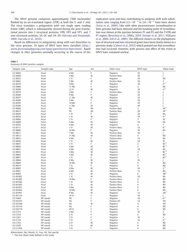
Table 1Summary of HPeV positive samples.
Samples code Samples type Age Sex
CU-B402 Fecal 6 Mo FCU-B408 Fecal 3 Mo MCU-B433 Fecal 1 Yr FCU-B463 Fecal 5 Mo MCU-B478 Fecal 6 Mo FCU-B486 Fecal 11 Mo FCU-B500 Fecal 12 Yr MCU-B579 Fecal 2 Mo FCU-B580 Fecal 9 Mo FCU-B587 Fecal 11 Mo MCU-B588 Fecal 3 Mo FCU-B595 Fecal 10 Mo FCU-B596 Fecal 11 Mo MCU-B606 Fecal NS NSCU-B615 Fecal NS NSCU-B628 Fecal 1 Yr FCU-B636 Fecal 1 Yr MCU-B685 Fecal 1 Yr FCU-B688 Fecal 3 Mo MCU-B705 Fecal 6 Mo MCU-B711 Fecal 1 Yr MCU-B806 Fecal 10 Mo FCU-B811 Fecal 7 Mo MCU-B812 Fecal 11 Mo MCU-B825 Fecal 6 Mo FCU-B846 Fecal 1 Yr MCU-B847 Fecal 1 Yr FCU-B850 Fecal 5 Mo MCU-B851 Fecal 6 Mo MCU-B852 Fecal 5 Mo MCU-B865 Fecal 5 Mo MCU-B867 Fecal 1 Yr FCU-B868 Fecal 9 Mo MCU-B869 Fecal 10 Mo MCU-B874 Fecal 1 Yr FCU-B903 Fecal 6 Mo MCU-B961 Fecal 6 Mo MCU-B984 Fecal 1 Yr MCU-B1002 Fecal 1 Yr MCU-B1009 Fecal 10 Mo FCU-B1046 Fecal 1 Yr MCU-B1050 Fecal 1 Yr MCU-B1055 Fecal 9 Mo MCU-B1064 Fecal 10 Mo MCU-B1091 Fecal 8 Mo MCU-B1154 Fecal 16 Yr FCU-H403 NP sawab NS FCU-H2107 NP sawab NS FCU-H2108 NP sawab NS MCU-H2159 NP sawab NS FCU-H2779 NP sawab 49 Yr FCU-C315 NP sawab 9 Mo FCU-C534 NP sawab 2 Yr FCU-C561 NP sawab 2 Yr FCU-C724 NP sawab 7 Yr FCU-C763 NP sawab 2 Yr MCU-C936 NP sawab 1 Yr MCU-C1204 NP sawab 3 Yr F
Abbreviation: Mo; Month, Yr; Year, NS; Not specify.⁎ The new 3Dpol clade defined in this study.
replication cycle and thus contributing to antigenic drift with substi-tution rates ranging from 2.2×10⁻³ to 2.8×10⁻³ have been shown(Faria et al., 2009). Like with other picornaviruses, recombination intheir genome had been detected and the breaking point of recombina-tion was shown at the junction between P1 and P2 and the 5′UTR andP1 region (Benschop et al., 2008a, 2010; Drexler et al., 2011; Williamset al., 2009; Zoll et al., 2009). The different clusters on the phylogenetictree of structural and non-structural genes have been clearly shown in aprevious study (Calvert et al., 2010) which pointed out that recombina-tion had occurred; however, both process and effect of this event inHPeV have remained unclear.
Other virus HPeV type 3Dpol clade
Negative 1B JPositive Rota 1B JNegative 1B BAPositive Rota 1B ZNegative 1B O′⁎
Negative 5 BANegative 1B O′⁎
Positive Rota 1B JNegative 1B JNegative 1B JNegative 1B JNegative 1B JNegative 1B JPositive Rota 5 AE′⁎
Positive Rota 1B AE′⁎
Negative 1B BANegative 1B O′⁎
Negative 3 WNegative 1B BANegative 4 YPositive Rota 1A K′⁎
Negative 1B BAPositive Rota 1B JPositive Rota 6 BDPositive Rota 1A BANegative 1B WNegative 2 BC′⁎
Negative 2 BC′⁎
Negative 1B BANegative 2 BC′⁎
Negative 2 BC′⁎
Negative 2 BC′⁎
Negative 1B JNegative 6 BDNegative 6 BDNegative 1B G′⁎
Positive Rota 14 BDNegative 6 AB′⁎
Positive Rota 1B BAPositive Rota 6 BDPositive Rota 6 BDPositive Rota 6 BDPositive Rota 6 BDPositive Rota 6 BDNegative 14 AB′⁎
Negative 1B JNegative 1B O′⁎
Positive H3 1A ANNegative 4 ANNegative 6 BDNegative 6 BDNegative 1B ZNegative 6 BDNegative 1B ZNegative 10 K′⁎
Negative 6 BDNegative 6 BDNegative 6 BD

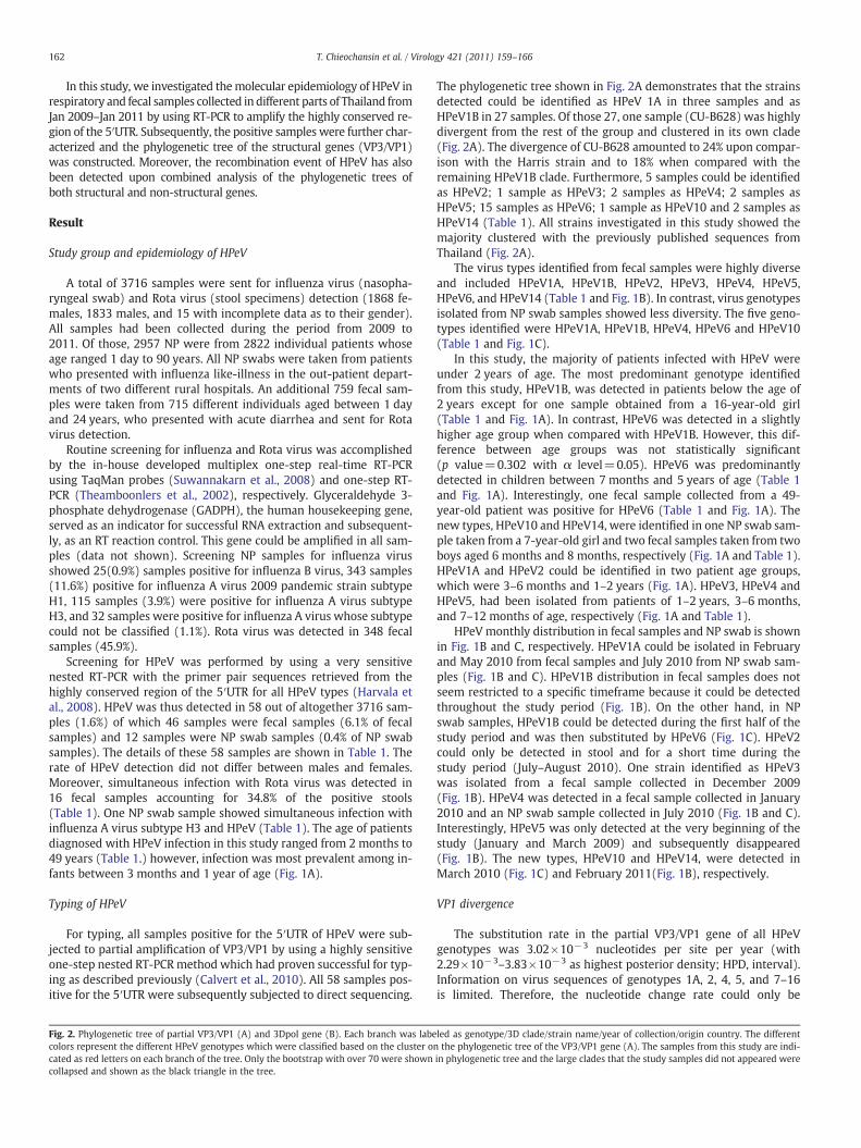
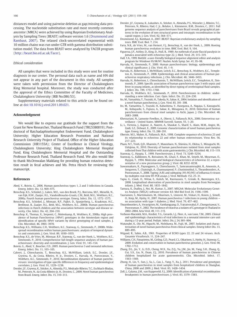
Fig. 1. Age distribution (A) and collection period of fecal (B) and NP swab (C) samples. The positive HPeV samples are shown as relative percentage of all samples collected.
161T. Chieochansin et al. / Virology 421 (2011) 159–166
162 T. Chieochansin et al. / Virology 421 (2011) 159–166
In this study, we investigated themolecular epidemiology of HPeV inrespiratory and fecal samples collected in different parts of Thailand fromJan 2009–Jan 2011 by using RT-PCR to amplify the highly conserved re-gion of the 5′UTR. Subsequently, the positive samples were further char-acterized and the phylogenetic tree of the structural genes (VP3/VP1)was constructed. Moreover, the recombination event of HPeV has alsobeen detected upon combined analysis of the phylogenetic trees ofboth structural and non-structural genes.
Result
Study group and epidemiology of HPeV
A total of 3716 samples were sent for influenza virus (nasopha-ryngeal swab) and Rota virus (stool specimens) detection (1868 fe-males, 1833 males, and 15 with incomplete data as to their gender).All samples had been collected during the period from 2009 to2011. Of those, 2957 NP were from 2822 individual patients whoseage ranged 1 day to 90 years. All NP swabs were taken from patientswho presented with influenza like-illness in the out-patient depart-ments of two different rural hospitals. An additional 759 fecal sam-ples were taken from 715 different individuals aged between 1 dayand 24 years, who presented with acute diarrhea and sent for Rotavirus detection.
Routine screening for influenza and Rota virus was accomplishedby the in-house developed multiplex one-step real-time RT-PCRusing TaqMan probes (Suwannakarn et al., 2008) and one-step RT-PCR (Theamboonlers et al., 2002), respectively. Glyceraldehyde 3-phosphate dehydrogenase (GADPH), the human housekeeping gene,served as an indicator for successful RNA extraction and subsequent-ly, as an RT reaction control. This gene could be amplified in all sam-ples (data not shown). Screening NP samples for influenza virusshowed 25(0.9%) samples positive for influenza B virus, 343 samples(11.6%) positive for influenza A virus 2009 pandemic strain subtypeH1, 115 samples (3.9%) were positive for influenza A virus subtypeH3, and 32 samples were positive for influenza A virus whose subtypecould not be classified (1.1%). Rota virus was detected in 348 fecalsamples (45.9%).
Screening for HPeV was performed by using a very sensitivenested RT-PCR with the primer pair sequences retrieved from thehighly conserved region of the 5′UTR for all HPeV types (Harvala etal., 2008). HPeV was thus detected in 58 out of altogether 3716 sam-ples (1.6%) of which 46 samples were fecal samples (6.1% of fecalsamples) and 12 samples were NP swab samples (0.4% of NP swabsamples). The details of these 58 samples are shown in Table 1. Therate of HPeV detection did not differ between males and females.Moreover, simultaneous infection with Rota virus was detected in16 fecal samples accounting for 34.8% of the positive stools(Table 1). One NP swab sample showed simultaneous infection withinfluenza A virus subtype H3 and HPeV (Table 1). The age of patientsdiagnosed with HPeV infection in this study ranged from 2 months to49 years (Table 1.) however, infection was most prevalent among in-fants between 3 months and 1 year of age (Fig. 1A).
Typing of HPeV
For typing, all samples positive for the 5′UTR of HPeV were sub-jected to partial amplification of VP3/VP1 by using a highly sensitiveone-step nested RT-PCRmethod which had proven successful for typ-ing as described previously (Calvert et al., 2010). All 58 samples pos-itive for the 5′UTR were subsequently subjected to direct sequencing.
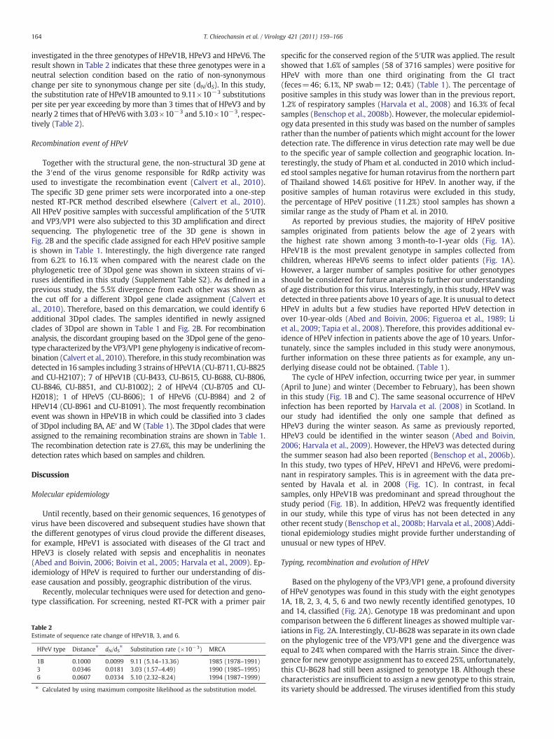
Fig. 2. Phylogenetic tree of partial VP3/VP1 (A) and 3Dpol gene (B). Each branch was labecolors represent the different HPeV genotypes which were classified based on the cluster ocated as red letters on each branch of the tree. Only the bootstrap with over 70 were showncollapsed and shown as the black triangle in the tree.
The phylogenetic tree shown in Fig. 2A demonstrates that the strainsdetected could be identified as HPeV 1A in three samples and asHPeV1B in 27 samples. Of those 27, one sample (CU-B628) was highlydivergent from the rest of the group and clustered in its own clade(Fig. 2A). The divergence of CU-B628 amounted to 24% upon compar-ison with the Harris strain and to 18% when compared with theremaining HPeV1B clade. Furthermore, 5 samples could be identifiedas HPeV2; 1 sample as HPeV3; 2 samples as HPeV4; 2 samples asHPeV5; 15 samples as HPeV6; 1 sample as HPeV10 and 2 samples asHPeV14 (Table 1). All strains investigated in this study showed themajority clustered with the previously published sequences fromThailand (Fig. 2A).
The virus types identified from fecal samples were highly diverseand included HPeV1A, HPeV1B, HPeV2, HPeV3, HPeV4, HPeV5,HPeV6, and HPeV14 (Table 1 and Fig. 1B). In contrast, virus genotypesisolated from NP swab samples showed less diversity. The five geno-types identified were HPeV1A, HPeV1B, HPeV4, HPeV6 and HPeV10(Table 1 and Fig. 1C).
In this study, the majority of patients infected with HPeV wereunder 2 years of age. The most predominant genotype identifiedfrom this study, HPeV1B, was detected in patients below the age of2 years except for one sample obtained from a 16-year-old girl(Table 1 and Fig. 1A). In contrast, HPeV6 was detected in a slightlyhigher age group when compared with HPeV1B. However, this dif-ference between age groups was not statistically significant(p value=0.302 with α level=0.05). HPeV6 was predominantlydetected in children between 7 months and 5 years of age (Table 1and Fig. 1A). Interestingly, one fecal sample collected from a 49-year-old patient was positive for HPeV6 (Table 1 and Fig. 1A). Thenew types, HPeV10 and HPeV14, were identified in one NP swab sam-ple taken from a 7-year-old girl and two fecal samples taken from twoboys aged 6 months and 8 months, respectively (Fig. 1A and Table 1).HPeV1A and HPeV2 could be identified in two patient age groups,which were 3–6 months and 1–2 years (Fig. 1A). HPeV3, HPeV4 andHPeV5, had been isolated from patients of 1–2 years, 3–6 months,and 7–12 months of age, respectively (Fig. 1A and Table 1).
HPeV monthly distribution in fecal samples and NP swab is shownin Fig. 1B and C, respectively. HPeV1A could be isolated in Februaryand May 2010 from fecal samples and July 2010 from NP swab sam-ples (Fig. 1B and C). HPeV1B distribution in fecal samples does notseem restricted to a specific timeframe because it could be detectedthroughout the study period (Fig. 1B). On the other hand, in NPswab samples, HPeV1B could be detected during the first half of thestudy period and was then substituted by HPeV6 (Fig. 1C). HPeV2could only be detected in stool and for a short time during thestudy period (July–August 2010). One strain identified as HPeV3was isolated from a fecal sample collected in December 2009(Fig. 1B). HPeV4 was detected in a fecal sample collected in January2010 and an NP swab sample collected in July 2010 (Fig. 1B and C).Interestingly, HPeV5 was only detected at the very beginning of thestudy (January and March 2009) and subsequently disappeared(Fig. 1B). The new types, HPeV10 and HPeV14, were detected inMarch 2010 (Fig. 1C) and February 2011(Fig. 1B), respectively.
VP1 divergence
The substitution rate in the partial VP3/VP1 gene of all HPeVgenotypes was 3.02×10⁻³ nucleotides per site per year (with2.29×10−3–3.83×10⁻³ as highest posterior density; HPD, interval).Information on virus sequences of genotypes 1A, 2, 4, 5, and 7–16is limited. Therefore, the nucleotide change rate could only be
led as genotype/3D clade/strain name/year of collection/origin country. The differentn the phylogenetic tree of the VP3/VP1 gene (A). The samples from this study are indi-in phylogenetic tree and the large clades that the study samples did not appeared were

163T. Chieochansin et al. / Virology 421 (2011) 159–166
164 T. Chieochansin et al. / Virology 421 (2011) 159–166
investigated in the three genotypes of HPeV1B, HPeV3 and HPeV6. Theresult shown in Table 2 indicates that these three genotypes were in aneutral selection condition based on the ratio of non-synonymouschange per site to synonymous change per site (dN/dS). In this study,the substitution rate of HPeV1B amounted to 9.11×10⁻³ substitutionsper site per year exceeding by more than 3 times that of HPeV3 and bynearly 2 times that of HPeV6 with 3.03×10⁻³ and 5.10×10⁻³, respec-tively (Table 2).
Recombination event of HPeV
Together with the structural gene, the non-structural 3D gene atthe 3′end of the virus genome responsible for RdRp activity wasused to investigate the recombination event (Calvert et al., 2010).The specific 3D gene primer sets were incorporated into a one-stepnested RT-PCR method described elsewhere (Calvert et al., 2010).All HPeV positive samples with successful amplification of the 5′UTRand VP3/VP1 were also subjected to this 3D amplification and directsequencing. The phylogenetic tree of the 3D gene is shown inFig. 2B and the specific clade assigned for each HPeV positive sampleis shown in Table 1. Interestingly, the high divergence rate rangedfrom 6.2% to 16.1% when compared with the nearest clade on thephylogenetic tree of 3Dpol gene was shown in sixteen strains of vi-ruses identified in this study (Supplement Table S2). As defined in aprevious study, the 5.5% divergence from each other was shown asthe cut off for a different 3Dpol gene clade assignment (Calvert etal., 2010). Therefore, based on this demarcation, we could identify 6additional 3Dpol clades. The samples identified in newly assignedclades of 3Dpol are shown in Table 1 and Fig. 2B. For recombinationanalysis, the discordant grouping based on the 3Dpol gene of the geno-type characterized by theVP3/VP1 genephylogeny is indicative of recom-bination (Calvert et al., 2010). Therefore, in this study recombinationwasdetected in 16 samples including 3 strains of HPeV1A (CU-B711, CU-B825and CU-H2107); 7 of HPeV1B (CU-B433, CU-B615, CU-B688, CU-B806,CU-B846, CU-B851, and CU-B1002); 2 of HPeV4 (CU-B705 and CU-H2018); 1 of HPeV5 (CU-B606); 1 of HPeV6 (CU-B984) and 2 ofHPeV14 (CU-B961 and CU-B1091). The most frequently recombinationevent was shown in HPeV1B in which could be classified into 3 cladesof 3Dpol including BA, AE′ and W (Table 1). The 3Dpol clades that wereassigned to the remaining recombination strains are shown in Table 1.The recombination detection rate is 27.6%, this may be underlining thedetection rates which based on samples and children.
Discussion
Molecular epidemiology
Until recently, based on their genomic sequences, 16 genotypes ofvirus have been discovered and subsequent studies have shown thatthe different genotypes of virus cloud provide the different diseases,for example, HPeV1 is associated with diseases of the GI tract andHPeV3 is closely related with sepsis and encephalitis in neonates(Abed and Boivin, 2006; Boivin et al., 2005; Harvala et al., 2009). Ep-idemiology of HPeV is required to further our understanding of dis-ease causation and possibly, geographic distribution of the virus.
Recently, molecular techniques were used for detection and geno-type classification. For screening, nested RT-PCR with a primer pair
Table 2Estimate of sequence rate change of HPeV1B, 3, and 6.
HPeV type Distance⁎ dN/dS⁎ Substitution rate (×10−3) MRCA
1B 0.1000 0.0099 9.11 (5.14–13.36) 1985 (1978–1991)3 0.0346 0.0181 3.03 (1.57–4.49) 1990 (1985–1995)6 0.0607 0.0334 5.10 (2.32–8.24) 1994 (1987–1999)
⁎ Calculated by using maximum composite likelihood as the substitution model.
specific for the conserved region of the 5′UTR was applied. The resultshowed that 1.6% of samples (58 of 3716 samples) were positive forHPeV with more than one third originating from the GI tract(feces=46; 6.1%, NP swab=12; 0.4%) (Table 1). The percentage ofpositive samples in this study was lower than in the previous report,1.2% of respiratory samples (Harvala et al., 2008) and 16.3% of fecalsamples (Benschop et al., 2008b). However, the molecular epidemiol-ogy data presented in this study was based on the number of samplesrather than the number of patients whichmight account for the lowerdetection rate. The difference in virus detection rate may well be dueto the specific year of sample collection and geographic location. In-terestingly, the study of Pham et al. conducted in 2010 which includ-ed stool samples negative for human rotavirus from the northern partof Thailand showed 14.6% positive for HPeV. In another way, if thepositive samples of human rotavirus were excluded in this study,the percentage of HPeV positive (11.2%) stool samples has shown asimilar range as the study of Pham et al. in 2010.
As reported by previous studies, the majority of HPeV positivesamples originated from patients below the age of 2 years withthe highest rate shown among 3 month-to-1-year olds (Fig. 1A).HPeV1B is the most prevalent genotype in samples collected fromchildren, whereas HPeV6 seems to infect older patients (Fig. 1A).However, a larger number of samples positive for other genotypesshould be considered for future analysis to further our understandingof age distribution for this virus. Interestingly, in this study, HPeV wasdetected in three patients above 10 years of age. It is unusual to detectHPeV in adults but a few studies have reported HPeV detection inover 10-year-olds (Abed and Boivin, 2006; Figueroa et al., 1989; Liet al., 2009; Tapia et al., 2008). Therefore, this provides additional ev-idence of HPeV infection in patients above the age of 10 years. Unfor-tunately, since the samples included in this study were anonymous,further information on these three patients as for example, any un-derlying disease could not be obtained. (Table 1).
The cycle of HPeV infection, occurring twice per year, in summer(April to June) and winter (December to February), has been shownin this study (Fig. 1B and C). The same seasonal occurrence of HPeVinfection has been reported by Harvala et al. (2008) in Scotland. Inour study had identified the only one sample that defined asHPeV3 during the winter season. As same as previously reported,HPeV3 could be identified in the winter season (Abed and Boivin,2006; Harvala et al., 2009). However, the HPeV3 was detected duringthe summer season had also been reported (Benschop et al., 2006b).In this study, two types of HPeV, HPeV1 and HPeV6, were predomi-nant in respiratory samples. This is in agreement with the data pre-sented by Havala et al. in 2008 (Fig. 1C). In contrast, in fecalsamples, only HPeV1B was predominant and spread throughout thestudy period (Fig. 1B). In addition, HPeV2 was frequently identifiedin our study, while this type of virus has not been detected in anyother recent study (Benschop et al., 2008b; Harvala et al., 2008).Addi-tional epidemiology studies might provide further understanding ofunusual or new types of HPeV.
Typing, recombination and evolution of HPeV
Based on the phylogeny of the VP3/VP1 gene, a profound diversityof HPeV genotypes was found in this study with the eight genotypes1A, 1B, 2, 3, 4, 5, 6 and two newly recently identified genotypes, 10and 14, classified (Fig. 2A). Genotype 1B was predominant and uponcomparison between the 6 different lineages as showed multiple var-iations in Fig. 2A. Interestingly, CU-B628 was separate in its own cladeon the phylogenic tree of the VP3/VP1 gene and the divergence wasequal to 24% when compared with the Harris strain. Since the diver-gence for new genotype assignment has to exceed 25%, unfortunately,this CU-B628 had still been assigned to genotype 1B. Although thesecharacteristics are insufficient to assign a new genotype to this strain,its variety should be addressed. The viruses identified from this study

165T. Chieochansin et al. / Virology 421 (2011) 159–166
were grouped together on the phylogenetic tree of VP3/VP1 gene andalso clustered with viruses previously reported from Thailand(Fig. 2A). This indicates a geographically specific common determi-nant that appears to be found on this VP3/VP1 phylogenetic tree ofHPeV. Moreover, difference of the HPeV genotype could be detectedin children below 2 years of age.
Altogether, the genotypes have not evolved quite as rapidly as thesubstitution rate calculated by BEAST suggested (3.02×10⁻³ substi-tutions per site per year) which was slightly higher than that previ-ously proposed for the VP1 gene (Faria et al., 2009). However, alongwith phylogenetic analysis, a vast difference in substitution rate wasshown among different genotypes of virus (Table 2). Incorporationof much more samples used for the calculation may be the reasonwhy the evolution rate in this study appeared slightly higher thanthat previously estimated (Calvert et al., 2010). Similarly, the dN/dScalculated from this study also were slightly higher than previouslymentioned by Calvert et al. (2010). In contrast, the MRCA shown inour study (Table 2.) did not show any difference to the previous re-port (Calvert et al., 2010). Further studies on larger sample sizeswill provide further understanding of virus evolution.
Interestingly, as defined in the previous study, more than 5.5% di-vergence was shown as the cut off for a different 3Dpol gene clade as-signment (Calvert et al., 2010). Hence 6 additional new 3Dpol cladescould be defined based on our study samples (Table 1.).
Recombination of HPeV had previously been addressed with the re-combination breaking points established at the junction between 5′UTRand P1 and the P1 and P2 polyprotein (Benschop et al., 2008a, 2010;Drexler et al., 2011; Williams et al., 2009; Zoll et al., 2009). Despite thephylogenetic analysis comparison between structural and non-structur-al genes such as the 3Dpol gene was successfully used to investigate re-combination (Calvert et al., 2010). The discordance cluster in the 3Dpolgene when compared with the VP3/VP1 gene phylogenetic tree provesthat recombination in the virus genome has occurred (Calvert et al.,2010). Most of the samples from our study were classified as the sameclade for 3Dpol phylogenetic tree as the reported in Thai samples thatreported by Calvert et al. (2010) (Table 1). Sixteen samples (27.6%)showed discordance between two genetic regions on the phylogenetictree (Fig. 2) serving as evidence for recombination from this study.HPeV1B was the predominant genotype with a recombination frequen-cy of 27%. This study was in agreement with previous research empha-sizing the high recombination frequency of HPeV type 1B (Calvert etal., 2010). In contrast, HPeV6 exhibited less recombination frequency(6%)with only one sample identified (CU-B984, Fig. 2.). Other genotypesincluding HPeV1A, HPeV4, HPeV5, and HPeV14 had alsoundergone recombination (Fig. 2.), however, the frequency of recombi-nation had not been done due to the small identification numbers fromthe study. HPeV3 isolated from this study clustered in the correlatedphylogenetic tree cladewith both VP3/VP1 and 3Dpol geneswhen com-pared to the previous report (Fig. 2.) (Calvert et al., 2010). Therefore, therecombinationmight not have been detectable in this study. The under-lying factors ormechanisms for the recombination event are still unclearand should be addressed by further investigation.
In conclusion, although HPeV has been discovered a long time ago,its biology, epidemiology, evolution and pathogenicity are stillunclear. Therefore, ongoing molecular research will further our un-derstanding of this virus which can also be applied to other relatedviruses.
Method
Clinical samples
The nasopharyngeal (NP) swabs were collected from patients pre-senting with influenza-like illness. A total of 2957 of NP swab sampleswere collected between July 14, 2009 and March 9, 2011. All sampleswere sent from local hospitals including; 1703 samples fromChumphae
hospital, Chumphae district, Khonkaen province, 1254 samples fromThungsong hospital, Thungsong district, Nakorn Sri Thammarat prov-ince. Samples were kept in transport media which contain phosphate-buffered saline (PBS) with antibiotics (2×10⁶ U/l of penicillin G and200 mg/l of streptomycin). All were kept on ice then sent to Center ofExcellence in Clinical Virology, Department of Pediatrics, Faculty ofMedicine, Chulalongkorn University for the routine screening. Sampleswere processed immediately upon arrival, the remaining were keptat−70 °C for further use.
Fecal samples were sent to our center for Human Rota virus rou-tine screening. A total of 715 fecal samples had been collected inthe 2-year period of January 7, 2009 to April 18, 2011. Of those, 12samples were sent from Umphang hospital, Umphang district, Takprovince; 597 samples from Chumphae hospital, Chumphae district,Khonkaen province and 150 samples from King Chulalongkorn Me-morial hospital, Bangkok. Upon arrival, fecal samples were diluted1:10 with PBS, thoroughly mixed on a vortex and centrifuged at8000 rpm. Supernatants were collected and stored at −70 °C untilfurther use.
RNA extraction and HPeV detection
All samples were subjected to RNA extraction by conventionalGTC-phenol-chloroform method performed on 200 μl each of clinicalsamples. The extracted RNA was stored at −70 °C until used.
All extracted RNA was subjected to reverse transcription intocDNA using the ImProm-II™ Reverse Transcription system (Promega,Madison, WI). Reverse transcription (RT) was performed by followingthe manufacturer's specifications and a random hexamer primer wasused for the RT step. cDNA was stored at −20 °C until used.
Nested-PCR for HPeV detection was modified from a previousstudy (Harvala et al., 2010). The PerfectTaq Plus MasterMix (5PRIME, Darmstadt, Germany) was used as the amplification mixture.The predicted positive result was shown as a single band of 243 bpwhen visualized under UV light after 2% agarose gel electrophoresisand staining with ethidium bromine.
Partial VP3/VP1 and 3Dpol gene amplification
For HPeV characterization, the samples positive for the 5′UTRweresubjected to nested PCR to amplify the VP3/VP1 junction for HPeV typ-ing as previously described (Harvala et al., 2010). The expected PCRproduct was 304 bp when virtualized under UV light after 2% agarosegel electrophoresis and staining with ethidium bromine.
The partial 3Dpol gene was amplified by nested PCR modifiedfrom a previous report (Calvert et al., 2010). The expected secondround PCR product of 700 bp was virtualized under UV light after2% agarose gel electrophoresis and staining with ethidium bromine.
All VP3/VP1 and 3Dpol positive amplicons were purified by usingAgarose Gelextract Mini Kit (5 PRIME, Darmstadt, Germany). Directsequencing was performed under 1st BASE DNA Sequencing Services(1st BASE Laboratories, Malaysia). Sequencing results were annotat-ed, aligned, and managed by using combination of software includingBasic Local Alignment Search Tool (BLAST) (http://blast.ncbi.nlm.nih.gov/Blast.cgi), Simmonics version 1.7 (www.virus-evolution.org),Chromas Lite (http://www.technelysium.com.au/chromas_lite.html),and BioEdit version 7.0.4.1(Hall, 1999).
Molecular and evolution analysis
All study sequences had been submitted to GenBank data base.The accession number that included in this study was show in Sup-plement Table (S1). Phylogenetic trees were constructed by usingMEGA 4 software (Tamura et al., 2007) in which neighbor-joiningmethod with 1000 replication for bootstrapping was implemented.Trees contracted under maximum-composite-likelihood (MCL) as
166 T. Chieochansin et al. / Virology 421 (2011) 159–166
distances model and using pairwise deletion as gap/missing data pro-cessing. The nucleotide substitution rate and most recently commonancestor (MRCA) were achieved by using Bayesian Evolutionary Anal-ysis by Sampling Trees (BEAST) software version 1.6 (Drummond andRambaut, 2007). The relaxed clock-uncorrelated exponential with10 million chains was run under GTR with gamma distribution substi-tution model. The data from BEAST were analyzed by TACER program(http://beast.bio.ed.ac.uk/Tacer).
Ethical consideration
All samples that were included in this study were sent for routinediagnosis in our center. The personal data such as name and HN didnot appear in any part of the document in this study. All sampleswere taken with permission from the Director of ChulalongkornKing Memorial hospital. Moreover, the study was conducted afterthe approval of the Ethics Committee of the Faculty of Medicines,Chulalongkorn University (IRB No.086/53).
Supplementary materials related to this article can be found on-line at doi:10.1016/j.virol.2011.09.021.
Acknowledgment
We would like to express our gratitude for the support from theGrant for NewResearcher, Thailand Research Fund (TRG5380019); Post-doctoral of Ratchadaphiseksomphot Endowment Fund, ChulalongkornUniversity; Higher Education Research Promotion and NationalResearch University Project of Thailand, Office of the Higher EducationCommission (HR1155A); Center of Excellence in Clinical Virology,Chulalongkorn University; King Chulalongkorn Memorial HospitalFund, King Chulalongkorn Memorial Hospital and the OutstandingProfessor Research Fund, Thailand Research Fund. We also would liketo thank Ms.Ornwalan Maiklang for providing human rotavirus detec-tion result in fecal achieves and Ms. Petra Hirsch for reviewing themanuscript.
References
Abed, Y., Boivin, G., 2006. Human parechovirus types 1, 2 and 3 infections in Canada.Emerg. Infect. Dis. 12, 969–975.
Benschop, K.S., Schinkel, J., Luken, M.E., van den Broek, P.J., Beersma, M.F., Menelik, N.,van Eijk, H.W., Zaaijer, H.L., VandenBroucke-Grauls, C.M., Beld, M.G., Wolthers, K.C.,2006a. Fourth human parechovirus serotype. Emerg. Infect. Dis. 12, 1572–1575.
Benschop, K.S., Schinkel, J., Minnaar, R.P., Pajkrt, D., Spanjerberg, L., Kraakman, H.C.,Berkhout, B., Zaaijer, H.L., Beld, M.G., Wolthers, K.C., 2006b. Human parechovirusinfections in Dutch children and the association between serotype and disease se-verity. Clin. Infect. Dis. 42, 204–210.
Benschop, K., Thomas, X., Serpenti, C., Molenkamp, R., Wolthers, K., 2008a. High prev-alence of human Parechovirus (HPeV) genotypes in the Amsterdam region andidentification of specific HPeV variants by direct genotyping of stool samples. J.Clin. Microbiol. 46, 3965–3970.
Benschop, K.S., Williams, C.H., Wolthers, K.C., Stanway, G., Simmonds, P., 2008b. Wide-spread recombination within human parechoviruses: analysis of temporal dynam-ics and constraints. J. Gen. Virol. 89, 1030–1035.
Benschop, K.S., de Vries, M., Minnaar, R.P., Stanway, G., van der Hoek, L., Wolthers, K.C.,Simmonds, P., 2010. Comprehensive full-length sequence analyses of human par-echoviruses: diversity and recombination. J. Gen. Virol. 91, 145–154.
Boivin, G., Abed, Y., Boucher, F.D., 2005. Human parechovirus 3 and neonatal infections.Emerg. Infect. Dis. 11, 103–105.
Calvert, J., Chieochansin, T., Benschop, K.S., McWilliam, Leitch, E.C., Drexler, J.F.,Grywna, K., da Costa, Ribeiro, H. Jr., Drosten, C., Harvala, H., Poovorawan, Y.,Wolthers, K.C., Simmonds, P., 2010. Recombination dynamics of human parecho-viruses: investigation of type-specific differences in frequency and epidemiologicalcorrelates. J. Gen. Virol. 91, 1229–1238.
Drexler, J.F., Grywna, K., Stöcker, A., Almeida, P.S., Medrado-Ribeiro, T.C., Eschbach-Bludau,M., Petersen, N., da Costa-Ribeiro-Jr, H., Drosten, C., 2009. Novel human parechovirusfrom Brazil. Emerg. Infect. Dis. 15, 310–313.
Drexler, J.F., Grywna, K., Lukashev, A., Stöcker, A., Almeida, P.S., Wieseler, J., Ribeiro, T.C.,Petersen, N., Ribeiro, Had, C. Jr., Belalov, I., Kümmerer, B.M., Drosten, C., 2011. Fullgenome sequence analysis of parechoviruses from Brazil reveals geographical pat-terns in the evolution of non-structural genes and intratypic recombination in thecapsid region. J. Gen. Virol. 92, 564–571.
Drummond, A.J., Rambaut, A., 2007. BEAST: Bayesian evolutionary analysis by samplingtrees. BMC Evol. Biol. 7, 214.
Faria, N.R., de Vries, M., van Hemert, F.J., Benschop, K., van der Hoek, L., 2009. Rootinghuman parechovirus evolution in time. BMC Evol. Biol. 9, 164.
Figueroa, J.P., Ashley, D., King, D., Hull, B., 1989. An outbreak of acute flaccid paralysis inJamaica associated with echovirus type 22. J. Med. Virol. 29, 315–319.
Hall, T.A., 1999. BioEdit: a user-friendly biological sequence alignment editor and analysisprogram for Windows 95/98/NT. Nucleic Acids Symp. Ser. 41, 95–98.
Harvala, H., Simmonds, P., 2009. Human parechoviruses: biology, epidemiology andclinical significance. J. Clin. Virol. 45, 1–9.
Harvala, H., Robertson, I., McWilliam, Leitch, E.C., Benschop, K., Wolthers, K.C., Temple-ton, K., Simmonds, P., 2008. Epidemiology and clinical associations of human par-echovirus respiratory infections. J. Clin. Microbiol. 46; 3446–3453.
Harvala, H., Robertson, I., Chieochansin, T., McWilliam, Leitch, E.C., Templeton, K., Sim-monds, P., 2009. Specific association of human parechovirus type 3 with sepsis andfever in young infants, as identified by direct typing of cerebrospinal fluid samples.J. Infect. Dis. 199, 1753–1760.
Harvala, H., Wolthers, K.C., Simmonds, P., 2010. Parechoviruses in children: under-standing a new infection. Curr. Opin. Infect. Dis. 23, 224–230.
Ito, M., Yamashita, T., Tsuzuki, H., Takeda, N., Sakae, K., 2004. Isolation and identification ofa novel human parechovirus. J. Gen. Virol. 85, 391–398.
Ito, M., Yamashita, T., Tsuzuki, H., Kabashima, Y., Hasegawa, A., Nagaya, S., Kawaguchi,M., Kobayashi, S., Fujiura, A., Sakae, K., Minagawa, H., 2010. Detection of humanparechoviruses from clinical stool samples in Aichi, Japan. J. Clin. Microbiol. 48,2683–2688.
Khetsuriani, N., Lamonte-Fowlkes, A., Oberst, S., Pallansch, M.A., 2006. Enterovirus sur-veillance : United States. MMWR Surveill. Summ. 55, 1–20.
Li, L., Victoria, J., Kapoor, A., Naeem, A., Shaukat, S., Sharif, S., Alam, M.M., Angez, M.,Zaidi, S.Z., Delwart, E., 2009. Genomic characterization of novel human parechovirustype. Emerg. Infect. Dis. 15, 288–291.
Oberste, M.S., Maher, K., Pallansch, M.A., 1998. Complete sequence of echovirus 23 andits relationship to echovirus 22 and other human enteroviruses. Virus Res. 56,217–223.
Pham, N.T., Trinh, Q.D., Khamrin, P., Maneekarn, N., Shimizu, H., Okitsu, S., Mizuguchi, M.,Ushijima, H., 2010. Diversity of human parechoviruses isolated from stool samplescollected fromThai childrenwith acute gastroenteritis. J. Clin. Microbiol. 48, 115–119.
Stanway, G., Hyypia, T., 1999. Parechoviruses. J. Virol. 73, 5249–5254.Stanway, G., Kalkkinen, N., Roivainen, M., Ghazi, F., Khan, M., Smyth, M., Meurman, O.,
Hyypiä, T., 1994. Molecular and biological characteristics of echovirus 22, a repre-sentative of a new picornavirus group. J. Virol. 68, 8232–8238.
Suwannakarn, K., Payungporn, S., Chieochansin, T., Samransamruajkit, R., Amonsin, A.,Songserm, T., Chaisingh, A., Chamnanpood, P., Chutinimitkul, S., Theamboonlers, A.,Poovorawan, Y., 2008. Typing (A/B) and subtyping (H1/H3/H5) of influenza A virusesby multiplex real-time RT-PCR assays. J. Virol. Methods 152, 25–31.
Tapia, G., Cinek, O., Witsø, E., Kulich, M., Rasmussen, T., Grinde, B., Rønningen, K.S.,2008. Longitudinal observation of parechovirus in stool samples from Norwegianinfants. J. Med. Virol. 80, 1835–1842.
Tamura, K., Dudley, J., Nei, M., Kumar, S., 2007. MEGA4: Molecular Evolutionary Genet-ics Analysis (MEGA) software version 4.0. Mol Biol Evol 24, 1596–1599.
Tauriainen, S., Martiskainen, M., Oikarinen, S., Lönnrot, M., Viskari, H., Ilonen, J., Simell,O., Knip, M., Hyöty, H., 2007. Human parechovirus 1 infections in young children —
no association with type 1 diabetes. J. Med. Virol. 79, 457–462.Theamboonlers, A., Veravigrom,M., Yambangyang,O., Trairatvorakul, P., Chongsrisawat, V.,
Poovorawan, Y., 2002. The incidence of rotavirus a isolates of G genotype in Thailand in2002–2004. Acta Virol. 49, 111–115.
Verboon-Maciolek, M.A., Krediet, T.G., Gerards, L.J., Fleer, A., van Loon, T.M., 2005. Clinicaland epidemiologic characteristics of viral infections in a neonatal intensive care unitduring a 12-year period. Pediatr. Infect. Dis. J. 24, 901–904.
Watanabe, K., Oie, M., Higuchi, M., Nishikawa, M., Fujii, M., 2007. Isolation and charac-terization of novel human parechovirus from clinical samples. Emerg Infect Dis. 13,889–895.
Wigand, R., Sabin, A.B., 1961. Properties of ECHO types 22, 23 and 24 viruses. Arch.Gesamte Virusforsch. 11, 224–247.
Williams, C.H., Panayiotou,M., Girling, G.D., Peard, C.I., Oikarinen, S., Hyöty,H., Stanway, G.,2009. Evolution and conservation in human parechovirus genomes. J. Gen. Virol. 90,1702–1712.
Zhang, D.L., Jin, Y., Li, D.D., Cheng, W.X., Xu, Z.Q., Yu, J.M., Jin, M., Yang, S.H., Zhang, Q.,Cui, S.X., Liu, N., Duan, Z.J., 2010. Prevalence of human parechovirus in Chinesechildren hospitalized for acute gastroenteritis. Clin. Microbiol. Infect. 17,1563–1569.
Zhong, H., Lin, Y., Sun, J., Su, L., Cao, L., Yang, Y., Xu, J., 2011. Prevalence and genotypesof human parechovirus in stool samples from hospitalized children in Shanghai,China, 2008 and 2009. J. Med. Virol. 83, 1428–1434.
Zoll, J., Galama, J.M., van Kuppeveld, F.J., 2009. Identification of potential recombinationbreakpoints in human parechoviruses. J. Virol. 83, 3379–3383.