molecular identification of an arabidopsis s ... · eccellenza geni in campo biosanitario e...
TRANSCRIPT

Molecular Identification of an ArabidopsisS-Adenosylmethionine Transporter. Analysis of OrganDistribution, Bacterial Expression, Reconstitution intoLiposomes, and Functional Characterization1
Luigi Palmieri2, Roberto Arrigoni2, Emanuela Blanco, Fernando Carrari3, Maria Ines Zanor,Claudia Studart-Guimaraes, Alisdair R. Fernie, and Ferdinando Palmieri*
Department of Pharmaco-Biology, Laboratory of Biochemistry and Molecular Biology, University of Bari,70125 Bari, Italy (L.P., R.A., E.B., F.P.); Consiglio Nazionale delle Ricerche, Institute of Biomembranesand Bioenergetics, 70125 Bari, Italy (L.P., R.A., F.P.); and Department Willmitzer, Max-Planck-Institut furMolekulare Pflanzenphysiologie, Potsdam-Golm 14476, Germany (F.C., M.I.Z., C.S.-G., A.R.F.)
Despite much study of the role of S-adenosylmethionine (SAM) in the methylation of DNA, RNA, and proteins, and as a cofactorfor a wide range of biosynthetic processes, little is known concerning the intracellular transport of this essential metabolite.Screening of the Arabidopsis (Arabidopsis thaliana) genome yielded two potential homologs of yeast (Saccharomyces cerevisiae) andhuman SAM transporters, designated as SAMC1 and SAMC2, both of which belong to the mitochondrial carrier protein family.The SAMC1 gene is broadly expressed at the organ level, although only in specialized tissues of roots with high rates of celldivision, and appears to be up-regulated in response to wounding stress, whereas the SAMC2 gene is very poorly expressed in allorgans/tissues analyzed. Direct transport assays with the recombinant and reconstituted SAMC1 were utilized to demonstratethat this protein displays a very narrow substrate specificity confined to SAM and its closest analogs. Further experimentsrevealed that SAMC1 was able to function in uniport and exchange reactions and characterized the transporter as highly active,but sensitive to physiologically relevant concentrations of S-adenosylhomocysteine, S-adenosylcysteine, and adenosylornithine.Green fluorescent protein-based cell biological analysis demonstrated targeting of SAMC1 to mitochondria. Previous proteomicanalyses identified this protein also in the chloroplast inner envelope. In keeping with these results, bioinformatics predicted duallocalization for SAMC1. These findings suggest that the provision of cytosolically synthesized SAM to mitochondria and possiblyalso to plastids is mediated by SAMC1 according to the relative demands for this metabolite in the organelles.
S-adenosylmethionine (SAM), the second most widelyused enzyme substrate after ATP (Cantoni, 1975), isthe methyl group donor for almost all biological meth-ylation reactions. It is also used as a source of meth-ylene groups (in the synthesis of cyclopropyl fatty acids),amino groups (in the synthesis of 7,8-diaminoperla-gonic acid), ribosyl groups (in the synthesis of epoxy-
queuosine), and aminopropyl groups (in the synthesisof ethylene and polyamines; Fontecave et al., 2004).
Both in chloroplasts and mitochondria, SAM isneeded for the methylation of DNA, RNA, and pro-teins (Montasser-Kouhsari et al., 1978; Black et al., 1987;Kobayashi et al., 1990; Block et al., 2002) and in chloro-plasts for the methylation of Rubisco and for the activ-ity of the methyltransferases involved in tocopheroland plastoquinone synthesis (d’Harlingue and Camara,1985; Grimm et al., 1997; Ying et al., 1999; Cheng et al.,2003). Moreover, SAM is required as an essential co-factor in mitochondria for the activity of biotin syn-thase and lipoic acid synthase (Miller et al., 2000;Picciocchi et al., 2001, 2003; Yasuno and Wada, 2002),and in plastids for the activity of lipoic acid synthaseand Thr synthase (Wallsgrove et al., 1983; Curien et al.,1996; Laber et al., 1999; Yasuno and Wada, 2002).
In plants, the genes and the enzymes involved in themetabolism of SAM have been characterized (Ravanelet al., 1998; Hanson and Roje, 2001; Hesse and Hoefgen,2003); however, their subcellular distribution requiresa network connection between cytosol, mitochondria,and chloroplasts. Because in Arabidopsis (Arabidopsisthaliana) SAM is synthesized from ATP and Met by foursynthetases that are widely accepted to be localized
1 This work was supported by grants from Ministero dell’Istru-zione, dell’Universita e della Ricerca (MIUR), Consiglio Nazionaledelle Ricerche-MIUR project ‘‘Functional genomics,’’ the Centro diEccellenza Geni in campo Biosanitario e Agroalimentare, and theConsorzio interuniversitario per le biotecnologie.
2 These authors contributed equally to the paper.3 Present address: Instituto de Biotecnologia, Centro de Investi-
gacion en Ciencias Veterinarias y Agronomicas, Instituto Nacionalde Tecnologia Agricola B1712WAA, Castelar, Buenos Aires, Argen-tina.
* Corresponding author; e-mail [email protected]; fax 39–080–544–2770.
The author responsible for distribution of materials integral to thefindings presented in this article in accordance with the policydescribed in the Instructions for Authors (www.plantphysiol.org) is:Ferdinando Palmieri ([email protected]).
www.plantphysiol.org/cgi/doi/10.1104/pp.106.086975
Plant Physiology, November 2006, Vol. 142, pp. 855–865, www.plantphysiol.org � 2006 American Society of Plant Biologists 855 www.plantphysiol.orgon February 16, 2019 - Published by Downloaded from
Copyright © 2006 American Society of Plant Biologists. All rights reserved.

exclusively in the cytosol (Schroder et al., 1997; Ravanelet al., 1998; Hanson and Roje, 2001), the SAM re-quired in the chloroplasts and mitochondria must beimported from the cytosol. Furthermore, S-adenosyl-homocysteine (SAHC) produced from SAM duringmethylation reactions, is a potent competitive inhibitorof methyltransferases (Weretilnyk et al., 2001) and, inplants, as shown in yeast (Saccharomyces cerevisiae)SAHC hydrolase is most probably present exclusivelyin the cytosol (Ravanel et al., 1998; Hanson et al., 2000;Hanson and Roje, 2001). Therefore, it was proposedthat chloroplasts and mitochondria have to exportSAHC to the cytosol to maintain the SAM-to-SAHCratio that regulates methyltransferase activity (Ravanelet al., 2004). Recently, in our laboratory, a mitochon-drial carrier, which is able to import SAM and exportSAHC into and from the mitochondria in yeast andman (Marobbio et al., 2003; Agrimi et al., 2004), hasbeen cloned and characterized; however, in plant mi-tochondria or chloroplasts, the one or more proteinsresponsible for this transport activity have not beenhitherto isolated or identified at the genetic level.
In this study, we provide evidence that the geneproducts of At4g39460 and At1g34065, named SAMC1and SAMC2, respectively, are two isoforms of a SAMtransporter in Arabidopsis. These proteins are 325 and321 amino acids long, respectively, possess the char-acteristic sequence features of the mitochondrial car-rier family (Millar and Heazlewood, 2003; Fernie et al.,2004; Palmieri, 2004; Picault et al., 2004) and display ahigh degree (75%) of homology. The SAMC1 proteinwas expressed in Escherichia coli, purified, reconstitutedin phospholipid vesicles, and identified from its trans-port properties as a carrier for SAM. SAMC1 mRNA isexpressed at higher levels than SAMC2 RNA in auto-trophic and heterotrophic tissues. The green fluores-cent protein (GFP) fused to SAMC1 was found to betargeted to mitochondria. However, a proteomic studydemonstrated previously that SAMC1 is a componentof the chloroplast envelope (Ferro et al., 2002). Thesefindings are consistent with the subcellular localiza-tions predicted by targeting prediction programs andsuggest that SAMC1 could be dual targeted.
RESULTS
Isolation and Characterization of SAMC1 and SAMC2
By screening the Arabidopsis genome (http://www.arabidopsis.org) with the sequences of yeast Sam5p(Marobbio et al., 2003) and human SAMC (Agrimi et al.,2004), two homologs have been identified: At4g39460(GenBank AAK76726) and At1g34065 (GenBank NP_680566), hereafter named SAMC1 and SAMC2, respec-tively. The SAMC1 and SAMC2 coding sequences wereamplified by PCR from an Arabidopsis cDNA library(Minet et al., 1992). They encoded proteins of 325 and321 amino acids, respectively, which are 64% identicalto one another. The amplified SAMC2 differs from thesequence NP_680566 derived from conceptual trans-
lation in that it lacks residues 91 to 96 (NFSGWW) ofthe previously annotated sequence. The two novelArabidopsis proteins belong to the mitochondrial car-rier family because their amino acid sequences arecomposed of three tandem repeats of about 100 aminoacids, each containing two transmembrane a-helices,linked by an extensive loop, and a conserved signaturemotif (see Fig. 1). However, unlike many members ofthis family, including Sam5p and SAMC, SAMC1 andSAMC2 appear to have a presequence of nearly 50amino acids. They share 28% to 29% identical aminoacids with Sam5p and SAMC, a percentage that in-creases to 31% to 34% based on the sequences ofSAMC1 and SAMC2 without the first 49 residues. InThe Arabidopsis Information Resource database, 28 ex-pressed sequence tags (ESTs) were found for SAMC1,whereas no ESTs were found for SAMC2. During thepreparation of this article, an EST has been released inthe National Center for Biotechnology Informationdatabase (GenBank BP837715) that corresponds to 1 to377 bp of the SAMC2 coding sequence, except for a fewbase discrepancies most likely due to the different eco-types sequenced (Columbia [Col] versus Landsberg).Taken together, these data indicate that SAMC1 is ex-pressed at a significantly higher level than SAMC2,although both genes are transcriptionally active.
Expression Patterns of SAMC1 and SAMC2in Arabidopsis
In a preliminary northern-blot analysis, SAMC1 ex-pression was found in flowers, leaves, stems, and seed-lings, whereas no SAMC2 expression was detected(data not shown). We then determined gene expres-sion levels by real-time reverse transcription (RT)-PCRusing the housekeeping elongation factor (EF) 1a geneas an internal control (Fig. 2). In all organs analyzed,SAMC1 was expressed at higher levels than SAMC2,consistent with digital and conventional northern anal-yses. The SAMC1 mRNA was expressed most stronglyin seedlings. It was also expressed in leaves and flowersand, to a lesser extent, in stems and roots. SAMC2mRNA was expressed at comparable low levels in allorgans analyzed. These results are in close agreementwith the data housed in publicly available microarraydata collections (Zimmermann et al., 2004), except forstems, where we found a lower level of expression forSAMC1 than that predicted by Affimetrix and ATH1gene chip arrays. To investigate organ specificity inmore detail, the SAMC1 promoter region was fusedtranscriptionally to the b-glucuronidase (GUS) gene.During the first stage of vegetative growth, strong ex-pression was observed in cotyledons of postgerminat-ing seedlings in the base of the primary root and in theroot tips (Fig. 3A, a). In young seedlings containingtwo to four true leaves (Fig. 3A, b–d), significant ex-pression was found along the entire primary root, inthe base and tips of the lateral roots (but not in the roothair), in the first two true leaves, and, to a lesser extent,in the cotyledons. Progressively reduced staining was
Palmieri et al.
856 Plant Physiol. Vol. 142, 2006 www.plantphysiol.orgon February 16, 2019 - Published by Downloaded from
Copyright © 2006 American Society of Plant Biologists. All rights reserved.

observed in young and adult leaves on aging (Figs. 3A,c and e) and virtually no expression was found insenescent leaves, unless they were wounded or dam-aged (Fig. 3B), or stems (Fig. 3A, f). Expression was,however, also observed in sepals during the earlystage of flower development and in mature flowers(Figs. 3A, f–h). No expression was found in petals,pollen grains, and the central septum of pollen tubes,whereas expression was clearly seen at the stigma ofthe pollen tubes (Figs. 3A, g–h). Furthermore, GUSexpression was found in the silique vasculature (Fig.3A, i). Similar studies performed with SAMC2 did notlead to any significant staining, confirming thatSAMC2 is very weakly expressed.
Bacterial Expression of SAMC Proteins
SAMC1 and SAMC2 proteins were expressed athigh levels in E.coli BL21(DE3) (Fig. 4, lanes 2 and 5).
They accumulated as inclusion bodies and were puri-fied by centrifugation and washing (Fig. 4, lanes 3 and6). The apparent molecular masses of the recombinantproteins were about 35 kD (the calculated values withinitiator Met were 34,844 and 34,407 D). The identity ofthe purified proteins was confirmed by N-terminalsequence analysis. About 100 mg of each purifiedprotein were obtained per liter of culture. The proteinswere not detected in cells harvested after induction ofexpression but lacking the coding sequence in thevector (Fig. 4, lanes 1 and 4) nor in bacteria harvestedimmediately before induction (data not shown).
Functional Characterization of SAMC1
The recombinant SAMC1 protein was reconstitutedinto liposomes and its transport activities for a varietyof potential substrates were tested in homoexchangeexperiments (i.e. with the same substrate inside and
Figure 1. Alignment of the Arabidopsis SAMC1 and SAMC2 with the yeast Sam5p and the human SAMC. Black regions denoteidentical amino acids and gray regions conserved amino acids. Solid lines were placed below the conserved motif that ischaracteristic of the mitochondrial carrier protein family. AtSAMC1, Arabidopsis SAMC1; AtSAMC2, Arabidopsis SAMC2;ScSam5p, yeast Sam5p; hSAMC, human SAMC.
S-Adenosylmethionine Transporter in Arabidopsis
Plant Physiol. Vol. 142, 2006 857 www.plantphysiol.orgon February 16, 2019 - Published by Downloaded from
Copyright © 2006 American Society of Plant Biologists. All rights reserved.

outside). Using external and internal substrate con-centrations of 1 and 10 mM, respectively, the recombi-nant and reconstituted protein catalyzed an active[3H]SAM/SAM exchange, which was inhibited by amixture of pyridoxal-5#-P and bathophenanthroline. Itdid not catalyze homoexchanges for phosphate, car-nitine, Glu, Asp, Met, Orn, malate, AMP, and ADP. Ofimportance, no [3H]SAM/SAM exchange activity wasdetected if SAMC1 had been boiled before incorpora-tion into liposomes or if sarkosyl-solubilized materialwas reconstituted from bacterial cells either lackingthe expression vector for SAMC1 or harvested imme-diately before induction of expression. In contrast toSAMC1, recombinant and reconstituted SAMC2 showedno activity under any of the experimental conditionstested, which include variation of the parameters thatinfluence solubilization of the inclusion bodies andreconstitution of the protein into liposomes.
The substrate specificity of reconstituted SAMC1 wasfurther investigated by measuring the rate of [3H]SAMuptake into proteoliposomes that had been preloadedwith various potential substrates (Fig. 5A). The highestactivity was observed in the presence of internal SAM.[3H]SAM was also efficiently taken up by proteoli-posomes containing SAHC and S-adenosylcysteine.Negligible or zero activity was found in the absenceof internal substrate (NaCl present) or in the presenceof internal Met, Cys, adenosine, cAMP, dAMP, Orn,citrate, and malate. Alternative internal compounds5#-deoxyadenosine, phosphate, pyruvate, oxoglutarate,Glu, Gln, thiamine, thiamine pyrophosphate, ADP, andNAD1 were also incapable of supporting [3H]SAM up-take (data not shown). Therefore, reconstituted SAMC1displays a very narrow substrate specificity confinedto SAM and its closest analogs.
The [3H]SAM/SAM exchange reaction catalyzed byreconstituted SAMC1 was inhibited strongly by tannicacid, bromocresol purple, and the sulfydryl reagentsmercuric chloride, mersalyl, and p-hydroxymercuri-benzoate (Fig. 5B). It was also inhibited, to a lesser extent,by 2 mM N-ethylmaleimide, 2 mM bathophenanthro-line, and 2 mM pyridoxal-5#-P. The latter two inhibitorsdisplayed a synergistic effect, causing complete in-hibition of transport activity when added togetherat higher concentrations. Carboxyatractyloside andbongkrekic acid, which are specific and powerfulinhibitors of the mitochondrial ADP/ATP carrier(Klingenberg, 1989; Fiore et al., 1998), exhibited dif-ferent behavior: carboxyatractyloside inhibited theactivity of SAMC1 partially, whereas bongkrekic acidhad little effect at a concentration (10 mM) that com-pletely inhibits the ADP/ATP carrier. No inhibitionwas observed with the pyruvate carrier-specific inhib-itor a-cyano-4-hydroxycinnamate (Halestrap, 1975),whereas [3H]SAM uptake was inhibited strongly byexternal addition of SAHC, S-adenosylcysteine, andadenosylornithine (sinefungin; Fig. 5C). In contrast, nosignificant inhibition was observed with external Met,Cys, adenosine, AMP, ATP, Orn, malate (Fig. 5C), orpyruvate, phosphate, oxoglutarate, and Asp (data notshown), when used at a concentration 20 times that ofthe labeled substrate.
Kinetic Characteristics of Recombinant SAMC1
The uptake of [3H]SAM into proteoliposomes byexchange (in the presence of 10 mM internal SAM)followed first-order kinetics, isotopic equilibrium be-ing approached exponentially (Fig. 6A). The rate con-stant and the initial rate of SAM exchange deducedfrom the time course (Palmieri et al., 1995) were 0.16min21 and 0.4 mmol min21 g21 protein, respectively. Incontrast, the uniport uptake of SAM was very low inproteoliposomes preloaded with NaCl in the absenceof internal substrate. The uniport mode of transportwas further investigated by measuring the efflux of[3H]SAM from prelabeled active proteoliposomes be-cause this provides a more convenient assay for uni-directional transport (Palmieri et al., 1995). In theseexperiments, little, yet significant, efflux of [3H]SAMfrom prelabeled proteoliposomes was observed in theabsence of external substrate (Fig. 6B). The addition of5 mM SAM induced a greater efflux of SAM (Fig. 6B),and the efflux either with or without added substratewas prevented by inhibitors of SAM transport (datanot shown). Therefore, SAMC1 is able to catalyze bothuniport and exchange reactions.
The kinetic constants of the recombinant SAMC1were determined by measuring the initial transportrate at various external [3H]SAM concentrations in thepresence of a constant saturating internal concentra-tion of 10 mM SAM. The transport affinity (Km) andspecific activity (Vmax) values for SAM/SAM exchangeat 25�C were 95 6 19 mM and 1.2 6 0.2 mmol min21 g21
protein, respectively, in 18 experiments. Externally
Figure 2. Expression of SAMC1 and SAMC2 in various organs. Real-time PCR experiments were conducted on cDNAs prepared by RT oftotal RNAs from various Arabidopsis organs, using gene-specificprimers. Three independent preparations of total RNA (100 ng) fromeach organ were assayed in triplicate (SEs were less than 10%). Therelative quantification of SAMC1 (black bars) and SAMC2 (white bars)was performed according to the comparative method (22DDCt). EF1a
was employed as a reference gene. For the calibrator (SAMC2stem), DDCt 5 0 and 22DDCt 5 1. For the remaining organs, the valueof 22DDCt indicates the fold change in gene expression relative to thecalibrator.
Palmieri et al.
858 Plant Physiol. Vol. 142, 2006 www.plantphysiol.orgon February 16, 2019 - Published by Downloaded from
Copyright © 2006 American Society of Plant Biologists. All rights reserved.

added SAHC, S-adenosylcysteine, and adenosylorni-thine were competitive inhibitors of [3H]SAM uptakebecause they increased the apparent Km without chang-ing the Vmax (data not shown). The inhibition constants(Ki) of SAHC, S-adenosylcysteine, and adenosylorni-thine were 20.8 6 3.51 mM, 0.20 6 0.08 mM, and 1.48 60.33 mM, respectively, in at least three experiments foreach inhibitor.
Subcellular Localization of SAMC1
Some members of the mitochondrial carrier familyare localized in nonmitochondrial membranes (Palmieriet al., 2001b; Bedhomme et al., 2005; Leroch et al., 2005;Weber et al., 2005). Using different computer predic-tion programs, the subcellular localization of SAMC1was not unequivocal. For example, the ChloroP programpredicted plastidial localization with high probability(score 0.85), whereas the iPSort program predictedmitochondrial localization also with high probability(score 0.75). Furthermore, the scores for the chloro-plastic and mitochondrial localization obtained by theTargetP and Predotar programs were similar to thosefound for other proteins that are targeted to both or-ganelles in Arabidopsis (Duchene et al., 2005). Wetherefore investigated the intracellular localization of
ectopically expressed SAMC1 using a GFP-taggedprotein (Fig. 7). Cells expressing SAMC1-GFP showeda punctate pattern of green fluorescence typical of themitochondrial network that overlapped with the fluo-rescence displayed by the mitochondrial-specific dye,MitoTracker Red (Fig. 7A). This situation was similarto that obtained with GFP fused with the functionallycharacterized mitochondrial malate dehydrogenase(mMDH; Nunes-Nesi et al., 2005; Fig. 7A). In contrast,the green fluorescence of either SAMC1-GFP ormMDH-GFP did not colocalize with the red autofluo-rescence of chlorophyll (Fig. 7B). These data show thatSAMC1 is efficiently targeted to mitochondria eventhough it does not exhibit a canonical presequence; theydo not, however, refute earlier evidence (Ferro et al.,2002), suggesting that the transporter is also present inthe chloroplast envelope.
DISCUSSION
In this work, SAMC1 was shown, by direct transportassays, to be capable of transporting SAM and SAHCupon expression in E. coli and reconstitution intoliposomes. This approach has been used for the iden-tification of mitochondrial carriers from yeast (Palmieriet al., 2006a) and mammals (Palmieri, 2004), and has
Figure 3. GUS staining of Arabidopsis plants trans-formed with the SAMC1 promoter-GUS fusion. A,Histochemical analysis of promoter activity in seed-lings (a–d), adult leaf (e), inflorescence stems (f),flowers (g and h), and siliques (i). Plants wereharvested 8 (a), 11 (b), 13 (c), and 38 (e–i) d aftergermination. Details of the roots from the seedling inc are shown in d. B, GUS staining of wounded leavesfrom adult (a) and senescent (b and c) plants.
S-Adenosylmethionine Transporter in Arabidopsis
Plant Physiol. Vol. 142, 2006 859 www.plantphysiol.orgon February 16, 2019 - Published by Downloaded from
Copyright © 2006 American Society of Plant Biologists. All rights reserved.
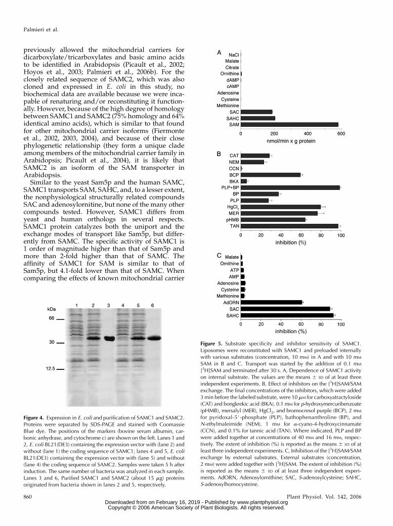
previously allowed the mitochondrial carriers fordicarboxylate/tricarboxylates and basic amino acidsto be identified in Arabidopsis (Picault et al., 2002;Hoyos et al., 2003; Palmieri et al., 2006b). For theclosely related sequence of SAMC2, which was alsocloned and expressed in E. coli in this study, nobiochemical data are available because we were inca-pable of renaturing and/or reconstituting it function-ally. However, because of the high degree of homologybetween SAMC1 and SAMC2 (75% homology and 64%identical amino acids), which is similar to that foundfor other mitochondrial carrier isoforms (Fiermonteet al., 2002, 2003, 2004), and because of their closephylogenetic relationship (they form a unique cladeamong members of the mitochondrial carrier family inArabidopsis; Picault et al., 2004), it is likely thatSAMC2 is an isoform of the SAM transporter inArabidopsis.
Similar to the yeast Sam5p and the human SAMC,SAMC1 transports SAM, SAHC, and, to a lesser extent,the nonphysiological structurally related compoundsSAC and adenosylornitine, but none of the many othercompounds tested. However, SAMC1 differs fromyeast and human orthologs in several respects.SAMC1 protein catalyzes both the uniport and theexchange modes of transport like Sam5p, but differ-ently from SAMC. The specific activity of SAMC1 is1 order of magnitude higher than that of Sam5p andmore than 2-fold higher than that of SAMC. Theaffinity of SAMC1 for SAM is similar to that ofSam5p, but 4.1-fold lower than that of SAMC. Whencomparing the effects of known mitochondrial carrier
Figure 4. Expression in E. coli and purification of SAMC1 and SAMC2.Proteins were separated by SDS-PAGE and stained with CoomassieBlue dye. The positions of the markers (bovine serum albumin, car-bonic anhydrase, and cytochrome c) are shown on the left. Lanes 1 and2, E. coli BL21(DE3) containing the expression vector with (lane 2) andwithout (lane 1) the coding sequence of SAMC1; lanes 4 and 5, E. coliBL21(DE3) containing the expression vector with (lane 5) and without(lane 4) the coding sequence of SAMC2. Samples were taken 5 h afterinduction. The same number of bacteria was analyzed in each sample.Lanes 3 and 6, Purified SAMC1 and SAMC2 (about 15 mg) proteinsoriginated from bacteria shown in lanes 2 and 5, respectively.
Figure 5. Substrate specificity and inhibitor sensitivity of SAMC1.Liposomes were reconstituted with SAMC1 and preloaded internallywith various substrates (concentration, 10 mM) in A and with 10 mM
SAM in B and C. Transport was started by the addition of 0.1 mM
[3H]SAM and terminated after 30 s. A, Dependence of SAMC1 activityon internal substrate. The values are the means 6 SD of at least threeindependent experiments. B, Effect of inhibitors on the [3H]SAM/SAMexchange. The final concentrations of the inhibitors, which were added3 min before the labeled substrate, were 10 mM for carboxyatractyloside(CAT) and bongkrekic acid (BKA), 0.1 mM for p-hydroxymercuribenzoate(pHMB), mersalyl (MER), HgCl2, and bromocresol purple (BCP), 2 mM
for pyridoxal-5#-phosphate (PLP), bathophenanthroline (BP), andN-ethylmaleimide (NEM), 1 mM for a-cyano-4-hydroxycinnamate(CCN), and 0.1% for tannic acid (TAN). Where indicated, PLP and BPwere added together at concentrations of 40 mM and 16 mM, respec-tively. The extent of inhibition (%) is reported as the means 6 SD of atleast three independent experiments. C, Inhibition of the [3H]SAM/SAMexchange by external substrates. External substrates (concentration,2 mM) were added together with [3H]SAM. The extent of inhibition (%)is reported as the means 6 SD of at least three independent experi-ments. AdORN, Adenosylornithine; SAC, S-adenosylcysteine; SAHC,S-adenosylhomocysteine.
Palmieri et al.
860 Plant Physiol. Vol. 142, 2006 www.plantphysiol.orgon February 16, 2019 - Published by Downloaded from
Copyright © 2006 American Society of Plant Biologists. All rights reserved.

inhibitors on the three orthologs, the Arabidopsis SAMcarrier exhibits a sensitivity to tannic acid and bromo-cresol purple much stronger than the yeast ortholog,but similar to that displayed by the human carrier.Carboxyatractyloside and bathophenantroline inhibitSAMC1 more than Sam5p and SAMC, whereas sen-sitivity to p-hydroxymercuribenzoate, mersalyl, andmercuric chloride is about the same for all three trans-porters. In addition, like its yeast and human coun-terparts, the Arabidopsis carrier is not inhibited bybongkrekic acid, a-cyano-4-hydroxycinnamate, and manycompounds structurally related to SAM, such as aminoacids, nucleosides, and nucleotides.
The green fluorescence of the GFP-tagged SAMC1completely overlapped with the fluorescence mito-chondrion-selective dye MitoTracker Red, demonstrat-ing that the Arabidopsis SAMC1 transporter has amitochondrial localization. However, the data avail-able suggest that SAMC1 may have a dual mitochon-drial and chloroplastic localization, as demonstratedfor several plant proteins (for reviews, see Peeters andSmall, 2001; Karniely and Pines, 2005; Mackenzie, 2005).First, using an elegant strategy based on the use of
highly purified and characterized membrane frac-tions, extraction of hydrophobic proteins with organicsolvents, SDS-PAGE separation, and tandem mass spec-trometry analysis, Ferro et al. (2002) identified SAMC1as a component of the chloroplast envelope. Second,the results reported in this article based on the use ofGFP fused proteins show that this protein is colocal-ized with a mitochondrial marker. Whereas we wereunable to detect fluorescence in the plastid, this maymerely reflect a lack of sensitivity in the GFP assay. Ithas been shown that, in the presence of an uneven
Figure 6. Kinetics of [3H]SAM transport in proteoliposomes reconsti-tuted with SAMC1. A, Uptake of SAM. 1 mM [3H]SAM was added toproteoliposomes containing 10 mM SAM (exchange, d) or 5 mM NaCland no substrate (uniport, s). Data are means 6 SD for three indepen-dent experiments. B, Efflux of [3H]SAM from proteoliposomes recon-stituted in the presence of 1 mM SAM. The internal substrate pool waslabeled with [3H]SAM by carrier-mediated exchange equilibration.Then the proteoliposomes were passed through Sephadex G-75. Theefflux of [3H]SAM was started by adding buffer A alone (d) or 5 mM
SAM in buffer A (s). Data are means 6 SD for three independentexperiments.
Figure 7. Subcellular localization of SAMC1. Transient expression inArabidopsis protoplasts of GFP fused in frame with the full-lengthSAMC1 or mMDH. GFP alone and chlorophyll were used as controlsfor targeting to cytosol and plastids, respectively; GFP fused to mMDHand MitoTracker Red for targeting to mitochondria. Merged, Mergedimage of GFP fluorescence with MitoTracker Red fluorescence (A) andof GFP fluorescence and chlorophyll autofluorescence (B). The barsindicated in the figures represent 8 mm (GFP alone and SAMC1-GFP)and 20 mm (mMDH-GFP).
S-Adenosylmethionine Transporter in Arabidopsis
Plant Physiol. Vol. 142, 2006 861 www.plantphysiol.orgon February 16, 2019 - Published by Downloaded from
Copyright © 2006 American Society of Plant Biologists. All rights reserved.

dual distribution, the large amount in one localizationmakes difficult the detection of the small amount inthe other (Duchene et al., 2005; Regev-Rudzki et al.,2005). It is also possible that the GFP promoter fusionmissed essential cis-elements that aid its targeting tothe plastid, but are not necessary for mitochondrialtargeting (Christensen et al., 2005; Kabeya and Sato,2005). Two further lines of evidence support the duallocalization of SAMC1. First, it was recently shownthat, in isolated spinach (Spinacia oleracea) leaf chloro-plasts, SAM is taken up by a transport system (Ravanelet al., 2004) that exhibits properties very similar tothose displayed by the recombinant SAMC1. Second,bioinformatic analysis of the protein sequence re-vealed a long N-terminal extension of SAMC1, indic-ative of joint plastid and mitochondrial targeting.
Because mitochondrial and plastidic demands forthis metabolite must be met by transport of cytosoli-cally derived SAM (Schroder et al., 1997; Ravanel et al.,1998; Hanson and Roje, 2001), our data strongly sug-gest that a primary physiological role of SAMC1 inautotrophic and heterotrophic tissues is probably tocatalyze the uptake of SAM in exchange for SAHC,which is produced from SAM in the mitochondrialmatrix and plastidial stroma by methyltransferaseactivities. By fulfilling this task, SAMC1 allows regen-eration of the methyl group of SAM in the cytosol,provides the organelles with methyl groups, and reg-ulates methyltransferase activities in the organelles byremoving the potent competitive inhibitor SAHC.
In addition, SAM is also a substrate for many otherenzyme-catalyzed reactions in plant organelles. Forexample, in plants, lipoic acid synthase is localizedboth in chloroplasts and mitochondria (Yasuno andWada, 2002), biotin synthase only in mitochondria(Picciocchi et al., 2001, 2003), and Thr synthase only inplastids (Wallsgrove et al., 1983; Curien et al., 1996;Laber et al., 1999). Because all of these enzymes requireSAM as an essential cofactor, a further important roleof SAMC1 is probably to catalyze the uniport of SAMinto mitochondria and possibly also into chloroplasts.Whereas further experimentation is required to con-firm the putative plastidial localization, it should benoted that the other metabolites produced from SAMwithin the mitochondria or plastids by the action ofbiotin synthetase, lipoate synthetase, and Thr synthase(i.e. 5#-deoxyadenosine and Met) cannot be exportedby SAMC1 because they are not substrates of this car-rier, implying that the presence of further transport-ers is responsible for their removal. Identification ofthese proteins should be the focus of further studiesaimed at understanding the cellular network of SAMmetabolism.
By activating Thr synthase, SAM modulates carbonflux partition between members of the Asp family ofamino acids and regulates its own biosynthesis (Hesseand Hoefgen, 2003). Because de novo Met synthesis isexclusively chloroplastic, whereas SAM synthetase iscytosolic, SAMC1 may play a key role in the interac-tion between Met synthesis and SAM metabolism.
Transgenic plants with reduced SAMC1 activity mightenable us to determine the physiological influence ofSAMC1 on de novo Met biosynthesis. First investiga-tions of SAMC1 Arabidopsis knockout plants revealedthat homozygous seeds of these knockout lines areunable to germinate (data not shown). Interestingly, ithas been shown that de novo synthesis of Met plays animportant role in seed germination and seedlinggrowth (Gallardo et al., 2002). The strong up-regulationof Met synthase and SAM synthetase observed dur-ing seed germination and seedling establishmentby Gallardo et al. (2002) parallels the high levels ofSAMC1 expression in the early stages of plant devel-opment. SAM metabolism could affect plant growthand development in several ways. For example, SAMserves as a substrate for SAM decarboxylase, the keyregulatory enzyme responsible for the production ofthe polyamines spermidine and spermine, which arebelieved to be involved in plant development (Alcazaret al., 2005; Cona et al., 2006) and stress response(Panicot et al., 2002; Perez-Amador et al., 2002; Zapataet al., 2004). Such studies would be expected to com-plement the findings of Moffatt and coworkers, whodemonstrated that a reduction in adenosine kinaseactivity by antisense inhibition resulted in decreasedcellular levels of SAM and developmental abnormal-ities (Moffatt et al., 2002). In addition, they would likelyprovide more direct proof as to whether these effectsare directly ascribable to altered SAM metabolism. Theimportance of SAM in ethylene biosynthesis and theobservation that proteins involved in SAM metabo-lism are up-regulated on wounding and herbivory(this study; Arimura et al., 2002) suggest that it willalso be interesting to analyze how the plant prioritizesthe gene regulatory and biosynthetic functions of SAMin response to both biotic and abiotic stresses.
MATERIALS AND METHODS
Materials
[3H]SAM was purchased from NEN Life Science Products. Cardiolipin and
sarkosyl (N-lauroylsarcosine) were supplied by Sigma. Egg-yolk phospho-
lipids (egg lecithin) were obtained from Fluka and Amberlite XAD-2 from
Supelco. All other reagents were analytical grade.
Sequence Search and Analysis
The genome of Arabidopsis (Arabidopsis thaliana) was screened with the
sequences of the yeast (Saccharomyces cerevisiae) Sam5p (Marobbio et al.,
2003) and the human SAMC (Agrimi et al., 2004) using BLASTP and
TBLASTN. The amino acid sequences were aligned with the ClustalW
multiple sequence alignment program (version 1.8; http://www.clustalw.
genome.ad.jp). The SAMC1 (accession no. AM260490) and SAMC2 (accession
no. AM260491) sequences, showing the lowest e-values, were selected for
further study.
RNA Isolation and Northern-Blot Analysis
Total RNA was isolated from frozen organs using TRIzol (Gibco BRL)
according to the manufacturer’s instructions. RNA concentration was mea-
sured and its integrity was checked on a 1.5% agarose gel (w/v). Hybridization
Palmieri et al.
862 Plant Physiol. Vol. 142, 2006 www.plantphysiol.orgon February 16, 2019 - Published by Downloaded from
Copyright © 2006 American Society of Plant Biologists. All rights reserved.

was performed as described in Sambrook et al. (1989), using specific probes of
237 bp (from 2110 to 1127 nucleotides of the SAMC1 cDNA) and 176 bp (from
11 to 1176 nucleotides of the SAMC2 cDNA) for SAMC1 and SAMC2,
respectively. The probes were labeled with a [32P]dCTP by random priming,
using the random prime labeling system (Amersham Bioscience). RNA
loading was checked with ethidium bromide. Membranes were autoradio-
graphed at 270�C for several days with an intensifying screen.
Expression Analysis by Real-Time RT-PCR
Total RNAs from different organs were reverse transcribed using the
GeneAmp RNA PCR core kit (Applied Biosystems) with random hexamers as
primers. For real-time PCRs, primers based on the cDNA sequences of SAMC1
and SAMC2 were designed with Primer Express (Applied Biosystems). The
forward and reverse primers corresponded to nucleotides 318 to 340 and 398
to 418 for SAMC1, nucleotides 386 to 406 and 406 to 436 for SAMC2, and
nucleotides 618 to 639 and 698 to 718 for EF1a. Real-time PCR was performed
in an optical 96-well plate using the automated ABI Prism 7000 sequence
detection system (Applied Biosystems). Fifty microliters of reaction volume
contained 2 mL of template (reverse transcribed first-strand cDNA), 25 mL
SYBR Green PCR master mix (Applied Biosystems), and 300 nM each primer.
The specificity of PCR amplification was checked with the heat dissociation
protocol following the final cycle of PCR. Separation of real-time PCR pro-
ducts on 4% (w/v) agarose gel revealed single bands of the expected size
whose identity was confirmed by direct sequencing. To correct for differences
in the amount of starting first-strand cDNAs, the Arabidopsis EF1a gene was
amplified in parallel as a reference gene. The relative quantification of SAMC1
and SAMC2 in various organs was performed according to the comparative
method (22DDCt; Bustin, 2000; Fiermonte et al., 2002; user bulletin no. 2 [P/N
4303859]; Applied Biosystems), with the stem DCt for SAMC2 as calibrator.
22DDCt 5 22(DCt sample2DCt calibrator), where DCt sample is Ct sample 2 Ct reference
gene and Ct is the threshold cycle (i.e. the PCR cycle number at which emitted
fluorescence exceeds 10 times the SD of baseline emissions).
Promoter-GUS Fusion Experiments
For promoter-GUS fusion experiments, the promoter regions of SAMC1
(from 21,146 bp to 16 bp) and SAMC2 (from 21,396 bp to 112 bp) were
amplified by PCR from Arabidopsis Col-0 genomic DNA using the follow-
ing primers: forward, 5#-CACCGGGAGATAATTGAAAGC-3# and reverse,
5#-AGCCATGAGAAACGCCTCTGACCTAAT-3# for SAMC1; and forward,
5#-CACCTGAGAGAAAAAGAAAGAAGAAAAAGAAGAGA-3# and reverse,
5#-GTCACTATCCATCTAAAACCATT-3# for SAMC2. The PCR products were
first cloned into the shuttle vector pENTR/D-TOPO (Invitrogen) and then
transferred into the binary Gateway vector pKGWFS7 in frame with the GUS
gene (Karimi et al., 2002). The resulting constructs were introduced into
Arabidopsis Col-0 plants by Agrobacterium-mediated transformation accord-
ing to the floral-dip method (Clough and Bent, 1998). To select transgenic
plants, seeds were germinated on Murashige and Skoog plates (Murashige
and Skoog, 1962) containing 1% Suc and supplemented with kanamycin in a
growth chamber (250 mmol photons m22 s21; 22�C) under a 16-h light/8-h
dark regime before transfer to soil in a climate-controlled chamber under the
same photoperiod. Samples for GUS activity analysis were harvested 8, 11, 13,
or 38 d after germination and stained overnight according to standard
protocols (Clough and Bent, 1998).
Construction of Expression Plasmids
PCR primers for SAMC1 (forward, 5#-GGATCCATGGCTCCTCTTACTCT-
CTCCGT-3#; reverse, 5#-AAGCTTTTATTCTTCTTTGGTTTCTTTAACCGT-3#)
and for SAMC2 (forward, 5#-CATAAGCTTATGGATAGTGACATTGTTT-
CCAGTAGCATA-3#; reverse, 5#-GAATTCTTAAGCATTGTGACTCTTTTGG-
CTTCT-3#) carrying BamHI and HindIII and HindIII and EcoRI sites,
respectively, were used to amplify the predicted open reading frames from
an Arabidopsis cDNA library (Minet et al., 1992). The resulting products were
cloned into the pRUN vector (derived from pKN172; Fiermonte et al., 1993) for
expression in Escherichia coli.
For subcellular localization of SAMC1 and mMDH, the SAMC1-GFP and
the mMDH-GFP fusion constructs were prepared. The coding sequences of
SAMC1 and of mMDH without the terminal codon were amplified by PCR
using the following primers: forward, 5#-CACCATGGCTCCTCTTACTCT-
CTCGT-3# and reverse, 5#-TTCTTCTTTGGTTTCTTTAACCGTATT-3# for
SAMC1; and forward, 5#-CACCATGAGGACCTCCATGTTG-3# and reverse,
5#-GTTTTCTTTGGCAAACTTGATTC-3# for tomato (Lycopersicon esculentum)
mMDH (encoded by AY725474). The resulting products were cloned into the
entry vector pENTR/D-TOPO (Invitrogen) using the Gateway recombination
system. Subsequently, the SAMC1 or the mMDH coding sequence was re-
combined into the destination vector pK7FWG2 encoding a C-terminal en-
hanced GFP under the cauliflower mosaic virus 35S promoter (Karimi et al.,
2002; CLONTECH). The inserts were sequenced to confirm identity prior to
plant transformation.
In Vivo Targeting of GFP Fusion Constructs
Protoplasts were prepared from Arabidopsis Col-0 plants (Jin et al., 2001)
grown as described above in the absence of kanamycin. The pK7FWG2 vector
containing the coding sequence of GFP, SAMC1-GFP, or mMDH-GFP was
introduced into Arabidopsis protoplasts by the polyethylene glycol-mediated
transformation method (Kang et al., 1998). After 36 to 48 h of incubation in the
dark at 22�C, protoplasts were imaged using a laser-scanning confocal micro-
scope (Leica DM IRBE microscope; TCS SPII confocal scanner). In some ex-
periments, the transformed protoplasts were incubated in the presence of
400 nM MitoTracker Red CMXRos (Molecular Probes) at 25�C for 30 min and
then extensively washed before imaging following the protocol of Studart-
Guimaraes et al. (2005).
Bacterial Expression and Purification of SAMC1
and SAMC2
The overproduction of SAMC1 and SAMC2 as inclusion bodies in the
cytosol of E. coli BL21(DE3) was accomplished as described (Fiermonte et al.,
1993). Control cultures with empty vector were processed in parallel. Inclu-
sion bodies were purified on a Suc density gradient (Fiermonte et al., 1993),
washed at 4�C with Tris-EDTA buffer (10 mM Tris/HCl, 1 mM EDTA, pH 7.0),
then twice with a buffer containing Triton X-114 (3%, w/v), 1 mM EDTA, and
10 mM PIPES/NaOH, pH 7.0, and once again with Tris-EDTA buffer (Palmieri
et al., 2001a; Picault et al., 2002; Hoyos et al., 2003). The SAMC1 and SAMC2
proteins were solubilized in 1.6% sarkosyl (w/v). Small residues were
removed by centrifugation (258,000g for 20 min at 4�C).
Reconstitution of Recombinant SAMC1 and SAMC2into Liposomes
The recombinant proteins were diluted 7-fold with buffer containing 0.6%
Triton X-114, 0.2 mM EDTA, 10 mM PIPES, pH 7.0, and then reconstituted by
cyclic removal of detergent (Palmieri et al., 1995). The reconstitution mixture
consisted of protein solution (0.12 mg), 10% Triton X-114, 10% phospholipids
as sonicated liposomes, 10 mM SAM (except where otherwise indicated),
cardiolipin (0.7 mg), 20 mM PIPES, pH 7.0, and water (final volume 700 mL).
The mixture was recycled 13 times through an Amberlite column (3.2 cm 3
0.5 cm) preequilibrated with buffer containing 10 mM PIPES, pH 7.0, and
substrate at the same concentration as in the reconstitution mixture. All op-
erations were performed at 4�C, except the passages through Amberlite,
which were carried out at room temperature.
Transport Measurements
External substrate was removed from the proteoliposomes on Sephadex
G-75 columns preequilibrated with a buffer containing 50 mM NaCl and 10 mM
PIPES, pH 7.0 (buffer A). Transport at 25�C was started by adding [3H]SAM to
substrate-loaded proteoliposomes (exchange) or to empty proteoliposomes
(uniport) and terminated, after the desired time, by the addition of 40 mM
pyridoxal-5#-P and 16 mM bathophenantroline (the inhibitor stop method;
Palmieri et al., 1995). In controls, inhibitors were added together with the
labeled substrate. Finally, the external radioactivity was removed on Sephadex
G-75 and the radioactivity in the liposomes was measured (Palmieri et al.,
1995). The experimental values were corrected by subtracting control values.
The initial transport rate was calculated from the radioactivity taken up by
proteoliposomes in the initial linear range of substrate uptake (Palmieri et al.,
1995). For efflux measurements, proteoliposomes containing 1 mM SAM were
labeled with 5 mM [3H]SAM by carrier-mediated exchange equilibration
(Palmieri et al., 1995). After 60 min, external radioactivity was removed by
passing the proteoliposomes through Sephadex G-75. Efflux was started by
S-Adenosylmethionine Transporter in Arabidopsis
Plant Physiol. Vol. 142, 2006 863 www.plantphysiol.orgon February 16, 2019 - Published by Downloaded from
Copyright © 2006 American Society of Plant Biologists. All rights reserved.

adding unlabeled external substrate or buffer A alone and terminated
by adding the inhibitors indicated above.
Other Methods
Proteins were separated by SDS-PAGE and stained with Coomassie Blue.
The N termini were sequenced and the amount of purified proteins was
estimated by laser densitometry of stained samples using carbonic anhydrase
as a protein standard (Fiermonte et al., 1998). The amount of protein incor-
porated into liposomes was measured as described previously (Fiermonte
et al., 1998). In all cases, it was about 25% of the protein added to the
reconstitution mixture.
Sequence data from this article can be found in the GenBank/EMBL data
libraries under accession numbers AM260490 (SAMC1) and AM260491
(SAMC2).
Received July 19, 2006; accepted August 29, 2006; published September 1,
2006.
LITERATURE CITED
Agrimi G, Di Noia MA, Marobbio CMT, Fiermonte G, Lasorsa
FM, Palmieri F (2004) Identification of the human mitochondrial
S-adenosylmethionine transporter: bacterial expression, reconstitution,
functional characterization and tissue distribution. Biochem J 379: 183–190
Alcazar R, Garcia-Martinez JL, Cuevas JC, Tiburcio AF, Altabella T (2005)
Overexpression of ADC2 in Arabidopsis induces dwarfism and late-
flowering through GA deficiency. Plant J 43: 425–436
Arimura G, Ozawa R, Nishioka T, Boland W, Koch T, Kuhnemann F,
Takabayashi J (2002) Herbivore-induced volatiles induce the emission
of ethylene in neighboring lima bean plants. Plant J 29: 87–98
Bedhomme M, Hoffmann M, McCarthy EA, Gambonnet B, Moran RG,
Rebbeille F, Ravanel S (2005) Folate metabolism in plants: an Arabi-
dopsis homolog of the mammalian mitochondrial folate trans-
porter mediates folate import into chloroplasts. J Biol Chem 280: 34823–
34831
Black MT, Meyer D, Widger WR, Cramer WA (1987) Light-regulated
methylation of chloroplast proteins. J Biol Chem 262: 9803–9807
Block MA, Tewari AK, Albrieux C, Marechal E, Joyard J (2002) The plant
S-adenosyl-L-methionine: Mg-protoporphyrin IX methyltransferase is
located in both envelope and thylakoid chloroplast membranes. Eur J
Biochem 269: 240–248
Bustin SA (2000) Absolute quantification of mRNA using real-time reverse
transcription polymerase chain reaction assays. J Mol Endocrinol 25:
169–193
Cantoni GL (1975) Biological methylation: selected aspects. Annu Rev
Biochem 44: 435–451
Cheng Z, Sattler S, Maeda H, Sakuragi Y, Bryant DA, DellaPenna D
(2003) Highly divergent methyltransferases catalyze a conserved reac-
tion in tocopherol and plastoquinone synthesis in cyanobacteria and
photosynthetic eukaryotes. Plant Cell 15: 2343–2356
Christensen AC, Lyznik A, Mohammed S, Elowsky CG, Elo A, Yule R,
Mackenzie SA (2005) Dual-domain, dual-targeting organellar protein
presequences in Arabidopsis can use non-AUG start codons. Plant Cell
17: 2805–2816
Clough SJ, Bent AF (1998) Floral dip: a simplified method for Agrobacterium-
mediated transformation of Arabidopsis thaliana. Plant J 16: 735–743
Cona A, Rea G, Angelini R, Federico R, Tavladoraki P (2006) Functions of
amine oxidases in plant development and defence. Trends Plant Sci 11:
80–88
Curien G, Dumas R, Ravanel S, Douce R (1996) Characterization of an
Arabidopsis thaliana cDNA encoding an S-adenosylmethionine-sensitive
threonine synthase: threonine synthase from higher plants. FEBS Lett
390: 85–90
d’Harlingue A, Camara B (1985) Plastid enzymes of terpenoid biosynthe-
sis: purification and characterization of g-tocopherol methyltransferase
from Capsicum chromoplasts. J Biol Chem 260: 15200–15203
Duchene AM, Giritch A, Hoffmann B, Cognat V, Lancelin D, Peeters NM,
Zaepfel M, Marechal-Drouard L, Small ID (2005) Dual targeting is the
rule for organellar aminoacyl-tRNA synthetases in Arabidopsis thaliana.
Proc Natl Acad Sci USA 102: 16484–16489
Fernie AR, Carrari F, Sweetlove LJ (2004) Respiratory metabolism: glycol-
ysis, the TCA cycle and mitochondrial electron transport. Curr Opin
Plant Biol 7: 254–261
Ferro M, Salvi D, Riviere-Rolland H, Vermat T, Seigneurin-Berny D,
Grunwald D, Garin J, Joyard J, Rolland N (2002) Integral membrane
proteins of the chloroplast envelope: identification and subcellular
localization of new transporters. Proc Natl Acad Sci USA 99: 11487–
11492
Fiermonte G, De Leonardis F, Todisco S, Palmieri L, Lasorsa FM, Palmieri
F (2004) Identification of the mitochondrial ATP-Mg/Pi transporter:
bacterial expression, reconstitution, functional characterization, and
tissue distribution. J Biol Chem 279: 30722–30730
Fiermonte G, Dolce V, David L, Santorelli FM, Dionisi-Vici C, Palmieri F,
Walker JE (2003) The mitochondrial ornithine transporter: bacterial
expression, reconstitution, functional characterization, and tissue dis-
tribution of two human isoforms. J Biol Chem 278: 32778–32783
Fiermonte G, Dolce V, Palmieri F (1998) Expression in Escherichia coli,
functional characterization and tissue distribution of isoforms A and B
of the phosphate carrier from bovine mitochondria. J Biol Chem 273:
22782–22787
Fiermonte G, Palmieri L, Todisco S, Agrimi G, Palmieri F, Walker JE
(2002) Identification of the mitochondrial glutamate transporter: bacte-
rial expression, reconstitution, functional characterization, and tissue
distribution of two human isoforms. J Biol Chem 277: 19289–19294
Fiermonte G, Walker JE, Palmieri F (1993) Abundant bacterial expression
and reconstitution of an intrinsic membrane transport protein from
bovine mitochondria. Biochem J 294: 293–299
Fiore C, Trezeguet V, Le Saux A, Roux P, Schwimmer C, Dianoux AC,
Noel F, Lauquin GJ, Brandolin G, Vignais PV (1998) The mitochondrial
ADP/ATP carrier: structural, physiological and pathological aspects.
Biochimie 80: 137–150
Fontecave M, Atta M, Mulliez E (2004) S-adenosylmethionine: nothing
goes to waste. Trends Biochem Sci 29: 243–249
Gallardo K, Job C, Groot SP, Puype M, Demol H, Vandekerckhove J, Job
D (2002) Importance of methionine biosynthesis for Arabidopsis seed
germination and seedling growth. Physiol Plant 116: 238–247
Grimm R, Grimm M, Eckerskorn C, Pohlmeyer K, Rohl T, Soll J (1997)
Postimport methylation of the small subunit of ribulose-1,5-biphosphate
carboxylase in chloroplasts. FEBS Lett 408: 350–354
Halestrap AP (1975) The mitochondrial pyruvate carrier: kinetics and
specificity for substrates and inhibitors. Biochem J 148: 85–96
Hanson AD, Gage DA, Shachar-Hill Y (2000) Plant one-carbon metabo-
lism and its engineering. Trends Plant Sci 5: 206–213
Hanson AD, Roje S (2001) One-carbon metabolism in higher plants. Annu
Rev Plant Physiol Plant Mol Biol 52: 119–137
Hesse H, Hoefgen R (2003) Molecular aspects of methionine biosynthesis.
Trends Plant Sci 8: 259–262
Hoyos ME, Palmieri L, Wertin T, Arrigoni R, Polacco JC, Palmieri F (2003)
Identification of a mitochondrial transporter for basic amino acids in
Arabidopsis thaliana by functional reconstitution into liposomes and
complementation in yeast. Plant J 33: 1027–1035
Jin JB, Kim YA, Kim SJ, Lee SH, Kim DH, Cheong GW, Hwang I (2001) A
new dynamin-like protein, ADL6, is involved in trafficking from the
trans-Golgi network to the central vacuole in Arabidopsis. Plant Cell 13:
1511–1525
Kabeya Y, Sato N (2005) Unique translation initiation at the second AUG
codon determines mitochondrial localization of the phage-type RNA
polymerases in the moss Physcomitrella patens. Plant Physiol 138: 369–382
Kang SG, Jin JB, Piao HL, Pih KT, Jang HJ, Lim JH, Hwang I (1998)
Molecular cloning of an Arabidopsis cDNA encoding a dynamin-like
protein that is localized to plastids. Plant Mol Biol 38: 437–447
Karimi M, Inze D, Depicker A (2002) GATEWAY vectors for Agrobacterium-
mediated plant transformation. Trends Plant Sci 7: 193–195
Karniely S, Pines O (2005) Single translation—dual destination: mecha-
nisms of dual protein targeting in eukaryotes. EMBO Rep 6: 420–425
Klingenberg M (1989) Molecular aspects of the adenine nucleotide carrier
from mitochondria. Arch Biochem Biophys 270: 1–14
Kobayashi H, Ngernprasirtsiri J, Akazawa T (1990) Transcriptional
regulation and DNA methylation in plastids during transitional con-
version of chloroplasts to chromoplasts. EMBO J 9: 307–313
Laber B, Maurer W, Hanke C, Grafe S, Ehlert S, Messerschmidt A,
Clausen T (1999) Characterization of recombinant Arabidopsis thaliana
threonine synthase. Eur J Biochem 263: 212–221
Palmieri et al.
864 Plant Physiol. Vol. 142, 2006 www.plantphysiol.orgon February 16, 2019 - Published by Downloaded from
Copyright © 2006 American Society of Plant Biologists. All rights reserved.

Leroch M, Kirchberger S, Haferkamp I, Wahl M, Neuhaus HE, Tjaden J
(2005) Identification and characterization of a novel plastidic adenine
nucleotide uniporter from Solanum tuberosum. J Biol Chem 280: 17992–
18000
Mackenzie SA (2005) Plant organellar protein targeting: a traffic plan still
under construction. Trends Cell Biol 15: 548–554
Marobbio CM, Agrimi G, Lasorsa FM, Palmieri F (2003) Identification
and functional reconstitution of yeast mitochondrial carrier of
S-adenosylmethionine. EMBO J 22: 5975–5982
Millar AH, Heazlewood JL (2003) Genomic and proteomic analysis of
mitochondrial carrier proteins in Arabidopsis. Plant Physiol 131: 443–453
Miller JR, Busby RW, Jordan SW, Cheek J, Henshaw TF, Ashley GW,
Broderick JB, Cronan JE, Marletta MA (2000) Escherichia coli LipA is a
lipoyl synthase: in vitro biosynthesis of lipoylated pyruvate dehydro-
genase complex from octanoyl-acyl carrier protein. Biochemistry 39:
15166–15178
Minet M, Dufour ME, Lacroute F (1992) Complementation of Saccharo-
myces cerevisiae auxotrophic mutants by Arabidopsis thaliana cDNAs.
Plant J 2: 417–422
Moffatt BA, Stevens YY, Allen MS, Snider JD, Pereira LA, Todorova MI,
Summers PS, Weretilnyk EA, Martin-McCaffrey L, Wagner C (2002)
Adenosine kinase deficiency is associated with developmental abnor-
malities and reduced transmethylation. Plant Physiol 128: 812–821
Montasser Kouhsari S, Keith G, Weil GH (1978) Methylation of yeast
tRNAPhe by enzymes from cytoplasm, chloroplasts and mitochondria
of Phaseolus vulgaris. Biochim Biophys Acta 521: 576–583
Murashige T, Skoog F (1962) A revised medium for rapid growth and
bioassays with tobacco tissue cultures. Plant Physiol 15: 473–497
Nunes-Nesi A, Carrari F, Lytovchenko A, Smith AM, Loureiro ME,
Ratcliffe RG, Sweetlove LJ, Fernie AR (2005) Enhanced photosynthetic
performance and growth as a consequence of decreasing mitochondrial
malate dehydrogenase activity in transgenic tomato plants. Plant
Physiol 137: 611–622
Palmieri F (2004) The mitochondrial transporter family (SLC25): physio-
logical and pathological implications. Pflugers Arch 447: 689–709
Palmieri F, Agrimi G, Blanco E, Castegna A, Di Noia MA, Iacobazzi V,
Lasorsa FM, Marobbio CMT, Palmieri L, Scarcia P, et al (2006a)
Identification of mitochondrial carriers in Saccharomyces cerevisiae by
transport assay of reconstituted recombinant proteins. Biochim Biophys
Acta doi/10.1016/j.bbabio.2006.05.023
Palmieri F, Indiveri C, Bisaccia F, Iacobazzi V (1995) Mitochondrial
metabolite carrier proteins: purification, reconstitution, and transport
studies. Methods Enzymol 260: 349–369
Palmieri L, Pardo B, Lasorsa FM, Del Arco A, Kobayashi K, Iijima M,
Runswick MJ, Walker JE, Saheki T, Satrustegui J, et al (2001a) Citrin
and Aralar1 are Ca21-stimulated aspartate/glutamate transporters in
mitochondria. EMBO J 20: 5060–5069
Palmieri L, Rottensteiner H, Girzalsky W, Scarcia P, Palmieri F, Erdmann
R (2001b) Identification and functional reconstitution of the yeast
peroxisomal adenine nucleotide transporter. EMBO J 20: 5049–5059
Palmieri L, Todd CD, Arrigoni R, Hoyos ME, Santoro A, Polacco CJ,
Palmieri F (2006b) Arabidopsis mitochondria have two basic amino acid
transporters with partially overlapping specificities and differential
expression in seedling development. Biochim Biophys Acta doi/
10.1016/j.bbabio.2006.03.025
Panicot M, Minguet EG, Ferrando A, Alcazar R, Blazquez MA, Carbonell
J, Altabella T, Koncz C, Tiburcio AF (2002) A polyamine metabolon
involving aminopropyl transferase complexes in Arabidopsis. Plant
Cell 14: 2539–2551
Peeters N, Small I (2001) Dual targeting to mitochondria and chloroplasts.
Biochim Biophys Acta 1541: 54–63
Perez-Amador MA, Leon J, Green PJ, Carbonell J (2002) Induction of the
arginine decarboxylase ADC2 gene provides evidence for the involve-
ment of polyamines in the wound response in Arabidopsis. Plant
Physiol 130: 1454–1463
Picault N, Hodges M, Palmieri L, Palmieri F (2004) The growing family of
mitochondrial carrier in Arabidopsis. Trends Plant Sci 9: 138–146
Picault N, Palmieri L, Pisano I, Hodges M, Palmieri F (2002) Identification
of a novel transporter for dicarboxylates and tricarboxylates in plant
mitochondria: bacterial expression, reconstitution, functional charac-
terization and tissue distribution. J Biol Chem 277: 24204–24211
Picciocchi A, Douce R, Alban C (2001) Biochemical characterization of the
Arabidopsis biotin synthase reaction: the importance of mitochondria in
biotin synthesis. Plant Physiol 127: 1224–1233
Picciocchi A, Douce R, Alban C (2003) The plant biotin synthase reaction:
identification and characterization of essential mitochondrial accessory
protein components. J Biol Chem 278: 24966–24975
Ravanel S, Block MA, Rippert P, Jabrin S, Curien G, Rebeille F, Douce R
(2004) Methionine metabolism in plants: chloroplasts are autonomous
for de novo methionine synthesis and can import S-adenosylmethionine
from the cytosol. J Biol Chem 279: 22548–22557
Ravanel S, Gakiere B, Job D, Douce R (1998) The specific features of
methionine biosynthesis and metabolism in plants. Proc Natl Acad Sci
USA 95: 7805–7812
Regev-Rudzki N, Karniely S, Ben-Haim NN, Pines O (2005) Yeast aco-
nitase in two location and two metabolic pathways: seeing small amount
is believing. Mol Biol Cell 16: 4163–4171
Sambrook J, Fritsch EF, Maniatis T (1989) Molecular Cloning: A Labora-
tory Manual, Ed 2. Cold Spring Harbor Laboratory Press, Cold Spring
Harbor, NY
Schroder G, Eichel J, Breinig S, Schroder J (1997) Three differentially
expressed S-adenosylmethionine synthetases from Catharanthus roseus:
molecular and functional characterization. Plant Mol Biol 33: 211–222
Studart-Guimeraes C, Gibon Y, Frankel N, Wood CC, Zanor MI, Fernie
AR, Carrari F (2005) Identification and characterisation of the alfa and
beta subunits of the succinyl CoA ligase of tomato. Plant Mol Biol 59:
781–791
Wallsgrove RM, Lea PJ, Miflin BJ (1983) Intracellular localization of
aspartate kinase and the enzyme of threonine and methionine biosyn-
thesis in green leaves. Plant Physiol 71: 780–784
Weber AP, Schwacke R, Flugge UI (2005) Solute transporters of the plastid
envelope membrane. Annu Rev Plant Biol 56: 133–164
Weretilnyk EA, Alexander KJ, Drebenstedt M, Snider JD, Summers PS,
Moffatt BA (2001) Maintaining methylation activities during salt stress:
the involvement of adenosine kinase. Plant Physiol 125: 856–865
Yasuno R, Wada H (2002) The biosynthetic pathway for lipoic acid is
present in plastids and mitochondria in Arabidopsis thaliana. FEBS Lett
517: 110–114
Ying Z, Mulligan RM, Janney N, Houtz RL (1999) Rubisco small and large
subunit N-methyltransferases: bi- and mono-functional methyltransfer-
ases that methylate the small and large subunits of Rubisco. J Biol Chem
274: 36750–36756
Zapata PJ, Serrano M, Pretel MT, Amoros A, Botella MA (2004) Poly-
amines and ethylene changes during germination of different plant
species under salinity. Plant Sci 167: 781–788
Zimmermann P, Hirsch-Hoffmann M, Hennig L, Gruissem W (2004)
GENEVESTIGATOR: Arabidopsis microarray database and analysis
toolbox. Plant Physiol 136: 2621–2632
S-Adenosylmethionine Transporter in Arabidopsis
Plant Physiol. Vol. 142, 2006 865 www.plantphysiol.orgon February 16, 2019 - Published by Downloaded from
Copyright © 2006 American Society of Plant Biologists. All rights reserved.