molecular mechanism of immunoglobulin gene …mediatum.ub.tum.de/doc/603746/603746.pdfsummary...
TRANSCRIPT
Technische Universität München
GSF - Forschungszentrum für Umwelt und Gesundheit
Institut für Molekulare Strahlenbiologie
Direktor: apl. Prof. Dr. Jean-Marie Buerstedde
Neuherberg
Molecular Mechanism of Immunoglobulin Gene Conversion In Chicken DT40 Cells
Huseyin Saribasak
Vollständiger Abdruck der von der Fakultät Wissenschaftszentrum Weihenstephan für
Ernährung, Landnutzung und Umwelt der Technische Universität München zur Erlangung
des akademischen Grades eines
Doktors der Naturwissenschaften (Dr. rer. nat.)
genehmigte Dissertation.
Vorsitzender: Univ.- Prof. Dr. Siegfried Scherer
Prüfer der Dissertation: 1. Univ.- Prof. Dr. Wolfgang Wurst
2. apl. Prof. Dr. Jean-Marie Buerstedde
Die Dissertation wurde am 15.05.2006 bei der Technischen Universität München
eingereicht und durch die Fakultät Wissenschaftszentrum Weihenstephan für Ernährung,
Landnutzung und Umwelt am 04.09.2006 angenommen.
Summary
Immunoglobulin (Ig) gene conversion is one of three B cell specific processes which
create the repertoire of antigen receptors in B cells. The process involves the unidirectional
transfer of sequences from pseudogene V segments into the rearranged V gene. There are
general and lymphoid-specific trans-acting factors as well as cis-acting elements involved
in this process.
Gene conversion is most likely initiated by AID mediated cytosine deamination. If the
resulting uracils need to be further processed by uracil glycosylase (UNG), UNG
inactivation should block gene conversion and induce transition mutations. We report in
this thesis that is indeed the phenotype in the B cell line DT40. Ig gene conversion is
almost completely extinguished in the UNG-/- mutant and large numbers of transition
mutations at C/G bases accumulate within the rearranged Ig light chain gene. The mutation
rate of UNG-/- cells is about seven times higher than of pseudo V gene deleted (ψV-) cells
in which mutations arise presumably after uracil excision. In addition, UNG-/- cells show
relatively more mutations upstream and downstream of the VJ segment. This suggests that
hypermutating B cells process AID-induced uracils with approximately one seventh of
uracils giving rise to mutations depending on their position.
Besides gene conversion DT40 is also a good tool to analyze somatic hypermutation.
Whereas in human and mice all of the nucleotides are target for mutations, in DT40 C/G
bases are the main targets. Proliferating cell nuclear antigen (PCNA) coordinates DNA
synthesis during replication. Mono-ubiquination of PCNA by RAD18 is induced by DNA
damage and leads to the engagement of translesion DNA polymerases in S. cerevisiae. In
this thesis we showed that RAD18 is sensitive to DNA damaging agents and somatic
mutations are reduced in the absence of RAD18. The results demonstrate that PCNA
ubiquitination by RAD18 as well as by some other factors in vertebrate cells are needed,
most likely by recruitment of error-prone translesion polymerases.
ii

Zusammenfassung
Immunglobulin (Ig) Genkonversion ist einer der drei B-Zellspezifischen Prozesse,
welche für die große Variabilität der Antigenrezeptoren in den B-Zellen verantwortlich
sind. Maßgebend für den Prozess ist der unidirektionale Transfer von Sequenzen eines
strangaufwärts gelegenen V-Segment-Pseudogens in das exprimierte, umgebaute V-
Region-Gen. Der Prozess läuft unter der Beteiligung allgemeiner und lymphoid-
spezifischer trans-wirkender Faktoren, sowie cis-wirkender Elemente ab.
Genkonversion wird wahrscheinlich durch die AID-Vermittelte Desaminierung von
Cytosin eingeleitet. Wird das resultierende Uracil im weiteren Verlauf durch Uracil
Glykosylase (UNG) modifiziert, sollte die Inaktivierung von UNG die Genkonversion
stoppen und Transition-Mutationen einführen. In dieser Doktorarbeit konnte nachgewiesen
werden, dass in einer UNG-/- Mutante der B-Zelllinie DT40 die Genkonversion fast
komplett zum Erliegen kommt und sich Transitionen an C/G Basen im umgebauten Ig Gen
der leichten Kette häufen. Die Mutationsrate von UNG-/- Zellen ist ungefähr siebenmal
höher als in Zellen mit einer Deletion der V-Pseudogene (ψV-), bei welchen Mutationen
vermutlich nach dem Ausschneiden des Uracils auftreten. Darüber hinaus zeigen UNG-/-
Zellen relativ mehr Mutationen stromaufwärts und stromabwärts des VJ-Segments. Das
weist darauf hin, dass in Hypermutierenden B-Zellen etwa ein Siebtel des durch AID
eingeführten Uracils in Abhängigkeit von seiner Position zu Mutationen führt.
Neben der Genkonversion lässt sich in DT40 auch die somatische Hypermutation
untersuchen. Während beim Menschen und in der Maus alle Nukleotide Ziel für
Mutationen sind, werden in DT40 hauptsächlich C/G Basen mutiert. „Proliferating cell
nuclear antigen“ (PCNA) koordiniert die DNA-Synthese während der Replikation. Mono-
Ubiquitinierung von PCNA durch RAD18 wird durch Schädigung der DNA eingeleitet und
führt zur Rekrutierung der Translesion DNA Polymerasen in S. cerevisiae. In der
vorliegenden Arbeit haben wir gezeigt, dass RAD18 sensitiv für DNA schädigende
Agenzien ist und somatische Mutationen in Abwesenheit von RAD18 abnehmen. Die
Ergebnisse veranschaulichen, dass die Ubiquitinierung von PCNA durch RAD18 und
einige andere Faktoren in Zellen von Wirbeltieren für die somatische Hypermutation
notwendig ist, wahrscheinlich weil es Error-Prone Translesion Polymerasen rekrutiert.
iii
Abbreviations:
AID Activation Induced Cytidine Deaminase
AP Apurinic/ apyrimidinic
APOBEC-1 Apolipoprotein B mRNA Editing Catalytic Polypeptide 1
BDT Big Dye Terminator
BSR Blasticidine S Resitance gene
CIP Calf Intestine Phosphatase
CSR Class Switch Recombination
C region Immunoglobulin Constant region
dNTP Deoxynucleotide Triple Phosphate
DMSO Dimethyl sulfoxide
D region Immunoglobulin Diversity region
DSB Double Strand Break
EDTA Ethylene di-Amine Tetra Acetic Acid
EF Elongation Factor
EST Expressed Sequence Tag
EtBr Ethidium Bromide
FBS Fetal Bovine Serum
GC Gene Conversion
GPT Guanine Phosphoribosyl Transferase
HR Homologues Recombination
4-HT 4 Hydroxy Tamoxifen
Ig Immunoglubulin
IRES Internal Ribosome Entry Site
J region Immunoglobulin Joining region
LB Luria Broth
NHEJ Non Homologues End Joining
iv
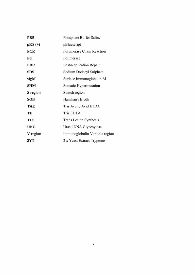
PBS Phosphate Buffer Saline
pKS (+) pBluescript
PCR Polymerase Chain Reaction
Pol Polimerase
PRR Post-Replication Repair
SDS Sodium Dodecyl Sulphate
sIgM Surface Immunoglobulin M
SHM Somatic Hypermutation
S region Switch region
SOB Hanahan's Broth
TAE Tris Acetic Acid ETDA
TE Tris EDTA
TLS Trans Lesion Synthesis
UNG Urasil DNA Glycosylase
V region Immunoglobulin Variable region
2YT 2 x Yeast Extract Tryptone
v
Table of Contents
Summary ii
Zuzammenfassung iii
Abbreviations iv
INTRODUCTION:
I. Immunity and Generation of Antibody Diversity 1
A. V(D)J Recombination 1
B. Somatic Hypermutation 2
C. Class Switch Recombination 4
II. Chicken B-Cell Repertoire Development and Bursal B-Cell Line DT40 5
A. Immunoglobulin Gene Conversion 5
B. Chicken B Cell Development 7
C. DT40 7
III. Molecular Mechanism of Gene Conversion 9
A. Factors involved in initiation 9
1. AID 9
2. UNG 11
B. The effect of homologous recombination (HR) 12
C. Somatic Hypermutation Besides Gene Conversion in DT40 13
* Pseudogene (ψV) Knockout 13
D. Translesion Synthesis (TLS) 16
IV. Objectives: 18
MATERIALS
I. Instruments 19
II. Experimental Kits 19
III. Oligonucleotides 19
vi
IV. Enzymes 21
V. Immuno-staining antibodies & anti-antibodies 22
VI. DNA Size Marker for electrophoresis 22
VII. Bacterial Strain 22
VIII. Mammalian Cell Line 22
IX. Plasmids 22
METHODS
I. Molecular Biology 23
A. Culture of E.coli 23
B. E.coli DH5α competent cell preparation 23
C. Polymerase Chain Reaction (PCR) 25
D. Analysis of DNA by electrophoresis 25
E. Purification & Gel Purification of DNA 25
F. DNA Ligation 26
G. Transformation 26
H. Colony PCR 26
I. Plasmid preparation 27
J. Restriction Enzyme Digestion 28
K. First strand cDNA synthesis 28
L. Determination of DNA and RNA concentration 28
M. Genomic DNA Isolation 29
N. Total RNA Isolation 29
O. Cell Extract Preparation 30
II. Cell Culture 31
A. Basic Cell Culture Conditions 31
B. Thawing of the cells 31
C. Freeze down of cells 31
D. Transfection 31
E. Identifying Targeted Events By PCR 32
vii
F. Subcloning of DT40 cells 32
G. Analysis of IgM expression by FACS 33
H. Drug Resistance Marker Recycling 33
I. Sorting: 33
J. Colony Survial Assay 34
RESULTS
Section I. UNG 35
I. Genomic Identification of UNG Locus 35
II. Vector Construction 35
A. Knockout Vectors 35
B. Complementation Vector 38
III. Knockout and Complementation of UNG 38
IV. UNG Activity Test 39
V. Quantification of Surface IgM Levels 41
A. Ig Reversion Assay 41
B. Ig Mutation Assay 42
VI. Ig Light Chain Sequencing 45
Section II. RAD18 51
I. Genomic locus of RAD18 51
II. Knockout of RAD18 52
III. Mutagen Sensitivity Test 53
IV. Quantification of Surface IgM Levels 54
V. Ig Light Chain Sequencing 55
DISCUSSION 56
REFERENCES 62
Acknowledgements 74Publication List 75
viii

1
INTRODUCTION:
I. Immunity and Generation of Antibody Diversity:
The function of the immune system is to protect vertebrates from foreign substances.
Two types of responses have evolved to accomplish this task: Innate and adaptive
(acquired) immunity. Innate immunity involves hereditary components which provide an
immediate "first-line" of defense to frequently encountered pathogens whereas acquired
immunity involves particular type of cells and proteins which provide a protection to
particular type of agents (Tortora et al., 1995).
A component of the blood, white blood cells are produced in the bone marrow and help
to defend the body against infectious disease and foreign materials as part of the immune
system. Lymphocytes are a type of white blood cells and they are the primary effectors in
the adaptive immune system. There are two broad categories of lymphocytes, namely T
cells and B cells. T cells are chiefly responsible for cell-mediated immunity whereas B cells
are primarily responsible for humoral immunity (antibody related). Antibodies
(immunoglobulins-Ig) are proteins synthesized and secreted by B cells during immune
response within processes like phagocytosis or inflammation. Each antibody recognizes a
specific antigen unique to its target (Janeaway et al., 2001).
To fight against such a variety of invaders, the immune system needs to generate vast
numbers of receptors. To do this, immune system has evolved different strategies like
V(D)J recombination, somatic hypermutation (SHM) and class switch recombination
(CSR).
A. V(D)J Recombination:
As a primary immune repertoire, V(D)J recombination (Fig. 1) involves combinatorial
gene rearrangement of functional immunoglobulin genes (Tonegawa et al, 1983). It is
antigen independent and occurs in the bone marrows. Initiated by lymphoid and sequence
specific recombination activating genes (RAG1 and RAG2) (Schatz et al., 1988; Schatz et
al., 1989), it recruits Non Homologous End Joining (NHEJ) mechanism for resolution
(Chaudhuri et al., 2004). B cells then migrate to secondary lymphoid organs like spleen and
lymph node, where they can undergo further antigen driven Ig gene diversification through
Somatic Hypermutation and Class Switch Recombination.
2
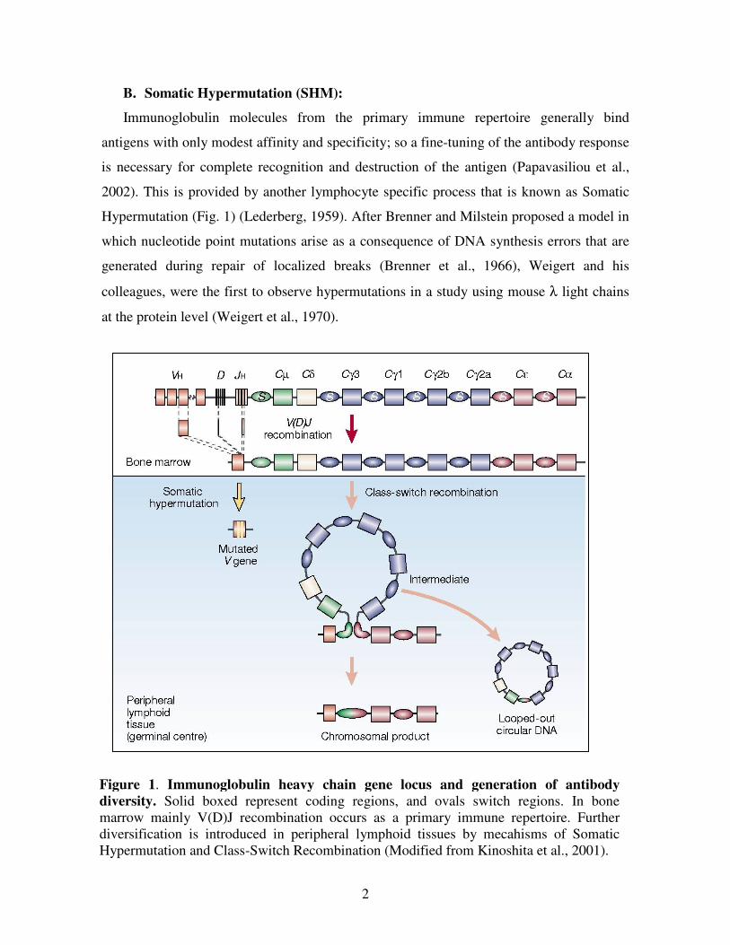
B. Somatic Hypermutation (SHM):
Immunoglobulin molecules from the primary immune repertoire generally bind
antigens with only modest affinity and specificity; so a fine-tuning of the antibody response
is necessary for complete recognition and destruction of the antigen (Papavasiliou et al.,
2002). This is provided by another lymphocyte specific process that is known as Somatic
Hypermutation (Fig. 1) (Lederberg, 1959). After Brenner and Milstein proposed a model in
which nucleotide point mutations arise as a consequence of DNA synthesis errors that are
generated during repair of localized breaks (Brenner et al., 1966), Weigert and his
colleagues, were the first to observe hypermutations in a study using mouse λ light chains
at the protein level (Weigert et al., 1970).
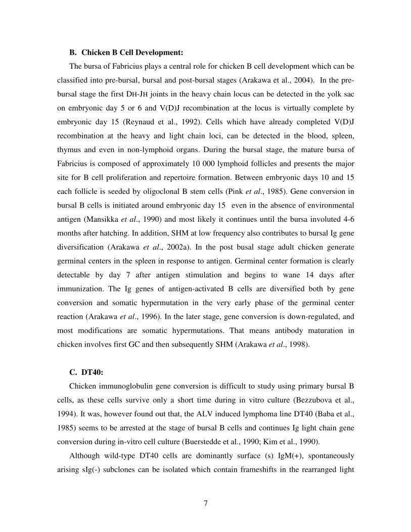
Figure 1. Immunoglobulin heavy chain gene locus and generation of antibody
diversity. Solid boxed represent coding regions, and ovals switch regions. In bone
marrow mainly V(D)J recombination occurs as a primary immune repertoire. Further
diversification is introduced in peripheral lymphoid tissues by mecahisms of Somatic
Hypermutation and Class-Switch Recombination (Modified from Kinoshita et al., 2001).
3
Somatic Hypermutation is triggered when the surface Ig of B cell encounters with an
antigen. Following this, point mutations are introduced at a high rate specifically into
immunoglobulin V genes of activated B cells (Kinoshita et al., 2001). In the germinal
centers, successive cycle of these mutations and selection lead B cells produce antibodies
with higher affinity which is called as affinity maturation (Fig. 2) (Siskind et al., 1969).
Owing to the strong effects of selection, the true rates of SHM in vivo are unclear. However
it is thought generally that SHM occurs at an extraordinarily high frequency (10-3
bases per
generation). This frequency is almost one million times higher than the level of mutation in
housekeeping genes (Martin et al., 2002). Transition mutations occur more frequently than
transversions (Golding et al., 1987) and using a huge collection of mutation data it is found
out that there is a hotspot motif for SHM (Rogozin et al., 1992). That is, mutations of G-C
base pairs that are in RGYW (R=A or G, Y=C or T, W=A or T) and the complementary
WRCY motifs occur at a high frequency. These mutational properties are probably the
result of the biochemical specificities of the mutator enzymes (Martin et al., 2002).
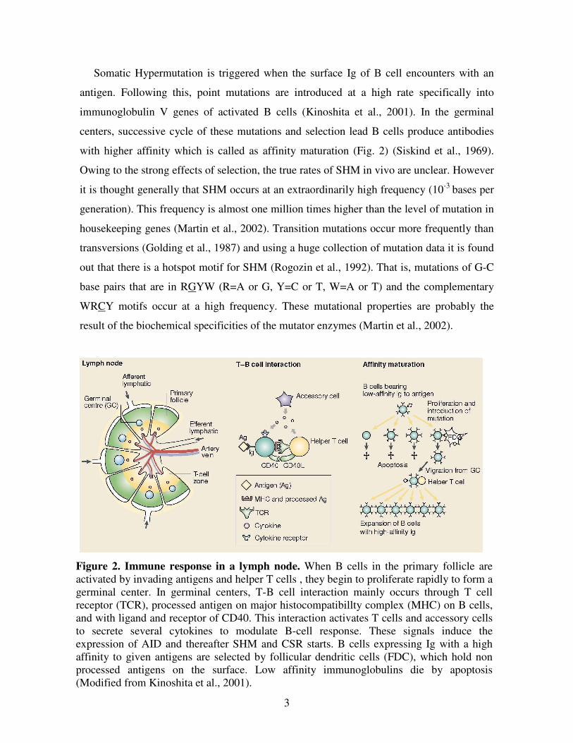
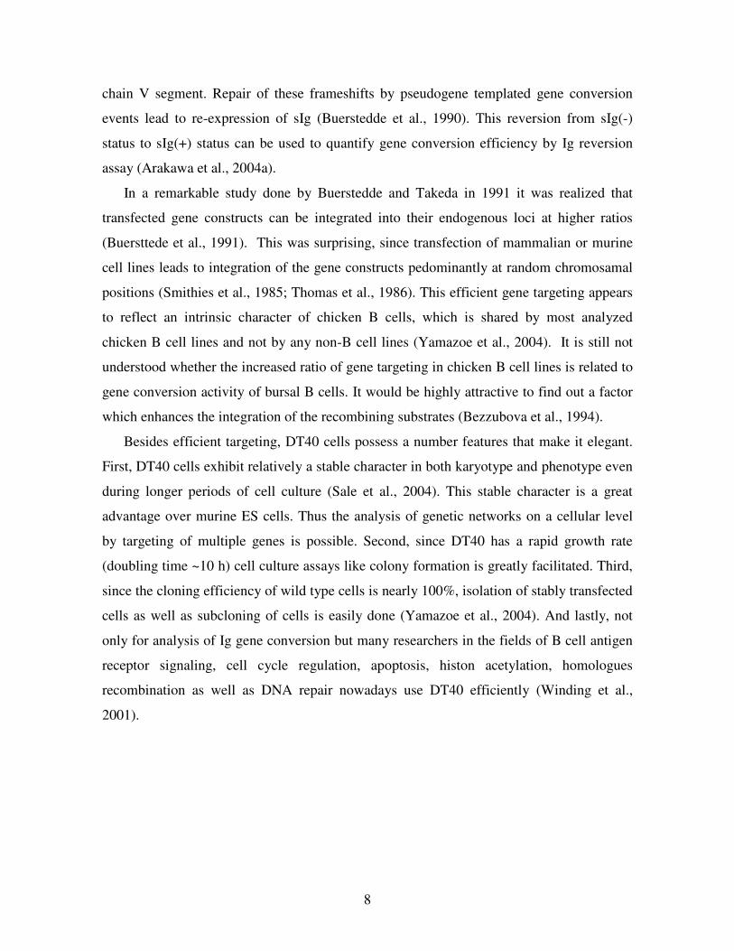
Figure 2. Immune response in a lymph node. When B cells in the primary follicle are
activated by invading antigens and helper T cells , they begin to proliferate rapidly to form a
germinal center. In germinal centers, T-B cell interaction mainly occurs through T cell
receptor (TCR), processed antigen on major histocompatibillty complex (MHC) on B cells,
and with ligand and receptor of CD40. This interaction activates T cells and accessory cells
to secrete several cytokines to modulate B-cell response. These signals induce the
expression of AID and thereafter SHM and CSR starts. B cells expressing Ig with a high
affinity to given antigens are selected by follicular dendritic cells (FDC), which hold non
processed antigens on the surface. Low affinity immunoglobulins die by apoptosis
(Modified from Kinoshita et al., 2001).
4
One of the questions for SHM is how the target genes are selected for mutations. In
several studies, it has been shown that transcription of the target locus is essential for
somatic hypermutation (Peters et al., 1996; Fukita et al., 1998), and that the frequency of
mutation directly correlated to the rate of transcription (Goyenechea et al., 1997; Bachl et
al., 2001). Mutations start downstream of the promoter and lie around 1.5kb to 2 kb along
Ig locus (Lebecque et al., 1990). In addition, Ig enhancer as well as transcription regulatory
elements were also shown to be important for SHM (Betz et al., 1994; Klix et al., 1998;
Michael et al., 2003).
It could well be that SHM start with a single strand (Brenner et al., 1966) or a double
strand DNA breaks (Bross et al., 2000; Papavasiliou et al., 2000) or both (Sale et al., 1998),
although the association of double strand breaks are controversial (Bross et al., 2002; Chua
et al., 2002).
Mutations can arise actively from the error-prone processing of DNA breaks or
passively from the absence of normal repair machineries (Papavasiliou et al., 2002). It has
been documented in many articles that part of the Base Excision Repair and Mismatch
Repair System is involved in SHM (reviewed in Martin et al., 2002; Neuberger et al.,
2005). The proteins shown to be important for SHM up to now are including a lymphoid
specific factor Activation Induced Cytidine Deamniase (AID) (Muramatsu et al., 2000),
Uracil DNA Glycosylase (UNG) (Di Noia et al., 2002; Rada et al., 2002), MSH2 (Rada et
al., 1998; Rada et al., 2004) as well as error-prone polymerases like pol η (Zeng et al.,
2001) and REV1 (Simpson et al. 2003; Jansen et al., 2006).
C. Class Switch Recombination:
Class Switch Recombination (Fig. 1) allows the expression of antibodies that have the
same antigen specificity but are of a secondary isotype like (IgG, IgA, or IgE). The
immunoglobulin locus contains CH genes, which encode proteins that are capable of
different effector functions (Chaudhuri et al., 2004). CSR involves recombination between
switch regions, which are highly repetitive GC-rich sequences of 1-10 kb in length that lie
upstream of Ig C-region genes (Martin et al., 2002). Signal transduction through surface
IgM, CD40 and cytokine receptors of a B cell induce the reaction of CSR and the repair
step is mediated by non-homologues end-joining repair system (Kinoshita et al., 2001).
5
Mechanistically, CSR is a deletional recombination event like V(D)J recombination,
however because neither consensus nor homologues sequence is generally found around
junctions, it is different from it. Despite being distinct processes targeted to distinct Ig
regions, CSR and SHM are similar in that they both occur in antigen-simulated B cells,
require transcription as well as AID for initiation (Chaudhuri et al., 2004). The comparison
between SHM and CSR is summarized in Table 1.
II. Chicken B-Cell Repertoire Formation and Bursal B-Cell Line DT40:
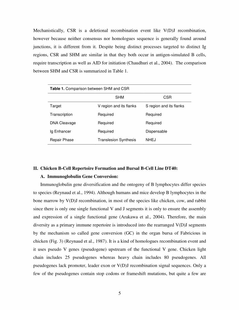
A. Immunoglobulin Gene Conversion:
Immunoglobulin gene diversification and the ontogeny of B lymphocytes differ species
to species (Reynaud et al., 1994). Although humans and mice develop B lymphocytes in the
bone marrow by V(D)J recombination, in most of the species like chicken, cow, and rabbit
since there is only one single functional V and J segments it is only to ensure the assembly
and expression of a single functional gene (Arakawa et al., 2004). Therefore, the main
diversity as a primary immune repertoire is introduced into the rearranged V(D)J segments
by the mechanism so called gene conversion (GC) in the organ bursa of Fabricious in
chicken (Fig. 3) (Reynaud et al., 1987). It is a kind of homologues recombination event and
it uses pseudo V genes (pseudogene) upstream of the functional V gene. Chicken light
chain includes 25 pseudogenes whereas heavy chain includes 80 pseudogenes. All
pseudogenes lack promoter, leader exon or V(D)J recombination signal sequences. Only a
few of the pseudogenes contain stop codons or frameshift mutations, but quite a few are
Table 1. Comparison between SHM and CSR
SHM CSR
Target V region and its flanks S region and its flanks
Transcription Required Required
DNA Cleavage Required Required
Ig Enhancer Required Dispensable
Repair Phase Translesion Synthesis NHEJ
6
truncated in their 5’ or 3’ ends (Reynaud et al., 1989). Unlike V(D)J recombination or
CSR, after gene conversion pseudogenes do not disappear that is, it is not a cut & paste
mechanism but a copy & paste mechanism (Reynaud et al., 1987; Carlson et al., 1990).
Only the pseudogenes on the same chromosome are used as donors (Carlson et al., 1990)
and pseudogenes that are either more homologous, closer or in the opposite orientation to
the rearranged V segment are preferred (McCormack et al., 1990; Sayegh et al., 1999).
Conversion tracts range from 8 bp to around 200 bp (McCormack et al., 1990). The 5' ends
of the gene conversion tracts always begin in regions of homology between the pseudogene
donor and recipient V segment, whereas the 3' ends can occur in regions of non-homology
and often encompass nucleotide insertions or deletions (McCormack et al., 1990).
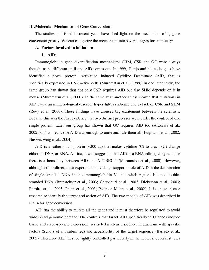
Figure 3. Chicken Ig gene diversification. Antibody diversity in chicken mainly
involves gene conversion and somatic hypermutation. Chicken has only a single V and J
segment in the light chain locus which can be functionally rearranged by V(D)J
recombination. The rearranged VJ segment is diversified by segmental gene conversion
using pseudo V genes as donors. Rearranged VJ segments also undergoes somatic
hypermutation in the bursa and spleen.
7
B. Chicken B Cell Development:
The bursa of Fabricius plays a central role for chicken B cell development which can be
classified into pre-bursal, bursal and post-bursal stages (Arakawa et al., 2004). In the pre-
bursal stage the first DH-JH joints in the heavy chain locus can be detected in the yolk sac
on embryonic day 5 or 6 and V(D)J recombination at the locus is virtually complete by
embryonic day 15 (Reynaud et al., 1992). Cells which have already completed V(D)J
recombination at the heavy and light chain loci, can be detected in the blood, spleen,
thymus and even in non-lymphoid organs. During the bursal stage, the mature bursa of
Fabricius is composed of approximately 10 000 lymphoid follicles and presents the major
site for B cell proliferation and repertoire formation. Between embryonic days 10 and 15
each follicle is seeded by oligoclonal B stem cells (Pink et al., 1985). Gene conversion in
bursal B cells is initiated around embryonic day 15 even in the absence of environmental
antigen (Mansikka et al., 1990) and most likely it continues until the bursa involuted 4-6
months after hatching. In addition, SHM at low frequency also contributes to bursal Ig gene
diversification (Arakawa et al., 2002a). In the post busal stage adult chicken generate
germinal centers in the spleen in response to antigen. Germinal center formation is clearly
detectable by day 7 after antigen stimulation and begins to wane 14 days after
immunization. The Ig genes of antigen-activated B cells are diversified both by gene
conversion and somatic hypermutation in the very early phase of the germinal center
reaction (Arakawa et al., 1996). In the later stage, gene conversion is down-regulated, and
most modifications are somatic hypermutations. That means antibody maturation in
chicken involves first GC and then subsequently SHM (Arakawa et al., 1998).
C. DT40:
Chicken immunoglobulin gene conversion is difficult to study using primary bursal B
cells, as these cells survive only a short time during in vitro culture (Bezzubova et al.,
1994). It was, however found out that, the ALV induced lymphoma line DT40 (Baba et al.,
1985) seems to be arrested at the stage of bursal B cells and continues Ig light chain gene
conversion during in-vitro cell culture (Buerstedde et al., 1990; Kim et al., 1990).
Although wild-type DT40 cells are dominantly surface (s) IgM(+), spontaneously
arising sIg(-) subclones can be isolated which contain frameshifts in the rearranged light
8
chain V segment. Repair of these frameshifts by pseudogene templated gene conversion
events lead to re-expression of sIg (Buerstedde et al., 1990). This reversion from sIg(-)
status to sIg(+) status can be used to quantify gene conversion efficiency by Ig reversion
assay (Arakawa et al., 2004a).
In a remarkable study done by Buerstedde and Takeda in 1991 it was realized that
transfected gene constructs can be integrated into their endogenous loci at higher ratios
(Buersttede et al., 1991). This was surprising, since transfection of mammalian or murine
cell lines leads to integration of the gene constructs pedominantly at random chromosamal
positions (Smithies et al., 1985; Thomas et al., 1986). This efficient gene targeting appears
to reflect an intrinsic character of chicken B cells, which is shared by most analyzed
chicken B cell lines and not by any non-B cell lines (Yamazoe et al., 2004). It is still not
understood whether the increased ratio of gene targeting in chicken B cell lines is related to
gene conversion activity of bursal B cells. It would be highly attractive to find out a factor
which enhances the integration of the recombining substrates (Bezzubova et al., 1994).
Besides efficient targeting, DT40 cells possess a number features that make it elegant.
First, DT40 cells exhibit relatively a stable character in both karyotype and phenotype even
during longer periods of cell culture (Sale et al., 2004). This stable character is a great
advantage over murine ES cells. Thus the analysis of genetic networks on a cellular level
by targeting of multiple genes is possible. Second, since DT40 has a rapid growth rate
(doubling time ~10 h) cell culture assays like colony formation is greatly facilitated. Third,
since the cloning efficiency of wild type cells is nearly 100%, isolation of stably transfected
cells as well as subcloning of cells is easily done (Yamazoe et al., 2004). And lastly, not
only for analysis of Ig gene conversion but many researchers in the fields of B cell antigen
receptor signaling, cell cycle regulation, apoptosis, histon acetylation, homologues
recombination as well as DNA repair nowadays use DT40 efficiently (Winding et al.,
2001).
9
III. Molecular Mechanism of Gene Conversion:
The studies published in recent years have shed light on the mechanism of Ig gene
conversion greatly. We can categorize the mechanism into several stages for simplicity:
A. Factors involved in initiation:
1. AID:
Immunoglobulin gene diversification mechanisms SHM, CSR and GC were always
thought to be different until one AID comes out. In 1999, Honjo and his colleagues have
identified a novel protein, Activation Induced Cytidine Deaminase (AID) that is
specifically expressed in CSR active cells (Muramatsu et al., 1999). In one later study, the
same group has shown that not only CSR requires AID but also SHM depends on it in
mouse (Muramatsu et al., 2000). In the same year another study showed that mutations in
AID cause an immunological disorder hyper IgM syndrome due to lack of CSR and SHM
(Revy et al., 2000). These findings have aroused big excitement between the scientists.
Because this was the first evidence that two distinct processes were under the control of one
single protein. Later our group has shown that GC requires AID too (Arakawa et al.,
2002b). That means one AID was enough to unite and rule them all (Fugmann et al., 2002;
Nussenzweig et al., 2004).
AID is a rather small protein (~200 aa) that makes cytidine (C) to uracil (U) change
either on DNA or RNA. At first, it was suggested that AID is a RNA-editing enzyme since
there is a homology between AID and APOBEC-1 (Muramatsu et al., 2000). However,
although still indirect, most experimental evidence support a role of AID in the deamination
of single-stranded DNA in the immunoglobulin V and switch regions but not double-
stranded DNA (Bransteitter et al., 2003; Chaudhuri et al., 2003; Dickerson et al., 2003;
Ramiro et al., 2003; Pham et al., 2003; Peterson-Mahrt et al., 2002). It is under intense
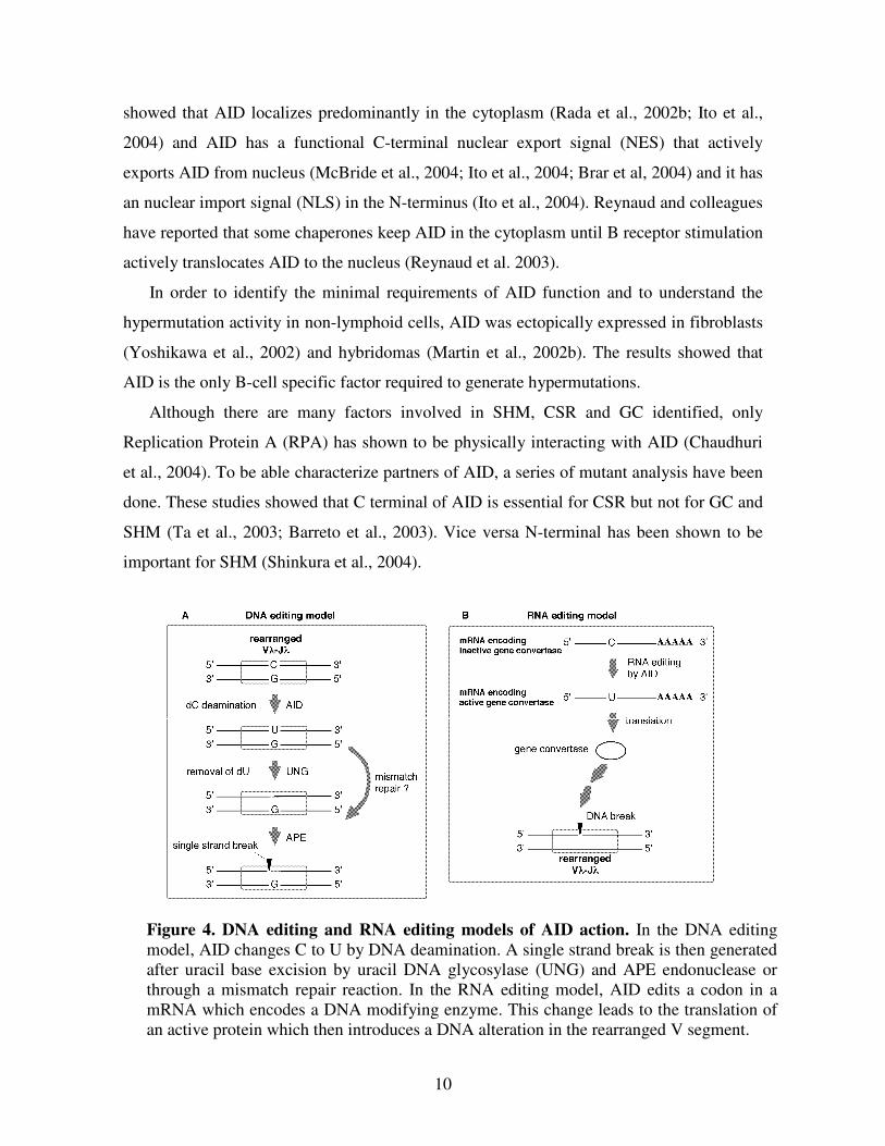
research to identify the target and action of AID. The two models of AID was described in
Fig. 4 for gene conversion.
AID has the ability to mutate all the genes and it must therefore be regulated to avoid
widespread genomic damage. The controls that target AID specifically to Ig genes include
tissue and stage-specific expression, restricted nuclear residence, interactions with specific
factors (Schotz et al., submitted) and accessibility of the target sequence (Barreto et al.,
2005). Therefore AID must be tightly controlled particularly in the nucleus. Several studies
10
showed that AID localizes predominantly in the cytoplasm (Rada et al., 2002b; Ito et al.,
2004) and AID has a functional C-terminal nuclear export signal (NES) that actively
exports AID from nucleus (McBride et al., 2004; Ito et al., 2004; Brar et al, 2004) and it has
an nuclear import signal (NLS) in the N-terminus (Ito et al., 2004). Reynaud and colleagues
have reported that some chaperones keep AID in the cytoplasm until B receptor stimulation
actively translocates AID to the nucleus (Reynaud et al. 2003).
In order to identify the minimal requirements of AID function and to understand the
hypermutation activity in non-lymphoid cells, AID was ectopically expressed in fibroblasts
(Yoshikawa et al., 2002) and hybridomas (Martin et al., 2002b). The results showed that
AID is the only B-cell specific factor required to generate hypermutations.
Although there are many factors involved in SHM, CSR and GC identified, only
Replication Protein A (RPA) has shown to be physically interacting with AID (Chaudhuri
et al., 2004). To be able characterize partners of AID, a series of mutant analysis have been
done. These studies showed that C terminal of AID is essential for CSR but not for GC and
SHM (Ta et al., 2003; Barreto et al., 2003). Vice versa N-terminal has been shown to be
important for SHM (Shinkura et al., 2004).
Figure 4. DNA editing and RNA editing models of AID action. In the DNA editing
model, AID changes C to U by DNA deamination. A single strand break is then generated
after uracil base excision by uracil DNA glycosylase (UNG) and APE endonuclease or
through a mismatch repair reaction. In the RNA editing model, AID edits a codon in a
mRNA which encodes a DNA modifying enzyme. This change leads to the translation of
an active protein which then introduces a DNA alteration in the rearranged V segment.
11
2. UNG:
Uracil may arise as an aberrant base in DNA either through misincorporation of dUMP
opposite adenine during DNA replication (Brynolf et al., 1978; Tye et al., 1978) or by
spontaneous deamination of cytosine residues in DNA generating a U:G mispair (Lindahl,
1993; Nilsen et al., 2000). If left not repaired, these uracils may give rise to transition
mutations C to T or G to A, in a subsequent replication (Duncan et al., 1982; Impellizzeri et
al., 1991) or may result in mutant proteins after transcriptional bypass by RNA polymerase
(Viswanathan et al., 1999).
Uracil residues in DNA are substrates for base excision repair (Lindahl, 2000). In this
repair, uracil DNA glycosylase (UNG - in all organisms) as well as SMUG1, TDG and
MBD4 (in human and mice) are the responsible enzymes to remove uracils from DNA
(Lindahl, 1974; Pearl, 2000). Mammalian UNG gene encodes two isoforms, nuclear
(UNG1) and mitochondrial (UNG2), which are generated via transcription from different
promoters and alternative RNA splicing (Nilsen et al., 1997).
Although UNG has been identified more than 30 years since now, a link between UNG
and Ig gene diversification has never been thought. It was just 4 years ago Neuberger and
his colleagues have published three papers and discovered uracils as the main intermediates
of antibody diversity (Petersen-Mahrt et al., 2002; DiNoia et al., 2002; Rada et al., 2002).
Later, it was also showed in the human patients that are deficient for UNG that CSR is
decreased and SHM is perturbed (Imai et al., 2003). However, direct evidence for the
presence of uracils in DNA that were produced by AID, came just recently in the cells that
are induced to express AID (Martomo et al., 2005).
The DNA deamination model of antibody diversification predicts that recruitment of
AID to the Ig locus causes localized deamination of dC → dU; repair of this uracil in DNA
then leads to SHM, GC or CSR depending on the pathway used for resolving the dU lesion,
as well as on the precise location of the lesion (either the IgV domain or in the vicinity of
the switch region) (Neuberger et al., 2003). In the case for gene conversion (Fig. 4), these
uracils are removed by UNG, and the abasic sites are repaired in a recombination mediated
manner and pseudogenes as donors.
12
B. The effect of homologous recombination (HR):
Studies in yeast S. cerevisiae have shown that gene conversion and targeted integration
depends on the RAD52 pathway and these genes mediate the repair of double-strand break
(DSB) repair by homologous recombination (Haber et al., 1999). RAD52 epistasis group
encode proteins which function either in the recognition of the double-strand break
(RAD50, MRE11 and XRS2) or in the promotion of homology search and strand invasion
(RAD51, RAD52, RAD54, RAD55, RAD57) (Symington, 2002).
The mechanism of chicken Ig gene conversion and HR might be related, because both
processes occur at high levels in DT40 and other chicken B-cell lines (Buerstedde et al.,
1990). Analysis of HR in vertebrates was initiated by the discovery of mammalian
orthologs of the yeast recombination gene RAD51 (Shinohara et al., 1993). Homologues of
the RAD51, RAD52 and RAD54 genes were first cloned from chicken bursal cells by
reverse PCR using degenerate primers derived for conservative sequence motifs of the
yeast proteins. Gene disruptions in DT40 revealed that RAD51 deficient cells do not
survive, most likely because they cannot repair replication induced DSBs (Sonoda et al.,
1998). In contrast to the severe recombination and repair defect of the S. cereviseae RAD52
mutants, the DT40 RAD52 deficient cells have only modestly reduced targeted integration
frequencies and are not hyper-sensitive to DNA damage (Yamaguchi-Iwai et al., 1998).
However, RAD54 deficient DT40 cells are highly X-ray sensitive compared to wild type
cells, have 100 fold decreased targeted integration ratios and also show reduced Ig light
chain gene conversion activity (Bezzubova et al., 1997). The disruption of the Nbs1 gene
which is the counterpart of the yeast XRS2 gene produces a phenotype very similar to the
RAD54 phenotype in DT40 (Tauchi et al., 2002). Vertebrates have five RAD51 paralogues
(RAD51B, RAD51C, RAD51D, XRCC2 and XRCC3) and DT40 mutants of each of these
genes have reduced targeted integration frequencies and DSB repair deficiencies (Takata et
al., 2001). Interestingly, some of the functions of the XRCC3 may be compensated by
RAD52, because inactivation of XRCC3 in cells lacking RAD52 results in chromosomal
instability and cell death (Fujimori et al., 2001).
The studies of the RAD52 pathway in DT40 prove that DSB repair is well conserved
during eukaryotic evolution and that targeted integration in vertebrate cells is a side effect
of DSB repair. The high ratios of targeted integration in DT40 most likely reflects the up-
13
regulation of the RAD52 pathway in bursal B cells compared to other chicken and
mammalian cells. This increased general homologous recombination activation is required
for efficient Ig gene conversion, but it cannot explain how gene conversion events are
specifically initiated in the Ig loci.
C. Somatic Hypermutation Besides Gene Conversion in DT40:
In wild type DT40 cells besides gene conversion there seems to occur also single point
mutations close to GC tracts. These non templated mutations were first suggested as they
may arise in consequence of errors in recombination (Buersttedde et al., 1991; Reynaud et
al., 1987). However, they were also suggested in several studies that these are not related
with gene conversions (Sale, 2004; Carlson et al., 1990). This led the idea that SHM and
GC might be mechanistically related since both of the processes occur mainly in
immunoglobulin V gene (Reynaud et al., 1987; Maizels, 1995; Weill et al., 1996). Direct
evidence for the relationship between SHM and GC came with the inactivation of the
RAD51 paralogues in DT40 cells. Normally, these proteins involved in HR but knockout
study revealed a reduction in frequency of the GC events along with increase in the
frequency of SHM (Sale et al., 2001). This finding showed exactly that both GC and SHM
can occur in the same cell line and that the initiating events of GC and SHM are related but
they differ in the manner by which most likely U:G mispairs are resolved.
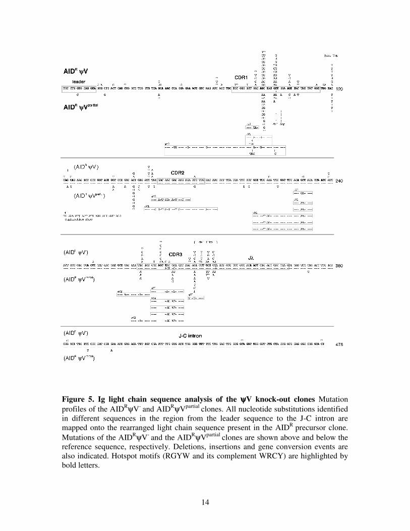
• Pseudogene (ψψψψV) Knockout:
In order to get further insight into how SHM and GC is being regulated and to get
intermediates of gene conversion reaction, we deleted ψV genes in DT40 (Arakawa et al.,
2004b). The results demonstrated that the deletion of the nearby pseudogene donors
abolishes Ig gene conversion in DT40 and activates a mutation activity which closely
resembles Ig hypermutation (SHM). The features shared between the new activity and
SHM include 1) AID dependence, 2) a predominance of single nucleotide substitutions, 3)
distribution of the mutations within the 5' transcribed region, 4) a preference for hotspots
and 5) Ig gene specificity. The only difference with regard to Ig hypermutation in vivo is
the relative lack of mutations in A/T bases and a predominance of transversion mutations in
the ψV knock-out clones. However, this difference is also seen in
14
Figure 5. Ig light chain sequence analysis of the ψψψψV knock-out clones Mutation
profiles of the AIDRψV
- and AID
RψVpartial
clones. All nucleotide substitutions identified
in different sequences in the region from the leader sequence to the J-C intron are
mapped onto the rearranged light chain sequence present in the AIDR
precursor clone.
Mutations of the AIDRψV
- and the AID
RψVpartial
clones are shown above and below the
reference sequence, respectively. Deletions, insertions and gene conversion events are
also indicated. Hotspot motifs (RGYW and its complement WRCY) are highlighted by
bold letters.
15
hypermutating EBV transformed B cell lines (Bachl et al., 1996; Faili et al., 2002) and
DT40 mutants of RAD51-paralogues (Sale et al., 2001) indicating that part of the Ig
hypermutator activity is missing in transformed B cell lines.
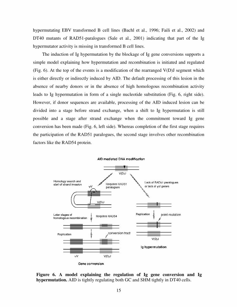
The induction of Ig hypermutation by the blockage of Ig gene conversions supports a
simple model explaining how hypermutation and recombination is initiated and regulated
(Fig. 6). At the top of the events is a modification of the rearranged V(D)J segment which
is either directly or indirectly induced by AID. The default processing of this lesion in the
absence of nearby donors or in the absence of high homologous recombination activity
leads to Ig hypermutation in form of a single nucleotide substitution (Fig. 6, right side).
However, if donor sequences are available, processing of the AID induced lesion can be
divided into a stage before strand exchange, when a shift to Ig hypermutation is still
possible and a stage after strand exchange when the commitment toward Ig gene
conversion has been made (Fig. 6, left side). Whereas completion of the first stage requires
the participation of the RAD51 paralogues, the second stage involves other recombination
factors like the RAD54 protein.
Figure 6. A model explaining the regulation of Ig gene conversion and Ig
hypermutation. AID is tightly regulating both GC and SHM tightly in DT40 cells.
16
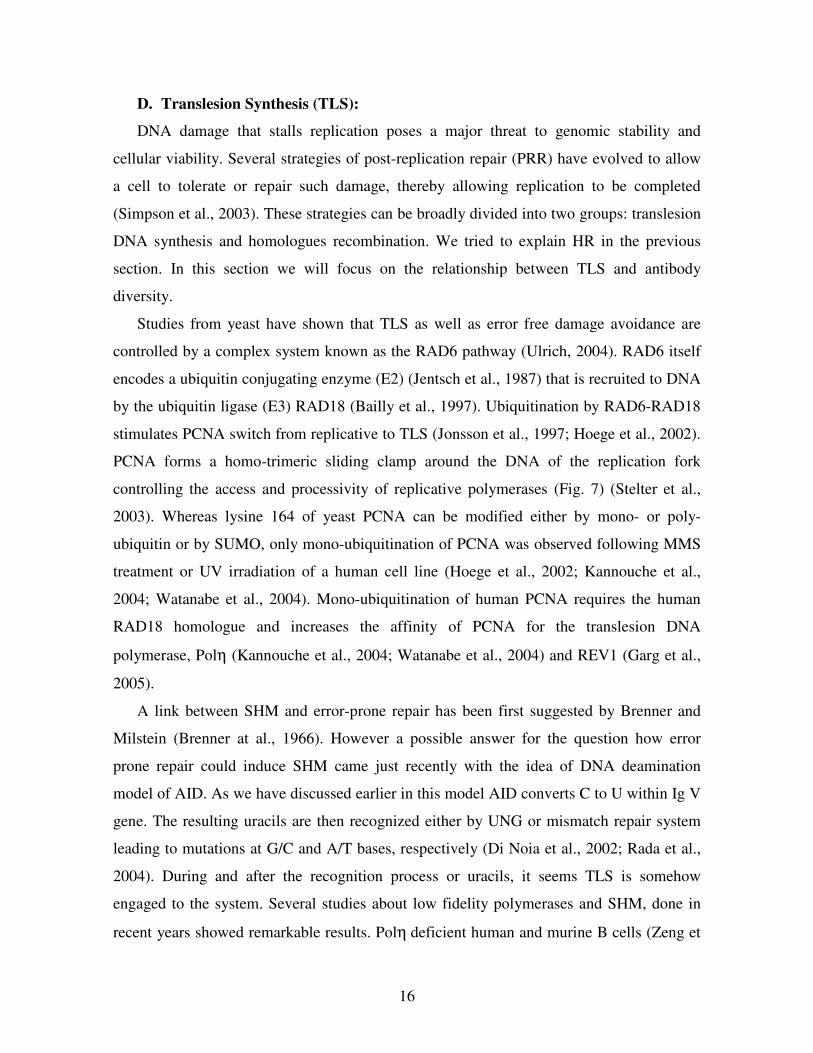
D. Translesion Synthesis (TLS):
DNA damage that stalls replication poses a major threat to genomic stability and
cellular viability. Several strategies of post-replication repair (PRR) have evolved to allow
a cell to tolerate or repair such damage, thereby allowing replication to be completed
(Simpson et al., 2003). These strategies can be broadly divided into two groups: translesion
DNA synthesis and homologues recombination. We tried to explain HR in the previous
section. In this section we will focus on the relationship between TLS and antibody
diversity.
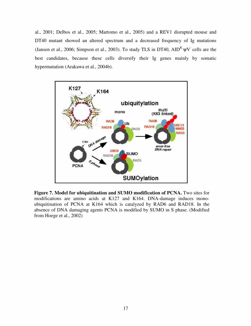
Studies from yeast have shown that TLS as well as error free damage avoidance are
controlled by a complex system known as the RAD6 pathway (Ulrich, 2004). RAD6 itself
encodes a ubiquitin conjugating enzyme (E2) (Jentsch et al., 1987) that is recruited to DNA
by the ubiquitin ligase (E3) RAD18 (Bailly et al., 1997). Ubiquitination by RAD6-RAD18
stimulates PCNA switch from replicative to TLS (Jonsson et al., 1997; Hoege et al., 2002).
PCNA forms a homo-trimeric sliding clamp around the DNA of the replication fork
controlling the access and processivity of replicative polymerases (Fig. 7) (Stelter et al.,
2003). Whereas lysine 164 of yeast PCNA can be modified either by mono- or poly-
ubiquitin or by SUMO, only mono-ubiquitination of PCNA was observed following MMS
treatment or UV irradiation of a human cell line (Hoege et al., 2002; Kannouche et al.,
2004; Watanabe et al., 2004). Mono-ubiquitination of human PCNA requires the human
RAD18 homologue and increases the affinity of PCNA for the translesion DNA
polymerase, Polη (Kannouche et al., 2004; Watanabe et al., 2004) and REV1 (Garg et al.,
2005).
A link between SHM and error-prone repair has been first suggested by Brenner and
Milstein (Brenner at al., 1966). However a possible answer for the question how error
prone repair could induce SHM came just recently with the idea of DNA deamination
model of AID. As we have discussed earlier in this model AID converts C to U within Ig V
gene. The resulting uracils are then recognized either by UNG or mismatch repair system
leading to mutations at G/C and A/T bases, respectively (Di Noia et al., 2002; Rada et al.,
2004). During and after the recognition process or uracils, it seems TLS is somehow
engaged to the system. Several studies about low fidelity polymerases and SHM, done in
recent years showed remarkable results. Polη deficient human and murine B cells (Zeng et
17
al., 2001; Delbos et al., 2005; Martomo et al., 2005) and a REV1 disrupted mouse and
DT40 mutant showed an altered spectrum and a decreased frequency of Ig mutations
(Jansen et al., 2006; Simpson et al., 2003). To study TLS in DT40, AIDR ψV
- cells are the
best candidates, because these cells diversify their Ig genes mainly by somatic
hypermutation (Arakawa et al., 2004b).
Figure 7. Model for ubiquitination and SUMO modification of PCNA. Two sites for
modifications are amino acids at K127 and K164. DNA-damage induces mono-
ubiquitination of PCNA at K164 which is catalyzed by RAD6 and RAD18. In the
absence of DNA damaging agents PCNA is modified by SUMO in S phase. (Modified
from Hoege et al., 2002)
18
IV. Objectives:
Many studies suggest that hypermutation and switch recombination is initiated by AID-
induced cytosine deamination in the rearranged V(D)J segments, and the resulting uracils
are removed by UNG (Muramatsu et al., 2000; Di Noia et al., 2002; Imai et al., 2003). It is
an interesting question whether gene conversion, in analogy to hypermutation and switch
recombination, requires the processing of uracils by UNG. It was reported that expression
of an Ugi transgene in DT40 reduced gene conversion to 30% of the wild-type level (Di
Noia et al., 2004), suggesting that uracil glycosylase activity enhances gene conversion.
However this phenotype remains difficult to interpret, because contrary to expectation no
evidence for an increased mutation rate was found and it was unclear how efficiently and
specifically Ugi transgene expression inhibited UNG activity. Therefore, our first objective
includes disruption of the UNG gene in DT40 and analysis of the phenotype with regard to
gene conversion.
PCNA mono-ubiquitination by RAD18 has been shown to be important for DNA
damage tolerance in yeast (Ulrich et al., 2004). Although it has been reported RAD18 is
dispensable for hypermutation in DT40 (Simpson et al., 2005), we wondered this also holds
true in a background which mainly diversifies V region by hypermutations (Arakawa et al.,
2004b). Thus our second objective includes knockout of RAD18 in DT40 (AIDR ψV
-) cells.
As an experimental overview, the targeting constructs were prepared, transfections were
done and phenotypes were analyzed by either Ig reversion and or Ig mutation assay as well
as by sequencing of the immunoglobulin V genes.
19
MATERIALS :
I. Instruments:
• PCR machines: GeneAmp PCR System 9700 [Applied Biosystems, CA; USA]
• Centrifuges: Heraeus [Kendro Lab. Products, Osterode, GERMANY]
• Electrophoresis chambers: Horizontal & Vertical [Bio-Rad Lab., CA; USA]
• Gel Visualization: Gel Doc 2000TM
[Bio-Rad Lab., California; USA]
• Spectrophotometer: BioPhotometer [Eppendorf GmbH, Hamburg; GERMANY]
• Incubators: Heraeus [Kendro Lab. Products, Osterode; GERMANY]
• Culture Shaker: Innova 4430 [New Brunswick Scientific, Nürtingen; GERMANY]
• Electroporator: Gene Pulse XcellTM
[Bio-Rad Lab., CA; USA]
• Vacuum Pump: [Vacuubrand GmbH, Wertheim; GERMANY]
• Vacuum Manifold: [Millipore GmbH, Schwalbach; GERMANY]
• Analytical FACS: BDTM
LSRII Flow Cytometer [Becton Dickinson, CA; USA]
• Quantitative FACS: MoFloTM
Cell Sorter [Dako Colorado Inc., Colorado; USA]
• Light Microscope: Axiovert 25 [Zeiss, Göttingen; GERMANY]
• Cell Viability Analyzer: Vi-CellTM
[Beckman Coulter GmbH, Krefeld; GERMANY]
• Phosphoimager: Fuji, FLA-3000 [Fuji Photo Film Corp., Tokyo; JAPAN]
• DNA Sequencer: ABI 3730 DNA Analyzer [Applied Biosystems & Hitachi; Hitachi
High Tech. Corp., Tokyo; JAPAN.
II. Experimental Kits:
• PCR Purification: [Qiagen GmbH, Hilden; GERMANY]
• Gel Extraction: [Qiagen GmbH, Hilden; GERMANY]
• DNA Ligation: Version 2.1 [Takara Bio Inc., Shiga; JAPAN]
• Plasmid Isolation (Mini): [Qiagen GmbH, Hilden; GERMANY]
• Plasmid Isolation (Maxi): [Qiagen GmbH, Hilden; GERMANY]
• First strand cDNA Synthesis: Super Script IIITM
[Invirogen GmbH, Karlsruhe;
GERMAY]
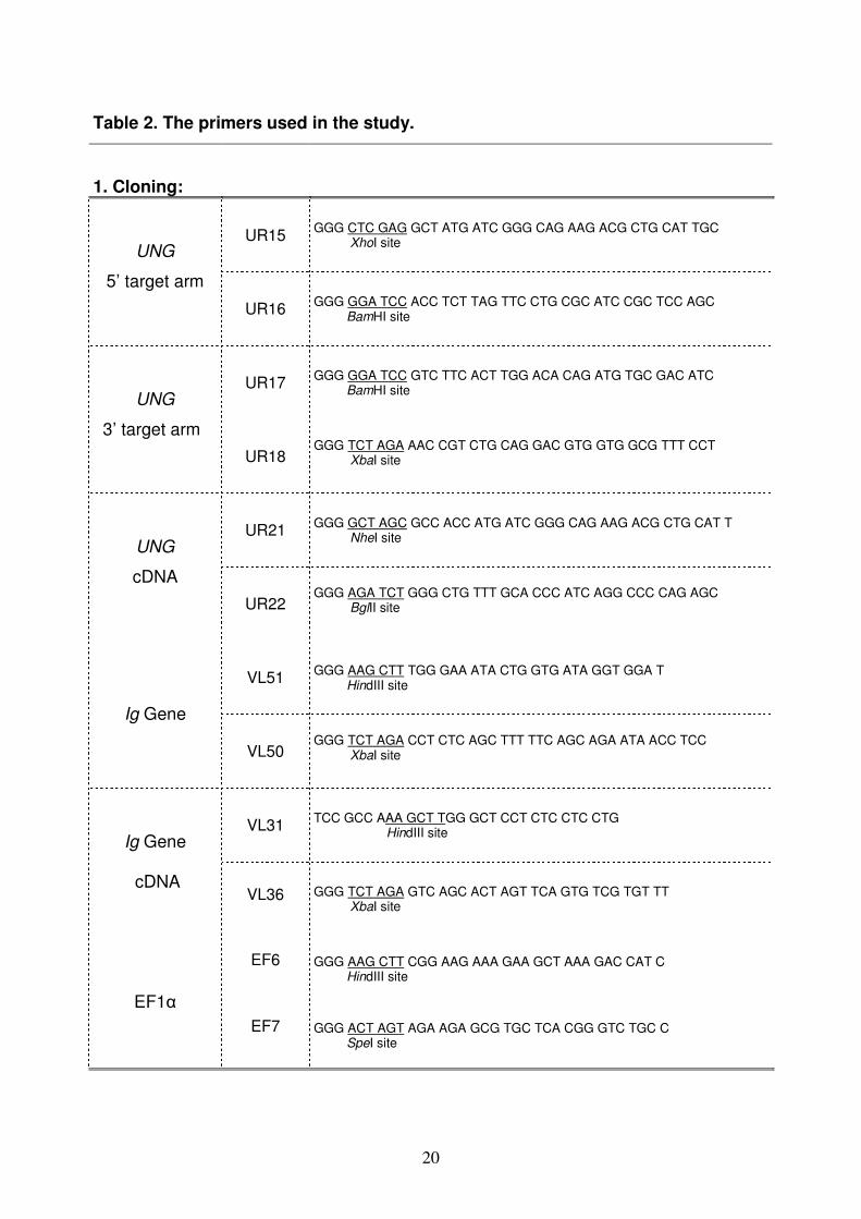
III. Oligonucleotides:
Oligomers (5’ to 3’) used throughout the study were summarized in Table 2.
[Invitrogene GmbH, Karlsruhe; GERMANY].
20
Table 2. The primers used in the study.
1. Cloning:
UR15 GGG CTC GAG GCT ATG ATC GGG CAG AAG ACG CTG CAT TGC XhoI site
UNG
5’ target arm
UR16 GGG GGA TCC ACC TCT TAG TTC CTG CGC ATC CGC TCC AGC BamHI site
UR17 GGG GGA TCC GTC TTC ACT TGG ACA CAG ATG TGC GAC ATC BamHI site
UNG
3’ target arm
UR18 GGG TCT AGA AAC CGT CTG CAG GAC GTG GTG GCG TTT CCT XbaI site
UR21 GGG GCT AGC GCC ACC ATG ATC GGG CAG AAG ACG CTG CAT T NheI site
UNG
cDNA
UR22 GGG AGA TCT GGG CTG TTT GCA CCC ATC AGG CCC CAG AGC BglII site
VL51 GGG AAG CTT TGG GAA ATA CTG GTG ATA GGT GGA T HindIII site
Ig Gene
VL50 GGG TCT AGA CCT CTC AGC TTT TTC AGC AGA ATA ACC TCC XbaI site
VL31 TCC GCC AAA GCT TGG GCT CCT CTC CTC CTG HindIII site
Ig Gene
cDNA VL36 GGG TCT AGA GTC AGC ACT AGT TCA GTG TCG TGT TT
XbaI site
EF6
GGG AAG CTT CGG AAG AAA GAA GCT AAA GAC CAT C HindIII site
EF1α
EF7
GGG ACT AGT AGA AGA GCG TGC TCA CGG GTC TGC C SpeI site
21
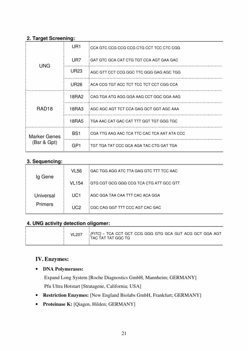
2. Target Screening:
UR1 CCA GTC CCG CCG CCG CTG CCT TCC CTC CGG
UR7 GAT GTC GCA CAT CTG TGT CCA AGT GAA GAC
UR23 AGC GTT CCT CCG GGC TTC GGG GAG AGC TGG
UNG
UR26 ACA CCG TGT ACC TCT TCC TCT CCT CGG CCA
18RA2 CAG TGA ATG AGG GGA AAG CCT GGC GGA AAG
18RA3 AGC AGC AGT TCT CCA GAG GCT GGT AGC AAA RAD18
18RA5 TGA AAC CAT GAC CAT TTT GGT TGT GGG TGC
BS1 CGA TTG AAG AAC TCA TTC CAC TCA AAT ATA CCC Marker Genes
(Bsr & Gpt) GP1 TGT TGA TAT CCC GCA AGA TAC CTG GAT TGA
3. Sequencing:
VL56 GAC TGG AGG ATC TTA GAG GTC TTT TCC AAC
Ig Gene
VL154 GTG CGT GCG GGG CCG TCA CTG ATT GCC GTT
UC1 AGC GGA TAA CAA TTT CAC ACA GGA Universal
Primers UC2 CGC CAG GGT TTT CCC AGT CAC GAC
4. UNG activity detection oligomer:
VL207 [FITC] – TCA CCT GCT CCG GGG GTG GCA GUT ACG GCT GGA AGT TAC TAT TAT GGC TG
IV. Enzymes:
• DNA Polymerases:
Expand Long System [Roche Diagnostics GmbH, Mannheim; GERMANY]
Pfu Ultra Hotstart [Stratagene, California; USA]
• Restriction Enzymes: [New England Biolabs GmbH, Frankfurt; GERMANY]
• Proteinase K: [Qiagen, Hilden; GERMANY]
22
• Calf Intestinal Phosphatase: [New England Biolabs GmbH, Frankfurt;
GERMANY]
• RNaseA: [Qiagen, Hilden; GERMANY]
V. Immuno-staining antibodies & anti-antibodies:
In order to quantify the surface IgM on B cells, Mouse Anti Chicken IgM (Clone M-
1) and Goat Anti Mouse IgG (H+L)-RPE Human Adsorbed were used. [Southern
Biotech. Ass. Inc., Biozol, Eching, Germany]
VI. DNA Size Marker:
A DNA marker which was manually prepared from φX174 DNA-HaeIII digest and
λ DNA-HindIII digest was used throughout the study [New England Biolabs GmbH,
Frankfurt; GERMANY]. It was prepared as follows:
The mixture was incubated for 15 min at 70oC without mixing and then it was taken
into ice for 5 min.
VII. Bacterial Strain:
During cloning of the constructs E. coli-DH5α cells were used as competent cells.
VIII. Mammalian Cell Line:
A chicken B-cell line DT40 was used to transfect constructs.
IX. Plasmids:
UNG: In order to construct vectors for knockout and complementation for UNG study,
pBluescript II KS (+) phagemid vector [Stratagene, California; USA] was used. In this
vector blue/white selection is possible and it has ampicillin resitance gene (ampR).
RAD18: RAD18 targeting vectors were requested from group of Prof. Dr. Shunichi
Takeda, Kyoto University, Japan (Yamashita et al., 2002).
λ DNA (500 ng/µl) 400 µl
φX174 DNA (1000 ng/µl) 100 µl
6X Loading buffer 334 µl
MiliQ water 1166 µl
23
METHODS :
I. Molecular Biology:
A. Culture of E.coli:
Luria Broth (LB)* agar plates and 2YT Broth* medium were used for culture of E.coli.
The plates were kept at 4°C for about 1 month. For longer storages glycerol stocks were
prepared in a following way: (2 volumes (v.) of cells + 1 v. of 50% glycerol). Stocks
were frozen at -80°C deep freezer.
2YT Broth: The mixture was autoclaved and 1ml ampicillin (250mg/ml) was added to
the mixture before use.
LB Agar Plates: The mixture was autoclaved. Before pouring onto petri plates, 1 ml
ampicillin (250mg/ml) and 1 ml 4% X-gal was added to the mixture.
LB Broth Base 20 g
BactoTM
Agar 15 g
Distilled water 1000 ml
B. E.coli DH5α competent cell preparation:
1) A streak of bacterial strain DH5α was made on a LB agar plate without antibiotics for
single colony isolation. The plate was incubated over night at 37oC.
2) A single colony was picked up and was cultured in 5 ml L broth or SOB broth*
medium at 37oC for over night.
3) 1 ml of SOB broth was taken and the background was measured at OD600.
4) 2.5 ml of overnight culture was transferred into 500 ml of SOB broth which had been
kept at 25oC and bacteria were cultured at 25
oC.
5) OD600 was measured every 30 min or every 1 h.
6) When the OD600 was 0.4, the bacterial culture was cooled down immediately by ice
and was kept on ice for 10 min.
7) The culture was transferred into the large centrifuge tubes and was centrifuged at
3000 rpm at 4oC for 10 min.
Bacto tryptone 16 g
Yeast extract 10 g
NaCl 5 g
10N NaOH 250 µl
Distilled water 1000 ml
24
8) Supernatant was discarded and the pellet was resuspended into 330 ml of ice-cold
FTB*. Vortex should not be used during this step.
9) The culture was kept on ice for 10 min, and was centrifuged at 3000 rpm at 4oC for
10 min.
10) Supernatant was discarded and the pellet was resuspended into 50 ml of ice-cold
FTB. Vortex should not be used. 3.5 ml of DMSO was added, mixed gently.
11) 400 µl of aliquots was dispensed into 1.5 ml tubes. Tubes were then frozen
immediately by liquid nitrogen and the competent cells were kept at -80oC.
12) The efficiency of competent cells was tested by using 1 ng of Bluescript plasmid for
transformation to one competent cell tube (400 µl). After heat shock, 600 ml of L broth
was added to the tube. Before spreading, 10 µl (1/100) and 100 µl (1/10) of bacterial
liquid was taken onto agar plates. The left (89/100) was centrifuged for 1 min at 13000
rpm. Then, the bacteria were spreaded by a loop.
13) The number of colonies was counted. The titer of competent cells was calculated
(Titer of competent cells is the number of colonies which would be produced by
transfection of 1 µg plasmid).
* SOB Broth: MgCl2 and MgSO4 were added after autoclave of mixture.
* FTB: The pH was adjusted to 6.7 after KCl and MnCl2.4H2O treatments respectively.
Bacto tryptone 20 g
Yeast extract 5 g
NaCl 0.585 g
KCl 0.l86 g
Distilled water 1000 ml
MgCl2 Final 10 mM
MgSO4 Final 10 mM
0,5 M PIPES 20 ml
CaCl2.2H2O 2.2 g
Distilled Water 1000 ml
KCl 18.64 g
MnCl2.4H2O 10,88 g
25
C. Polymerase Chain Reaction (PCR):
For the amplification of genomic DNA, hot start was preferred in order to prevent
unspecific bands. For this reason, upper and lower mixes were prepared separately and
upper mix was added onto lower mix at 80oC after pre-incubation on the machine. In
the case for high fidelity polymerases, since pfu Ultra was inhibited by a taq-directed
antibody no upper and lower mixes were prepared.
Amplifications were done by using Long-range PCR protocol as follows:
D. Analysis of DNA by electrophoresis:
Up to the purpose, 0.8% to 2% agarose gel which contains 0.1 µg/ml EtBr was used to
visualize DNA by Gel Doc 2000TM
[Bio-Rad Lab., California; USA]. DNA samples
were run for 35 min at 120 V. For gel purification, they were kept around 1 h in
electrophoresis. In the case for UNG activity test 15% TBE-urea polyacrylamide gel
was used and the DNA samples were run at 200 V for 40 min.
E. Purification & Gel Purification of DNA:
After PCR or restriction enzyme digestion, DNA was purified by Qiagen PCR
Purification kit [Qiagen GmbH, Hilden; GERMANY Qiagen] according to
manufacturer’s instructions.
Lower Mix:
DNA 1 – 100 ng
dNTP mix 0.2 mM
Primer forward 5 pmol
Primer reverse 5 pmol
10x Expand buffer 0.5 µl
MiliQ water rest
Total 5 µl
Upper Mix:
10x Expand buffer 0.5 µl
Expand System 0.5 U
10X Crezol Red 1 µl
MiliQ water rest
Total 5 µl
Pre-denaturation 93oC, 2 min
Denaturation 93oC, 10 sec
Annealing 65oC, 30 sec
Extension 68oC, 5 min*
Final extension 68oC, 7 min
}
35 cycles
* 20 sec. increases after each cycle
26
F. DNA Ligation:
Ligation was done by using a kit [Takara Bio Inc., Shiga; JAPAN] according to
manufacturer. In principle, around 1 volume of vector (100ng/µl), 9 volume of insert
(100ng/µl) and 10 volume of kit were used.
G. Transformation:
A heat shock protocol was preferred during transformation of plasmids into bacterial
cells:
1) The plasmid (>1ng) was added into 50-400 µl of competent cells, depending on the
efficiency of the cells.
2) The tube was incubated on ice for 20 (-30) min and then was heat shocked at 42oC
for 45 sec on a heat block, and was incubated again on ice for 2 min.
3) 2YT Broth was added onto tube until the mixture reaches 1 ml and the tube was
incubated at 37oC for at least 10 min.
4) After mixing, 100 µl (1/10) was taken from bacterial liquid and was put onto half of
LB agar plate. The rest (9 /10) was centrifuged at 13.000 rpm for 1 min, the supernatant
was discarded and the rest (~50 µl) was put onto another half of the plate. The cells
were then spreaded by using L-loop.
7) The plates were incubated overnight at 37oC.
H. Colony PCR:
The ligation of the DNA was first checked by colony PCR. Single bacterial colonies
were taken into U-Bottom 96 well plates which contains 20 µl 2YT Broth. From this, 1
µl was used as a DNA template for PCR.
Mix:
DNA 1 µl
10X Expand buffer 1 µl
dNTP mix 0.2 mM
Primer forward 5 pmol
Primer reverse 5 pmol
Expand System 0.5 U
10X Crezol red 1 µl
MiliQ water rest
Total 10 µl
PCR Programme:
Pre-denaturation 94oC, 1 min
Denaturation 94oC, 20 sec
Annealing 55oC, 1 min
Extension 72oC, 2 min
Final extension 72oC, 6 min
*Amplifications were done 30 cycles.
27
I. Plasmid preparation:
During construction of the vectors for small number of samples, plasmid extraction was
done by using a kit according to manufacturer’s protocol [Qiagen GmbH, Hilden;
GERMANY]. After constructs were confirmed by enzyme digest, a maxi-prep was done
[Qiagen GmbH, Hilden; GERMANY]. For higher number of samples or during cloning
of Ig gene PCR products for sequencing, large-scale miniprep preparation was used by
using deep-well blocks and plates as follows:
1) The bacterial colonies were picked up into a 96 well U-Bottom plate each well
containing 70µl of 2YT Broth. 25µl of each individual sample were transferred into two
96 well blocks containing 1.3 ml 2YT Broth.
2) The blocks were incubated overnight at 37 oC at 220 rpm.
3) The next day, the blocks were centrifuged at 3300 rpm for 10 min.
4) Supernatant was discarded by tapping and 80 µl of Solution-I* was added into each
well. The pellet was resuspended, vortexed and incubated for 1 min.
5) 80 µl of Solution-II* was added and vortexed immediately. The samples were
incubated for an additional 5 min at room temperature.
6) 80 µl Solution-III* was added and vortexed.
7) After this, the deep well block was centrifuged at 3.300 rpm for 5 min. (During this
time Millipore FB plate was placed in the bottom, and NA lysate clearing plate was put
on top of the manifold.
8) 200 µl of supernatants were transferred onto the clearing plate.
9) The vacuum was adjusted to not exceed 8 inches Hg in order to ensure uniform
filtration. The vacuum was applied for 5 min until the lysate is drawn.
10) The FB plate was taken from inside the manifold and placed on top of the empty
manifold. A plastic tray was placed in the bottom of the manifold for waste.
11) 150 µl of Binding Solution* was added to wells and mixed immediately by
pipetting. Vacuum was applied at full strength for 2 min.
12) 200µl of 80% ethanol was added to each well. The vacuum was applied at full
strength for 2 min. The waste accumulated in the plastic tray at the bottom of the
manifold was discarded and the FB plate was centrifuged at 1.000 rpm for 30sec. (This
step was repeated two times)
13) The FB plate was put on top of a new 96-well microtiter plate and 100µl of 1x TE
Buffer was added to each well. The plate was incubated for 5 min and centrifuged at
3500 rpm for 5 min. The plasmid was kept at -20OC.
28
J. Restriction Enzyme Digestion:
Before ligation: To prepare vector and insert fragments, DNA samples were incubated
at 37oC for at least 3 h. After ligation: DNA samples were incubated for 1 h to check
cloning. In general 1-5 U of enzyme was enough to digest 1µg of DNA in 1 h.
K. First strand cDNA synthesis:
Super Script III (Invitrogen) was used to synthesize first strand cDNA according to
manufacturer’s intructions.
L. Determination of DNA and RNA concentration:
The purity and the concentration of the DNA were checked by a spectrophotometer
BioPhotometer [Eppendorf GmbH, Hamburg; GERMANY]. Generally, the intensity of
absorbance at the wavelength 260 nm was used for calculating concentration and the
ratio of 260/280 nm was used to estimate the purity. For pure DNA samples this value
should be between 1.8, 2.0.
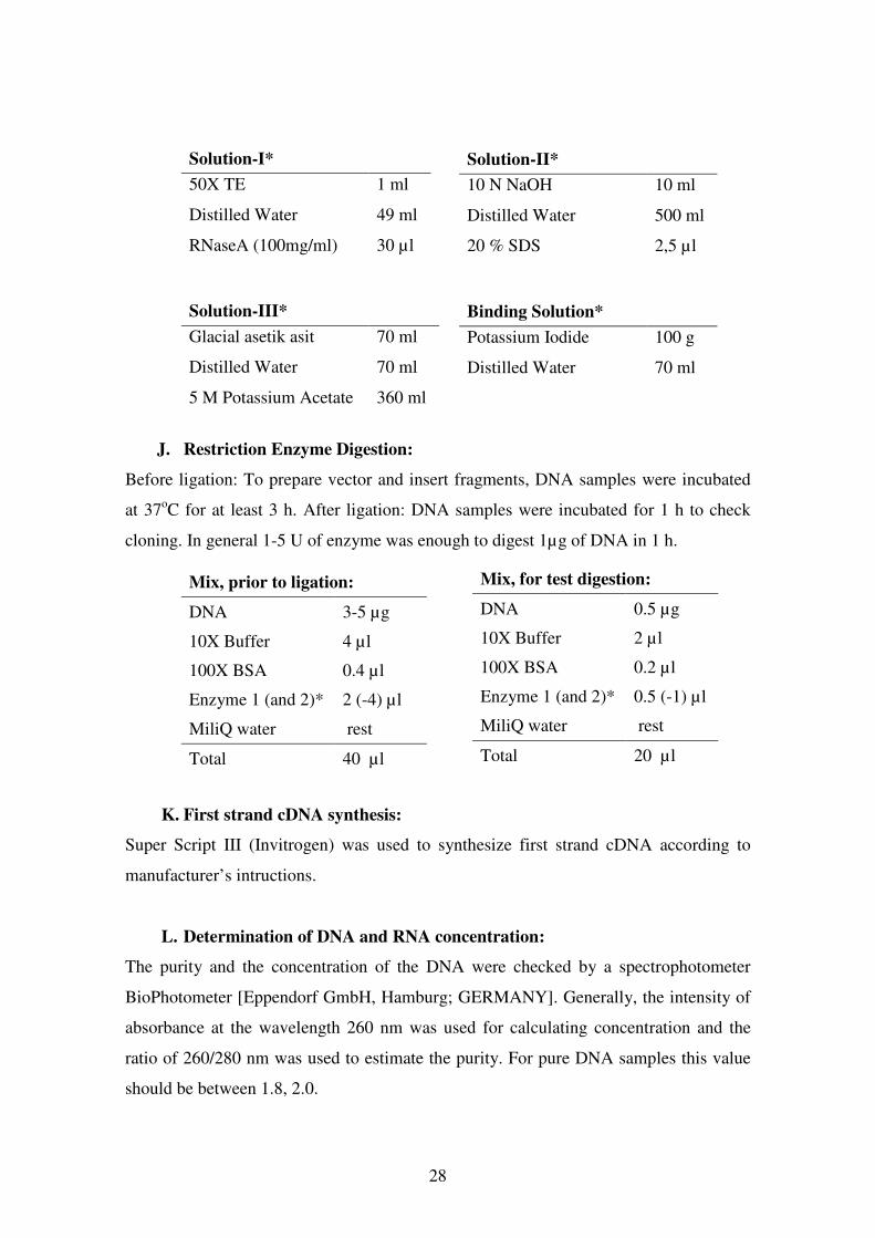
Solution-I*
50X TE 1 ml
Distilled Water 49 ml
RNaseA (100mg/ml) 30 µl
Solution-II*
10 N NaOH 10 ml
Distilled Water 500 ml
20 % SDS 2,5 µl
Solution-III*
Glacial asetik asit 70 ml
Distilled Water 70 ml
5 M Potassium Acetate 360 ml
Binding Solution*
Potassium Iodide 100 g
Distilled Water 70 ml
Mix, for test digestion:
DNA 0.5 µg
10X Buffer 2 µl
100X BSA 0.2 µl
Enzyme 1 (and 2)* 0.5 (-1) µl
MiliQ water rest
Total 20 µl
Mix, prior to ligation:
DNA 3-5 µg
10X Buffer 4 µl
100X BSA 0.4 µl
Enzyme 1 (and 2)* 2 (-4) µl
MiliQ water rest
Total 40 µl
29
M. Genomic DNA Isolation:
1) 50 ml of cells (~ 50x106
cells) in good condition were centrifuged at 1500 rpm for 5
min. The supernatant was discarded, the pellet was washed with 1-2 ml 1X PBS and
again was centrifuged at 1500 rpm and for 5 min.
2) The supernatant was discarded, the pellet was resuspended in 500µl proteinase K
Buffer* containing 0.1 mg/ml of proteinase K. The mixture was transferred into a 2 ml
tube and 12.5 µl of SDS (20%) was added, mixed by inverting and spinned down
shortly. Protein degradation was carried out overnight at 56oC.
3) The next day, 1 volume of phenol was added onto the DNA extract and mixed
carefully. After this the tube was centrifuged at 13000 rpm for 5 min.
4) The upper phase was transferred into a new tube and 1 volume of phenol/ chloroform
was added and mixed. Again it was centrifuged at 13000 rpm for 5 min.
5) The upper phase was transferred onto a new tube and 1 volume of chloroform was
added and mixed. It was centrifuged again at 13.000 rpm for 5 min.
6) The upper phase was transferred onto a new tube and 2 µl of RNAseA (100mg/ml)
was added to digest RNA. And then, it was incubated for 2 h at 37oC.
7) DNA was transferred into dialysis membrane and the ends were closed with clamps.
The membrane was put into a glass of cold 1X TE and kept at 4oC for 2 h.
(Alternatively, the reaction can be stopped by adding 50µl of 0,5M EDTA pH 8.0).
TE was changed at least three times with 2-4 h intervals.
8) DNA was transferred to a new tube and concentration was measured.
N. Total RNA Isolation:
1) 20 ml cells (~ 20x106 cells) were centrifuged at 1500 rpm for 5 min.
2) The supernatant was discarded and the pellet was resuspended in 1.5 ml of TRIzol
reagent. After that, it was transferred into a 2 ml eppendorf tube and was incubated for 5
min at room temperature to permit the complete dissociation of nucleoprotein
complexes.
*Proteinase K Buffer:
5 M NaCl 2 ml
1 M Tris-HCl (pH 8,0) 1 ml
0.5 M EDTA 5 ml
MiliQ water 92 ml
Total 100 ml
30
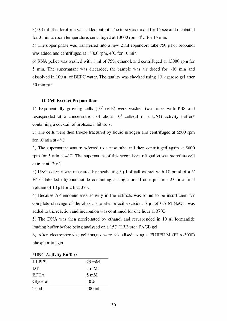
3) 0.3 ml of chloroform was added onto it. The tube was mixed for 15 sec and incubated
for 3 min at room temperature, centrifuged at 13000 rpm, 4oC for 15 min.
5) The upper phase was transferred into a new 2 ml eppendorf tube 750 µl of propanol
was added and centrifuged at 13000 rpm, 4oC for 10 min.
6) RNA pellet was washed with 1 ml of 75% ethanol, and centrifuged at 13000 rpm for
5 min. The supernatant was discarded, the sample was air droed for ~10 min and
dissolved in 100 µl of DEPC water. The quality was checked using 1% agarose gel after
50 min run.
O. Cell Extract Preparation:
1) Exponentially growing cells (106
cells) were washed two times with PBS and
resuspended at a concentration of about 103 cells/µl in a UNG activity buffer*
containing a cocktail of protease inhibitors.
2) The cells were then freeze-fractured by liquid nitrogen and centrifuged at 6500 rpm
for 10 min at 4°C.
3) The supernatant was transferred to a new tube and then centrifuged again at 5000
rpm for 5 min at 4°C. The supernatant of this second centrifugation was stored as cell
extract at -20°C.
3) UNG activity was measured by incubating 5 µl of cell extract with 10 pmol of a 5'
FITC–labelled oligonucleotide containing a single uracil at a position 23 in a final
volume of 10 µl for 2 h at 37°C.
4) Because AP endonuclease activity in the extracts was found to be insufficient for
complete cleavage of the abasic site after uracil excision, 5 µl of 0.5 M NaOH was
added to the reaction and incubation was continued for one hour at 37°C.
5) The DNA was then precipitated by ethanol and resuspended in 10 µl formamide
loading buffer before being analysed on a 15% TBE-urea PAGE gel.
6) After electrophoresis, gel images were visualised using a FUJIFILM (FLA-3000)
phosphor imager.
*UNG Activity Buffer:
HEPES 25 mM
DTT 1 mM
EDTA 5 mM
Glycerol 10%
Total 100 ml

31
II. Cell Culture:
A. Basic Cell Culture Conditions:
DT40 cells can be cultured in cultures flasks, petri dishes, or in 24 well plates.
Microtiter plates are suitable for transfection or subcloning. The optimum culture
condition for the cells is 41°C with 5% CO2. Chicken medium* can be used to culture
DT40 cells and Freezing medium* can be used to free down.
B. Thawing of the cells:
1) The tube which was taken from nitrogen tank was melted at 41 O
C and centrifuged at
1.500 rpm for 5 min.
2) The supernatant was discarded; the pellet was resuspended with chicken medium and
transferred to a flask or plate.
C. Freeze down of cells:
1) The cells were transferred to a flask and centrifuged at 1.500 rpm for 5 min.
2) The supernatant was discarded, the cells were resuspended with freezing medium,
first stored in - 80 O
C for overnight afterwards transferred to nitrogen tank.
D. Transfection:
Plasmid Preparation:
1) The plasmid DNA was linearized outside of the genes of interest by an appropriate
restriction enzyme within 500 µl total reaction volume. Following day the digestion was
checked by using 0.5 µg of DNA on an agarose gel.
2) The DNA was purified first with phenol/chloroform, and then with chloroform,
precipitated with propanol and rinced with 70% ethanol.
3) The pellet was dried up naturally for 10 min inside the laminar and resuspended in
distilled water for final 1 µg/ml concentration.
Chicken Medium:
RPMI 500 ml
FBS 50 ml
Penicillin / Streptoycin 10 ml
L-Glutamine 5 ml
Chicken Serum 5 ml
1 M β-mercaptoethanol 50 µl
Freezing Medium:
RPMI 70 ml
FBS 20 ml
DMSO 10 ml
32
Electroporation:
1) The concentration of the cells was determined (10 million cells were used per each
electroporation). Calculated amount was transferred into a 50 ml tube and centrifuged
for 5 min at 1500 rpm, 4oC.
2) The supernatant was removed, the pellet was resuspended in 800 µl of chicken
medium and transferred into electroporation cuvette together with DNA.
3) Electroporation was done using 25 µF and 700 V.
4) The mixture was put onto the tube containing 9.5 ml of chicken medium and the
solution was distributed into the wells (100µl) of a flat-bottom microtiter plates.
5) The following day (12-24 h after electroporation), 100 µl of selective medium
(containing twice the final concentration of the drug) was added to each well.
6) The plates were left for about seven to ten days in the incubator without changing the
medium. Drug resistant colonies should be visible by then.
E. Identifying Targeted Events By PCR:
After 7-10 days of electroporation, the visible colonies were picked up into 300 µl of
chicken medium in flat-bottom 96-well plates and incubated for 3 days.
Crude extract preparation:
1) The cells were transferred to 96 well PCR plate, washed with PBS and centrifuged 5
min at 1500 rpm.
2) The supernatant was discarded, the pellet was resuspended into 10 µl of K buffer [1 x
PCR buffer, 0.1 mg/ml proteinase K and 0.5% Tween 20] for proteinase K-mediated
proteolysis .
3) The cells were incubated for 45 min at 56˚C and to inactivate the proteinase K, they
were incubated at 95˚C for 10 min.
4) 1 µl of the crude extract was used for targeting screening PCR.
F. Subcloning of DT40 cells:
1) The viability of the cells was counted using cell viability analyzer.
2) Four wells of a six well plate were prepared to contain 15, 50, 150 and 500 viable
cells in 5 ml chicken medium and each well is mixed well.
3) Using a stepper, 100 µl of the cells were separated into the 96 well flat bottom
microtiter plate (Half plate for one dilution).
33
4) The plates were incubated for 7-8 days without changing medium. Subclones should
be visible by then as round colonies.
5) 10µl of colonies were started to be picked up from lowest dilutions into 1 ml of
chicken medium in 24-well flat-bottom plates.
G. Analysis of IgM expression by FACS:
In order to quantify the amounts of surface IgM (sIgM) expression as well as for sIgM
loss of DT40 mutant cell lines, antibody staining for FACS analysis was done (The
antibodies were prepared by diluting 1/100 – 1/500 folds).
1) The cells were transferred into round-bottom 96-well plates, washed with PBS.
2) For first staining, the cells were incubated with anti-chicken Cµ monoclonal
antibody-M1 on ice for 30 min and then twice washed with PBS.
3) For second staining, R-PE-conjugated goat anti-mouse IgG polyclonal antibody was
used with same conditions. Then, the cells were washed with PBS three times.
4) The sIgM expression of cell lines were measured and analyzed by flow cytomer.
H. Drug Resistance Marker Recycling:
Up to do purpose, partial or complete excision of the marker genes is possible in DT40
cells.
1) Around 105 cells were cultured with fresh chicken medium containing 0.01 mM 4-
hydroxt tamoxifen (1 day for partial and 2 days for complete excision).
2) The cells were subcloned by limiting dilution for final concentration of 0.3, 1, 3 and
10 cells / well in 96-well flat-bottom plates.
3) 7 – 8 days after subcloning, 10 µl of the visible subclones were transferred into 1 ml
of chicken medium.
4) In order to test the excision of the drug-resistance genes, the cells were incubated
with drug-containing mediums. The cells deficient for markers were selected for further
studies.
I. Sorting:
1) Enough amounts of cells were stained, filtered, washed and collected in 1-2 ml of
PBS.
2) During sorting, for storage of the sorted cells, 50% chicken medium and 50% FBS
was used. The tubes were prepared beforehand.
34
J. Colony Survival Assay:
Preparation of methylcellulose medium:
1) 11.9g D-MEM powder and 2.44g sodium bicarbonate was dissolved in 500ml water
and sterilized by 0.2µm filtration.
2) 10g methylcellulose powder and a stir bar were autoclaved in a 1L bottle.
3) Approximately 220mls warm (50 – 65˚C) sterile water was added to the
methylcellulose powder. Well mixed, ensuring an even suspension.
4) When temperature of methylcellulose solution is ≤50˚C, DMEM/HAM solution from
(1) was added the little by little while mixing well. The bottle was mixed vigorously.
5) This solution was mixed overnight on a magnetic stirrer in a cold room.
6) The 0.2µm-filtered serum/antibiotic/2-mercaptoethanol cocktail was added.
7) The pH was adjusted 7.0 to 7.4 with NaOH (4N solution) as necessary.
8) The mix was stirred for at least 3 hours (preferably overnight) in a cold room and was
kept at 4˚C for up to 1 month.
Preparing the methylcellulose plates:
1) Appropriate number of 6 well plates was labeled. One 6 well plate will be required
for each dose for every cell line e.g. testing 4 cell lines at 5 different UV doses will
require 20 6 well plates.
2) 5 ml of methylcellulose was put into each well. The methylcellulose was stirred for
30 minutes at room temperature before use.
3) The central gaps were half filled between the wells with sterile water.
4) The plates were placed in a 37˚C incubator until ready for use.
Plating out samples:
1) 100µl of sample was pipetted onto the appropriate well in a figure of 8 pattern.
2) The plate was swirled when the all the samples have been pipetted on the plate to
ensure that all the cells are evenly distributed across the methylcellulose.
3) The plates are then incubated in a humid box in a CO2 incubator until the colonies are
clearly visible and countable. This usually takes 10 days to 2 weeks depending on the
growth characteristics of the cell line. For consistency it is generally best to take the
count from the pair of wells with between 0 and 100 colonies. Counting above 100 is
unlikely to be accurate and colony formation is likely to be compromised at high
densities. The whole experiment should be repeated three times. Survival is plotted as a
percentage of the cells surviving on the untreated control.
35
RESULTS – 1: ‘UNG’
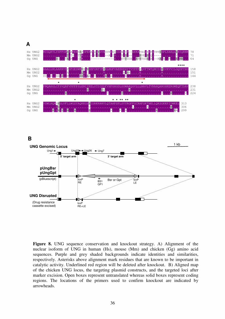
I. Genomic Identification of UNG Locus:
Using bursal EST database (Abdrakhmanov et al. 2000), an EST dkfz426_17p6r1 was
identified which shows a significant homology to murine and human UNG cDNA. This clone
was later sequenced and found to contain a 1096 bp cDNA insert including the full length
UNG open reading frame of 299 amino acids. The amino acid sequence of the chicken UNG
was found to be 79% and 75% identical to the human and murine nuclear UNG2, respectively
(Figure R1). The exact intron-exon structure of UNG genomic locus was tried to be deduced
from the PCR products using forward and reverse primers with 100 bp intervals (Figure R1).
After chicken genome sequence including the UNG locus on chromosome 15 was released,
this observation was confirmed (International Chicken Genome Seq. Consortium 2004).
II. Vector Construction:
B. Knockout Vectors: pUngBsr, pUngGpt
The 5’ and 3’ arms of the UNG knockout constructs were designed to insert and early in-
frame stop codon at codon 66 and delete the region between the codons 67-120 (Fig. 8A).
Two upstream (UR15-UR16) and two downstream primers (UR17-UR18) were selected to
amplify 5’ and 3’ target arms respectively using DT40 genomic DNA as a template. After
PCR amplification, 5’ target arm was digested with restriction enzymes XhoI and BamHI
whereas 3’ target arm was digested with BamHI and XbaI to be able to clone them into
pBluescript KS (+) basic vector. All of the clonings were first confirmed by colony PCR
using primers from insert and vector backbones and second by miniprep plasmid digest (Fig.
9). During digest, apart from the relevant restriction enzymes to distinguish vector and insert,
one more restriction enzyme NotI was used to linearize the plasmids in order to see the size
difference between the cloned and previous vector. During cloning, at first 5’ target arm was
cloned into the vector. Cloning was confirmed and clone was named as pUng5’Arm. For
second cloning, pUng5’Arm was digested with XhoI and BamHI to clone 3’ target arm. The
cloned vector, pUngArms, was prepared for last cloning by digesting only with BamHI. Drug
resistance marker genes, blasticidine S resistance gene (Bsr) and guanine phosphoribosyl
transferase gene (Gpt) as floxed BamHI cassettes, were cloned into pUngArms (Arakawa et al.
2001). The resultant clones were confirmed and named as pUngBsr and pUngGpt (Fig. 9A).
Finally, plasmids were amplified by maxi-prep and linearized by NotI for transfection.
36
Hs UNG2 MIGQKTLYSFFSPSPARKRHAPSPEPAVQGTGVAGVPEESGDAAAIP--AKKAPAGQEEPGTPPSSPLSAEQLDRIQRNK 78
Mm UNG2 .............T.TG..TTR....-.P.S...--A.IG...V.S.--....RVE.N.Q.----........V...... 71
Gg UNG .......HC...TA.P...-SR....-------------G...EVTDDA..RPKTSDH.A.--RA.A..P...E.MR... 64
****
Hs UNG2 AAALLRLAARNVPVGFGESWKKHLSGEFGKPYFIKLMGFVAEERKHYTVYPPPHQVFTWTQMCDIKDVKVVILGQDPYHG 158
Mm UNG2 .............A.......QQ.C........V..........N.HK.....E...........R.............. 151
Gg UNG EV..R..NE.S..P......RRQ.AA..T....VE..A.......R.......E...........R.............. 144
* * *
Hs UNG2 PNQAHGLCFSVQRPVPPPPSLENIYKELSTDIEDFVHPGHGDLSGWAKQGVLLLNAVLTVRAHQANSHKERGWEQFTDAV 238
Mm UNG2 ........................F.......DG.............R................................ 231
Gg UNG ............K......................T.......T.....................T....K.......V. 224
* * * ** **
Hs UNG2 VSWLNQNSNGLVFLLWGSYAQKKGSAIDRKRHHVLQTAHPSPLSVYRGFFGCRHFSKTNELLQKSGKKPIDWKEL 313
Mm UNG2 .......LS................V.......................L.......A............N.... 306
Gg UNG .....K.LH.V..M...A.......S...........V....F..N................K.........RA. 299
UNG Disrupted
pUngBsr
pUngGpt
(pBluescript)
(Drug resistance cassette excised)
UNG Genomic Locus
loxP RE+LE
loxP RE
Bsr or Gpt loxP LE
1 kb
Ung1 Ung7 Ung26 Ung23
BS1 GP1
5’ target arm 3’ target arm
A
B
Figure 8. UNG sequence conservation and knockout strategy. A) Alignment of the
nuclear isoform of UNG in human (Hs), mouse (Mm) and chicken (Gg) amino acid
sequences. Purple and grey shaded backgrounds indicate identities and similarities,
respectively. Asterisks above alignment mark residues that are known to be important in
catalytic activity. Underlined red region will be deleted after knockout. B) Aligned map
of the chicken UNG locus, the targeting plasmid constructs, and the targeted loci after
marker excision. Open boxes represent untranslated whereas solid boxes represent coding
regions. The locations of the primers used to confirm knockout are indicated by
arrowheads.
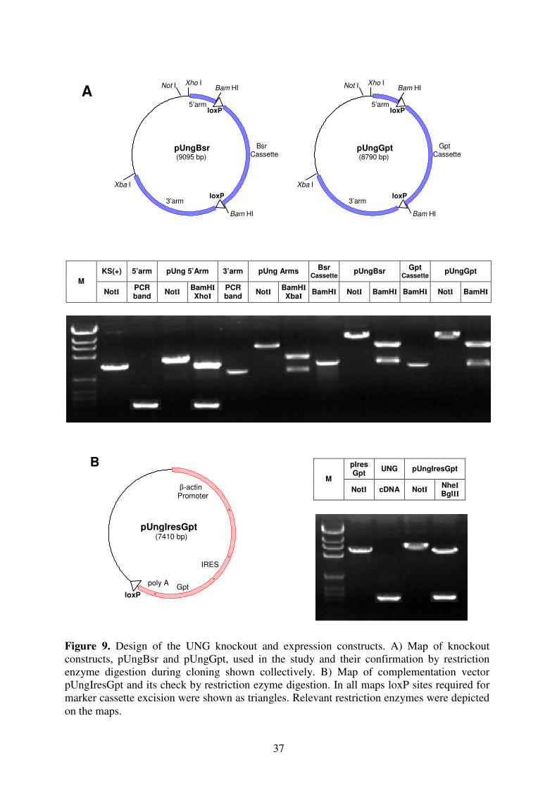
37
pUngIresGpt 7410 bps
pUngIresGpt
(7410 bp)
poly A loxP
Gpt
IRES
β-actin Promoter
B
KS(+) 5’arm pUng 5’Arm 3’arm pUng Arms Bsr
Cassette pUngBsr
Gpt
Cassette pUngGpt
M
NotI PCR
band NotI
BamHI
XhoI
PCR
band NotI
BamHI
XbaI BamHI NotI BamHI BamHI NotI BamHI
pIres
Gpt UNG pUngIresGpt
M
NotI cDNA NotI NheI
BglII
loxP
Xba I
Bam HI
loxP
Xho I Bam HINot I
Gpt Cassette
pUngGpt
(8790 bp)
5’arm
3’arm
loxP
Xba I
Bam HI
loxP
Xho I Bam HINot I
Bsr Cassette
pUngBsr
(9095 bp)
5’arm
3’arm
A
Figure 9. Design of the UNG knockout and expression constructs. A) Map of knockout
constructs, pUngBsr and pUngGpt, used in the study and their confirmation by restriction
enzyme digestion during cloning shown collectively. B) Map of complementation vector
pUngIresGpt and its check by restriction ezyme digestion. In all maps loxP sites required for
marker cassette excision were shown as triangles. Relevant restriction enzymes were depicted
on the maps.
38
C. Complementation Vector: pUngIresGpt
To clone UNG cDNA, two primers (UR21-UR22) which contains NheI and BglII restriction
sites respectively, were used. Using EST sequence of UNG cDNA, PCR was done, the
product was confirmed by sequence reaction and the product was cloned into the vector
pIresGpt. In this vector since cloned cDNA fragments and Gpt is under the control of one β-
actin promoter, it allows to express both UNG and marker cassette Gpt via Internal Ribosome
Entry Site (IRES) (Jang et al. 1988, Pelletier and Sonenberg 1988). The resultant clone was
named as pUngIresGpt (Fig. 9B). A maxi-prep was done and the vector was linearized by
NotI for stable transfection.
III. Knockout and Complementation of UNG:
UNG Inactivation Does Not Affect Cell Viability:
As a general rule, following all transfections 7-10 days later, the colonies were picked up and
a crude extract was prepared to check for targeted events using long range PCR. The positive
candidates were then stained for FACS analysis and 3 different clones were decided. Finally
the clones were frozen down into liquid nitrogen and genomic DNA was prepared.
For knockout of UNG, sIgM(-) variant of the DT40 clone AIDR
cells were used. In this cell
line AID is knocked out and the phenotype was complemented with AID overexpression. One
of the key features of this cell line, AID cDNA which is between loxP sites can be removed
upon induction of 4-HT and by this way gene conversion can be inactivated (Arakawa et al.
2002). Transfection of the first knockout construct, pUngBsr, was done and the result was
confirmed by long-range PCR using a primer located upstream of the 5’ targeting arm of the
construct along with a primer from the resistance marker (Fig. 8B; Fig. 10B). From the
positive clones 12 were stained for FACS analysis and after analysis 3 clones were decided as
AIDR
UNG+/-
Cl1, Cl2 and Cl3. Out of these, Clone 1 was taken and second construct,
pUngGpt, was transfected and knockout was confirmed as described above. The resultant
knockout clone was named as AIDR
UNG-/-
Cl1 (Fig. 10A; Fig. 10B).
Targeting of the first construct was observed ~1 in 10 events whereas for the second construct
it is dropped to ~1 in 30 clones. Upon screening from over 300 clones, 10 clones were found
to be targeted into the already targeted UNG allele but one clone was identified with two
targeting into two different alleles. The low frequency of targeting efficiency observed during
39
disruption of UNG as compare to knockout of other genes in DT40 could be first due to the
length of the target arms as a technical approach. Since genomic locus information was not
available when we started the study we were unable to take longer arms. Targeting efficiency
increases when the length of the target arms is between 1.5 kb to 2.5 kb. In our case 5’ arm is
0.8kb and 3’ arm is 2.4kb. A second probability could be due to a polymorphism between two
alleles. The identified UNG-deficient clone showed normal cell viability and cloning
efficiency, suggesting that the low recovery of homozygous clones was not related to a
growth defect of UNG knockout cells.
Recycling of Marker Genes and Complementation:
Since the number of drug marker cassettes is limited, they need to be recycled for further
transfections. Considering this, the cells were exposed to 4-HT to activate MerCreMer.
Following subcloning, the colonies were picked up, and a drug test was done in order to
identify the clones which are resistant to puromycin and sensitive to blasticidine and
mycophelolic acid. We want puromycin resistant clones because in overexpression construct
of AIDR cells puromycin exists. The positive candidates were checked by PCR, stained for
FACS analysis and 3 of them were selected for further analysis (Fig. 10C).
To one of the bsr and gpt negative clone, UNG expression construct pUngIresGpt was
transfected. This construct allows expression of UNG cDNA as a bicistronic message along
with the gpt transgene. The next day as a selective drug, different concentrations of
mycophenolic acid was spread. As usual, ~7-10 days later, colonies were picked up and
stained for FACS analysis. 3 of the complemented clones were decided and named as
AIDRUNG
R Cl1, Cl2, and Cl3 (Fig. 10A).
IV. UNG Activity Test:
UNG activity is strongly reduced in UNG-deficient cells:
To determine how disruption of UNG affects overall activity of uracyl glycosylases within
cells, one sophisticated method was used. For this, cell extracts of the AIDR, AID
RUNG
-/- and
AIDRUNG
R clones were incubated with an uracil containing oligonucleotide. Excision of the
uracil by uracil glycosylase leads to an abasic site which can be detected by the appearance of
a shorter fragment after NaOH or endonuclease mediated strand cleavage (Figure 10C). In the
absence of in vitro UNG treatment, the amount of cleavage product was strongly reduced in
extracts of AIDRUNG
-/- cells compared to extracts of wild-type and reconstituted AID
RUNG
R

40
Origin of
Cell Extracts AIDR AID
R
UNG-/-
AIDR
UNG-/-
AIDR
UNGR
No
extract
No
extract
In vitro
UNG treatment −−−− −−−− ++++ −−−− −−−− ++++
Cell Line AID
R UNG
-/-
(drug cas. not excised) Cell Lines AID
R AID
R
UNG-/-
AID
R
AIDR
UNG-/-
Primer Pairs Ung
23/26 Ung1 / BS1
Ung1 / GP1
Primer Pairs Ung
23/26 Ung
23/26 Ung 1/7
Ung 1/7
AIDR
sIgM(-)
AIDR
UNG+/-(Bsr)
pUngBsr
AIDR
UNG-(Bsr)/-(Gpt)
pUngGpt
Cre Induction
AIDR
UNG-/-
sIgM(-)
AIDR
UNG-/-
sIgM(-)
pUngIresGpt
AIDR
UNGR
sorting
AIDR
UNGR
sIgM(+)
Cre Induction
AIDR
UNG-/-
sIgM(+)
22mer
U FITC 3’ 5’
50mer −−−−
22mer −−−−
50mer
Figure 10. UNG knockout strategy and confirmation. A) Knockout and complementation
scheme step by step. B) Detection of marker cassettes after transfections by PCR. C)
Complete knockout of UNG (Removal of marker cassettes). In this PCR, AIDR precursor cell
line is compared with AIDR
UNG-/-
. D) UNG Assay using cell extracts. The structure of the
oligonucleotide substrate is shown below the image.
A
B
C
D
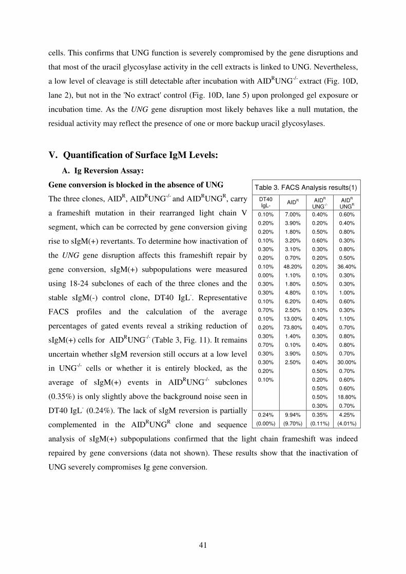
41
cells. This confirms that UNG function is severely compromised by the gene disruptions and
that most of the uracil glycosylase activity in the cell extracts is linked to UNG. Nevertheless,
a low level of cleavage is still detectable after incubation with AIDRUNG
-/- extract (Fig. 10D,
lane 2), but not in the 'No extract' control (Fig. 10D, lane 5) upon prolonged gel exposure or
incubation time. As the UNG gene disruption most likely behaves like a null mutation, the
residual activity may reflect the presence of one or more backup uracil glycosylases.
V. Quantification of Surface IgM Levels:
A. Ig Reversion Assay:
Gene conversion is blocked in the absence of UNG
The three clones, AIDR, AID
RUNG
-/- and AID
RUNG
R, carry
a frameshift mutation in their rearranged light chain V
segment, which can be corrected by gene conversion giving
rise to sIgM(+) revertants. To determine how inactivation of
the UNG gene disruption affects this frameshift repair by
gene conversion, sIgM(+) subpopulations were measured
using 18-24 subclones of each of the three clones and the
stable sIgM(-) control clone, DT40 IgL-. Representative
FACS profiles and the calculation of the average
percentages of gated events reveal a striking reduction of
sIgM(+) cells for AIDRUNG
-/- (Table 3, Fig. 11). It remains
uncertain whether sIgM reversion still occurs at a low level
in UNG-/-
cells or whether it is entirely blocked, as the
average of sIgM(+) events in AIDRUNG
-/- subclones
(0.35%) is only slightly above the background noise seen in
DT40 IgL- (0.24%). The lack of sIgM reversion is partially
complemented in the AIDRUNG
R clone and sequence
analysis of sIgM(+) subpopulations confirmed that the light chain frameshift was indeed
repaired by gene conversions (data not shown). These results show that the inactivation of
UNG severely compromises Ig gene conversion.
Table 3. FACS Analysis results(1)
DT40 IgL-
AIDR AID
R
UNG-/-
AID
R
UNGR
0.10% 7.00% 0.40% 0.60%
0.20% 3.90% 0.20% 0.40%
0.20% 1.80% 0.50% 0.80%
0.10% 3.20% 0.60% 0.30%
0.30% 3.10% 0.30% 0.80%
0.20% 0.70% 0.20% 0.50%
0.10% 48.20% 0.20% 36.40%
0.00% 1.10% 0.10% 0.30%
0.30% 1.80% 0.50% 0.30%
0.30% 4.80% 0.10% 1.00%
0.10% 6.20% 0.40% 0.60%
0.70% 2.50% 0.10% 0.30%
0.10% 13.00% 0.40% 1.10%
0.20% 73.80% 0.40% 0.70%
0.30% 1.40% 0.30% 0.80%
0.70% 0.10% 0.40% 0.80%
0.30% 3.90% 0.50% 0.70%
0.30% 2.50% 0.40% 30.00%
0.20% 0.50% 0.70%
0.10% 0.20% 0.60%
0.50% 0.60%
0.50% 18.80%
0.30% 0.70%
0.24% 9.94% 0.35% 4.25%
(0.00%) (9.70%) (0.11%) (4.01%)
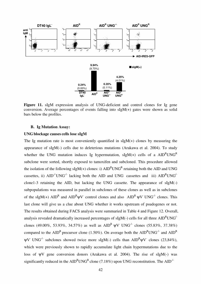
42
0.24%
(0.00%)
9.94%
(9.70%)
0.35%
(0.11%)
4.25%
(4.01%)
sIgM(+)
DT40 IgL- AID
R AID
R UNG
-/- AID
R UNG
R
B. Ig Mutation Assay:
UNG blockage causes
cells lose sIgM
The Ig mutation rate is most conveniently quantified in sIgM(+) clones by measuring the
appearance of sIgM(-) cells due to deleterious mutations (Arakawa et al. 2004). To study
whether the UNG mutation induces Ig hypermutation, sIgM(+) cells of a AIDRUNG
R
subclone were sorted, shortly exposed to tamoxifen and subcloned. This procedure allowed
the isolation of the following sIgM(+) clones: i) AIDRUNG
R retaining both the AID and UNG
cassettes, ii) AID-/-
UNG-/-
lacking both the AID and UNG cassettes and iii) AID
RUNG
/-
clone1-3 retaining the AID, but lacking the UNG cassette. The appearance of sIgM(-)
subpopulations was measured in parallel in subclones of these clones as well as in subclones
of the sIgM(+) AIDR and AID
RψV- control clones and also AID
R ψV- UNG
-/- clones. This
last clone will give us a clue about UNG whether it works upstream of psudogenes or not.
The results obtained during FACS analysis were summaried in Table 4 and Figure 12. Overall,
analysis revealed dramatically increased percentages of sIgM(-) cells for all three AIDRUNG
/-
clones (49.00%, 53.93%, 34.57%) as well as AIDR ψV
- UNG
-/- clones (55.83%, 37.38%)
compared to the AIDR
precursor clone (1.50%). On average both the AIDRUNG
-/- and AID
R
ψV-
UNG-/-
subclones showed twice more sIgM(-) cells than AIDRψV
- clones (23,84%),
which were previously shown to rapidly accumulate light chain hypermutations due to the
loss of ψV gene conversion donors (Arakawa et al. 2004). The rise of sIgM(-) was
significantly reduced in the AIDRUNG
R clone (7.18%) upon UNG reconstitution. The AID
-/-
DT40
IgL- AID
R AIDR
UNG-/-
AIDR
UNGR
anti
IgM
AID-IRES-GFP
Figure 11. sIgM expression analysis of UNG-deficient and control clones for Ig gene
conversion. Average percentages of events falling into sIgM(+) gates were shown as solid
bars below the profiles.
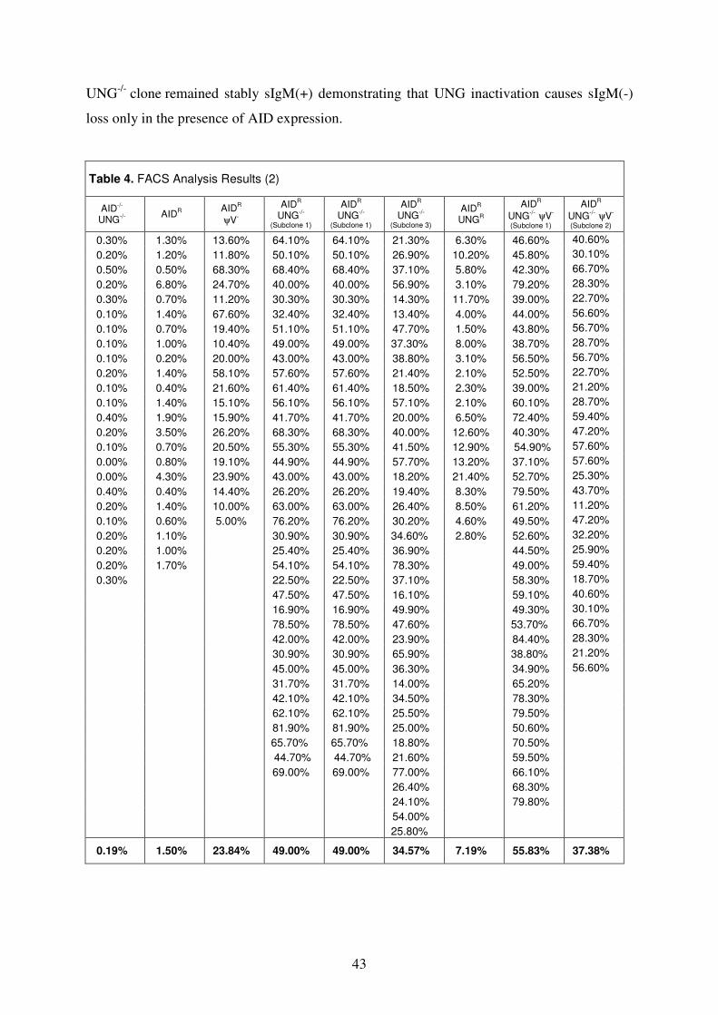
43
UNG-/-
clone remained stably sIgM(+) demonstrating that UNG inactivation causes sIgM(-)
loss only in the presence of AID expression.
Table 4. FACS Analysis Results (2)
AID-/-
UNG-/- AID
R AIDR
ψV-
AIDR
UNG-/-
(Subclone 1)
AIDR
UNG-/-
(Subclone 1)
AIDR
UNG-/-
(Subclone 3) AID
R
UNGR
AIDR
UNG-/-
ψV-
(Subclone 1)
AIDR
UNG-/-
ψV-
(Subclone 2) 0.30% 1.30% 13.60% 64.10% 64.10% 21.30% 6.30% 46.60% 40.60%
0.20% 1.20% 11.80% 50.10% 50.10% 26.90% 10.20% 45.80% 30.10%
0.50% 0.50% 68.30% 68.40% 68.40% 37.10% 5.80% 42.30% 66.70%
0.20% 6.80% 24.70% 40.00% 40.00% 56.90% 3.10% 79.20% 28.30%
0.30% 0.70% 11.20% 30.30% 30.30% 14.30% 11.70% 39.00% 22.70%
0.10% 1.40% 67.60% 32.40% 32.40% 13.40% 4.00% 44.00% 56.60%
0.10% 0.70% 19.40% 51.10% 51.10% 47.70% 1.50% 43.80% 56.70%
0.10% 1.00% 10.40% 49.00% 49.00% 37.30% 8.00% 38.70% 28.70%
0.10% 0.20% 20.00% 43.00% 43.00% 38.80% 3.10% 56.50% 56.70%
0.20% 1.40% 58.10% 57.60% 57.60% 21.40% 2.10% 52.50% 22.70%
0.10% 0.40% 21.60% 61.40% 61.40% 18.50% 2.30% 39.00% 21.20%
0.10% 1.40% 15.10% 56.10% 56.10% 57.10% 2.10% 60.10% 28.70%
0.40% 1.90% 15.90% 41.70% 41.70% 20.00% 6.50% 72.40% 59.40%
0.20% 3.50% 26.20% 68.30% 68.30% 40.00% 12.60% 40.30% 47.20%
0.10% 0.70% 20.50% 55.30% 55.30% 41.50% 12.90% 54.90% 57.60%
0.00% 0.80% 19.10% 44.90% 44.90% 57.70% 13.20% 37.10% 57.60%
0.00% 4.30% 23.90% 43.00% 43.00% 18.20% 21.40% 52.70% 25.30%
0.40% 0.40% 14.40% 26.20% 26.20% 19.40% 8.30% 79.50% 43.70%
0.20% 1.40% 10.00% 63.00% 63.00% 26.40% 8.50% 61.20% 11.20%
0.10% 0.60% 5.00% 76.20% 76.20% 30.20% 4.60% 49.50% 47.20%
0.20% 1.10% 30.90% 30.90% 34.60% 2.80% 52.60% 32.20%
0.20% 1.00% 25.40% 25.40% 36.90% 44.50% 25.90%
0.20% 1.70% 54.10% 54.10% 78.30% 49.00% 59.40%
0.30% 22.50% 22.50% 37.10% 58.30% 18.70%
47.50% 47.50% 16.10% 59.10% 40.60%
16.90% 16.90% 49.90% 49.30% 30.10%
78.50% 78.50% 47.60% 53.70% 66.70%
42.00% 42.00% 23.90% 84.40% 28.30%
30.90% 30.90% 65.90% 38.80% 21.20%
45.00% 45.00% 36.30% 34.90% 56.60%
31.70% 31.70% 14.00% 65.20%
42.10% 42.10% 34.50% 78.30%
62.10% 62.10% 25.50% 79.50%
81.90% 81.90% 25.00% 50.60%
65.70% 65.70% 18.80% 70.50%
44.70% 44.70% 21.60% 59.50%
69.00% 69.00% 77.00% 66.10%
26.40% 68.30%
24.10% 79.80%
54.00%
25.80%
0.19% 1.50% 23.84% 49.00% 49.00% 34.57% 7.19% 55.83% 37.38%
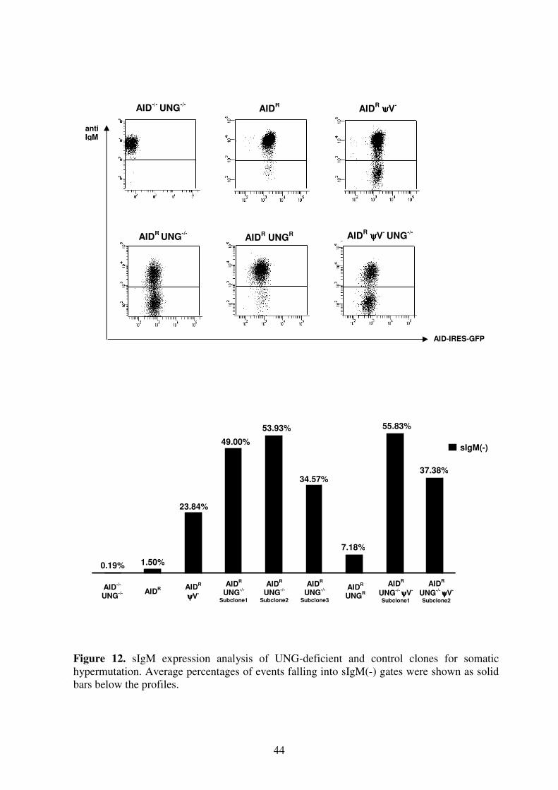
44
VI.
AID-/-
UNG-/- AID
R AIDR
ψψψψV-
AIDR
UNG-/-
Subclone1
AIDR
UNG-/-
Subclone2
AIDR
UNG-/-
Subclone3
AIDR
UNGR
AIDR
UNG-/-
ψψψψV-
Subclone1
AIDR
UNG-/-
ψψψψV-
Subclone2
anti
IgM
AID-IRES-GFP
Figure 12. sIgM expression analysis of UNG-deficient and control clones for somatic
hypermutation. Average percentages of events falling into sIgM(-) gates were shown as solid
bars below the profiles.
AID-/-
UNG-/-
AIDR AID
R ψψψψV
-
AIDR UNG
R AIDR
UNG-/-
-/- -/-
AIDR ψψψψV
- UNG
-/-
0.19% 1.50%
23.84%
49.00%
53.93%
34.57%
7.18%
55.83%
37.38%
sIgM(-)
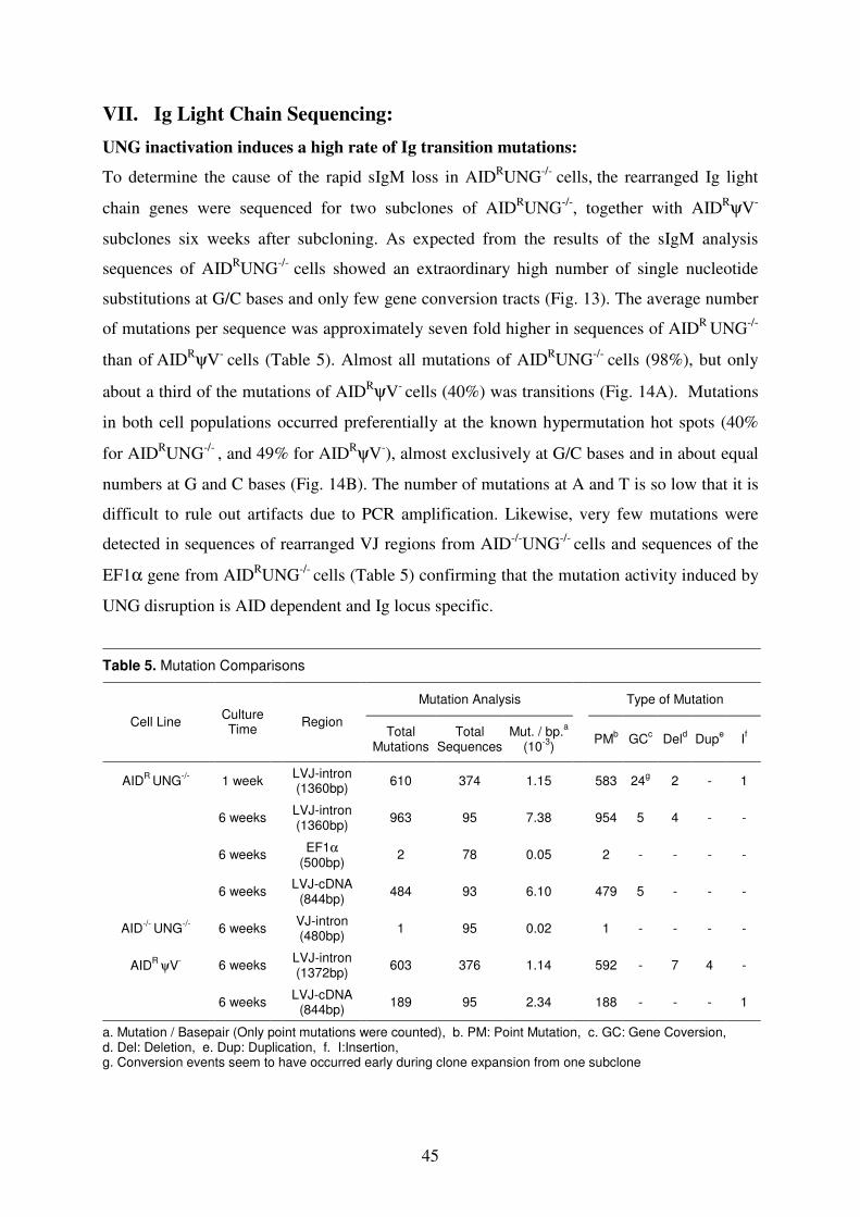
45
VII. Ig Light Chain Sequencing:
UNG inactivation induces a high rate of Ig transition mutations:
To determine the cause of the rapid sIgM loss in AIDRUNG
-/- cells,
the rearranged Ig light
chain genes were sequenced for two subclones of AIDRUNG
-/-, together with AID
RψV-
subclones six weeks after subcloning. As expected from the results of the sIgM analysis
sequences of AIDRUNG
-/- cells showed an extraordinary high number of single nucleotide
substitutions at G/C bases and only few gene conversion tracts (Fig. 13). The average number
of mutations per sequence was approximately seven fold higher in sequences of AIDR
UNG-/-
than of AID
RψV- cells (Table 5). Almost all mutations of AID
RUNG
-/- cells (98%), but only
about a third of the mutations of AIDRψV
- cells (40%) was transitions (Fig. 14A). Mutations
in both cell populations occurred preferentially at the known hypermutation hot spots (40%
for AIDRUNG
-/- , and 49% for AID
RψV-), almost exclusively at G/C bases and in about equal
numbers at G and C bases (Fig. 14B). The number of mutations at A and T is so low that it is
difficult to rule out artifacts due to PCR amplification. Likewise, very few mutations were
detected in sequences of rearranged VJ regions from AID-/-
UNG-/-
cells and sequences of the
EF1α gene from AIDRUNG
-/- cells (Table 5) confirming that the mutation activity induced by
UNG disruption is AID dependent and Ig locus specific.
Table 5. Mutation Comparisons
Mutation Analysis Type of Mutation
Cell Line Culture Time
Region
Total
Mutations Total
Sequences Mut. / bp.
a
(10-3
) PM
b GC
c Del
d Dup
e If
AIDR
UNG-/-
1 week LVJ-intron (1360bp)
610 374 1.15 583 24g
2 - 1
6 weeks LVJ-intron (1360bp)
963 95 7.38 954 5 4 - -
6 weeks EF1α
(500bp) 2 78 0.05 2 - - - -
6 weeks LVJ-cDNA
(844bp) 484 93 6.10 479 5 - - -
AID-/-
UNG-/-
6 weeks VJ-intron (480bp)
1 95 0.02 1 - - - -
AIDR
ψV- 6 weeks
LVJ-intron (1372bp)
603 376 1.14 592 - 7 4 -
6 weeks LVJ-cDNA
(844bp) 189 95 2.34 188 - - - 1
a. Mutation / Basepair (Only point mutations were counted), b. PM: Point Mutation, c. GC: Gene Coversion, d. Del: Deletion, e. Dup: Duplication, f. I:Insertion, g. Conversion events seem to have occurred early during clone expansion from one subclone
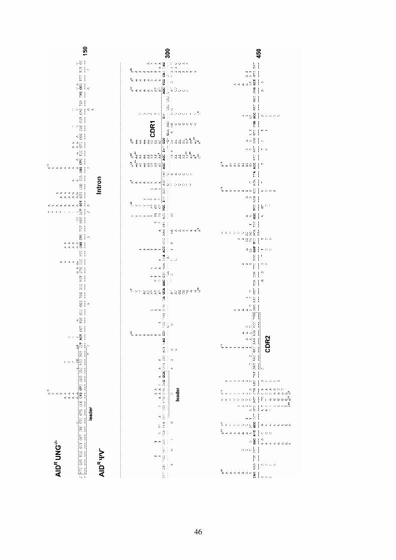
46
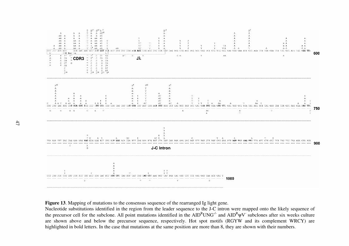
47
Figure 13. Mapping of mutations to the consensus sequence of the rearranged Ig light gene.
Nucleotide substitutions identified in the region from the leader sequence to the J-C intron were mapped onto the likely sequence of
the precursor cell for the subclone. All point mutations identified in the AIDRUNG
-/- and AID
RψV- subclones after six weeks culture
are shown above and below the precursor sequence, respectively. Hot spot motifs (RGYW and its complement WRCY) are
highlighted in bold letters. In the case that mutations at the same position are more than 8, they are shown with their numbers.
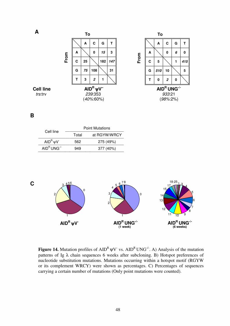
48
Cell line AIDR ψψψψV
−−−− AID
R UNG
-/-
trs:trv 239:353 933:21 (40%:60%) (98%:2%)
A C G T
A 0 6 0
C 5 1 415
G 510 10 5
T 0 2 0
A C G T
A 0 15 3
C 25 182 147
G 75 108 31
T 3 2 1
Point Mutations Cell line
Total at RGYW/WRCY
AIDR
ψV- 562 275 (49%)
AIDR
UNG-/- 949 377 (40%)
A
Fro
m
Fro
m
To To
B
C
Figure 14. Mutation profiles of AIDR ψV
- vs. AID
R UNG
-/-. A) Analysis of the mutation
patterns of Ig λ chain sequences 6 weeks after subcloning. B) Hotspot preferences of
nucleotide substitution mutations. Mutations occurring within a hotspot motif (RGYW
or its complement WRCY) were shown as percentages. C) Percentages of sequences
carrying a certain number of mutations (Only point mutations were counted).
AIDR ψψψψV
- AID
R UNG
-/-
(1 week)
AIDR
UNG-/-
(6 weeks)
0
1
2
34
5
0
1
3
4 5
2
6
7 8 6 4 1 3 4
5
6
7
8 9 10 11
12
13
14
15 17
18-25
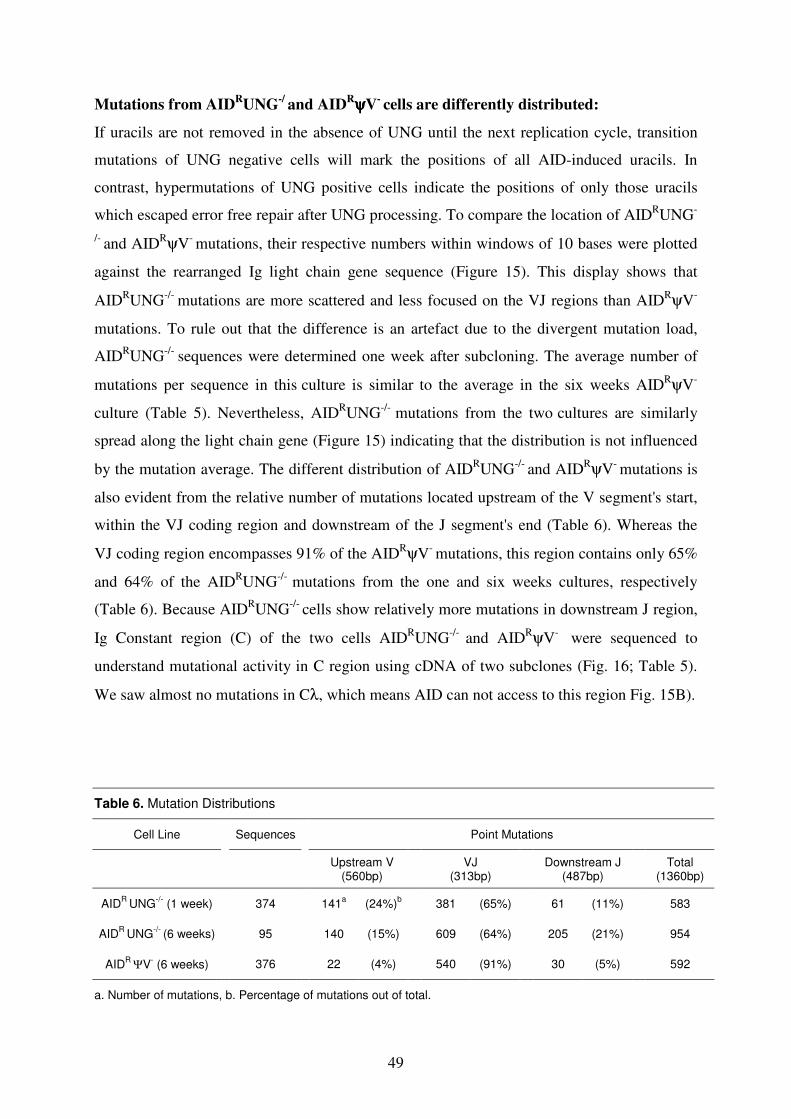
49
Mutations from AIDRUNG
-/ and AID
RψψψψV- cells are differently distributed:
If uracils are not removed in the absence of UNG until the next replication cycle, transition
mutations of UNG negative cells will mark the positions of all AID-induced uracils. In
contrast, hypermutations of UNG positive cells indicate the positions of only those uracils
which escaped error free repair after UNG processing. To compare the location of AIDRUNG
-
/- and AID
RψV- mutations, their respective numbers within windows of 10 bases were plotted
against the rearranged Ig light chain gene sequence (Figure 15). This display shows that
AIDRUNG
-/- mutations are more scattered and less focused on the VJ regions than AID
RψV-
mutations. To rule out that the difference is an artefact due to the divergent mutation load,
AIDRUNG
-/- sequences were determined one week after subcloning. The average number of
mutations per sequence in this culture is similar to the average in the six weeks AID
RψV-
culture (Table 5). Nevertheless, AIDRUNG
-/- mutations from the two
cultures are similarly
spread along the light chain gene (Figure 15) indicating that the distribution is not influenced
by the mutation average. The different distribution of AIDRUNG
-/- and AID
RψV- mutations is
also evident from the relative number of mutations located upstream of the V segment's start,
within the VJ coding region and downstream of the J segment's end (Table 6). Whereas the
VJ coding region encompasses 91% of the AIDRψV
- mutations, this region contains only 65%
and 64% of the AIDRUNG
-/- mutations from the one and six weeks cultures, respectively
(Table 6). Because AIDRUNG
-/- cells show relatively more mutations in downstream J region,
Ig Constant region (C) of the two cells AIDRUNG
-/- and AID
RψV-
were sequenced to
understand mutational activity in C region using cDNA of two subclones (Fig. 16; Table 5).
We saw almost no mutations in Cλ, which means AID can not access to this region Fig. 15B).
Table 6. Mutation Distributions
Cell Line Sequences Point Mutations
Upstream V
(560bp)
VJ (313bp)
Downstream J
(487bp)
Total (1360bp)
AIDR
UNG-/-
(1 week) 374 141a
(24%)b
381 (65%) 61 (11%) 583
AIDR
UNG-/-
(6 weeks)
95 140 (15%) 609 (64%) 205 (21%) 954
AIDR
ΨV- (6 weeks) 376 22 (4%) 540 (91%) 30 (5%) 592
a. Number of mutations, b. Percentage of mutations out of total.
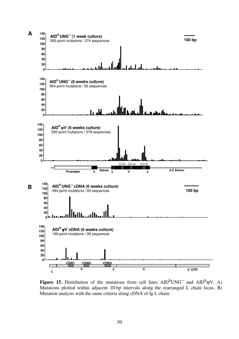
50
0
20
40
60
80
100
120
140
0
20
40
60
80
100
120
140
0
20
40
60
80
100
120
140
0
20
40
60
80
100
120
140
0
20
40
60
80
100
120
140
100 bp
A
B
L V
CDR1 CDR2
J C 3’ UTR
CDR3
AIDR
ψψψψV- cDNA (6 weeks culture)
189 point mutations / 95 sequences
AIDR
UNG-/-
cDNA (6 weeks culture)
484 point mutations / 93 sequences
AIDR
ψψψψV- (6 weeks culture)
592 point mutations / 376 sequences
AIDR
UNG-/-
(6 weeks culture)
954 point mutations / 95 sequences
AIDR
UNG-/-
(1 week culture)
583 point mutations / 374 sequences
100 bp
Figure 15. Distribution of the mutations from cell lines AIDRUNG
-/- and AID
RψV. A)
Mutations plotted within adjacent 10-bp intervals along the rearranged L chain locus. B)
Mutation analysis with the same criteria along cDNA of Ig L chain.
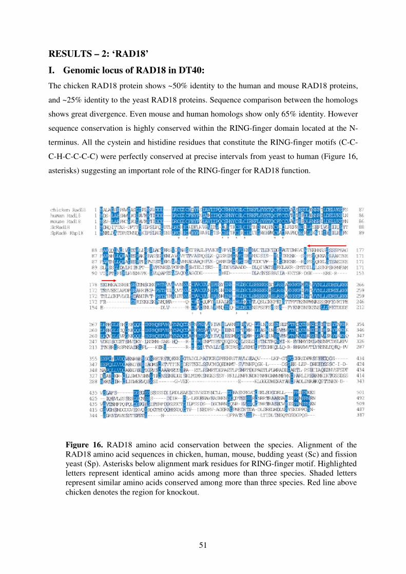
51
RESULTS – 2: ‘RAD18’
I. Genomic locus of RAD18 in DT40:
The chicken RAD18 protein shows ~50% identity to the human and mouse RAD18 proteins,
and ~25% identity to the yeast RAD18 proteins. Sequence comparison between the homologs
shows great divergence. Even mouse and human homologs show only 65% identity. However
sequence conservation is highly conserved within the RING-finger domain located at the N-
terminus. All the cystein and histidine residues that constitute the RING-finger motifs (C-C-
C-H-C-C-C-C) were perfectly conserved at precise intervals from yeast to human (Figure 16,
asterisks) suggesting an important role of the RING-finger for RAD18 function.
Figure 16. RAD18 amino acid conservation between the species. Alignment of the
RAD18 amino acid sequences in chicken, human, mouse, budding yeast (Sc) and fission
yeast (Sp). Asterisks below alignment mark residues for RING-finger motif. Highlighted
letters represent identical amino acids among more than three species. Shaded letters
represent similar amino acids conserved among more than three species. Red line above
chicken denotes the region for knockout.
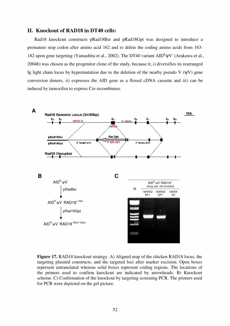
52
II. Knockout of RAD18 in DT40 cells:
Rad18 knockout constructs pRad18Bsr and pRad18Gpt was designed to introduce a
premature stop codon after amino acid 162 and to delete the coding amino acids from 163-
182 upon gene targeting (Yamashita et al., 2002). The DT40 variant AIDRψV- (Arakawa et al.,
2004b) was chosen as the progenitor clone of the study, because it, i) diversifies its rearranged
Ig light chain locus by hypermutation due to the deletion of the nearby pseudo V (ψV) gene
conversion donors, ii) expresses the AID gene as a floxed cDNA cassette and iii) can be
induced by tamoxifen to express Cre recombinase.
AIDR ψV
- RAD18
-/-
(drug cas. not excised)
M 18ARA2
BS1 18ARA2
GP1 18ARA
3/5
AIDR
ψV-
AIDR
ψV- RAD18
+/-Bsr
pRadBsr
AIDR
ψV- RAD18
-(Bsr)/-(Gpt)
pRad18Gpt
C
B
A
Figure 17. RAD18 knockout strategy. A) Aligned map of the chicken RAD18 locus, the
targeting plasmid constructs, and the targeted loci after marker excision. Open boxes
represent untranslated whereas solid boxes represent coding regions. The locations of
the primers used to confirm knockout are indicated by arrowheads. B) Knockout
scheme. C) Confirmation of the knockout by targeting screening PCR. The primers used
for PCR were depicted on the gel picture.
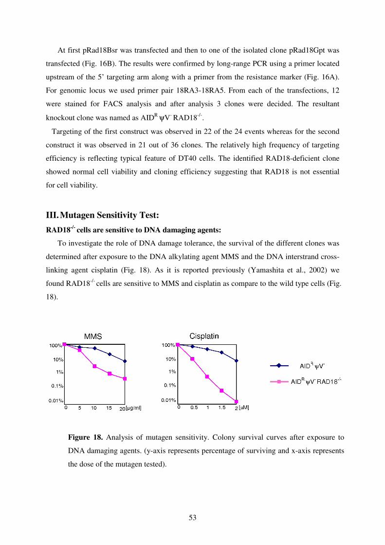
53
At first pRad18Bsr was transfected and then to one of the isolated clone pRad18Gpt was
transfected (Fig. 16B). The results were confirmed by long-range PCR using a primer located
upstream of the 5’ targeting arm along with a primer from the resistance marker (Fig. 16A).
For genomic locus we used primer pair 18RA3-18RA5. From each of the transfections, 12
were stained for FACS analysis and after analysis 3 clones were decided. The resultant
knockout clone was named as AIDR ψV
- RAD18
-/-.
Targeting of the first construct was observed in 22 of the 24 events whereas for the second
construct it was observed in 21 out of 36 clones. The relatively high frequency of targeting
efficiency is reflecting typical feature of DT40 cells. The identified RAD18-deficient clone
showed normal cell viability and cloning efficiency suggesting that RAD18 is not essential
for cell viability.
III. Mutagen Sensitivity Test:
RAD18-/-
cells are sensitive to DNA damaging agents:
To investigate the role of DNA damage tolerance, the survival of the different clones was
determined after exposure to the DNA alkylating agent MMS and the DNA interstrand cross-
linking agent cisplatin (Fig. 18). As it is reported previously (Yamashita et al., 2002) we
found RAD18-/- cells are sensitive to MMS and cisplatin as compare to the wild type cells (Fig.
18).
Figure 18. Analysis of mutagen sensitivity. Colony survival curves after exposure to
DNA damaging agents. (y-axis represents percentage of surviving and x-axis represents
the dose of the mutagen tested).
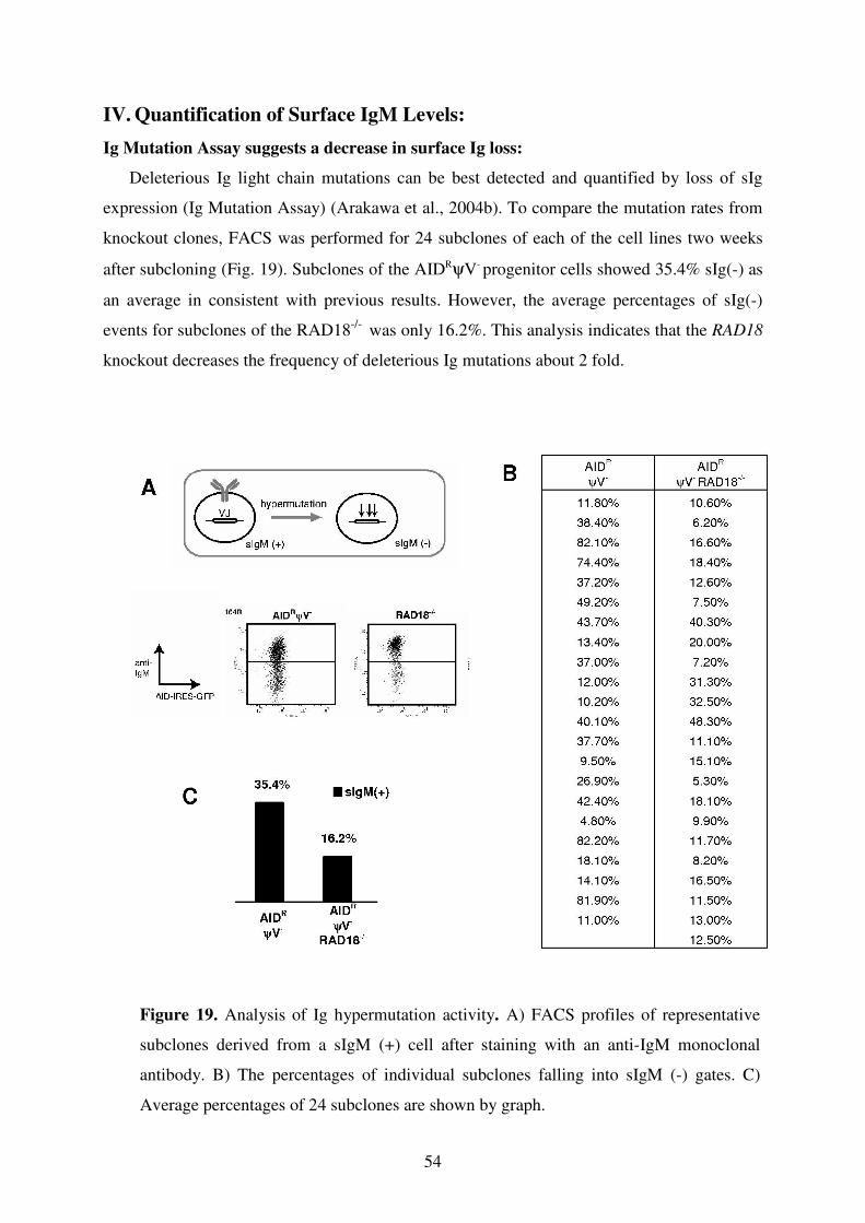
54
IV. Quantification of Surface IgM Levels:
Ig Mutation Assay suggests a decrease in surface Ig loss:
Deleterious Ig light chain mutations can be best detected and quantified by loss of sIg
expression (Ig Mutation Assay) (Arakawa et al., 2004b). To compare the mutation rates from
knockout clones, FACS was performed for 24 subclones of each of the cell lines two weeks
after subcloning (Fig. 19). Subclones of the AIDRψV- progenitor cells showed 35.4% sIg(-) as
an average in consistent with previous results. However, the average percentages of sIg(-)
events for subclones of the RAD18-/- was only 16.2%. This analysis indicates that the RAD18
knockout decreases the frequency of deleterious Ig mutations about 2 fold.
Figure 19. Analysis of Ig hypermutation activity. A) FACS profiles of representative
subclones derived from a sIgM (+) cell after staining with an anti-IgM monoclonal
antibody. B) The percentages of individual subclones falling into sIgM (-) gates. C)
Average percentages of 24 subclones are shown by graph.
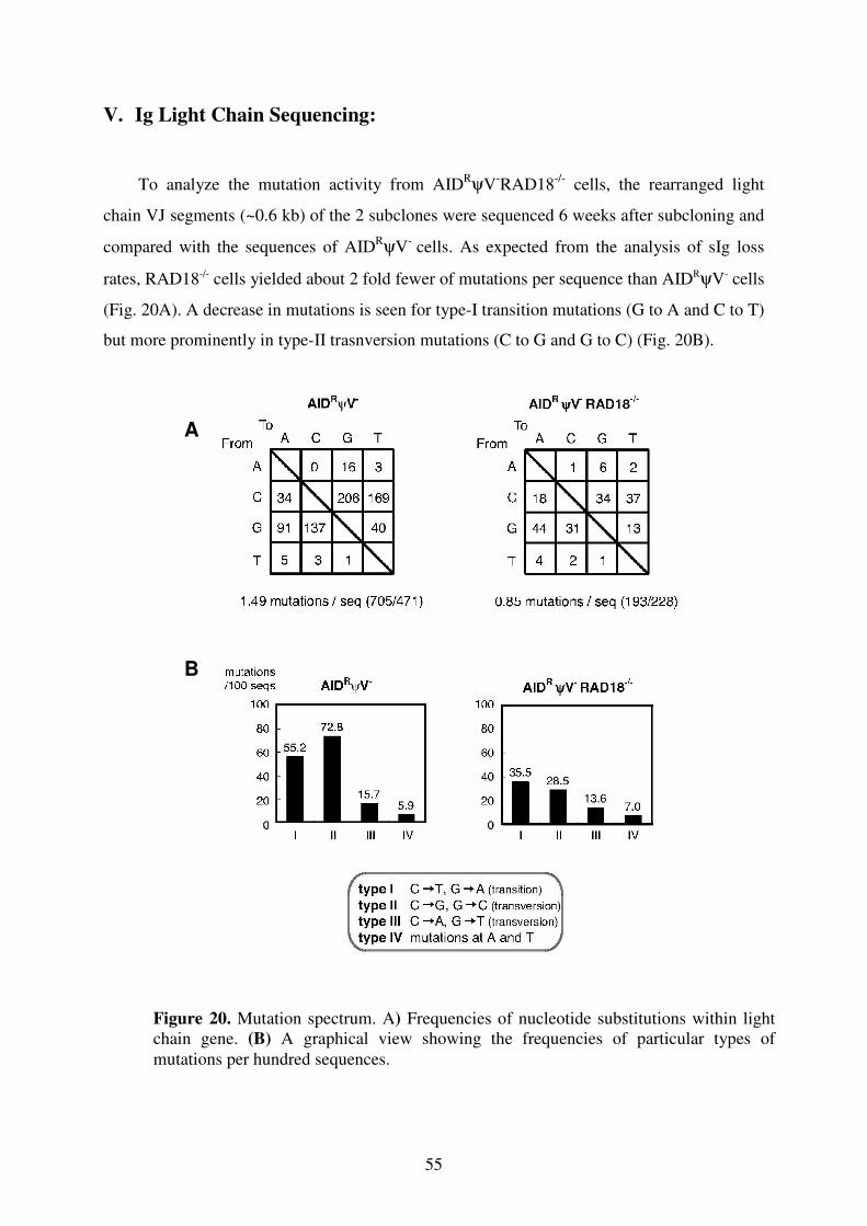
55
V. Ig Light Chain Sequencing:
To analyze the mutation activity from AIDRψV
-RAD18
-/- cells, the rearranged light
chain VJ segments (~0.6 kb) of the 2 subclones were sequenced 6 weeks after subcloning and
compared with the sequences of AIDRψV
- cells. As expected from the analysis of sIg loss
rates, RAD18-/- cells yielded about 2 fold fewer of mutations per sequence than AIDRψV- cells
(Fig. 20A). A decrease in mutations is seen for type-I transition mutations (G to A and C to T)
but more prominently in type-II trasnversion mutations (C to G and G to C) (Fig. 20B).
Figure 20. Mutation spectrum. A) Frequencies of nucleotide substitutions within light
chain gene. (B) A graphical view showing the frequencies of particular types of
mutations per hundred sequences.
B
A
56
DISCUSSION:
DNA deamination model of AID suggests a role for UNG downstream of AID to
remove uracils that are introduced by AID in immunoglobulin V genes or switch regions
(Petersen-Mahrt et al., 2002). Several studies have clarified this and showed that SHM and
CSR indeed need UNG (Di Noia et al., 2002; Rada et al., 2002a; Imai et al., 2003).
In the present study we could show that inactivation of the UNG gene in AID
expressing DT40 almost completely abolishes Ig gene conversions and activates a very
high rate of transition mutations at C/G bases in the rearranged light chain VJ region. This
demonstrates conclusively that Ig gene conversion like previously shown from
hypermutation and switch recombination requires UNG downstream of AID.
The results strongly support a model in which AID first deaminates cytosine within the
rearranged V(D)J segments and the resulting uracils are then processed by UNG to give rise
to first repair in an error free manner, second to gene conversions and third to
hypermutations in the absence of pseudogenes (Fig. 21). Inactivation of the UNG gene
blocks the processing of uracils and leads exclusively to transition mutations when uracil
pairs with adenine in the next replication cycle. So far, this model had been based on results
showing that Ugi (uracil DNA glycosylase inhibitor) expression shifts Ig hypermutations
toward transitions in a XRCC2-/-
mutant (Di Noia et al., 2002) and inhibits Ig gene
conversion in wild-type DT40 (Di Noia et al., 2004). However, the phenotype of the Ugi
gene transfectants differs from the phenotype of the UNG-/-
mutant in some features at odds
with the model. For example, Ugi expression increased transition mutations only to 86% in
the XRCC2-/-
mutant whereas this number was 98% from the UNG-/-
mutant (Di Noia et al.,
2002). Similarly, Ugi expression reduced Ig gene conversion to 30% of wild-type levels (Di
Noia et al., 2004), whereas the UNG gene disruption decreased gene conversion almost 100
fold based on the light chain frameshift repair assay. The most striking discrepancy to the
model is however the lack of detectable transition mutations after Ugi expression in wild-
type DT40. The difference between the results obtained after Ugi transfection and UNG
disruption could be due to the fact that UNG-deficient cells express a transfected AID,
which may differ both in abundance and cell cycle regulation from endogenous AID.
However, the most straightforward interpretation seems to be that Ugi expression in DT40
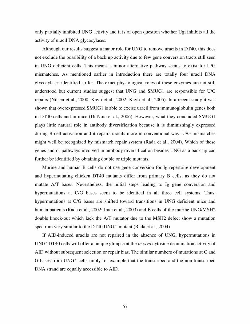
57
only partially inhibited UNG activity and it is of open question whether Ugi inhibits all the
activity of uracil DNA glycosylases.
Although our results suggest a major role for UNG to remove uracils in DT40, this does
not exclude the possibility of a back up activity due to few gene conversion tracts still seen
in UNG deficient cells. This means a minor alternative pathway seems to exist for U/G
mismatches. As mentioned earlier in introduction there are totally four uracil DNA
glycosylases identified so far. The exact physiological roles of these enzymes are not still
understood but current studies suggest that UNG and SMUG1 are responsible for U/G
repairs (Nilsen et al., 2000; Kavli et al., 2002; Kavli et al., 2005). In a recent study it was
shown that overexpressed SMUG1 is able to excise uracil from immunoglobulin genes both
in DT40 cells and in mice (Di Noia et al., 2006). However, what they concluded SMUG1
plays little natural role in antibody diversification because it is diminishingly expressed
during B-cell activation and it repairs uracils more in conventional way. U/G mismatches
might well be recognized by mismatch repair system (Rada et al., 2004). Which of these
genes and or pathways involved in antibody diversification besides UNG as a back up can
further be identified by obtaining double or triple mutants.
Murine and human B cells do not use gene conversion for Ig repertoire development
and hypermutating chicken DT40 mutants differ from primary B cells, as they do not
mutate A/T bases. Nevertheless, the initial steps leading to Ig gene conversion and
hypermutations at C/G bases seem to be identical in all three cell systems. Thus,
hypermutations at C/G bases are shifted toward transitions in UNG deficient mice and
human patients (Rada et al., 2002; Imai et al., 2003) and B cells of the murine UNG/MSH2
double knock-out which lack the A/T mutator due to the MSH2 defect show a mutation
spectrum very similar to the DT40 UNG-/-
mutant (Rada et al., 2004).
If AID-induced uracils are not repaired in the absence of UNG, hypermutations in
UNG-/-
DT40 cells will offer a unique glimpse at the in vivo cytosine deamination activity of
AID without subsequent selection or repair bias. The similar numbers of mutations at C and
G bases from UNG-/-
cells imply for example that the transcribed and the non-transcribed
DNA strand are equally accessible to AID.
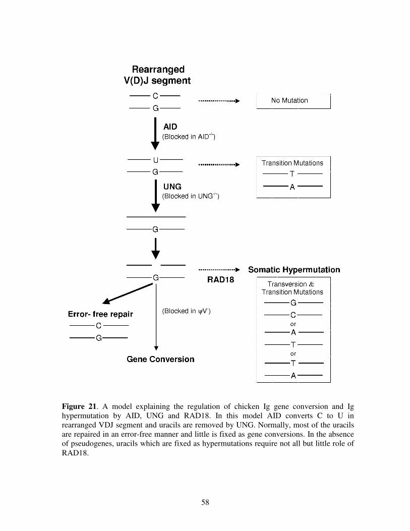
58
Figure 21. A model explaining the regulation of chicken Ig gene conversion and Ig
hypermutation by AID, UNG and RAD18. In this model AID converts C to U in
rearranged VDJ segment and uracils are removed by UNG. Normally, most of the uracils
are repaired in an error-free manner and little is fixed as gene conversions. In the absence
of pseudogenes, uracils which are fixed as hypermutations require not all but little role of
RAD18.
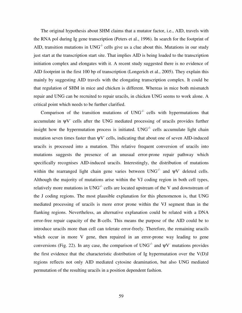
59
The original hypothesis about SHM claims that a mutator factor, i.e., AID, travels with
the RNA pol during Ig gene transcription (Peters et al., 1996). In search for the footprint of
AID, transition mutations in UNG-/-
cells give us a clue about this. Mutations in our study
just start at the transcription start site. That implies AID is being loaded to the transcription
initiation complex and elongates with it. A recent study suggested there is no evidence of
AID footprint in the first 100 bp of transcription (Longerich et al., 2005). They explain this
mainly by suggesting AID travels with the elongating transcription complex. It could be
that regulation of SHM in mice and chicken is different. Whereas in mice both mismatch
repair and UNG can be recruited to repair uracils, in chicken UNG seems to work alone. A
critical point which needs to be further clarified.
Comparison of the transition mutations of UNG-/-
cells with hypermutations that
accumulate in ψV- cells after the UNG mediated processing of uracils provides further
insight how the hypermutation process is initiated. UNG-/-
cells accumulate light chain
mutation seven times faster than ψV- cells, indicating that about one of seven AID-induced
uracils is processed into a mutation. This relative frequent conversion of uracils into
mutations suggests the presence of an unusual error-prone repair pathway which
specifically recognises AID-induced uracils. Interestingly, the distribution of mutations
within the rearranged light chain gene varies between UNG-/-
and ψV- deleted cells.
Although the majority of mutations arise within the VJ coding region in both cell types,
relatively more mutations in UNG-/-
cells are located upstream of the V and downstream of
the J coding regions. The most plausible explanation for this phenomenon is, that UNG
mediated processing of uracils is more error prone within the VJ segment than in the
flanking regions. Nevertheless, an alternative explanation could be related with a DNA
error-free repair capacity of the B-cells. This means the purpose of the AID could be to
introduce uracils more than cell can tolerate error-freely. Therefore, the remaining uracils
which occur in more V gene, then repaired in an error-prone way leading to gene
conversions (Fig. 22). In any case, the comparison of UNG-/-
and ψV- mutations provides
the first evidence that the characteristic distribution of Ig hypermutation over the V(D)J
regions reflects not only AID mediated cytosine deamination, but also UNG mediated
permutation of the resulting uracils in a position dependent fashion.
60
Studies in yeast suggest that PCNA mono-ubiquitination by RAD6/RAD18 induces a
switch from replicative to translesion DNA synthesis (Hoege et al., 2002; Ulrich et al.,
2004). It is of interesting question whether SHM in DT40 requires this pathway or not?
Our results show that at least partially SHM needs RAD18 induced
monoubiquitination (Fig. 21). RAD18-/-
cells are sensitive to DNA damaging agents MMS
and cisplatin and in RAD18-/-
cells only type II (majority) and type I mutations were
decreased. The decrease in type II mutations were also seen before in REV1-/-
cells (our
unpublished data; Simpson et al., 2003). The selective decrease of this type of
hypermutation may reflect the deoxycytidyl transferase activity of REV1, which would add
cytosine opposite to an abasic site in the template strand (Nelson et al., 1996). This means
that ubiquitinated PCNA recruits REV1 as an error-prone polymerase.
Why only partial of the mutations decreased has to be clarified. RAD18 is the only E3
ligase known to mono-ubiquitinate PCNA at lysine 164 in S. cerevisiae. The RAD18
knockout construct used in our study most likely generates a RAD18 null mutation
(Yamashita et al., 2002), although it does not delete the RAD18 RING finger coding
sequence. To rule out that the analysed RAD18 mutant still possessed enzymatic activity,
we generated a second RAD18 mutant in which the RING finger coding region is deleted.
Analysis of this new mutant confirmed the persistence of low-level PCNA ubiqitination
seen in the first RAD18 mutant (data not shown). These data point to the presence of a
RAD18 independent back-up pathway of PCNA ubiquitination in vertebrate cells. Most
Figure 22. A possible model for the initiation of gene conversion and or hypermutation.
61
likely, PCNA ubiquitination in RAD18-/- cells is mediated by another E3 ligase like, for
example, the one encoded by the FANCC or FANCL gene (Gurtan et al., 2006). It was
previously reported that RAD18 disruption in DT40 does not affect Ig hypermutation
(Simpson et al., 2005). However, we see an about 2 fold reduction of Ig hypermutation in
RAD18-/- cells. We believe this discrepancy may be caused by the difficulty to accurately
measure Ig hypermutation in wild-type DT40, which diversifies its Ig genes predominantly
by gene conversion.
If ubiquitinated PCNA by RAD18 functions as a link to the recruitment of error prone
polymerases during Ig hypermutation, it remains an intriguing question how it is coupled to
upstream events in the hypermutation process. The DNA editing model assumes that AID
first deaminates cytosine to uracil and that the resulting uracil is then excised by UNG-2
(Di Noia et al., 2002). We showed in thesis that, comparison of the mutation frequencies in
UNG disrupted and ψV deleted DT40 suggests about one in seven AID-induced uracils is
converted into a mutation. This high mutation rate indicates that the abasic sites produced
by uracil excision are not repaired by the standard base excision repair, but are deliberately
channeled into error-prone translesion synthesis. One of the possibilities is that UNG-2 is
recruited by PCNA (Otterlei et., 1999) and excises AID-induced uracils shortly before
DNA synthesis thereby precluding the possibility of base excision repair. Another
possibility is that the DNA lesions produced by the combined action of AID and UNG are
for some reason, perhaps by protein attachment or by another type of modification, guarded
from faithful repair until they encounter the PCNA clamp.
62
REFERENCES: 1. Abdrakhmanov I, Lodygin D, Geroth P, Arakawa H, Law A, Plachy J, Korn B,
Buerstedde JM. 2000. A large database of chicken bursal ESTs as a resource for the
analysis of vertebrate gene function. Genome Res. 10: 2062-2069.
2. Alting-Mees MA, Short JM. 1989. pBluescript II: gene mapping vectors. Nucleic Acids
Res. 17: 9494.
3. Arakawa H, Furusawa S, Ekino S, Yamagishi H. 1996. Immunoglobulin gene
hyperconversion ongoing in chicken splenic germinal centers. EMBO J. 15: 2540-2546.
4. Arakawa H, Kuma K, Yasuda M, Furusawa S, Ekino S, Yamagishi H. 1998.
Oligoclonal development of B cells bearing discrete Ig chains in chicken single
germinal centers. J Immunol. 160: 4232-4241.
5. Arakawa H, Lodygin D, Buerstedde JM. 2001. Mutant loxP vectors for selectable
marker recycle and conditional knock-outs. BMC Biotechnol. 1: 7.
6. Arakawa H, Kuma K, Yasuda M, Ekino S, Shimizu A, Yamagishi H. 2002a. Effect of
environmental antigens on the Ig diversification and the selection of productive V-J
joints in the bursa. J Immunol. 169: 818-828
7. Arakawa H, Hauschild J, Buerstedde JM. 2002b. Requirement of the activation-induced
deaminase (AID) gene for immunoglobulin gene conversion. Science. 295: 1301–1306.
8. Arakawa H, Buerstedde JM. 2004a. Immunoglobulin gene conversion: Insights from
Bursal B cells and the DT40 cell line. Dev Dyn. 229: 458:464.
9. Arakawa H, Saribasak H, Buerstedde JM. 2004b. Activation-induced cytidine
deaminase initiates immunoglobulin gene conversion and hypermutation by a common
intermediate. PLoS Biol. 2: 967-974.
10. Baba TW, Giroir BP, Humphries EH. 1985. Cell lines derived from avian lymphomas
exhibit two distinct phenotypes. Virology. 144: 139-151.
11. Bachl J, Wabl M. 1996. An immunoglobulin mutator that targets G.C base pairs. Proc.
Natl. Acad. Sci. U S A. 93: 851-855.
12. Bachl J, Carlson C, Gray-Schopfer V, Dessing M, Olsson C. 2001. Increased
transcription levels induce higher mutation rates in a hypermutating cell line. J
Immunol. 166: 5051-57.
13. Bailly V, Lauder S, Prakash S, Prakash L. 1997. Yeast DNA repair proteins Rad6 and
63
Rad18 form a heterodimer that has ubiquitin conjugating, DNA binding, and ATP
hydrolytic activities. J Biol Chem. 272: 23360- 23365.
14. Barreto VM, Reina-San-Martin B, Ramiro AR, McBride KM, Nussenzweig MC. 2003.
C-terminal deletion of AID uncouples class switch recombination from somatic
hypermutation and gene conversion. Mol Cell. 12: 501-508.
15. Barreto VM, Ramiro AR, Nussenzweig MC. 2005. Activation-induced deaminase:
controversies and open questions. Trends Immunol. 26: 90-96.
16. Betz AG, Milstein C, Gonzales-Fernandez A, Pannell R, Larson T, Neuberger MS.
1994. Elements regulating somatic hypermutation of an immunoglobulin kappa gene:
critical role for the intron enhancer / matrix attachment region. Cell. 77: 239-248.
17. Bezzubova OY, Buerstedde JM. 1994. Gene conversion in the chicken immunoglobulin
locus: a paradigm of homologous recombination in higher eukaryotes. Experientia.
50:270-276.
18. Bezzubova O, Silbergleit A, Yamaguchi-Iwai Y, Takeda S, Buerstedde JM. 1997.
Reduced X-ray resistance and homologous recombination frequencies in a RAD54-/-
mutant of the chicken DT40 cell line. Cell. 89: 185-193.
19. Bransteitter R, Pham P, Scharff MD, Goodman MF. 2003. Activation-induced cytidine
deaminase deaminates deoxycytidine on single-stranded DNA but requires the action of
RNase. Proc Natl Acad Sci U S A. 100: 4102-4107.
20. Brar SS, Watson M, Diaz M. 2004. Activation-induced cytosine deaminase (AID) is
actively exported out of the nucleus but retained by the induction of DNA breaks.
J Biol Chem. 279: 26395-26401.
21. Brenner S, Milstein C. 1966. Origin of antibody variation. Nature. 211: 242-243.
22. Bross L, Fukita Y, McBlane F, Demolliere C, Rajewski K, Jakobs H. 2000. DNA
double-strand breaks in immunoglobulin genes undergoing somatic hypermutation.
Immunity. 13: 589-97.
23. Bross L, Muramatsu M, Kinoshita K, Honjo T, Jakobs H. 2002. DNA double-strand
breaks: prior to but not sufficient in targeting hypermutation.J Exp Med.195:1187-1192.
24. Brynolf K, Eliasson R, Reichard P. 1978. Formation of Okazaki fragments in polyoma
DNA synthesis caused by misincorporation of uracil. Cell. 13: 573-580.
25. Buerstedde JM, Reynaud CA, Humphries EH, Olson W, Ewert DL, Weill JC. 1990.
64
Light chain gene conversion continues at high rate in an ALV-induced cell line. EMBO
J. 9: 921-927.
26. Buerstedde JM, Takeda S. 1991. Increased ratio of targeted to random integration after
transfection of chicken B cell lines. Cell. 67: 179-188.
27. Carlson LM, McCormack WT, Postema CE, Humphries EH, Thompson CB. 1990.
Templated insertions in the rearranged chicken IgL V gene segment arise by
intrachromosomal gene conversion. Genes Dev. 4: 536-547.
28. Chaudhuri J, Tian M, Khuong C, Chua K, Pinaud E, Alt FW. 2003. Transcription-
targeted DNA deamination by the AID antibody diversification enzyme. Nature.
422:726-730.
29. Chaudhuri J, Alt FW. 2004a. Class switch recombination: Interplay of transcription,
DNA deamination and DNA repair. Nat Rev Immunol. 4: 541-552.
30. Chaudhuri J, Khuong C, Alt FW. 2004. Replication protein A interacts with AID to
promote deamination of somatic hypermutation targets. Nature. 430: 992-998.
31. Chua KF, Alt FW, Manis JP. 2002. The function of AID in somatic mutation and class-
switch recombination: upstream or downstream of DNA breaks.J Exp Med.195:F37-41.
32. Delbos F, De Smet A, Faili A, Aoufouchi S, Weill JC, Reynaud CA. 2005. Contribution
of DNA polymerase eta to immunoglobulin gene hypermutation in the mouse. J. Exp.
Med. 201: 1191-1196.
33. Dickerson SK, Market E, Besmer E, Papavasiliou FN. 2003. AID mediates
hypermutation by deaminating single stranded DNA. J Exp Med. 197: 1291-1296.
34. Duncan BK, Weiss B. 1982. Specific mutator effects of ung (uracil-DNA glycosylase)
mutations in Escherichia coli. J Bacteriol. 151: 750-755.
35. Di Noia JM, Neuberger MS. 2002. Altering the pathway of immunoglobulin
hypermutation by inhibiting uracil-DNA glycosylase. Nature. 419: 43–48.
36. Di Noia JM, Neuberger MS. 2004. Immunoglobulin gene conversion in chicken DT40
cells largely proceeds through an abasic site intermediate generated by excision of the
uracil produced by AID-mediated deoxycytidine deamination. Eur J Immunol.34:504-8.
37. Di Noia JM, Rada C, Neuberger MS. 2006. SMUG1 is able to excise uracil from
immunoglobulin genes: insight into mutation versus repair. EMBO J. 25: 585-95.
38. Faili A, Aofouchi S, Gueranger Q, Zober C, Leon A, Bertocci B, Weill JC, Reynaud
65
CA. 2002. AID-dependent somatic hypermutation occurs as a DNA single-strand event
in the BL2 cell line. Nat. Immunol. 3: 815-821.
39. Fugmann SD, Schatz DG. 2002. Immunology: One AID to unite them all. Science.
295:1244-5.
40. Fujimori A, Tachiiri S, Sonoda E, Thompson LH, Dhar PK, Hiraoka M, Takeda S,
Zhang Y, Reth M, Takata M. 2001. Rad52 partially substitutes for the Rad51 paralog
XRCC3 in maintaining chromosomal integrity in vertebrate cells. EMBO J. 20: 5513-
5520.
41. Fukita Y, Jakobs H, Rajewski K. 1998. Somatic hypermutation in the heavy chain locus
correlates with transcription. Immunity. 9: 105-114.
42. Garg P, Burgers PM. 2005. Ubiquitinated proliferating cell nuclear antigen activates
translesion DNA polymerases eta and REV1. Proc. Natl. Acad. Sci. USA. 102: 18361-6.
43. Golding GB, Gearhart PJ, Glickman BW. 1987. Patterns of somatic mutations in
immunoglobulin variable regions. Genetics. 115: 169-176.
44. Goyenechea B, Klix N, Yelamos J, Williams G, Riddell A, Neuberger M, Milstein C.
1997. Cells strongly expressing Igκ transgenes show clonal recruitment of
hypermutation: a role for both MAR and the enhancers. EMBO J. 16: 3987-3994.
45. Gurtan AM, Stuckert P, D'Andrea AD. 2006. The WD40 Repeats of FANCL Are
Required for Fanconi Anemia Core Complex Assembly. J. Biol. Chem. 281:10896-905.
46. Haber JE. 1999. Gatekeepers of recombination. Nature. 398: 665-667.
47. Hoege C, Pfander B, Moldovan GL, Pyrowolakis G, Jentsch S. 2002. RAD6-dependent
DNA repair is linked to modification of PCNA by ubiquitin and SUMO. Nature.
419: 135-141.
48. Imai K, Slupphaug G, Lee WI, Revy P, Nonoyama S, Catalan N, Yel L, Forveille M,
Kavli B, Krokan HE, Ochs HD, Fischer A, Durandy A. 2003. Human uracil-DNA
glycosylase deficiency associated with profoundly impaired immunoglobulin class-
switch recombination. Nat. Immunol. 4:1023-1028.
49. Impellizzeri KJ, Anderson B, Burgers PM. 1991. The spectrum of spontaneous
mutations in a Saccharomyces cerevisiae uracil-DNA-glycosylase mutant limits the
function of this enzyme to cytosine deamination repair. J Bacteriol. 173: 6807-6810.
50. International Chicken Genome Sequencing Consortium. 2004. Sequence and
66
comparative analysis of the chicken genome provide unique perspectives on vertebrate
evolution. Nature. 432:695-716.
51. Ito S, Nagaoka H, Shinkura R, Begum N, Muramatsu M, Nakata M, Honjo T. 2004.
Activation induced cytidine deaminase shuttles between nucleus and cytoplasm like
apolipoprotein B mRNA editing catalytic polypeptide 1. Proc Natl Acad Sci U S A.
101: 1975-1980.
52. Janeaway CA, Travers P, Walport M, Shlomchik M. 2001. Immunobiology. Garland
Publishing.
53. Jang SK, Krausslich HG, Nicklin MJ, Duke GM, Palmenberg AC, Wimmer E. 1988. A
segment of the 5' nontranslated region of encephalomyocarditis virus RNA directs
internal entry of ribosomes during in vitro translation. J Virol. 62: 2636-2643.
54. Jansen JG, Langerak P, Tsaalbi-Shtylik A, van den Berk P, Jakobs H, de Wind N. 2006.
Strand-biased defect in C/G transversions in hypermutating immunogolobulin genes in
Rev1- deficient genes. J Exp Med. 203: 319-323.
55. Jentsch S, McGrath GP, Varshavsky A. 1987. The yeast DNA repair gene RAD6
encodes a ubiquitin-conjugating enzyme. Nature. 329: 131-134.
56. Jonsson ZO, Hubscher U. 1997. Proliferating cell nuclear antigen: more than a clamp
for DNA polymerases. Bioessays. 19: 967-975.
57. Kannouche PL, Wing J, Lehmann AR. 2004. Interaction of human DNA polymerase eta
with monoubiquitinated PCNA: a possible mechanism for the polymerase switch in
response to DNA damage. Mol. Cell. 14: 491-500.
58. Kavli B, Sundheim O, Akbari M, Otterlei M, Nilsen H, Skorpen F, Aas PA, Hagen L,
Krokan HE, Slupphaug G. hUNG2 is the major repair enzyme for removal of uracil
from U:A matches, U:G mismatches, and U in single-stranded DNA, with hSMUG1 as
a broad specificity backup. J Biol Chem. 277: 39926-39936.
59. Kavli B, Andersen S, Otterlei M, Liabakk NB, Imai K, Fischer A, Durandy A, Krokan
HE, Slupphaug G. 2005. B cells from hyper-IgM patients carrying UNG mutations lack
ability to remove uracil from ssDNA and have elevated genomic uracil. J Exp Med.
201: 2011-2021.
60. Kim S, Humphries EH, Tjoelker L, Carlson L, Thompson CB. 1990. Ongoing
diversification of the rearranged immunoglobulin light-chain gene in a bursal
67
lymphoma cell line. Mol Cell Biol. 10:3224-3231.
61. Kinoshita K, Honjo T. 2001. Linking class switch recombination with somatic
hypermutation. Nat Rev Mol Cell Biol. 2:493-503.
62. Klix N, Jolly CJ, Davies SL, Bruggemann M, Williams GT, Neuberger MS. 1998.
Multiple sequences from downstream of the J kappa cluster can combine to recruit
somatic hypermutation to a heterologous, upstream mutation domain. Eur J Immunol.
28: 317-326.
63. Lebecque SG, Gearhart PJ. 1990. Boundaries of somatic mutation in rearranged
immunoglobulin genes: 5’ boundary is near the promoter and 3’ boundary is
approximately 1kb from V(D)J gene. J Exp Med. 172: 1717:1727.
64. Lederberg J. 1959. Genes and antibodies. Science. 129: 1649-1653.
65. Lindahl T. 1974. An N-glycosidase from Escherichia coli that releases free uracil from
DNA containing deaminated cytosine residues. Proc Natl Acad Sci USA. 71:3649-3653.
66. Lindahl T. 1993. Instability and decay of the primary structure of the DNA. Nature.
362: 709-715.
67. Lindahl T. 2000. Suppression of spontaneous mutagenesis in human cells by DNA base
excision-repair. Mutat Res. 462: 129-135.
68. Longeich S, Tanaka A, Bozek G, Nicolea D, Storb U. 2005. The very 5' end and the
constant region of Ig genes are spared from somatic mutation because AID does not
access these regions. J Exp Med. 202: 1443-54.
69. Maizels N. 1995. Somatic hypermutation: how many mechanisms diversify V region
sequences? Cell. 83: 9-12.
70. Mansikka A, Sandberg M, Lassila O, Toivanen P. 1990. Rearrangement of
immunoglobulin light chain genes in the chicken occurs prior to colonization of the
embryonic bursa of Fabricius. Proc Natl Acad Sci USA. 87: 9416-9420.
71. Martin A, Scharff MD. 2002a. AID and mismatch repair in antibody diversification.
Nat Rev Immunol. 2:605-614.
72. Martin A, Bardwell PD, Woo CJ, Fan M, Shulman MJ, Scharff MD. 2002b. Activation-
induced cytidine deaminase turns on somatic hypermutation in hybridomas. Nature.
415: 802-806.
73. Martomo SA, Fu D, Yang WW, Joshi NS, Gearhart PJ. 2005. Deoxyuridine is
68
generated preferentially in the nontranscribed strand of DNA from cells expressing
activation induced cytidine deaminase. J Immunol. 174: 7787-7791.
74. Martomo SA, Yang WW, Wersto RP, Ohkumo T, Kondo Y, Yokoi M, Masutani C,
Hanaoka F, Gearhart PJ. 2005. Different mutation signatures in DNA polymerase eta-
and MSH6-deficient mice suggest separate roles in antibody diversification. Proc Natl
Acad Sci U S A. 102: 8656-8661.
75. McBride KM, Barreto V, Ramiro AR, Stavropoulos P, Nussenzweig MC. 2004.
Somatic hypermutation is limited by CRM1-dependent nuclear export of activation-
induced deaminase. J Exp Med. 199: 1235-1244.
76. McCormack WT, Thompson CB. 1990. Chicken IgL variable region gene conversions
display pseudogene donor preference and 5' to 3' polarity. Genes Dev. 4: 548-558.
77. Michael M, Shen HM, Longerich S, Kim N, Longacre A, Storb U. 2003. The E Box
motif CAGGTG enhances somatic hypermutation without enhancing transcription.
Immunity. 19: 235-242.
78. Muramatsu M, Sankaranand VS, Anant S, Sugai M, Kinoshita K, Davidson NO, Honjo
T. 1999. Specific expression of activation-induced cytidine deaminase (AID), a novel
member of the RNA-editing deaminase family in germinal center B cells. J Biol Chem.
274:18470-18476.
79. Muramatsu M., Kinoshita K, Fagarasan S, Yamada S, Shinkai Y, Honjo T. 2000. Class
switch recombination and hypermutation require activation-induced cytidine deaminase
(AID), a potential RNA editing enzyme. Cell. 102: 553–563.
80. Nelson JR, Lawrence CW, Hinkle DC. 1996. Deoxycytidyl transferase activity of yeast
REV1 protein. Nature. 382: 729-731.
81. Neuberger MS, Harris RS, Di Noia JM, Petersen-Mahrt SK. 2003. Immunity through
DNA deamination. Trends Biochem Sci. 28: 305-312.
82. Neuberger MS, Di Noia JM, Beale RCL, Williams GT, Yang Z, Rada C. 2005. Somatic
hypermutation at A.T pairs: polymerase error versus dUTP incorporation. Nat Rev
Immunol. 5:171-178.
83. Nilsen H, Otterlei M, Haug T, Solum K, Nagelhaus TA, Skorpen F, Krokan HE. 1997.
Nuclear and mitochondrial uracil-DNA glycosylases are generated by alternative
splicing and transcription from different positions in the UNG gene. Nucleic Acids Res.
69
25:750-55
84. Nilsen H, Rosewell I, Robins P, Skjelbred GF, Andersen S, Slupphaug G, Dly G,
Krokan HE, Lindahl T, Barnes DE. 2000. Uracil-DNA Glycosylase (UNG)-deficient
mice reveal a primary role of the enzyme during DNA replication. Mol Cell. 5: 1059-
1065.
85. Nussenzweig MC, Alt FW. 2004. Antibody diversity: one enzyme to rule them all. Nat
Med. 10: 1304-1305.
86. Otterlei M, Warbrick E, Nagelhus TA, Haug T, Slupphaug G, Akbari M, Ass PA,
Steinsbekk K, Bakke O, Krokan HE. 1999. Post-replicative base excision repair in
replication foci. EMBO J. 18: 3834-3844.
87. Papavasiliou FN, Schatz DG. 2002. Somatic hypermutation of immunoglobulin genes:
Merging mechanism for genetic diversity. Cell. 109: S35-S44.
88. Pearl LH. 2000. Structure and function in the uracil-DNA glycosylase super family.
Mutat Res. 460: 165-181.
89. Pelletier J, Sonenberg N. 1988. Internal initiation of translation of eukaryotic mRNA
directed by a sequence derived from poliovirus RNA. Nature 334: 320-325.
90. Peters A, Storb U. 1996. Somatic hypermutation of immunoglobulin genes is linked to
transcription initiation. Immunity. 4: 57-65.
91. Petersen-Mahrt SK, Harris RS, Neuberger MS. 2002. AID mutates E. coli suggesting a
DNA deamination mechanism for antibody diversification. Nature. 418: 99-103.
92. Pham P, Bransteitter R, Petruska J, Goodman MF. 2003. Processive AID-catalysed
cytosine deamination on single-stranded DNA simulates somatic hypermutation.
Nature. 424:103-107.
93. Pink JR, Vainio O, Rijnbeek AM. 1985. Clones of B lymphocytes in individual follicles
of the bursa of Fabricius. Eur J Immunol. 15:83-87
94. Rada C, Ehrenstein MR, Neuberger MS, Milstein C. 1998. Hot spot focusing of
somatic hypermutation in MSH2-deficient mice suggests two stages of mutational
targeting. Immunity. 9:135-141.
95. Rada C, Williams GT, Nilsen H, Barnes DE, Lindahl T, Neuberger MS. 2002a.
Immunoglobulin isotype switching is inhibited and somatic hypermutation perturbed in
UNG-deficient mice. Curr Biol. 12:1748-1755.
70
96. Rada C, Jarvis JM, Milstein C. 2002b. AID-GFP chimeric protein increases
hypermutation of Ig genes with no evidence of nuclear localization. Proc. Natl. Acad.
Sci. U S A. 99: 7003-7008.
97. Rada C, Di Noia JM, Neuberger MS. 2004. Mismatch recognition and uracil excision
provide complementary paths to both Ig switching and the A/T-focused phase of
somatic mutation. Mol Cell. 2004. 16:163-171.
98. Ramiro AR, Stavropoulos P, Jankovic M, Nussenzweig MC. 2003. Transcription
enhances AID-mediated cytidine deamination by exposing single-stranded DNA on the
nontemplate strand. Nat Immunol. 4: 452-456.
99. Revy P, Muto T, Levy Y, Geissmann F, Plebani A, Sanal O, Catalan N, Forveille M,
Dufourcq-Labelouse R, Gennery A, Tezcan I, Ersoy F, Kayserili H, Ugazio AG,
Brousse N, Muramatsu M, Notarangelo LD, Kinoshita K, Honjo T, Fischer A, Durandy
A. 2000. Activation-induced cytidine deaminase (AID) deficiency causes the autosomal
recessive form of the Hyper-IgM syndrome. Cell. 102:565-575.
100. Reynaud CA, Anquez V, Grimal H, Weill JC. 1987. A hyperconversion mechanism
generates the chicken light chain immune repertoire. Cell. 48: 379-388.
101. Reynaud CA, Imhof BA, Anquez V, Weill JC. 1992. Emergence of committed B
lymphoid progenitors in the developing chicken embryo. EMBO J. 11:4349-4358.
102. Reynaud CA, Dahan A, Anquez V, Weill JC. 1989. Somatic hyperconversion
diversifies the single Vh gene of the chicken with a high incidence in the D region. Cell.
59: 171-183
103. Reynaud CA, Bertocci B, Dahan A, Weill JC. 1994. Formation of the chicken B cell
repertoire: ontogenesis, regulation, of Ig gene rearrangement, and diversification by
gene conversion. Adv. Immunol. 57: 353-378.
104. Reynaud CA, Aoufouchi S, Faili A, Weill JC. 2003. What role for AID: mutator, or
assembler of the immunoglobulin mutasome? Nat Immunol. 4:631-638.
105. Rogozin IB, Kolchanov NA. 1992. Somatic hypermutagenesis in immunoglobulin
genes. II. Influence of neighboring base sequences on mutagenesis. Biochem Biophys
Acta. 1171: 11-18.
106. Sale JE, Calandrini DM, Takata M, Takeda S, Neuberger MS. 2001. Ablation of
XRCC2/3 transforms immunoglobulin V-gene conversion into somatic hypermutation.
71
Nature. 412: 921-926.
107. Sale JE. 2004. Immunoglobulin diversification in DT40: a model for vertebrate DNA
damage tolerance. DNA Repair. 3: 693-702.
108. Sayegh CE, Drury G, Ratcliffe MJ. 1999. Efficient antibody diversification by gene
conversion in vivo in the absence of selection for V(D)J-encoded determinants. EMBO
J. 18:6319-6328.
109. Schotz U, Cervelli M, Wang YD, Fiedler P, Buerstedde JM. 2006. E2A expression
stimulates immunoglobulin hypermutation. J Immunol. Accepted.
110. Shinkura R, Ito S, Begum NA, Nagaoka H, Muramatsu M, Kinoshita K, Sakakibara Y,
Hijikata H, Honjo T. 2004. Separate domains of AID are required for somatic
hypermutation and class-switch recombination. Nat Immunol. 5:707-712.
111. Shinohara A, Ogawa H, Matsuda Y, Ushio N, Ikeo K, Ogawa T. 1993. Cloning of
human, mouse and fission yeast recombination genes homologues to RAD51 and recA.
Nat Genet. 4: 239-243.
112. Short JM, Fernandez JM, Sorge JA Huse WD. 1988. Lambda ZAP: A bacteriophage
lambda expression vector with in vivo excision properties. Nucleic Acids Res.
16: 7583-600
113. Simpson LJ, Sale JE. 2003. Rev1 is essential for DNA damage tolerance and non-
templated immunoglobulin gene mutation in a vertebrate cell line.EMBO J. 22:1654-64.
114. Simpson LJ, Sale JE. 2005. UBE2V2 (MMS2) is not required for effective
immunoglobulin gene conversion or DNA damage tolerance in DT40. DNA Repair
(Amst). 4: 503-510.
115. Siskind GW, and Benacerraf B. 1969. Cell selection by antigen in the immune
response. Adv Immunol. 10:1-50.
116. Sonoda E, Sasaki MS, Buerstedde JM, Bezzubova O, Shinohara A, Ogawa H, Takata
M, Yamaguchi-Iwai Y, Takeda S. 1998. Rad51-deficient vertebrate cells accumulate
chromosomal breaks prior to cell death. EMBO J. 17:598-608.
117. Smithies O, Gregg RG, Boggs SS, Koralewski MA, Kucherlapati RS. 1985. Insertion of
DNA sequences into the human chromosomal beta-globin locus by homologous
recombination. Nature. 317:230-234.
118. Stelter P, Ulrich HD. 2003. Control of spontaneous and damage-induced mutagenesis

72
by SUMO and ubiquitin conjugation. Nature. 425: 188-191.
119. Symington LS. Role of RAD52 epistasis group genes in homologous recombination
and double-strand break repair. Microbiol Mol Biol Rev. 66: 630-670.
120. Ta VT, Nagaoka H, Catalan N, durandy A, Fischer A, Imai K, Nonoyama S, Tashiro J,
Ikegawa M, Ito S, Kinoshita K, Muramatsu M, Honjo T. 2003. AID mutant analyses
indicate requirement for class-switch-specific cofactors. Nat Immunol. 4:843-848.
121. Takata M, Sasaki MS, Tachiiri S, Fukushima T, Sonoda E, Schild D, Thompson LH,
Takeda S. 2001. Chromosome instability and defective recombinational repair in
knockout mutants of the five Rad51 paralogs. Mol Cell Biol. 21: 2858-2866.
122. Tauchi H, Kobayashi J, Morishima K, Van Gent DC, Shiraishi T, Verkaik NS,
VanHeems D, Ito E, Nakamura A, Sonoda E, Takata M, Takeda S, Matsuura S,
Komatsu K. 2002. Nbs1 is essential for DNA repair by homologous recombination in
higher vertebrate cells. Nature. 420: 93-98.
123. Thomas KR, Folger KR, Capecchi MR. 1986. High frequency targeting of genes to
specific sites in the mammalian genome. Cell. 44:419-28.
124. Tonegawa S. 1983. Somatic generation of antibody diversity. Nature. 302: 575-581.
125. Tortora GJ, Funke BR, Case CL. 1995. Microbiology: An Introduction. The Benjamin/
Cummings Publishing Company, Inc.
126. Tye BK, Chien B, Lehman IR, Duncan BK, Warner HR. 1978. Uracil incorporation: a
source of pulse-labeled DNA fragments in the replication of the Escherichia coli
chromosome. Proc Natl Acad Sci U S A. 75: 233-237.
127. Ulrich HD. 2004. How to activate a damage-tolerant polymerase: Consequences of
PCNA Modifications by Ubiquitin and SUMO. Cell Cycle. 3: 15-18.
128. Viswanathan A, You HJ, Doetsch PW. 1999. Phenotypic change caused by
transcriptional bypass of uracil in nondividing cells. Science. 284: 159-162.
129. Watanabe K, Tateishi S, Kawasuji M, Tsurimoto T, Inoue H, Yamaizumi M. 2004.
Rad18 guides poleta to replication stalling sites through physical interaction and PCNA
monoubiquitination. EMBO J. 28: 3886-3896.
130. Weigert MG, Cesari IM, Yonkovich SJ, Cohn M. 1970. Variability in the lambda light
chain sequences of mouse antibody. Nature. 228: 1045-1047.
131. Weill JC, Reynaud CA. 1996. Reaarangement-hypermutation-gene conversion: when,
73
where and why? Immunol. Today. 17: 92-97.
132. Winding P, Berchtold MW. 2001. The chicken B cell line DT40: A novel tool for gene
disruption experiments. J Immunol Methods. 249: 1-16.
133. Yamaguchi-Iwai Y, Sonoda E, Buerstedde JM, Bezzubova O, Morrison C, Takata M,
Shinohara A, Takeda S. 1998. Homologous recombination, but not DNA repair, is
reduced in vertebrate cells deficient in RAD52. Mol Cell Biol. 18: 6430-6435.
134. Yamashita YM, Okada T, Matsusaka T, Sonoda E, Zhao GY, Araki K, Tateishi S,
Yamaizumi M, Takeda S. 2002. RAD18 and RAD54 cooperatively contribute to
maintenance of genomic stability in vertebrate cells. EMBO J. 21: 5558-5566.
135. Yamazoe M, Sonada E, Hochegger H, Takeda S. 2004. Reverse genetic studies of the
DNA damage response in the chicken B lymphocyte line DT40. DNA Repair. 3:1175-
85.
136. Yoshikawa K, Okazaki IM, Eto T, Kinoshita K, Muramatsu M, Nagaoka H, Honjo T.
2002. AID enzyme-induced hypermutation in an actively transcribed gene in
fibroblasts. Science. 296: 2033-2036.
137. Zeng X, Winter DB, Kasmer C, Kraemer KH, Lehmann A, Gearhart PJ. 2001. DNA
polymerase eta is an A-T mutator in somatic hypermutation of immunoglobulin
variable genes. Nat Immunol. 2: 537-541.
74
Acknowledgements:
I am really grateful to many people in my Institute of Molecular Radiobiology, GSF
who not only helped me during my thesis but also always being as nice friends.
I am deeply indebted to my supervisor dear Prof. Dr. Jean Marie Buerstedde for giving
me a chance to get my PhD degree in his group working on DT40, for his nice discussions,
clever suggestions, and great help in all of the obstacles. It was a great pleasure to me to
conduct this thesis under his group.
I would like to thank very much my master dear Dr. Hiroshi Arakawa who really taught
and showed me every detail of all experiments and encouraged me all the times. Besides
science, I want to thank him also for his sincere and great friendship.
I want to express my special gratitude to my dear colleagues Randy Caldwell and Fatih
M. Ipek for setup of UNG activity assay and to Claire Brellinger, Andrea Steiner, Ulrike
Schotz, Petra Fiedler for their supports, technical helps as well as again for their nice
friendships.
My special appreciations go to our dear secretaries Sonia Ehrt and Britta Klass, for the
technical assistance to Herbert Braselmann, Elke Konhauser and Anna Artati for their
endless help, smiling faces.
And the deepest thanks to my dear wife Nesibe for the patience, help, motivations, as
well as for discussions. She is the secret hero of this thesis. In other words she shared
everything with me in my life not only at home but also at work.
Lastly I would like to thank my dear family; my parents, my sisters and my brother for
always being there for me.
75
Publication List
• Arakawa H, Saribasak H, Buerstedde JM; “Activation-induced cytidine deaminase initiates
immunoglobulin gene conversion and hypermutation by a common intermediate”.
PLoS Biol. 2004 Jul;2(7).
• Saribasak H, Saribasak NN, Ipek FM, Ellwart JW, Arakawa H, Buerstedde JM; “UNG
disruption blocks immunoglobulin gene conversion and induces transtion mutations”.
J Immunol. 2006 Jan 1;176(1):365-7.
• Arakawa H, Moldovan L, Saribasak H, Saribasak NN, Jentsch S, Buerstedde JM; “A role
for PCNA ubiquitination in immunoglobulin hypermutation”.
Accepted by PloS Biology.

76
LEBENSLAUF
Vorname & Name: Huseyin Saribasak
Geburstag & -ort: 20.05.1977, Bartin / TURKEY
Nationalität: Turkish
Geschlecht: Männlich
Familienstand: Verhairatet
Anschrift: c/o Mahmut Akyol, Connollystr. 9 / J36; 80809, München
E-Mail: [email protected]
AUSBILDUNG:
• Promotion: Technische Universität München, Fakultät Wissenschaftszentrum
Weihenstephan für Ernährung, Landnutzung und Umwelt (2003-06) ; München /
DEUTSCHLAND.
• MSc: Fatih University, Department of Biology (1999-2002); Istanbul / TURKEY.
• BSc: Middle East Technical University, Department of Biology (1995-1999); Ankara /
TURKEY.
• Gymnasium: Istanbul Private Fatih High School (1988-1995); Istanbul / TURKEY.
PUBLIKATIONEN:
• Arakawa H, Moldovan L, Saribasak H, Saribasak NN, Jentsch S, Buerstedde JM; “A role
for PCNA ubiquitination in immunoglobulin hypermutation”.
Accepted by PLoS Biology.
• Saribasak H, Saribasak NN, Ipek FM, Ellwart JW, Arakawa H, Buerstedde JM; “UNG
disruption blocks immunoglobulin gene conversion and induces transtion mutations”.
J Immunol. 2006 Jan 1;176(1):365-71.
• Salih BA, Abasiyanik MF, Saribasak H, Huten O, Sander E; “ A follow-up study on the
effect of Helicobacter pylori eradication on the severity of gastric histology”.
Dig Dis Sci. 2005 Aug;50(8):1517-22.
• Arakawa H, Saribasak H, Buerstedde JM; “Activation-induced cytidine deaminase initiates
immunoglobulin gene conversion and hypermutation by a common intermediate”.
PLoS Biol. 2004 Jul;2(7).
77
• Saribasak H, Salih BA, Yamaoka Y, Sander E; “Analysis of Helicobacter pylori genotypes
and correlation with clinical outcome in Turkey”.
J Clin Microbiol. 2004 Apr;42(4):1648-51.
BERUFSERFAHRUNG:
• Wissenschaftler: GSF – Forschungszentrum für Umwelt und Gesundheit, Institut für
Molekulare Strahlenbiologie (IMS), 2002-2006; München / DEUTSCHLAND
• Tutor: Microbiology, Cell Biology & Molecular Biology. Fatih University; Dept. of Biology &
Dept. of Environmental Engineering, 1999-2002; Istanbul / TURKEY.
WISSENSCHAFTLICHE PRAKTIKA:
• Oregon Health Sciences University, (Jul.-Aug. 2000, Portland / USA): School of Medicine,
Department of Behavioral Neuroscience. Research on “Effects of therapeutic doses of clozapine
on c-Fos expression in rat brain”. Supervisor: Andrey E. RYABININ.
• Istanbul University (Aug.-Sep. 1998, Istanbul / TURKEY): Experimental Medical Research
Center, Molecular Biology Section. Research on “The frequency of maple syrup urine disease
in Turkish Patients”. Supervisor: Dr. Ali DURSUN, Dr. Naci CINE.