molecular methods to assess listeria monocytogenes route of contamination in a dairy processing...
TRANSCRIPT
International Journal of Food Microbiology 141 (2010) S156–S162
Contents lists available at ScienceDirect
International Journal of Food Microbiology
j ourna l homepage: www.e lsev ie r.com/ locate / i j foodmicro
Molecular methods to assess Listeria monocytogenes route of contamination in a dairyprocessing plant
Valentina Alessandria, Kalliopi Rantsiou, Paola Dolci, Luca Cocolin ⁎Di.Va.P.R.A., Faculty of Agriculture, University of Turin, Italy
⁎ Corresponding author. Via Leonardo da Vinci, 44, 1Tel.: +39 011 670 8553; fax: +39 011 670 8549.
E-mail address: [email protected] (L. Coco
0168-1605/$ – see front matter © 2010 Elsevier B.V. Adoi:10.1016/j.ijfoodmicro.2010.02.001
a b s t r a c t
a r t i c l e i n f oKeywords:
Listeria monocytogenesQuantitative PCRDairy processing plantMolecular characterizationBiofilmIn this study we investigated the occurrence of Listeria monocytogenes in a dairy processing plant during twosampling campaigns in 2007 and 2008. Samples represented by semifinished and finished cheeses, swabsfrom the equipment and brines from the salting step, were subjected to analysis by using traditional andmolecular methods, represented mainly by quantitative PCR. Comparing the results obtained by theapplication of the two approaches used, it became evident how traditional microbiological analysisunderestimated the presence of L. monocytogenes in the dairy plant. Especially samples of the brines and theequipment swabs were positive only with qPCR. For some equipment swabs it was possible to detect a loadof 104–105 cfu/cm2, while the modified ISO method employed gave negative results both before and after theenrichment step. The evidences collected during the first sampling year, highlighting a heavy contaminationof the brines and of the equipment, lead to the implementation of specific actions that decreased thecontamination in these samples during the 2008 campaign. However, no reduction in the number of L.monocytogenes positive final products was observed, suggesting that a more strict control is necessary toavoid the presence of the pathogen. All the isolates of L. monocytogenes were able to attach to abioticsurfaces, and, interestingly, considering the results obtained from their molecular characterization it becameevident how strains present in the brines, were genetically connected with isolates from the equipment andfrom the final product, suggesting a clear route of contamination of the pathogen in the dairy plant. Thisstudy underlines the necessity to use appropriate analytical tools, such as molecular methods, to fullyunderstand the spread and persistence of L. monocytogenes in food producing companies.
0095 Grugliasco-Torino, Italy.
lin).
ll rights reserved.
© 2010 Elsevier B.V. All rights reserved.
1. Introduction
Listeria monocytogenes is a foodborne pathogen of great concernfor the food producing companies. Due to its physiological character-istics, such as resistance to acidic and sodium chloride stress, ability togrow at low temperature and possibility to form biofims, it can persistand/or re-contaminate food products, thereby representing animportant risk for the safety of the consumers (Olesen et al., 2009;Phan-Thanh et al., 2000; Gardan et al., 2003; Liu et al., 2002; Pan et al.,2006). The term “Listeria hysteria” was coined towards the end of1980s following a series of listeriosis outbreaks due to the consump-tion of soft-cheese and ready-to-eat (RTE) meats in the UK. Recently,this emerged again in the large outbreaks in Canada caused by delimeats (Warriner and Namvar, 2009). The lack of decrease in theoccurrence of listeriosis, reported in the community summary reporton foodborne outbreaks in the European Union in 2007 (Anonymous,2009), warns for the need of special attention to this foodbornepathogen in order to combat its presence in foodstuffs.
Several reviews have been published focusing on: the epidemiol-ogy and pathogenesis (Ramaswamy et al., 2007), the survival inadverse conditions and the adaptation mechanisms (Gandhi andChikindas, 2007) and the methods for isolation and identification(Gasanov et al., 2005). Moreover, the data reported in a large numberof research studies in the recent years, suggests that L. monocytogenesis not an “emerging” pathogen anymore.
L. monocytogenes, although less frequent in humans compared tocampylobacters and salmonellas, shows a high mortality rate of 20%,particularly amongst vulnerable groups such as the elderly. Listeriosisis also very dangerous to pregnant women as it can cause fetalinfections, miscarriages and stillbirths (Rocourt and Cossart, 1997).The foods most frequently associated with the outbreaks have beenidentified as RTE foods, smoked fish and other fishery products,followed by meat products and cheese (Lianou and Sofos, 2007).
In the effort of combating L. monocytogenes in foodstuffs, severalresearchers focused on the development of new methods, based onmolecular biology, which could detect this pathogenmore rapidly andreliably with respect to traditional microbiological methods. In thiscontext, a new group of methods, based on the polymerase chainreaction (PCR) could detect target pathogens without the need oftheir cultivation. Nowadays, with the second generation of PCR
S157V. Alessandria et al. / International Journal of Food Microbiology 141 (2010) S156–S162
methods, in which a quantification of the target microorganisms isalso possible, new applications become available. Several quantitativePCR (qPCR) protocols have been recently published, highlighting thatthis method can be advantageously used to detect and quantify L.monocytogenes in food (O'Grady et al., 2009; Rantsiou et al., 2008;Long et al., 2008; Pan and Breidt, 2007; Berrada et al., 2006; Rudi et al.,2005; Rodríguez-Lázaro et al., 2004).
In this study, in the frame of the 6th EU Framework program, thepresence of L. monocytogenes was monitored in a dairy company byusing traditional and molecular methods. Cheese samples, as well asequipment swabs and brines used in the salting process, werecollected in two sampling campaigns in 2007 and 2008, respectively,and they were analyzed with both approaches. Lastly, isolated strainswere molecularly identified and characterized and their capability toattach to abiotic surfaces was investigated.
2. Materials and methods
2.1. Samples
The dairy company considered in this study was sampled for thepresence of L.monocytogenes for two consecutive years (2007 and2008)for a period of three weeks each year. More specifically, samples werecollectedduringweeks 48, 49and50 in2007, andweeks47, 48 and49 in2008, spanning the last two weeks of November and the first week ofDecember. The samples analyzed in this study were represented bysemifinished and finished fresh cheeses, brines used in the saltingprocess and swabs from machinery and equipment in contact with thecheeses during processing and packaging. A total of 151 and 50 sampleswere processed in 2007 and 2008, respectively (Tables 1 and 2). Thecheese produced by the dairy company considered in this study was asoft cheese produced by pasteurized milk, added of a starter cultures(Streptococcus thermophilus) and salted in brine containing 19% NaCl(w/vol).Maturation is carried out for 3 to 5 days in controlled chamberswith a temperature of 5 to 10 °C and a relative humidity of 90%. Thecheese is characterized by a final pH ranging from 5.2 to 5.6 and a saltcontent of about 1% (w/w).
Samples were collected using sterile gloves, knifes and pipettes,and transferred in sterile plastic bags and tubes. For the swabs, 5 mlRinger solution (Oxoid, Milan, Italy) were added in the tube contain-ing the cotton flock at least 24 h prior the sampling, and a surface of10 cm2 was wiped off. All the samples were maintained at 4 °C andtransported within 12 h in the laboratory for analyses.
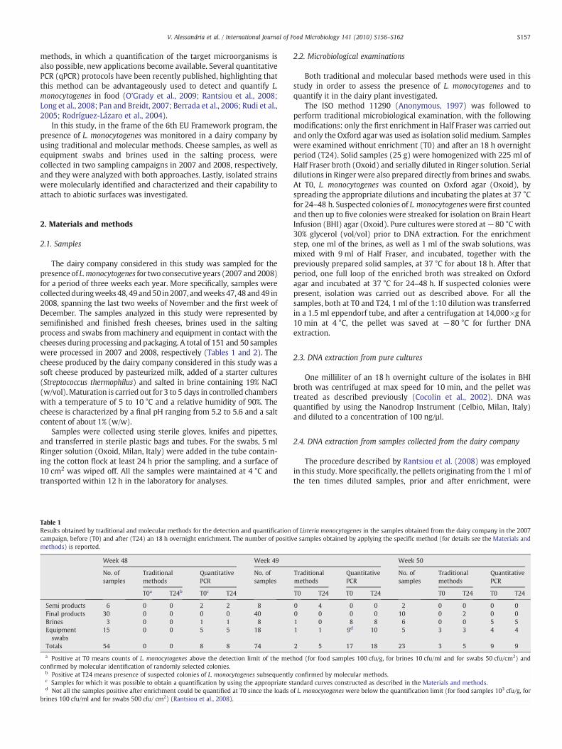
Table 1Results obtained by traditional and molecular methods for the detection and quantificationcampaign, before (T0) and after (T24) an 18 h overnight enrichment. The number of positivmethods) is reported.
Week 48 Week 49
No. ofsamples
Traditionalmethods
QuantitativePCR
No. ofsamples
T0a T24b T0c T24
Semi products 6 0 0 2 2 8Final products 30 0 0 0 0 40Brines 3 0 0 1 1 8Equipmentswabs
15 0 0 5 5 18
Totals 54 0 0 8 8 74
a Positive at T0 means counts of L. monocytogenes above the detection limit of the metconfirmed by molecular identification of randomly selected colonies.
b Positive at T24 means presence of suspected colonies of L. monocytogenes subsequentlyc Samples for which it was possible to obtain a quantification by using the appropriate sd Not all the samples positive after enrichment could be quantified at T0 since the loads o
brines 100 cfu/ml and for swabs 500 cfu/ cm2) (Rantsiou et al., 2008).
2.2. Microbiological examinations
Both traditional and molecular based methods were used in thisstudy in order to assess the presence of L. monocytogenes and toquantify it in the dairy plant investigated.
The ISO method 11290 (Anonymous, 1997) was followed toperform traditional microbiological examination, with the followingmodifications: only the first enrichment in Half Fraser was carried outand only the Oxford agar was used as isolation solid medium. Sampleswere examined without enrichment (T0) and after an 18 h overnightperiod (T24). Solid samples (25 g) were homogenized with 225 ml ofHalf Fraser broth (Oxoid) and serially diluted in Ringer solution. Serialdilutions in Ringer were also prepared directly from brines and swabs.At T0, L. monocytogenes was counted on Oxford agar (Oxoid), byspreading the appropriate dilutions and incubating the plates at 37 °Cfor 24–48 h. Suspected colonies of L. monocytogeneswere first countedand then up to five colonies were streaked for isolation on Brain HeartInfusion (BHI) agar (Oxoid). Pure cultures were stored at−80 °C with30% glycerol (vol/vol) prior to DNA extraction. For the enrichmentstep, one ml of the brines, as well as 1 ml of the swab solutions, wasmixed with 9 ml of Half Fraser, and incubated, together with thepreviously prepared solid samples, at 37 °C for about 18 h. After thatperiod, one full loop of the enriched broth was streaked on Oxfordagar and incubated at 37 °C for 24–48 h. If suspected colonies werepresent, isolation was carried out as described above. For all thesamples, both at T0 and T24, 1 ml of the 1:10 dilution was transferredin a 1.5 ml eppendorf tube, and after a centrifugation at 14,000×g for10 min at 4 °C, the pellet was saved at −80 °C for further DNAextraction.
2.3. DNA extraction from pure cultures
One milliliter of an 18 h overnight culture of the isolates in BHIbroth was centrifuged at max speed for 10 min, and the pellet wastreated as described previously (Cocolin et al., 2002). DNA wasquantified by using the Nanodrop Instrument (Celbio, Milan, Italy)and diluted to a concentration of 100 ng/µl.
2.4. DNA extraction from samples collected from the dairy company
The procedure described by Rantsiou et al. (2008) was employedin this study. More specifically, the pellets originating from the 1 ml ofthe ten times diluted samples, prior and after enrichment, were
of Listeria monocytogenes in the samples obtained from the dairy company in the 2007e samples obtained by applying the specific method (for details see the Materials and
Week 50
Traditionalmethods
QuantitativePCR
No. ofsamples
Traditionalmethods
QuantitativePCR
T0 T24 T0 T24 T0 T24 T0 T24
0 4 0 0 2 0 0 0 00 0 0 0 10 0 2 0 01 0 8 8 6 0 0 5 51 1 9d 10 5 3 3 4 4
2 5 17 18 23 3 5 9 9
hod (for food samples 100 cfu/g, for brines 10 cfu/ml and for swabs 50 cfu/cm2) and
confirmed by molecular methods.tandard curves constructed as described in the Materials and methods.f L. monocytogenes were below the quantification limit (for food samples 103 cfu/g, for
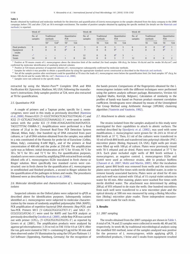
Table 2Results obtained by traditional and molecular methods for the detection and quantification of Listeria monocytogenes in the samples obtained from the dairy company in the 2008campaign, before (T0) and after (T24) an 18 h overnight enrichment. The number of positive samples obtained by applying the specific method (for details see the Materials andmethods) is reported.
Week 47 Week 48 Week 49
No. ofsamples
Traditionalmethods
QuantitativePCR
No. ofsamples
Traditionalmethods
QuantitativePCR
No. ofsamples
Traditionalmethods
QuantitativePCR
T0a T24b T0c T24 T0 T24 T0 T24 T0 T24 T0 T24
Final products 9 0 0 1d 4 5 0 0 3 3 6 0 0 0d 1Equipmentswabs
5 0 0 0d 4 7 0 0 0d 1 6 0 0 0d 1
Brines –e – – – 12 0 0 0d 3 – – – – –
Totals 14 0 0 1 8 24 0 0 3 7 12 0 0 0 2
a Positive at T0 means counts of L. monocytogenes above the detection limit of the method (for food samples 100 cfu/g, for brines 10 cfu/ml and for swabs 50 cfu/cm2) andconfirmed by molecular identification of randomly selected colonies.
b Positive at T24 means presence of suspected colonies of L. monocytogenes subsequently confirmed by molecular methods.c Samples for which it was possible to obtain a quantification by using the appropriate standard curves constructed as described in the Materials and methods.d Not all the samples positive after enrichment could be quantified at T0 since the loads of L. monocytogenes were below the quantification limit (for food samples 103 cfu/g, for
brines 100 cfu/ml and for swabs 500 cfu/ cm2) (Rantsiou et al., 2008).e Samples were not collected in the specific week.
S158 V. Alessandria et al. / International Journal of Food Microbiology 141 (2010) S156–S162
extracted by using the Master-Pure™ Complete DNA and RNAPurification Kit (Epicentre, Madison, WI, USA) following the manufac-turer's instructions. Only samples positive at T24, were also extractedat T0 for quantification.
2.5. Quantitative PCR
A couple of primers and a Taqman probe, specific for L. mono-cytogenes, were used in this study as previously described (Rantsiouet al., 2008). Primers IGS1 (5′-GGCCTATAGCTCAGCTGGTTAGAG-3′) andIGS2 (5′-GCTGAGCTAAGGCCCCGTAAAAGGG-3′) were used in combi-nation with the probe IGS (5′-FAM-ATAAGAAATACAAATAATCA-TACCCTTTTAC-TAMRA-3′). Amplifications were performed in a finalvolume of 25 μl in the Chromo4 Real-Time PCR Detection System(Biorad, Milan, Italy). One hundred ng of DNA extracted from purecultures, or 1 μl of DNA solution obtained from the samples, was addedto a mix constituted of the Fluomix for probe kit of Euroclone (Celbio,Milan, Italy), containing 8 mM MgCl2, and of the primers at finalconcentration of 400 nM and the probe at 250 nM. The amplificationcycle was as follows: initial denaturation at 95 °C for 10 min, 95 °C for30 s, 56 °C for 30 s and 72 °C for 30 s. For quantification purposes,standard curveswere created by amplifyingDNAextracted from seriallydiluted cells of L. monocytogenes EGDe inoculated in fresh cheese orRinger solution. More specifically two standard curves were con-structed: one in fresh cheese for the quantification of L. monocytogenesin semifinished and finished products, a second in Ringer solution forthe quantification of the pathogen in brines and swabs. The proceduresfollowed were as described by Rantsiou et al. (2008).
2.6. Molecular identification and characterization of L. monocytogenesisolates
Suspected colonies on the Oxford plates were subjected to qPCR asdescribed above for identification purposes. Subsequently isolatesidentified as L. monocytogenes were subjected to molecular character-ization by the means of randomly amplified polymorphic DNA (RAPD),PCR amplification of repetitive bacterial DNA elements (Rep-PCR) andSau-PCR. Primers M13 (5′-GAGGGTGGCGGTTCT-3′) and SAG1 (5′-CCGCCGCGATCAG-3′) were used for RAPD and Sau-PCR analysis aspreviously described byCocolin et al. (2005),while Rep-PCRwas carriedout with primer (GTG)5 (5′-GTGGTGGTGGTGGTG-3′) as reported byGevers et al. (2001). The amplification products were analyzed byagarose gel electrophoresis (1.5%w/vol) in TAE 1X for 4 h at 120 V. Afterthe run, gelswere stained in TAE 1× containing0.5 μg/ml for 30 min andthen observed under UV illumination by using the UVI pro Platinum 1.1Gel Software (Eppendorg, Hamburg, Germany) for the recognition of
the bands present. Comparisons of the fingerprints obtained for the L.monocytogenes isolates with the different techniques were performedusing the pattern analysis software package, Bionumerics, Version 4.6(Applied Maths, Kortrijk, Belgium). Calculation of similarity in theprofiles of bands was based on Pearson product–moment correlationcoefficient. Dendrograms were obtained by means of the UnweightedPair Group Method using Arithmetic Average (UPGMA) clusteringalgorithm (Vauterin and Vauterin, 1992).
2.7. Attachment to abiotic surfaces
The strains isolated from the samples analyzed in this study wereinvestigated for their capabilities to attach to abiotic surfaces. Themethod described by Djordjevic et al. (2002), was used with somemodifications. L. monocytogenes were grown for 18–24 h in 10 ml ofBHI broth at 37 °C. Then, 0.1 ml of the cultures were transferred in10 ml of fresh BHI broth and transferred into polyvinyl chloride (PVC)microtiter plates (Biolog, Hayward, CA, USA). Eight wells per strainwere filled up with 100 µL of culture. Plates were previously rinsedwith 70 % ethanol and air dried. Plates were incubated at 37 °C for24 h. Each plate included eight wells of BHI broth without L.monocytogenes, as negative control. L. monocytogenes EGDe andScottA were used as reference strains, able to produce biofilms(Chavant et al., 2007; Moltz and Martin, 2005). After the incubationperiod, spent BHI broth was removed from wells and the microtiterplates were washed five times with sterile distilled water, in order toremove loosely associated bacteria. Plates were air dried for 45 minand each well was stained with 150 µL of 1% crystal violet solution inwater for 45 min. After staining, plates were washed five times withsterile distilled water. The attachment was determined by adding200 µL of 95% ethanol to de-stain the wells. One hundred microlitersfrom each well were transferred to a new microtiter plate and theoptical density at 590 nm was measured by using a Biolog MicroSta-tion (Biolog) microtiter plate reader. Three independent measure-ments were made for each strain.
3. Results
3.1. 2007 sampling
The results obtained from the 2007 campaign are shown in Table 1.A total of 54, 74 and 23 samples were collected inweeks 48, 49 and 50,respectively. In week 48, by traditional microbiological analysis usingthe modified ISO method, none of the samples analyzed was positivefor the presence of L. monocytogenes, while applying qPCR, 2semifinished products, 1 brine and 5 swabs from equipment resulted
S159V. Alessandria et al. / International Journal of Food Microbiology 141 (2010) S156–S162
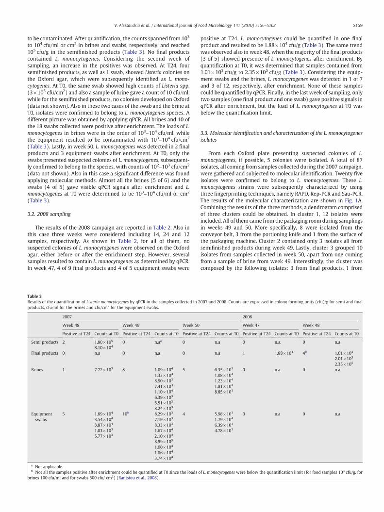
to be contaminated. After quantification, the counts spanned from 103
to 104 cfu/ml or cm2 in brines and swabs, respectively, and reached105 cfu/g in the semifinished products (Table 3). No final productscontained L. monocytogenes. Considering the second week ofsampling, an increase in the positives was observed. At T24, foursemifinished products, as well as 1 swab, showed Listeria colonies onthe Oxford agar, which were subsequently identified as L. mono-cytogenes. At T0, the same swab showed high counts of Listeria spp.(3×10⁵ cfu/cm2) and also a sample of brine gave a count of 10 cfu/ml,while for the semifinished products, no colonies developed on Oxford(data not shown). Also in these two cases of the swab and the brine atT0, isolates were confirmed to belong to L. monocytogenes species. Adifferent picture was obtained by applying qPCR. All brines and 10 ofthe 18 swabs collected were positive after enrichment. The loads of L.monocytogenes in brines were in the order of 103–104 cfu/ml, whilethe equipment resulted to be contaminated with 103–104 cfu/cm2
(Table 3). Lastly, in week 50, L. monocytogenes was detected in 2 finalproducts and 3 equipment swabs after enrichment. At T0, only theswabs presented suspected colonies of L. monocytogenes, subsequent-ly confirmed to belong to the species, with counts of 102–103 cfu/cm2
(data not shown). Also in this case a significant difference was foundapplying molecular methods. Almost all the brines (5 of 6) and theswabs (4 of 5) gave visible qPCR signals after enrichment and L.monocytogenes at T0 were determined to be 103–104 cfu/ml or cm2
(Table 3).
3.2. 2008 sampling
The results of the 2008 campaign are reported in Table 2. Also inthis case three weeks were considered including 14, 24 and 12samples, respectively. As shown in Table 2, for all of them, nosuspected colonies of L. monocytogenes were observed on the Oxfordagar, either before or after the enrichment step. However, severalsamples resulted to contain L. monocytogenes as determined by qPCR.In week 47, 4 of 9 final products and 4 of 5 equipment swabs were
Table 3Results of the quantification of Listeria monocytogenes by qPCR in the samples collected in 2products, cfu/ml for the brines and cfu/cm2 for the equipment swabs.
2007
Week 48 Week 49 Week 50
Positive at T24 Counts at T0 Positive at T24 Counts at T0 Positive a
Semi products 2 1.80×10⁵ 0 n.aa 08.10×10⁴
Final products 0 n.a 0 n.a 0
Brines 1 7.72×10³ 8 1.09×10⁴ 51.33×10⁴
8.90×10³
7.41×10³
1.10×10⁴
6.39×10³
5.51×10³
8.24×10³
Equipmentswabs
5 1.89×10⁴ 10b 8.29×10³ 43.54×10⁴ 7.19×10³
3.87×10⁴ 8.33×10³
1.03×10³ 1.67×10⁴
5.77×10³ 2.10×10⁴
8.59×10³
1.00×10⁴
1.86×10⁴
3.74×10⁴
a Not applicable.b Not all the samples positive after enrichment could be quantified at T0 since the loads o
brines 100 cfu/ml and for swabs 500 cfu/ cm2) (Rantsiou et al., 2008).
positive at T24. L. monocytogenes could be quantified in one finalproduct and resulted to be 1.88×10⁴ cfu/g (Table 3). The same trendwas observed also in week 48, when the majority of the final products(3 of 5) showed presence of L. monocytogenes after enrichment. Byquantification at T0, it was determined that samples contained from1.01×103 cfu/g to 2.35×10⁵ cfu/g (Table 3). Considering the equip-ment swabs and the brines, L. monocytogenes was detected in 1 of 7and 3 of 12, respectively, after enrichment. None of these samplescould be quantified by qPCR. Finally, in the last week of sampling, onlytwo samples (one final product and one swab) gave positive signals inqPCR after enrichment, but the load of L. monocytogenes at T0 wasbelow the quantification limit.
3.3. Molecular identification and characterization of the L. monocytogenesisolates
From each Oxford plate presenting suspected colonies of L.monocytogenes, if possible, 5 colonies were isolated. A total of 87isolates, all coming from samples collected during the 2007 campaign,were gathered and subjected to molecular identification. Twenty fiveisolates were confirmed to belong to L. monocytogenes. These L.monocytogenes strains were subsequently characterized by usingthree fingerprinting techniques, namely RAPD, Rep-PCR and Sau-PCR.The results of the molecular characterization are shown in Fig. 1A.Combining the results of the three methods, a dendrogram comprisedof three clusters could be obtained. In cluster 1, 12 isolates wereincluded. All of them came from the packaging room during samplingsin weeks 49 and 50. More specifically, 8 were isolated from theconveyor belt, 3 from the portioning knife and 1 from the surface ofthe packaging machine. Cluster 2 contained only 3 isolates all fromsemifinished products during week 49. Lastly, cluster 3 grouped 10isolates from samples collected in week 50, apart from one comingfrom a sample of brine from week 49. Interestingly, the cluster wascomposed by the following isolates: 3 from final products, 1 from
007 and 2008. Counts are expressed in colony forming units (cfu)/g for semi and final
2008
Week 47 Week 48
t T24 Counts at T0 Positive at T24 Counts at T0 Positive at T24 Counts at T0
n.a 0 n.a. 0 n.a
n.a 1 1.88×10⁴ 4b 1.01×10³
2.01×10³
2.35×10⁵
6.35×10³ 0 n.a 0 n.a1.08×104
1.23×10⁴
1.81×10⁴
8.85×10³
5.98×10³ 0 n.a 0 n.a1.79×104
6.39×10³
4.78×10³
f L. monocytogenes were below the quantification limit (for food samples 103 cfu/g, for
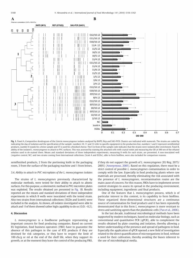
Fig. 1. Panel A, Compositive dendrogram of the Listeria monocytogenes isolates analyzed by RAPD, Rep and SAU-PCR. Clusters are indicated with numerals. The strains are coded byindicating the day of isolation and the specification of the sample: numbers 10, 11 and 12 refer to specific equipment in the production line, numbers 1 and 2 represent semifinishedproducts, number 8 stands for a brine sample and ST is used for a finished cheese. The E in from of the sample code indicates that the strains were isolated after enrichment. Panel B,ability of the isolated L. monocytogenes to attach to PVC surfaces. This was assessed by staining the attached cells with crystal violet and measuring the OD at 590 nm of the ethanolsolution used to de-stained them. Means and standard deviations of three independent experiments, measuring 8 wells for each strain, are presented. A non-inoculated well(negative control, NC) and two strains coming from International collections (Scott A and EGDe), able to form biofilms, were also included for comparison reasons.
S160 V. Alessandria et al. / International Journal of Food Microbiology 141 (2010) S156–S162
semifinished products, 2 from the portioning knife in the packagingroom, 3 from the surface of the packaging machine and 1 from brines.
3.4. Ability to attach to PVC microplates of the L. monocytogenes isolates
The strains of L. monocytogenes previously characterized bymolecular methods, were tested for their ability to attach to abioticsurfaces. For this purpose, a colorimetricmethod in PVCmicrotiter plateswas exploited. The results obtained are presented in Fig. 1B. Resultsreported are the means and standard deviations of three independentexperiments in which 8 wells were inoculated with the tested strain.Also two strains from international collections (EGDe and ScottA) wereincluded in the analysis. As shown, all isolates investigated were able toattach and 11of them showed rather high capability to do so (O.D.N0.2).
4. Discussion
L. monocytogenes is a foodborne pathogen representing animportant concern for food producing companies. Based on currentEU legislation, food business operators (FBO) have to guarantee theabsence of this pathogen in the case of RTE products if they aredestined for risk categories, or they have to respect the limit of100 cfu/g or ml at the end of the shelf life, when they support itsgrowth, or at themoment they leave the control of the producing FBO,
if they do not support the growth of L. monocytogenes (EU Reg. 2073/2005) (Anonymous, 2005). Based on this regulation, there must be astrict control of possible L. monocytogenes contamination in order tocomply with the law. Especially in food producing plants where rawmaterials are processed, thereby eliminating the risk associated withthe presence of L. monocytogenes, recontamination routes are themain cause of concern. For this reason, FBOs have to implement severecontrol strategies to assess its spread in the producing environment,including equipment, ingredients and final products.
One of the features that L. monocytogenes possess, which is ofparticular interest in this context, is its capability to form biofilms.These organized three-dimensional structures are a continuoussource of contamination for food products and it has been repeatedlydemonstrated that in this form L. monocytogenes is more resistant tostress and sanitizing agents than the planktonic cells (Pan et al., 2006).
In the last decade, traditional microbiological methods have beensupported bymodern techniques, based onmolecular biology, such asconventional and quantitative PCR (qPCR), able to complement theresults obtained by plate counting and isolation, thereby allowing abetter understanding of the presence and spread of pathogens in food.Especially the application of qPCR opened a new field of investigationrelated to the direct quantification of microorganisms in food, withouta need for their cultivation, thereby avoiding the biases inherent tothe use of microbiological media.
S161V. Alessandria et al. / International Journal of Food Microbiology 141 (2010) S156–S162
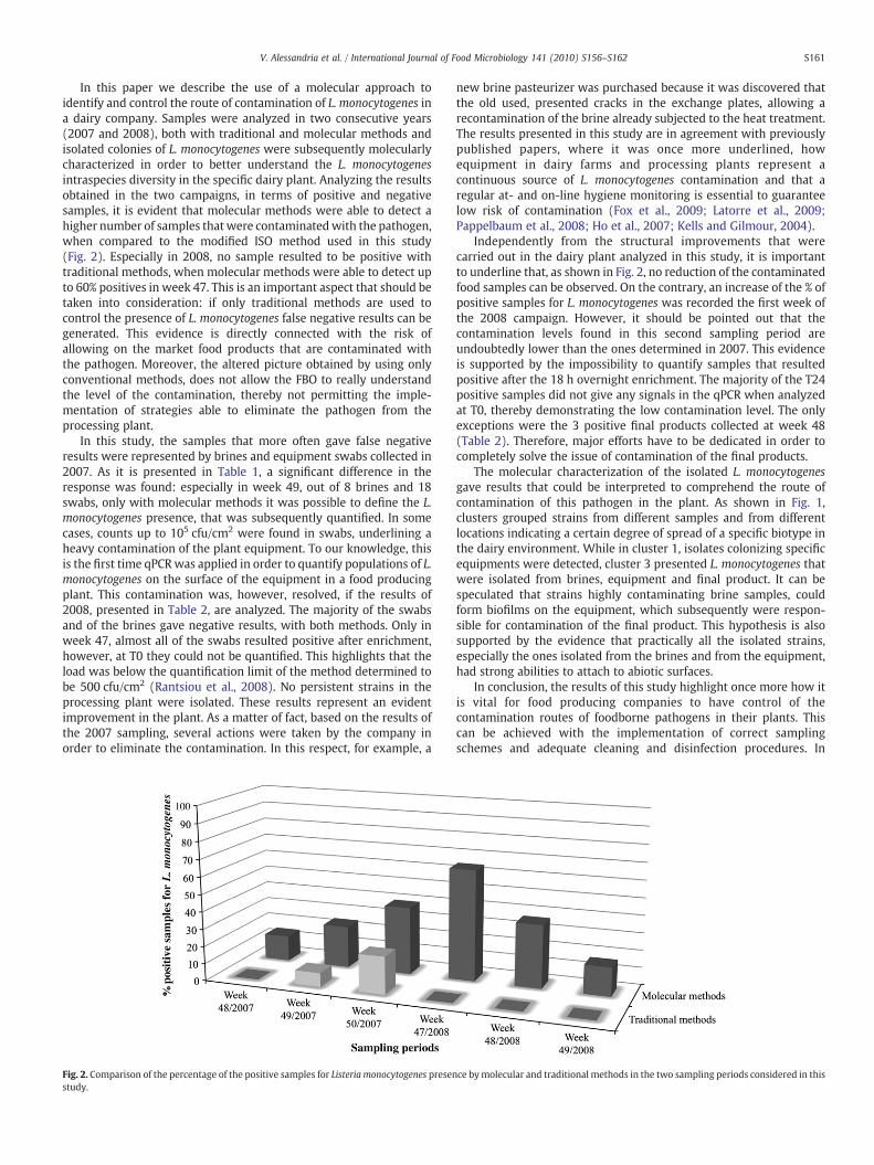
In this paper we describe the use of a molecular approach toidentify and control the route of contamination of L. monocytogenes ina dairy company. Samples were analyzed in two consecutive years(2007 and 2008), both with traditional and molecular methods andisolated colonies of L. monocytogenes were subsequently molecularlycharacterized in order to better understand the L. monocytogenesintraspecies diversity in the specific dairy plant. Analyzing the resultsobtained in the two campaigns, in terms of positive and negativesamples, it is evident that molecular methods were able to detect ahigher number of samples that were contaminatedwith the pathogen,when compared to the modified ISO method used in this study(Fig. 2). Especially in 2008, no sample resulted to be positive withtraditional methods, when molecular methods were able to detect upto 60% positives in week 47. This is an important aspect that should betaken into consideration: if only traditional methods are used tocontrol the presence of L. monocytogenes false negative results can begenerated. This evidence is directly connected with the risk ofallowing on the market food products that are contaminated withthe pathogen. Moreover, the altered picture obtained by using onlyconventional methods, does not allow the FBO to really understandthe level of the contamination, thereby not permitting the imple-mentation of strategies able to eliminate the pathogen from theprocessing plant.
In this study, the samples that more often gave false negativeresults were represented by brines and equipment swabs collected in2007. As it is presented in Table 1, a significant difference in theresponse was found: especially in week 49, out of 8 brines and 18swabs, only with molecular methods it was possible to define the L.monocytogenes presence, that was subsequently quantified. In somecases, counts up to 105 cfu/cm2 were found in swabs, underlining aheavy contamination of the plant equipment. To our knowledge, thisis the first time qPCRwas applied in order to quantify populations of L.monocytogenes on the surface of the equipment in a food producingplant. This contamination was, however, resolved, if the results of2008, presented in Table 2, are analyzed. The majority of the swabsand of the brines gave negative results, with both methods. Only inweek 47, almost all of the swabs resulted positive after enrichment,however, at T0 they could not be quantified. This highlights that theload was below the quantification limit of the method determined tobe 500 cfu/cm2 (Rantsiou et al., 2008). No persistent strains in theprocessing plant were isolated. These results represent an evidentimprovement in the plant. As a matter of fact, based on the results ofthe 2007 sampling, several actions were taken by the company inorder to eliminate the contamination. In this respect, for example, a
Fig. 2. Comparison of the percentage of the positive samples for Listeria monocytogenes presenstudy.
new brine pasteurizer was purchased because it was discovered thatthe old used, presented cracks in the exchange plates, allowing arecontamination of the brine already subjected to the heat treatment.The results presented in this study are in agreement with previouslypublished papers, where it was once more underlined, howequipment in dairy farms and processing plants represent acontinuous source of L. monocytogenes contamination and that aregular at- and on-line hygiene monitoring is essential to guaranteelow risk of contamination (Fox et al., 2009; Latorre et al., 2009;Pappelbaum et al., 2008; Ho et al., 2007; Kells and Gilmour, 2004).
Independently from the structural improvements that werecarried out in the dairy plant analyzed in this study, it is importantto underline that, as shown in Fig. 2, no reduction of the contaminatedfood samples can be observed. On the contrary, an increase of the % ofpositive samples for L. monocytogenes was recorded the first week ofthe 2008 campaign. However, it should be pointed out that thecontamination levels found in this second sampling period areundoubtedly lower than the ones determined in 2007. This evidenceis supported by the impossibility to quantify samples that resultedpositive after the 18 h overnight enrichment. The majority of the T24positive samples did not give any signals in the qPCR when analyzedat T0, thereby demonstrating the low contamination level. The onlyexceptions were the 3 positive final products collected at week 48(Table 2). Therefore, major efforts have to be dedicated in order tocompletely solve the issue of contamination of the final products.
The molecular characterization of the isolated L. monocytogenesgave results that could be interpreted to comprehend the route ofcontamination of this pathogen in the plant. As shown in Fig. 1,clusters grouped strains from different samples and from differentlocations indicating a certain degree of spread of a specific biotype inthe dairy environment. While in cluster 1, isolates colonizing specificequipments were detected, cluster 3 presented L. monocytogenes thatwere isolated from brines, equipment and final product. It can bespeculated that strains highly contaminating brine samples, couldform biofilms on the equipment, which subsequently were respon-sible for contamination of the final product. This hypothesis is alsosupported by the evidence that practically all the isolated strains,especially the ones isolated from the brines and from the equipment,had strong abilities to attach to abiotic surfaces.
In conclusion, the results of this study highlight once more how itis vital for food producing companies to have control of thecontamination routes of foodborne pathogens in their plants. Thiscan be achieved with the implementation of correct samplingschemes and adequate cleaning and disinfection procedures. In
ce bymolecular and traditional methods in the two sampling periods considered in this
S162 V. Alessandria et al. / International Journal of Food Microbiology 141 (2010) S156–S162
addition, the choice of the analytical procedure should also beconsidered as a relevant decision. As demonstrated in this study, themodified ISO method only partially can monitor the presence of L.monocytogenes in the processing plant. Especially when environmen-tal conditions influence the fitness and behavior of the pathogen, suchas in the brines (with 15–20% salt) or in the equipment swabs (wherecells have to respond to several stresses such as disinfectants,starvation and dried conditions), molecular methods should be usedto properly detect it. It has been previously proven that L.monocytogenes undergoing stresses enters a viable but not culturablestate (VBNC) that makes impossible its recovery by culture dependentmethods (Rowan, 2004). The application of molecular methods canresult in a better comprehension of the spread of a specific pathogenin a processing plant, thereby allowing the implementation ofcorrective actions to eliminate or decrease the risk associated withits presence in the final product.
Acknowledgements
This study was funded by the European Commission within the VIFramework Program, contract n. 007081, “Pathogen Combat: controland prevention of emerging and future pathogens at cellular andmolecular level throughout the food chain”. Authors express theirgratitude to the staff of the dairy company for their technical supportand help.
References
Anonymous, 1997. International Organization for Standardization. Microbiology—general guidance on methods for the detection of Listeria monocytogenes. DraftInternational Standard ISO/DIS 11290.
Anonymous, 2005. Commission Regulation (EC) No 2073/2005 of 15 November 2005on microbiological criteria for foodstuffs.
Anonymous, 2009. The community report on trends and sources of zoonoses andzoonotic agents on the European Union in 2007. The EFSA Journal 223.
Berrada, H., Soriano, J.M., Picó, Y., Mañes, J., 2006. Quantification of Listeriamonocytogenes in salads by real time quantitative PCR. International Journal ofFood Microbiology 107, 202–206.
Chavant, P., Gaillard-Martinie, B., Talon, R., Hébraud, M., Bernardi, T., 2007. A newdevice for rapid evaluation of biofilm formation potential by bacteria. Journal ofMicrobiological Methods 68, 605–612.
Cocolin, L., Rantsiou, K., Iacumin, L., Cantoni, C., Comi, G., 2002. Direct identification infood samples of Listeria spp. and Listeria monocytogenes by molecular methods.Applied and Environmental Microbiology 68, 6273–6282.
Cocolin, L., Stella, S., Nappi, R., Bozzetta, E., Cantoni, C., Comi, G., 2005. Analysis of PCR-based methods for characterization of Listeria monocytogenes strains isolated fromdifferent sources. International Journal of Food Microbiology 103, 167–178.
Djordjevic, D., Wiedmann, M., McLandsborough, L.A., 2002. Microtiter plate assay forassessment of Listeria monocytogenes biofilm formation. Applied and Environmen-tal Microbiology 68, 2950–2958.
Fox, E., O'Mahony, T., Clancy, M., Dempsey, R., O'Brien, M., Jordan, K., 2009. Listeriamonocytogenes in the Irish dairy farm environment. Journal of Food Protection 72,1450–1456.
Gandhi, M., Chikindas, M.L., 2007. Listeria: a foodborne pathogen that knows how tosurvive. International Journal of Food Microbiology 113, 1–15.
Gardan, R., Cossart, P., Labadie, J., The European Listeria Genome Consortium, 2003.Identification of Listeria monocytogenes genes involved in salt and alkaline-pHtolerance. Applied and Environmental Microbiology 69, 3137–3143.
Gasanov, U., Hughes, D., Hansbro, P.M., 2005. Methods for the isolation and identi-fication of Listeria spp. and Listeria monocytogenes: a review. FEMS MicrobiologyReviews 29, 851–875.
Ho, A.J., Lappi, V.R., Wiedmann, M., 2007. Longitudinal monitoring of Listeriamonocytogenes contamination patterns in a farmstead dairy processing facility.Journal of Dairy Science 90, 2517–2524.
Kells, J., Gilmour, A., 2004. Incidence of Listeria monocytogenes in two milk processingenvironments and assessment of Listeria monocytogenes blood agar for isolation.International Journal of Food Microbiology 91, 167–174.
Latorre, A.A., Van Kessel, J.A.S., Karns, J.S., Zurakowski, M.J., Pradhan, A.K., Zadoks, R.N.,Boor, K.J., Schukken, Y.H., 2009. Molecular ecology of Listeria monocytogenes:evidence for a reservoir in milking equipment on a dairy farm. Applied andEnvironmental Microbiology 75, 1315–1323.
Lianou, A., Sofos, J.N., 2007. A review of the incidence and transmission of Listeriamonocytogenes in ready-to-eat products in retail and food service environments.Journal of Food Protection 70, 2172–2198.
Liu, S., Graham, J.E., Bigelow, L., Morse, P.D., Wilkinson, B.J., 2002. Identification ofListeria monocytogenes genes expressed in response to growth at low temperature.Applied and Environmental Microbiology 68, 1697–1705.
Long, F., Zhu, X.N., Zhang, Z.M., Shi, X.M., 2008. Development of a quantitativepolymerase chain reaction method using a live bacterium as internal control for thedetection of Listeria monocytogenes. Diagnostic Microbiology and Infectious Disease62, 374–381.
Moltz, A.G., Martin, S.E., 2005. Formation of biofilms by Listeria monocytogenes undervarious growth conditions. Journal of Food Protection 68, 92–97.
O'Grady, J., Ruttledge, M., Sedano-Balbás, S., Smith, T.J., Barry, T., Maher, M., 2009. Rapiddetection of Listeria monocytogenes in food using culture enrichment combinedwith real-time PCR. Food Microbiology 26, 4–7.
Olesen, I., Vogensen, F.V., Jespersen, L., 2009. Gene tanscription and virulence potentialof Listeria monocytogenes strains after exposure to acidic and NaCl stress.Foodborne Pathogens and Disease 6, 669–680.
Pan, Y., Breidt Jr, F., 2007. Enumeration of viable Listeria monocytogenes cells by real-time PCR with propidium monoazide and ethidium monoazide in the presence ofdead cells. Applied and Environmental Microbiology 73, 8028–8031.
Pan, Y., Breidt Jr., F., Kathariou, S., 2006. Resistance of Listeria monocytogenes biofilms tosanitizing agents in a simulated food processing environment. Applied andEnvironmental Microbiology 72, 7711–7717.
Pappelbaum, K., Grif, K., Heller, I., Wüirzner, R., Hein, I., Ellerbroek, L., Wagner, M., 2008.Monitoring hygiene on- and at-line is critical for controlling Listeria monocytogenesduring produce processing. Journal of Food Protection 71, 735–741.
Phan-Thanh, L., Mahouin, F., Aligé, S., 2000. Acid responses of Listeria monocytogenes.International Journal of Food Microbiology 55, 121–126.
Ramaswamy, V., Cresence, V.M., Rejitha, J.S., Lekshmi, M.U., Dharsana, K.S., Prasad, S.P.,Vijila, H.M., 2007. Listeria—review of epidemiology and pathogenesis. Journal ofMicrobiology, Immunology and Infection 40, 4–13.
Rantsiou, K., Alessandria, V., Urso, R., Dolci, P., Cocolin, L., 2008. Detection,quantification and vitality of Listeria monocytogenes in food as determined byquantitative PCR. International Journal of Food Microbiology 121, 99–105.
Rocourt, J., Cossart, P., 1997. Listeria monocytogenes. In: Doyle, M.P., Beuchat, L.R.,Montville, T.J. (Eds.), Food Microbiology—Fundamentals and Frontiers, AmericanSociety for Microbiology (ASM), Washington, pp. 337–352.
Rodríguez-Lázaro, D., Jofré, A., Aymerich, T., Hugas, M., Pla, M., 2004. Rapid quantitativedetection of Listeria monocytogenes in meat products by real-time PCR. Applied andEnvironmental Microbiology 70, 6299–6301.
Rowan, N.J., 2004. Viable but non culturable forms of food and waterborne bacteria:Quo vadis? Trends in Food Science and Technology 15, 462–467.
Rudi, K., Naterstad, K., Drömtorp, S.M., Holo, H., 2005. Detection of viable and deadListeria monocytogenes on gouda-like cheeses by real-time PCR. Letters in AppliedMicrobiology 40, 301–306.
Vauterin, L., Vauterin, P., 1992. Computer-aided objective comparison of electropho-retic patterns for grouping and identification of microorganisms. European Journalof Clinical Microbiology 33, 633–641.
Warriner, K., Namvar, A., 2009. What is the hysteria with Listeria? Trends in FoodScience and Technology 20, 245–254.