molecular microbiology 88 doi:10.1111/mmi.12231 first
TRANSCRIPT

Bacterial DNA polymerases participate inoligonucleotide recombination
Xin-tian Li,1† Lynn C. Thomason,1,2†
James A. Sawitzke,1† Nina Costantino1 andDonald L. Court1*1Molecular Control and Genetics Section, GeneRegulation and Chromosome Biology, FrederickNational Laboratory for Cancer Research, Frederick,MD 21702, USA.2Basic Science Program, SAIC-Frederick, Inc.,Frederick, MD 21702, USA.
Summary
Synthetic single-strand oligonucleotides (oligos) withhomology to genomic DNA have proved to be highlyeffective for constructing designed mutations in tar-geted genomes, a process referred to as recom-bineering. The cellular functions important for thistype of homologous recombination have yet to bedetermined. Towards this end, we have identifiedEscherichia coli functions that process the recombin-ing oligo and affect bacteriophage l Red-mediatedoligo recombination. To determine the nature of oligoprocessing during recombination, each oligo con-tained multiple nucleotide changes: a single basechange allowing recombinant selection, and silentchanges serving as genetic markers to determine theextent of oligo processing during the recombination.Such oligos were often not incorporated into the hostchromosome intact; many were partially degraded inthe process of recombination. The position andnumber of these silent nucleotide changes within theoligo strongly affect both oligo processing andrecombination frequency. Exonucleases, especiallythose associated with DNA Polymerases I and III,affect inheritance of the silent nucleotide changes inthe oligos. We demonstrate for the first time that themajor DNA polymerases (Pol I and Pol III) and DNAligase are directly involved with oligo recombination.
Introduction
Homologous recombination-mediated genetic engineer-ing, recombineering, utilizes the phage l recombinationproteins Gam, Exo and Beta, collectively known as ‘Red’(Murphy, 1998; Zhang et al., 1998; Yu et al., 2000; Elliset al., 2001). For recombineering, a PCR product or single-strand oligonucleotide (oligo) that contains the desiredsequence change is introduced into Red-expressing, elec-trocompetent cells to obtain the alteration in the targetedDNA. Oligo recombination, the focus of this paper, requiresonly the l Beta protein (Ellis et al., 2001; Costantino andCourt, 2003). Beta is a single-strand DNA (ssDNA) bindingprotein that can protect oligos from exonuclease degrada-tion (Muniyappa and Radding, 1986; Karakousis et al.,1998) and promote annealing of complementary ssDNAs(Kmiec and Holloman, 1981). In a thorough screen ofE. coli host recombination functions, none is required for lRed-mediated oligo recombination (Ellis et al., 2001;Costantino and Court, 2003; Sawitzke et al., 2011).
A number of factors affect the efficiency of oligo recom-bination. For any target DNA sequence, either of twocomplementary oligos can be used to modify it. The recom-bination efficiency of the oligo corresponding in sequenceto the discontinuously replicated, lagging-strand is moreefficient than the leading-strand oligo (Ellis et al., 2001;Costantino and Court, 2003; Zhang et al., 2003; vanKessel and Hatfull, 2008; Swingle et al., 2010; Sawitzkeet al., 2011; Van Pijkeren et al., 2012).All models to explainoligo recombination must incorporate this bias caused byDNA replication. In our favoured model for oligo recombi-nation, Beta protein binds, protects and anneals the oligoto complementary regions of ssDNA existing transiently atthe DNA replication fork (Fig. 1) (Court et al., 2002). Thelagging/leading-strand bias can be explained by the pres-ence of a larger single-strand gap on the lagging-strand(Benkovic et al., 2001; Lajoie et al., 2012), which allowsthe lagging-strand oligo greater opportunity to anneal,thereby resulting in a higher recombination frequency (Elliset al., 2001; Costantino and Court, 2003; Sawitzke et al.,2011). An alternate model in which oligo recombinationinitiates by Beta-mediated DNAstrand invasion (Noirot andKolodner, 1998; Rybalchenko et al., 2004), giving rise to aD-loop, could also explain the replication bias, if thelagging- and leading-strand oligos are treated differently
Accepted 8 April, 2013. *For correspondence. E-mail [email protected]; Tel. (+1) 301 846 5940; Fax (+1) 301 846 6988. †Theseauthors contributed equally to this manuscript.
Molecular Microbiology (2013) 88(5), 906–920 � doi:10.1111/mmi.12231First published online 2 May 2013
Published 2013. This article is a U.S. Government work and is in the public domain in the USA

when the replication fork reaches the D-loop. Sawitzkeet al. (2011) found that proximity of the genetic marker tothe end of the oligo also affects recombination efficiency,as does the length and concentration of oligo used. TheE. coli methyl-directed mismatch repair (MMR) systemlowers the efficiency of oligo recombination at least 100-fold (Costantino and Court, 2003; Li et al., 2003) by remov-ing mismatches when an oligo is annealed to its targetDNA. Thus, elimination of the MMR system results in ahigher recombination frequency. Under optimal conditions,more than 50% of the cells are recombinant at the galKlocus (Sawitzke et al., 2011).
Wang et al. (2011) reported that not all markers in anoligo were inherited in the process of recombineering, butthe molecular details of oligo recombination and thismarker loss have not been elucidated. During oligorecombination, is the oligo processed while still single-stranded and/or after annealed to the target? How doesmarker distribution along the oligo affect recombination
frequency and marker inheritance? In principle, exonucle-ases that act on either ssDNA or dsDNA could affect oligorecombination. There are four major exonucleases (RecJ,ExoI, ExoVII and ExoX) that degrade ssDNA in E. coli(Burdett et al., 2001) and decrease Red-mediated oligorecombination, but only when oligo is limiting (Sawitzkeet al., 2011). Several dsDNA exonucleases may alsoaffect oligo recombination after the oligo has annealed tothe target. The RecBCD enzyme has multiple nucleaseactivities that collectively destroy linear dsDNA (Goldmarkand Linn, 1972). Other dsDNA exonucleases that couldbe involved in processing include Exonuclease III(Richardson et al., 1964), Exonuclease IX (Allen et al.,2009), and l Exo (Little et al., 1967).
Three DNA polymerases have inherent exonucleaseactivities. DNA Polymerase III (Pol III), the major replica-tive polymerase, has a 3′ → 5′ proofreading exonucleaseencoded by dnaQ (Echols et al., 1983). DNA PolymeraseI (Pol I), encoded by the polA gene, is a multifunctional
Fig. 1. Oligo processing at the replication fork.A. A replication fork with template (green) and newly synthesized (blue) DNA is shown. Pol III (yellow circles) extends from the 3′ ends of theOkazaki fragment and the leading-strand.B. A lagging-strand oligo (purple) is shown bound by Beta (grey ovals) to the template at a single-strand gap. Pol I, which degrades the RNAprimer of the Okazaki fragment, can also degrade the 5′ end of the lagging-strand oligo after replacing Pol III. DNA polymerization by Pol Ireadies the recombinant oligo for DNA ligation and incorporation into the genome.C. A leading-strand oligo (purple) is shown bound by Beta (grey ovals) to the leading-strand template at the single-strand gap ahead of thenewly synthesized leading-strand. An unknown exonuclease(s) (orange circle) processes the 3′ end of the oligo. Pol I and/or an unknownexonuclease (red circle) is involved in the degradation of the 5′ end of the leading-strand oligo.
Oligonucleotide processing during l Red recombination 907
Published 2013. This article is a U.S. Government work and is in the public domain in the USA, Molecular Microbiology, 88, 906–920

protein involved in DNA replication and repair. Pol I hastwo exonuclease activities: 3′ → 5′ proofreading and5′ → 3′ nick translation (Rigby et al., 1977; Kornberg,1990); the latter activity is responsible for removing RNAprimers from the 5′ end of Okazaki fragments on thelagging-strand. DNA Polymerase II (Pol II), encoded bypolB, is a repair polymerase with a 3′ → 5′ proofreadingexonuclease activity (Banach-Orlowska et al., 2005).
The present study used multiply marked oligos toexamine the effect of exonuclease activities on recombi-nation efficiency and the fate of the oligo. Sequenceanalysis of the recombinants reveals that nucleotides canbe lost from either one or both ends of the oligo, and thisprocessing occurs after the oligo is annealed to the targetDNA. The number and distribution of markers on the oligodirectly alter processing during incorporation and affectrecombination frequency. The major DNA polymerases,Pol I and Pol III, as well as DNA ligase affect recombina-tion frequency and marker loss.
Results and discussion
Genetic engineering in vivo by oligo recombination is apowerful tool that is becoming the method of choice forgenome modification. This technique routinely utilizesssDNA oligos with centrally located modifications. Werecently reported (Sawitzke et al., 2011) that the locationof the modifying base (marker) along the length of theoligo affects the efficiency of incorporation of that base.Oligos having the genetic marker less than nine basesfrom an end have lower recombination efficiencies thanthose with the marker further from an end. To betterunderstand how marker position affects recombination,we used oligos containing multiple markers arranged invarious configurations to monitor recombination efficiencyand marker recovery within recombinants.
Experimental design
The experimental design is illustrated in Fig. 2. We usedderivatives of the MMR-defective strains, HME63 andHME68 (Costantino and Court, 2003; Thomasonet al., 2007a), which contain a galK amber mutation,galKTYR145UAG. Use of MMR mutant strains ensures thatmarker loss from the recombining oligo is not the result ofmismatch repair, but is due to other host functions. Theselected genetic marker, which is always centred in theoligo, corrects the galK amber mutation at codon 145 andconfers the ability to grow on minimal galactose medium,allowing selection of Gal+ recombinants. Other bases in theoligo are used as markers in the sense that they vary fromwild type sequence (i.e. transitions and transversions) but,because of their placement in wobble positions, do not alterthe GalK protein. These unselected markers are mispaired
when the oligo is annealed to the target DNA, and theirinheritance can be followed by DNA sequencing.
In these experiments, the Red functions were inducedfor 15 min and cells were prepared for electroporation bystandard protocols (see Experimental procedures). Theperiod of incubation following oligo electroporation waslimited to 30 min, which allows for cell recovery (Sawitzkeet al., 2011).
Our previous paper demonstrates that functions in thelambda PL operon, when induced for the 15 min timeperiod used here, block cell division for 50 min and resultin cell synchrony without loss of cell viability (Sergueevet al., 2002). Hence in the experiments here, each Gal+
colony is derived from an independent recombinationevent. Sequence analysis was used to determine markerinheritance in these independent recombinant clones.Oligo concentration and the presence or absence of spe-cific host functions have been examined for their effectson recombination frequency and marker inheritance.
Oligo design affects recombination efficiency
We found that marker placement along the oligo affectedrecombination frequency. Oligo 144 contains only the
Fig. 2. Experimental outline for generating and analysing GalK+
recombinants. The experiments were performed as outlined.Although at least 48 independent recombinants were analysed, onoccasion an individual colony yielded poor sequence and thus wasnot added to the data.
908 X. Li et al. �
Published 2013. This article is a U.S. Government work and is in the public domain in the USA, Molecular Microbiology, 88, 906–920

selected galK+ mismatch and had the highest efficiency inthis study (Fig. 3A). Oligo XT13 had a similar efficiencydespite having six additional silent mismatches. OligoXT36, containing 12 silent wobble position markers sixbases apart, recombined at a 20-fold reduced efficiency. Incontrast, oligo XT21, with 23 silent markers spaced everythird base along its entire length, showed no recombinationactivity. For oligo XT21, sequencing revealed that the fewGal+ colonies obtained appeared to have spontaneousmutations eliminating the amber codon (see legends toFigs 3A and S1). These four oligos differ in both density of
markers and extent of homology at their respective ends,two factors which could influence recombination.
Keeping the marker density constant by using oligoswith silent markers spaced every third base, we askedhow the positioning of these markers along the oligoaffects recombination. Oligo XT18 contains such markersflanking the selected change but leaving at least 12 basesof uninterrupted homology at each end. Recombinationwith this oligo is at least 10 000-fold higher than that withXT21, demonstrating the importance of homology at theoligo ends. Oligo XT524 has the same sequence as XT21
Fig. 3. Recombination with multiply marked, lagging-strand oligos. A summary of oligos used to examine the effect of marker density andspacing is shown. Oligo 144 is 70 nt, oligo XT524 is 117 nt, and all others are 75 nt in length. Experiments were performed in the MMRmutant host, HME68. In (A) features of each oligo are shown with a diagram (to scale except XT524), indicating the number and placement ofmismatches. The ‘+’ denotes the selected base and the ‘|’ denotes the unselected markers. The number above markers indicates the distancein bases from the 5′ end. Recombination efficiency is normalized to 108 viable cells; efficiencies given here are a representative experimentfrom at least 3 independent experiments where variability was less than fivefold. 95% confidence limits can be found in Table S4. In (B), thepercentage of recombinants that lost markers from either the 5′ or 3′ end are enumerated for each oligo. If a recombinant lost markers fromboth ends it is not recorded in either column, but such events happen and are shown (5′ & 3′) in Figs 4 and S1. Panel C indicates the Figurethat contains the data for each of the oligos. NA: not applicable, either because no unselected markers are present on that end, or becausethere were no recombinants (XT21). For oligo XT21, 16 rare Gal+ colonies were recovered, 13 of which did not inherit the selected ‘T’ markerfrom the oligo, i.e. they were spontaneous revertants. The three isolates that have a TA‘T’ codon could either be derived from the oligo (truerecombinants) or be revertants (Fig. S1B and legend). These are unlikely to be true recombinants because no other oligo markers wereinherited, and they occur at the same frequency as other spontaneous revertants.
Oligonucleotide processing during l Red recombination 909
Published 2013. This article is a U.S. Government work and is in the public domain in the USA, Molecular Microbiology, 88, 906–920

but carries an additional 21 bases of perfect homology oneach end. In this respect, it is like XT18 and likewise,XT524 recombines at high efficiency (Fig. 3A).
To ask if homologies at the two ends are of equalimportance, we used oligos XT30 and XT29, which arecompletely homologous to the target but only at the 5′ or3′ end respectively. The 2000-fold difference in recom-bination frequency between these two oligos demon-strates that homology at the 5′ end is much moreimportant than at the 3′ end (Fig. 3A). This result is con-sistent with our previous data showing that 5′ homologyis critical for efficient recombination (Sawitzke et al.,2011).
Like oligo XT29, oligo XT351 contains silent markersextending from the 5′ end, but its markers span theselected base. The efficiency of XT351 was reduced~ 40-fold relative to oligo XT29. Like oligo XT30, oligoXT38 contains silent markers extending from the 3′ end,but its markers also span the selected base. The effi-ciency of XT38 was reduced ~ 80-fold relative to oligoXT30. However, recombination with XT30 and XT38 is> 1000-fold more than that with XT29 and XT351,respectively, again demonstrating the greater importanceof intact 5′ homology. In general, homology just 5′ to theselected marker is important for efficient recombination.The exceptions are oligo XT18 and XT524, which com-pensate by having adequate homology at both the 5′and 3′ ends.
XT37 and XT352, despite being mispaired at the 5′ends, recombine robustly (Fig. 3A). What distinguishesthem is that both oligos retain considerable homologyflanking the selected marker. To further investigate why 5′homology is important in some cases but not others, wesequenced Gal+ recombinants from each oligo recombi-nation reaction to determine the fate of unselectedmarkers.
Comparing marker loss from lagging- andleading-strand oligos
Sequencing of Gal+ recombinants allowed us to trace theinheritance of the silent markers and to determinewhether or not they were incorporated into final recom-binant products. Indeed, we found that oligos are notalways incorporated in their entirety but often suffer lossof silent markers. As we will discuss below, this markerloss is caused by exonuclease degradation. Figure 4Aillustrates the data collected for the lagging-strand oligoXT13 and defines the marker categories used. Amongindependent recombinants sequenced, all of the oligo-encoded silent markers were inherited in 67% of therecombinants, whereas 25% showed marker loss fromone end or the other. The extent of marker loss is variablein different recombinants and suggests sequential but
incomplete degradation from an oligo end. In contrast,with the complementary leading-strand oligo XT14(Fig. 4B), all the silent markers were inherited in 27% ofthe recombinants, whereas 57% of recombinants showedloss on one end or the other. In a few cases, markers werelost at both ends; this occurred in 2% and 7% of totalrecombinants for the lagging- and leading-strand oligosrespectively. Recovery of these recombinants is at a rateexpected if the processing events at the 5′ and 3′ endswere independent.
To examine marker loss from the ends in more detail,we used lagging- or leading-strand oligos with mis-matches six bases apart (XT36 and XT418). For eacholigo, independent recombinants were sequenced (FigsS1A and S2A), and per cent loss was determined foreach of the 12 silent markers (Fig. 5). As shown, theextreme 5′ and 3′ markers were lost in every recom-binant examined. There is a gradient of marker loss oneach side of the centrally selected marker. From the 5′ends, these gradients are similar for both oligos,whereas marker loss is greater for the leading-strand onthe 3′ side of the selected marker. We conclude thatlagging- and leading- strand oligos are treated differentlyafter they enter the replication fork during the recombi-nation event.
We also observed other rare recombinants where inter-nal markers were replaced by chromosomal bases (see‘Other’ in Figs 4, S1, S2 and Table 1). There are a total of60 instances of these ‘Other’ events, and surprisingly allbases in question are derived from the chromosome.Thus, spontaneous mutagenesis is not the cause, nor iserror during oligo production. Although MMR is notinvolved because these cells are deleted for mutS,another DNA repair system may be responsible. Yet, thisclass of recombinants was found at a similar frequencyeven in cells additionally mutant for very short patch repair(VSP) or the MutY repair system (data not shown). We donot understand how this class of recombinants occurs.They cannot be derived from recombination events involv-ing more than one oligo because they appear at a similarfrequency even when oligo concentration is limiting(compare Fig. 4A versus Fig. S1K).
Position of the silent markers affects marker loss
Figure 3B summarizes the markers lost by recombinantsobtained from the oligos described in Fig. 3A, and allowscomparison of oligos with different marker configurations.This information is derived from data like that shown inFig. 4A for oligo XT13. As illustrated in Fig. 4A and tabu-lated in Fig. 3B, 25% of the oligo XT13 recombinants hadlost markers from one or the other end, 10% from the 5′end and 15% from the 3′ end, while 2% had lost markersfrom both ends. Recombinants with oligo XT36, which has
910 X. Li et al. �
Published 2013. This article is a U.S. Government work and is in the public domain in the USA, Molecular Microbiology, 88, 906–920

markers spaced every 6 bases extending to within 2–3bases of the oligo ends, always lost markers from both the5′ and 3′ ends (Figs 3B and S1A). For oligo XT36, likeoligo XT13, the extent of marker loss varies in differentrecombinants, again suggesting sequential degradation.The Gal+ isolates found with oligo XT21, which containsmismatches every third base over the entire length of theoligo, are most easily explained by spontaneous mutation
and not recombination (Figs 3B and S1B), implying aninability of that oligo to pair with its target. In contrast, forXT36, enough homology remains among the mismatchesto allow sufficient pairing and recombination. Indeed, ourunpublished in vitro results show that Beta is unable toanneal two complementary oligos with every third basemismatched but is able to anneal two complementaryoligos with every sixth base mismatched. Similar results
Fig. 4. Marker loss pattern in wild type and polymerase mutant cells. Marker loss is compared for the lagging- and leading-strand oligorecombinants in ‘wild type’, HME68 (see A and B respectively). Similar experiments are shown for the Pol I mutant derivative of HME68,XTL70, which is defective for the 5′ → 3′ exonuclease activity (C and D), the Pol III mutant, XTL76 (E and F), and the ligase mutant, XTL47(G and H). The recombination efficiency of the illustrated experiment is shown at the top of each panel. Normal variation in recombinationfrequency is less than fourfold. In each panel, the ‘Position’ row indicates the distance of the markers from the left end of the oligo asdiagrammed, with the ‘+’ denoting the selected marker at position 39, which is the G of the TAG amber codon in the ‘Host’ sequence. The‘Oligo’ row shows the sequence changes present on the oligo at the indicated position. The sequences of Gal+ recombinants are shown belowthe ‘Oligo’ row; grey shaded spaces indicate those bases that remained unchanged from the host and show where markers were lost from theoligo, white spaces indicate those markers that were inherited from the oligo. For each panel, recombinants are grouped according to theirpattern of marker loss. The uppermost group of recombinants showed no marker loss. Groups with 3′, 5′, or 5′ & 3′ marker loss are indicated.The final group, ‘Other’, comprises recombinants with internal markers lost. In (A), (C), (E) and (G), the lagging-strand oligo XT13 (Table S2and Fig. 3) was used for recombination. In (B), (D), (F) and (H), the leading-strand oligo XT14 (Table S2) was used. The oligos XT13 andXT14 are designed to create the same changes in the final product and have complementary sequences; their 5′ → 3′ orientation is indicatedby the arrow above each panel. In all panels, the bases shown for the ‘Host’ are from the galKam coding sequence. For simplicity ofpresentation, the oligo bases in (B), (D), (F) and (H) are shown as identical to those in (A), (C), (E) and (G) but in reality the leading-strandoligo (XT14) for (B), (D), (F) and (H) contains markers that are the complement of the sequence shown.
Oligonucleotide processing during l Red recombination 911
Published 2013. This article is a U.S. Government work and is in the public domain in the USA, Molecular Microbiology, 88, 906–920

have been found by others (Martinsohn et al., 2008),showing Red-mediated recombination between 22%diverged sequences.
Recombinants obtained with oligo XT18 and XT524,which have markers every third base in the segmentflanking the selected marker, inherit the entire set of silentmutations 100% of the time (Fig. 3B, Fig. S1C and D).With these oligos, sufficient homology exists at both endsto allow robust recombination despite internal mis-matches at every third base. Our failure to observe anymarker loss with either oligo can be explained by the ideathat any degradation extending into the mismatch regionprevents sufficient pairing and thus, prevents the selectedrecombination event. The modest reduction in recombina-tion frequency observed with these two oligos is consist-ent with this idea.
It should be noted that oligo XT524 carries all 24 of thethird position markers used in this study. The efficient
recovery of Gal+ recombinants containing all the markersfrom this oligo shows that the 23 silent markers per se donot interfere with either the recombination process orGalK protein function (Fig. S1D). Oligos designed likeXT18 or XT524 could be extremely useful for creation ofclustered changes in a target sequence. Such changescan be screened for by direct PCR analysis or colonyhybridization (Swaminathan et al., 2001; Sharan et al.,2009).
Oligos XT30 and XT29 have complete homology to oneside of the selected marker, but contain unpaired markersevery third base from the selected marker to the other end(Fig. 3A). In oligos XT351 and XT38, the mismatchesextend from an end and span the selected marker(Fig. 3A). In all cases, the homology on one side mustpermit some pairing and recombination within the mis-paired region (Fig. 3B, Fig. S1E–H). Most of the recom-binants have lost markers from the mispaired end.
Fig. 4. cont.
912 X. Li et al. �
Published 2013. This article is a U.S. Government work and is in the public domain in the USA, Molecular Microbiology, 88, 906–920

Results described here agree with earlier experiments(Sawitzke et al., 2011), in which the selected marker wasthe only mispair but was positioned at different locationsalong the length of the oligo. There, recombination effi-ciency was greatly decreased if the selected marker wasless than nine bases from an end. The data suggests thata minimal homology region is necessary to allow efficientpairing, and when the oligo is paired at both ends, such aswith XT18, XT524 or XT13, it is readily incorporated intothe newly synthesized DNA. Closely spaced mismatchesat an oligo end inhibit recombination of that end, andmismatches at the 5′ end inhibit more than at the 3′ end.If the oligo has mismatches near the ends, but adequatehomology closer to the selected marker (oligos XT37 andXT352), efficient recombination can occur, but the termi-nal mismatches are removed (Fig. 3, Fig. S1I and J).
Host functions
Sawitzke et al. (2011) showed that E. coli recombinationfunctions had little or no effect on Red-mediated oligorecombination. Our observations of marker loss implicateexonucleases in the recombination process; therefore, weexamined the role of several exonucleases to determinehow they impact recombination frequency and markerloss.
Single-strand exonucleases. Several single-strand exo-nucleases affect oligo recombination that occurs in theabsence of the l Red proteins (Dutra et al., 2007). Whenwe eliminated four exonucleases, RecJ, ExoI, ExoVII andExoX by deletions in a single strain XTL51 (‘Quad’ mutant),
we saw little effect on Red-mediated oligo recombinationlevels except at limiting oligo concentrations where recom-bination frequency was enhanced (Sawitzke et al., 2011).These results suggest that the exonucleases, whenpresent, destroy oligos and eliminate the substrate forrecombination. In those experiments, the selectablemarker was the only base change on the oligo, so markerloss at other positions was impossible to monitor.
Here, those experiments were repeated using comple-mentary lagging- and leading-strand oligos (XT13 andXT14) containing six silent markers in addition to theselected marker, and the impact of oligo concentrationand single-strand exonucleases on recombination fre-quency and marker loss was monitored (Table 2A).Despite the additional markers, the efficiencies of recom-bination found with oligos XT13 and XT14 are similar tothose obtained in the previous study (Sawitzke et al.,2011), i.e. at high oligo concentration, there is no differ-ence between recombination frequency for wild type andthe ‘Quad’ mutant whereas at low oligo concentration the‘Quad’ mutant has higher frequencies.
However, sequence analysis of the Gal+ recombinantsfrom these crosses revealed a difference between wildtype and the ‘Quad’ mutant for the pattern of markerloss for both lagging- and leading-strands, which wasdependent on oligo concentration (Table 2A and relevantpanels in Figs S1 and S2). At low oligo concentration, the‘Quad’ mutant showed a dramatic increase in markerloss but only from the 3′ end of the lagging-strand oligoXT13 (Tables 1A and 2A; Fig. 4A, Fig. S1K, L and M).Finding an increased marker loss from the 3′ end of thelagging-strand oligo in the absence of the four exonucle-ases was unexpected. In explanation, we suggest that inthe presence of the four single-strand exonucleases,contributions from other minor nuclease(s) might bemasked. When the single-strand exonuclease activitiesare missing, the activity of a less aggressive 3′ exonu-clease, which removes only some markers, may berevealed. An alternative explanation could involve activa-tion of a previously silent exonuclease.
At high oligo concentration, the ‘Quad’ mutant showed aslight but significant decrease in marker loss but only fromthe 3′ end of the leading-strand oligo XT14, presumablydue to the loss of nuclease activity (Tables 1B and 2A,Fig. 4B versus Fig. S2C). The fact that this effect wasspecific for the leading-strand indicates that the degrada-tion occurs after the oligo anneals to its target. As Exo Xis the only ssDNA exonuclease with 3′ → 5′ dsDNA activ-ity, it was the most likely candidate (Viswanathan andLovett, 1999). However, when tested, a mutant of Exo X(XTL532) had no effect (data not shown).
In summary, the four single-strand exonucleases do notappear to be major players in marker loss, in agreementwith Mosberg et al. (2012), who saw a minor effect on the
Fig. 5. Marker loss pattern of lagging-strand oligo XT36 andleading-strand oligo XT418. Loss of markers at each position alongthe oligos was analysed. Each oligo has one marker every 6nucleotides, and the x-axis indicates those positions relative to the5′ end of each oligo. The per cent marker loss at each position isplotted on the y-axis. Note that 100% marker loss is at the origin.The lagging-strand oligo XT36 (Fig. S1A) is indicated by a solid linewith closed circles, and the leading-strand oligo XT418 (Fig. S2A)is indicated by a dashed line with open circles.
Oligonucleotide processing during l Red recombination 913
Published 2013. This article is a U.S. Government work and is in the public domain in the USA, Molecular Microbiology, 88, 906–920

lagging-strand. The main effect on recombination isdestruction of the oligos with a resulting reduction ofrecombination frequency, but only at limiting oligo concen-tration. Since single-strand exonucleases play only aminor role for marker loss (3′ leading-strand), and sincemarker loss occurs primarily after pairing, other dsDNAnucleases may be responsible.
Replicative DNA polymerases. Entry of an oligo into thereplication fork, paired either to the lagging- or leading-strand DNA template, might be expected to disrupt repli-cation elongation. During lagging-strand synthesis, theDNA polymerase holoenzyme, Pol III, is released during
discontinuous replication when it encounters the RNAprimer on the 5′ end of an Okazaki fragment, leaving onlya nick (Li and Marians, 2000; Leu et al., 2003). The Pol Ipolymerase will then load and remove the RNA primer bynick translation filling the gap to allow ligation. In Fig. 1B,the oligo is shown downstream of the replicating Pol IIIpolymerase. Thus, Pol III will encounter the annealed DNAoligo, which differs in structure from the normal RNA/DNAhybrid (Horton and Finzel, 1996). Pol III is likely to disso-ciate from the template, due to low strand displacementactivity, when it encounters the oligo and reinitiate at thenext RNA primer just as it would if it had encountered anOkazaki fragment (Benkovic et al., 2001; McHenry, 2011).
Table 1A. Summary of lagging-strand marker loss data in host mutants.a
Strain Genotype Oligob 5′ lossc 3′ lossc Otherc RecdP-value P-value P-value5′ loss 3′ loss Other
HME68 Wild type XT13 7 8 3 48 1.0 0.48 1.0XTL70 polA5� → 3�exo XT13 0 9 1 47 0.002 0.72 0.51XTL76 dnaQ<>cat XT13 13 10 2 40 0.02 0.85 1.0XTL47 lig7ts XT13 27 9 1 44 <0.0001 0.86 0.51XTL51 Quad mutant XT13 5 12 4 44 0.67 0.60 0.35HME69 polA resA1 XT13 4 7 4 42 0.38 0.46 0.33XTL92 xthA<>tet XT13 7 10 4 43 1.00 1.00 0.34XTL100 recBCD sbcC XT13 7 13 2 61 0.47 0.88 0.57XTL324 xni<>cat XT13 7 13 5 39 0.67 0.27 0.10SIMD90 l Dexo XT13 6 6 3 42 1.00 0.34 0.74XTL372 polB<>spec XT13 8 14 1 55 1.00 0.75 0.36HME70 DrecA XT13 7 4 4 42 0.83 0.08 0.34HME68 Wild type XT13/103 5 6 4 40 0.82 0.44 0.31XTL51 Quad mutant XT13/103 4 27 3 43 0.38 <0.0001 0.74XTL85 polA’5� → 3�exo XT13 0 9 0 46 0.002 0.72 0.10
107 157 41 676
Table 1B. Summary of leading-strand marker loss data in host mutants.a
Strain Genotype Oligob 5′ lossc 3′ lossc Otherc RecdP-value P-value P-value5′ loss 3′ loss Other
HME68 Wild type XT14 9 24 4 45 0.86 0.33 0.08XTL70 polA5� → 3�exo XT14 10 25 1 46 1.00 0.27 1.0XTL76 dnaQ<>cat XT14 23 18 1 41 0.0006 0.88 1.0XTL47 lig7ts XT14 12 19 0 40 0.46 0.66 0.39XTL51 Quad mutant XT14 10 8 0 47 0.86 0.009 0.39HME69 polA resA1 XT14 5 16 2 37 0.31 1.00 0.64XTL100 recBCD sbcC XT14 7 12 0 31 1.00 0.87 0.62XTL324 xni<>cat XT14 6 16 1 38 0.43 0.88 1.0SIMD90 l Dexo XT14 12 23 2 45 0.60 0.49 0.68XTL372 polB<>spec XT14 9 20 1 52 0.49 0.79 0.40HME70 DrecA XT14 6 16 3 44 0.26 0.66 0.22HME68 Wild type XT14/103 2 6 1 13 0.75 0.61 0.34XTL51 Quad mutant XT14/103 11 17 3 44 0.86 0.88 0.22
122 220 19 523
a. Statistical calculations for a Fisher’s exact test were made assuming no difference between any of the strains and conditions tested, andtherefore, results were compared for each strain to a data set including all the strains (totals shown beneath relevant columns). We omitted thevalue for the tested strain from the total in each case. P-values shown in bold indicate significant results.b. Oligo concentration was reduced 1000-fold for XT13/103 and XT14/103.c. 5′ loss and 3′ loss indicate the number of oligos sequenced that had any loss from the 5′ or 3′ end respectively. ‘Other’ represents the numberof rare recombinants in which an internal marker was lost.d. The total number of recombinants examined by sequence analysis.
914 X. Li et al. �
Published 2013. This article is a U.S. Government work and is in the public domain in the USA, Molecular Microbiology, 88, 906–920

If only a nick remained (Leu et al., 2003), DNA ligase coulddirectly join the newly synthesized strand to the oligo.However, if premature release of Pol III occurred as a resultof the different structure encountered, a gap would remainand another polymerase, like Pol I, would be required to fillthe gap (Li and Marians, 2000; Langston et al., 2009).
During leading-strand synthesis, the holoenzyme repli-cates continuously as the DnaB helicase opens up theDNA duplex ahead of the replication complex (Kurth andO’Donnell, 2009). The presence of an oligo betweenpolymerase and the helicase would be expected toimpede the progression of polymerase, probably causingPol III to release its template much as it does during
discontinuous replication (Fig. 1C). Other polymerasesmay be required to carry out repair replication until the forkis reassembled. Thus, the exonuclease activities of thereplicative polymerases might be expected to impact oligoincorporation and integrity. We examine below the effectson oligonucleotide recombination frequency and markerloss in various DNA polymerase mutants.
DNA polymerase I. Pol I is used during DNA replicationof the lagging-strand (Setlow et al., 1972) as well as forDNA repair (Sharon et al., 1975), and thus, might beexpected to affect oligo recombination. Pol I has multipleactivities that include a 5′ → 3′ exonuclease involved in
Table 2A. Effect of single-strand exonucleases on oligo recombination.
Genotypea Oligob Strandc #d Efficiencye
% Marker loss
None 5′ 3′ Both Other
+ 6000 Lag 48 2.2 ¥ 107 67 10 15 2 6+ 6 Lag 44 9.4 ¥ 103 66 11 14 0 9+ 6000 Lead 45 6.7 ¥ 105 27 13 44 7 9+ 6 Lead 14* 9.1 ¥ 101 36 14 43 0 7Quad 6000 Lag 48 1.2 ¥ 107 56 11 25 0 8Quad 6 Lag 46 1.8 ¥ 105 31 4 54 4 7Quad 6000 Lead 42 2.2 ¥ 106 53 14 29 2 2Quad 6 Lead 47 1.1 ¥ 104 45 13 25 11 6
Table 2B. Effect of DNA polymerase and ligase mutations on oligo recombination.
Genotypea Strandc #d Efficiencye
% Marker Loss
None 5′ 3′ Both Other
+ Lag 48 2.2 ¥ 107 67 10 15 2 6Lead 45 6.7 ¥ 105 27 13 44 7 9
polA resA1 Lag 42 1.3 ¥ 107 74 9 17 0 0Lead 39 3.6 ¥ 105 49 5 33 8 5
polA (5′→3′ exo)<>cat Lag 47 2.4 ¥ 106 79 0 19 0 2Lead 47 1.3 ¥ 105 30 15 46 7 2
dnaQ<>cat Lag 42 5.2 ¥ 105 48 24 17 7 5Lead 42 1.6 ¥ 104 24 31 24 19 2
polB<>spec Lag 56 4.1 ¥ 107 60 13 23 2 2Lead 53 5.9 ¥ 105 51 9 30 8 2
f lig7ts Lag 45 1.9 ¥ 105 24 53 14 7 2Lead 40 2.5 ¥ 103 37 15 33 15 0
a. The ‘+’ indicates strain HME68, whereas ‘Quad’ is XTL51, a derivative of HME68 deleted for four single-strand exonuclease genes. For theremaining strains, all derivatives of HME63 or HME68, the relevant genotype is indicated.b. Molecules of oligo per cell electroporated. In Table 2B, 6000 molecules per cell were used.c. Lagging-‘lag’ or leading-‘lead’ strand oligos are XTL13 and XTL14 respectively.d. For each data set 39–56 Gal+ isolates were analysed. In the case denoted by the asterisk a total of 48 Gal+ isolates were sequenced, but 34proved to be spontaneous revertants at the target TAG codon. The sequence data for experiments shown in Table 2A are in Figs 4A, S1K, 4B, S2B,S1L, S1M, S2C and S2D. The sequence data for experiments shown in Table 2B are in Figs 4A, 4B, S1N, S2E, 4C, 4D, 4E, 4F, S1S, S2I, 4G and4H.e. Efficiency was calculated as Gal+/108 viable cells. Recombination efficiencies given here are a representative experiment from at least threeindependent experiments where variability was less than fourfold. 95% confidence limits can be found in Table S4. Controls for DNA uptake areshown in Table S5.f. Following recovery at 30°C, recombinants were selected at 34°C instead of 32°C.
Oligonucleotide processing during l Red recombination 915
Published 2013. This article is a U.S. Government work and is in the public domain in the USA, Molecular Microbiology, 88, 906–920

removal of RNA primers on Okazaki fragments, a 3′ → 5′exonuclease proofreading activity conferring high fidelity,and a DNA polymerase activity (Fig. 6A). The resA1 ambermutation (Fig. 6B) results in a truncated protein lackingboth 3′ → 5′ exonuclease and DNA polymerase activitiesbut retaining 5′ → 3′ exonuclease activity (Vaccaro andSiegel, 1975; Kelley and Joyce, 1983). Recombinationfrequencies for the lagging- and leading-strand oligos areunaffected by the resA1 mutation (Table 2B). Amongrecombinants, the distribution of marker loss from thelagging- and leading-strand oligos in the resA1 strain wassimilar to wild type (Fig. 4A and B, Table 2B, Figs S1N andS2E).
The effect of the 5′ → 3′ exonuclease activity of Pol I wasdetermined by deleting that portion of the polA codingsequence and replacing it with a chloramphenicol resist-ance (cat) open reading frame expressed from the nativepolA promoter (Fig. 6C). This substitution fused the chlo-ramphenicol acetyltransferase protein to the Klenowfragment containing the 3′ → 5′ exonuclease and thepolymerase domain. With this mutant Pol I, the recombi-
nation frequency is reduced ninefold for the lagging-strandand fivefold for the leading-strand oligos (Table 2B).Marker loss at the 5′ end of the lagging-strand oligo iscompletely eliminated (Tables 1A and 2B, Fig. 4A versusC). The marker loss for the leading-strand is unchangedrelative to wild type (Table 2B, Fig. 4B versus D). To deter-mine whether these effects are due to poor functioning ofthe Klenow fragment in the fusion construct, a secondconstruct (XTL85) was made that contained the chloram-phenicol cassette but with a stop codon as well as a strongShine–Dalgarno sequence and a start codon for the down-stream Klenow fragment of polA (Fig. 6D). Similar recom-bination frequencies and lagging-strand marker lossresults were obtained with this mutant construct, whichdoes not make a fusion protein (Fig. S1U). None of thesemutants support replication of pBR322 (Table S5) asreported previously (Kogoma and Maldonado, 1997). Wewere able to generate a deletion mutant of the entire codingsequence of polA, however, growth was so poor that itcould not be propagated in culture, in agreement with aprevious result (Baba et al., 2006) and in contrast toanother (Joyce and Grindley, 1984).
Our results demonstrate that the 5′ → 3′ exonucleaseactivity of Pol I affects both oligo recombination frequencyand marker loss. This is most apparent with the lagging-strand oligo, and implicates the 5′ → 3′ exonuclease activ-ity in marker loss from the 5′ end of that oligo. We proposethat during a recombination event with a lagging-strandoligo annealed at the replication fork, Pol III is replaced byPol I and the 5′ → 3′ exonuclease activity of Pol I mayremove part of the 5′ end of the oligo via nick translationduring Pol I replication from the 3′ end of the upstreamOkazaki fragment (Fig. 1B). Although the Pol I nick trans-lation activity does not affect 5′ marker loss on the leading-strand, it has a modest effect on the recombinationfrequency (Tables 2B and S4). DNA Polymerase IIImolecules replicate the leading- and lagging-strandsco-ordinately as a complex (Maki et al., 1988) while Pol I isthought to act primarily on the lagging-strand (Setlow et al.,1972). Without the Pol I 5′ → 3′ exonuclease activity toremove the RNA primers on Okazaki fragments, synthesisof the lagging-strand may be slowed, thereby perturbingthe replication complex and consequently leading-strandsynthesis. Thus, recombination frequencies of both theleading- and lagging-strands may be affected in thismutant. Our recombination results are also consistent withthe Pol I 5′ → 3′ exonuclease activity directly aiding adiscontinuous type of replication on the leading-strand;results described below and by others (Wang, 2005;Langston et al., 2009; Lia et al., 2012) suggest that such aninteraction might occur.
We note that 5′ marker loss from the lagging-strand stilloccurs in a polA resA1 mutant lacking the Klenow frag-ment (Fig. S1N). This implies that the remaining portion of
Fig. 6. Structure of the polA gene and mutants. This figure isapproximately to scale.A. The polA gene encodes the three functional domains of the Pol Iprotein, which are indicated by the shaded boxes: (1) a 5′ → 3′exonuclease that mediates nick translation during lagging-strandsynthesis; (2) a 3′ → 5′ exonuclease that mediates proofreading;(3) a DNA polymerase activity.B. The resA1 amber mutation (TAG) is located between the twoexonuclease domains.C. The sequence that encodes the 5′ → 3′ exonuclease domainof Pol I is replaced by the cat open reading frame to create thepolA (5′ → 3′ exo)<>cat mutation (XTL70).D. The sequence that encodes the 5′ → 3′ exonuclease domain isreplaced by the cat cassette with a stop codon followed by ATG, atranslation restart (SD/AUG) for expression of the downstreamsequences.
916 X. Li et al. �
Published 2013. This article is a U.S. Government work and is in the public domain in the USA, Molecular Microbiology, 88, 906–920

the protein, the 5′ → 3′ exonuclease, still localizes cor-rectly either by itself or associated with another proteinsuch as Pol III.
DNA polymerase III. Pol III is the major replicative DNApolymerase in E. coli. This polymerase forms a dimer ortrimer with the leading- and lagging-strands each beingreplicated by one of the two active polymerase units witha possible third held in reserve (Maki et al., 1988;McInerney et al., 2007; Reyes-Lamothe et al., 2010; Liaet al., 2012). Among the different protein subunits com-posing the holoenzyme are the 3′ → 5′ proof-reading exo-nucleases encoded by the dnaQ gene (Echols et al.,1983). When we replaced the dnaQ gene with a cat cas-sette, there was a greater than 40-fold reduction in recom-bination with both the lagging- and leading-strand oligos(Tables 2B and S4) in contrast to a small effect whenrecombination was assayed on a plasmid (Huen et al.,2006). Thus, Pol III integrity is important for efficient oligorecombination on both the lagging- and leading-strands,indicating that the DNA replication machinery is critical foroligo recombination.
Among recombinants isolated from the dnaQ mutant,marker loss is altered for both strands. For the lagging-strand oligo, marker loss from the 5′ end increased com-pared with wild type, but the 3′ end of the oligo wasunaffected (Tables 1A and 2B; Fig. 4A and E). The latterresult is probably a consequence of the oligo being posi-tioned between Pol III and the downstream Okazaki frag-ment (Fig. 1B) with the oligo 3′ end shielded from Pol III.Pol I would be expected to fill in the gap between the oligo3′ end and the downstream Okazaki fragment irrespectiveof the dnaQ allele. The low level of processing at the 5′end in wild type (10%) and the increased processing ofthe 5′ end in the dnaQ mutant (24%) can be explained asfollows. If the wild type Pol III usually replicates up to theoligo before falling off, a nick directly repairable by DNAligase would be left in the DNA and no marker loss wouldoccur. The data suggest that Pol III dissociates prema-turely at least 10% of the time, leaving a gap for Pol I entryand removal of 5′ markers from the oligo (Table 2B,Fig. 4A). In the dnaQ mutant, the less processive Pol IIIenzyme dissociates more often (Studwell and O’Donnell,1990), leaving a gap between the newly synthesizedstrand and the 5′ end of the oligo at least 24% of the time(Table 2B, Fig. 4E).
Among leading-strand recombinants isolated from thednaQ mutant, marker loss from the 5′ end increased sub-stantially relative to wild type (Tables 1B and 2B, Fig. 4Band F). In a mechanism similar to that proposed for thelagging-strand oligo, Pol I may act to fill in the gapbetween the prematurely terminated leading-strand andthe oligo, explaining the increase in 5′ marker loss in thednaQ mutant (Fig. 1C). We tried to test the role of Pol I by
generating a double mutant defective for dnaQ andlacking the 5′ → 3′ exonuclease of Pol I; however, wecould not propagate the strain. It is also possible that anunknown 5′ → 3′ exonuclease is involved in the degrada-tion of the 5′ end of the leading-strand oligo.
DNA polymerase II. The SOS-inducible repairpolymerase Pol II also has a 3′ → 5′ exonuclease proof-reading activity (Langston et al., 2009). At the replicationfork, Pol II has been shown to replace Pol III and correctmistakes on either the leading- or lagging-strand(Banach-Orlowska et al., 2005). We replaced the polBgene, encoding Pol II, with a spectinomycin cassette.This mutation had no effect on recombination frequency,and although marker loss among recombinants wasslightly reduced on the 3′ end of the leading-strand oligo(from 44% to 30%), this decrease was not significant(Tables 1 and 2B, Figs 4A, S1S, 4B, S2I).
In summation, we have determined that the 5′ → 3′exonuclease activity of Pol I is responsible for all markerloss observed on the 5′ end of lagging-strand oligos.Although we see some reduction in 3′ marker loss whenthe other polymerases are mutated, we never eliminated3′ marker loss on the leading- or lagging-strands andredundant functions may be responsible. In order to lookat potential redundant functions, we tried to constructdouble mutants such as dnaQ polB and dnaQ polA;however, these double mutants were inviable. It is alsopossible some other 3′ → 5′ exonuclease is responsibleas diagrammed in Fig. 1C.
DNA ligase. We expect that DNA ligase is necessary toconnect the annealed oligo with the growing DNA, eitherleading- or lagging-strand. As DNA ligase is an essentialfunction, a temperature-sensitive mutation, lig7ts
(Gottesman et al., 1973) was used to examine the effectof ligase deficiency on recombination. Indeed, when thelig7ts cells are incubated at the semi-restrictive tempera-ture of 34°C after electroporation, the recombination fre-quency is reduced 100-fold for the lagging-strand and260-fold for the leading-strand oligos, demonstrating thatligase activity is necessary for oligo recombination(Tables 2B and S4, Fig. 4G and H). Five times as manylagging-strand recombinants experienced marker loss onthe 5′ end of the oligo and degradation from this end wasmore extensive under these semi-restrictive conditions(Tables 1 and 2B, Fig. 4A versus G). The straightforwardinterpretation of these data is that when ligase activity islimiting, the annealed lagging-strand oligo is not promptlyjoined to the upstream Okazaki fragment, leaving it morevulnerable to exonuclease digestion at the 5′ end. Wewere unable to test whether the observed marker losswas due to the Pol I 5′ → 3′ exonuclease, as the lig7ts
polA (5′ → 3′ exo)<>cat double mutant could not bemade.
Oligonucleotide processing during l Red recombination 917
Published 2013. This article is a U.S. Government work and is in the public domain in the USA, Molecular Microbiology, 88, 906–920
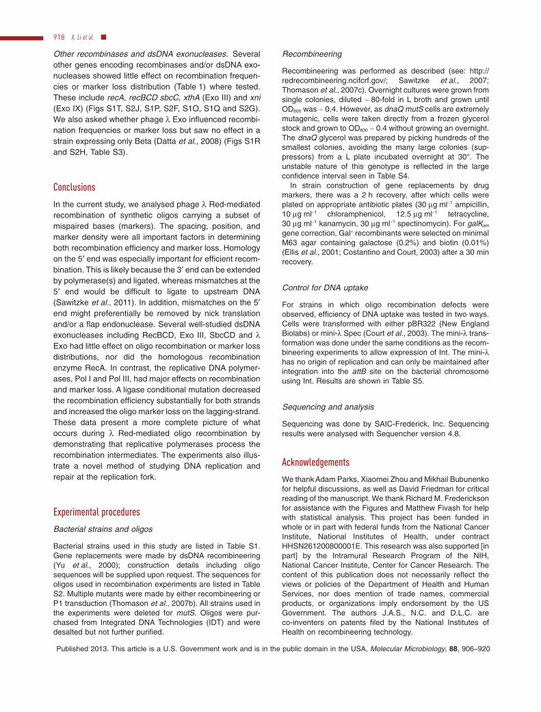
Other recombinases and dsDNA exonucleases. Severalother genes encoding recombinases and/or dsDNA exo-nucleases showed little effect on recombination frequen-cies or marker loss distribution (Table 1) where tested.These include recA, recBCD sbcC, xthA (Exo III) and xni(Exo IX) (Figs S1T, S2J, S1P, S2F, S1O, S1Q and S2G).We also asked whether phage l Exo influenced recombi-nation frequencies or marker loss but saw no effect in astrain expressing only Beta (Datta et al., 2008) (Figs S1Rand S2H, Table S3).
Conclusions
In the current study, we analysed phage l Red-mediatedrecombination of synthetic oligos carrying a subset ofmispaired bases (markers). The spacing, position, andmarker density were all important factors in determiningboth recombination efficiency and marker loss. Homologyon the 5′ end was especially important for efficient recom-bination. This is likely because the 3′ end can be extendedby polymerase(s) and ligated, whereas mismatches at the5′ end would be difficult to ligate to upstream DNA(Sawitzke et al., 2011). In addition, mismatches on the 5′end might preferentially be removed by nick translationand/or a flap endonuclease. Several well-studied dsDNAexonucleases including RecBCD, Exo III, SbcCD and lExo had little effect on oligo recombination or marker lossdistributions, nor did the homologous recombinationenzyme RecA. In contrast, the replicative DNA polymer-ases, Pol I and Pol III, had major effects on recombinationand marker loss. A ligase conditional mutation decreasedthe recombination efficiency substantially for both strandsand increased the oligo marker loss on the lagging-strand.These data present a more complete picture of whatoccurs during l Red-mediated oligo recombination bydemonstrating that replicative polymerases process therecombination intermediates. The experiments also illus-trate a novel method of studying DNA replication andrepair at the replication fork.
Experimental procedures
Bacterial strains and oligos
Bacterial strains used in this study are listed in Table S1.Gene replacements were made by dsDNA recombineering(Yu et al., 2000); construction details including oligosequences will be supplied upon request. The sequences foroligos used in recombination experiments are listed in TableS2. Multiple mutants were made by either recombineering orP1 transduction (Thomason et al., 2007b). All strains used inthe experiments were deleted for mutS. Oligos were pur-chased from Integrated DNA Technologies (IDT) and weredesalted but not further purified.
Recombineering
Recombineering was performed as described (see: http://redrecombineering.ncifcrf.gov/; Sawitzke et al., 2007;Thomason et al., 2007c). Overnight cultures were grown fromsingle colonies, diluted ~ 80-fold in L broth and grown untilOD600 was ~ 0.4. However, as dnaQ mutS cells are extremelymutagenic, cells were taken directly from a frozen glycerolstock and grown to OD600 ~ 0.4 without growing an overnight.The dnaQ glycerol was prepared by picking hundreds of thesmallest colonies, avoiding the many large colonies (sup-pressors) from a L plate incubated overnight at 30°. Theunstable nature of this genotype is reflected in the largeconfidence interval seen in Table S4.
In strain construction of gene replacements by drugmarkers, there was a 2 h recovery, after which cells wereplated on appropriate antibiotic plates (30 mg ml-1 ampicillin,10 mg ml-1 chloramphenicol, 12.5 mg ml-1 tetracycline,30 mg ml-1 kanamycin, 30 mg ml-1 spectinomycin). For galKam
gene correction, Gal+ recombinants were selected on minimalM63 agar containing galactose (0.2%) and biotin (0.01%)(Ellis et al., 2001; Costantino and Court, 2003) after a 30 minrecovery.
Control for DNA uptake
For strains in which oligo recombination defects wereobserved, efficiency of DNA uptake was tested in two ways.Cells were transformed with either pBR322 (New EnglandBiolabs) or mini-l Spec (Court et al., 2003). The mini-l trans-formation was done under the same conditions as the recom-bineering experiments to allow expression of Int. The mini-lhas no origin of replication and can only be maintained afterintegration into the attB site on the bacterial chromosomeusing Int. Results are shown in Table S5.
Sequencing and analysis
Sequencing was done by SAIC-Frederick, Inc. Sequencingresults were analysed with Sequencher version 4.8.
Acknowledgements
We thank Adam Parks, Xiaomei Zhou and Mikhail Bubunenkofor helpful discussions, as well as David Friedman for criticalreading of the manuscript. We thank Richard M. Fredericksonfor assistance with the Figures and Matthew Fivash for helpwith statistical analysis. This project has been funded inwhole or in part with federal funds from the National CancerInstitute, National Institutes of Health, under contractHHSN261200800001E. This research was also supported [inpart] by the Intramural Research Program of the NIH,National Cancer Institute, Center for Cancer Research. Thecontent of this publication does not necessarily reflect theviews or policies of the Department of Health and HumanServices, nor does mention of trade names, commercialproducts, or organizations imply endorsement by the USGovernment. The authors J.A.S., N.C. and D.L.C. areco-inventers on patents filed by the National Institutes ofHealth on recombineering technology.
918 X. Li et al. �
Published 2013. This article is a U.S. Government work and is in the public domain in the USA, Molecular Microbiology, 88, 906–920

ReferencesAllen, L.M., Hodskinson, M.R., and Sayers, J.R. (2009)
Active site substitutions delineate distinct classes of eubac-terial flap endonuclease. Biochem J 418: 285–292.
Baba, T., Ara, T., Hasegawa, M., Takai, Y., Okumura, Y.,Baba, M., et al. (2006) Construction of Escherichia coliK-12 in-frame, single-gene knockout mutants: the Keio col-lection. Mol Syst Biol 2: 2006–0008.
Banach-Orlowska, M., Fijalkowska, I.J., Schaaper, R.M., andJonczyk, P. (2005) DNA polymerase II as a fidelity factor inchromosomal DNA synthesis in Escherichia coli. MolMicrobiol 58: 61–70.
Benkovic, S.J., Valentine, A.M., and Salinas, F. (2001)Replisome-mediated DNA replication. Annu Rev Biochem70: 181–208.
Burdett, V., Baitinger, C., Viswanathan, M., Lovett, S.T., andModrich, P. (2001) In vivo requirement for RecJ, ExoVII,ExoI, and ExoX in methyl-directed mismatch repair. ProcNatl Acad Sci USA 98: 6765–6770.
Costantino, N., and Court, D.L. (2003) Enhanced levels of lRed-mediated recombinants in mismatch repair mutants.Proc Natl Acad Sci USA 100: 15748–15753.
Court, D.L., Sawitzke, J.A., and Thomason, L.C. (2002)Genetic engineering using homologous recombination.Annu Rev Genet 36: 361–388.
Court, D.L., Swaminathan, S., Yu, D., Wilson, H., Baker, T.,Bubunenko, M., et al. (2003) Mini-lambda: a tractablesystem for chromosome and BAC engineering. Gene 315:63–69.
Datta, S., Costantino, N., Zhou, X., and Court, D.L. (2008)Identification and analysis of recombineering functionsfrom Gram-negative and Gram-positive bacteria and theirphages. Proc Natl Acad Sci USA 105: 1626–1631.
Dutra, B.E., Sutera, V.A., and Lovett, S.T. (2007) RecA-independent recombination is efficient but limited by exo-nucleases. Proc Natl Acad Sci USA 104: 216–221.
Echols, H., Lu, C., and Burgers, P.M. (1983) Mutator strainsof Escherichia coli, mutD and dnaQ, with defective exonu-cleolytic editing by DNA polymerase III holoenzyme. ProcNatl Acad Sci USA 80: 2189–2192.
Ellis, H.M., Yu, D., DiTizio, T., and Court, D.L. (2001) Highefficiency mutagenesis, repair, and engineering of chromo-somal DNA using single-stranded oligonucleotides. ProcNatl Acad Sci USA 98: 6742–6746.
Goldmark, P.J., and Linn, S. (1972) Purification and proper-ties of the recBC DNase of Escherichia coli K-12. J BiolChem 247: 1849–1860.
Gottesman, M.M., Hicks, M.L., and Gellert, M. (1973) Genet-ics and function of DNA ligase in Escherichia coli. J MolBiol 77: 531–547.
Horton, N.C., and Finzel, B.C. (1996) The structure of anRNA/DNA hybrid: a substrate of the ribonuclease activity ofHIV-1 reverse transcriptase. J Mol Biol 264: 521–533.
Huen, M.S., Li, X.T., Lu, L.Y., Watt, R.M., Liu, D., and Huang,J.D. (2006) The involvement of replication in singlestranded oligonucleotide-mediated gene repair. NucleicAcids Res 34: 6183–6194.
Joyce, C.M., and Grindley, N.D. (1984) Method for determin-ing whether a gene of Escherichia coli is essential: appli-cation to the polA gene. J Bacteriol 158: 636–643.
Karakousis, G., Ye, N., Li, Z., Chiu, S.K., Reddy, G., andRadding, C.M. (1998) The Beta protein of phage l bindspreferentially to an intermediate in DNA renaturation. J MolBiol 276: 721–731.
Kelley, W.S., and Joyce, C.M. (1983) Genetic characteriza-tion of early amber mutations in the Escherichia coli polAgene and purification of the amber peptides. J Mol Biol164: 529–560.
van Kessel, J.C., and Hatfull, G.F. (2008) Efficient point muta-genesis in mycobacteria using single-stranded DNArecombineering: characterization of antimycobacterial drugtargets. Mol Microbiol 67: 1094–1107.
Kmiec, E., and Holloman, W.K. (1981) b protein of bacteri-ophage l promotes renaturation of DNA. J Biol Chem 256:12636–12639.
Kogoma, T., and Maldonado, R.R. (1997) DNA polymerase Iin constitutive stable DNA replication in Escherichia coli. JBacteriol 179: 2109–2115.
Kornberg, A. (1990) The private life of DNA polymerase I.Methods Enzymol 182: 783–788.
Kurth, I., and O’Donnell, M. (2009) Replisome dynamicsduring chromosome duplication. In Escherichia coli andSalmonella: Cellular and Molecular Biology. Böck, A.,Curtiss, R., III, Kaper, J.B., Karp, P.D., Neidhardt, F.C.,Nyström, T., et al. (eds). Washington, DC: ASM Press,pp. 1–47.
Lajoie, M.J., Gregg, C.J., Mosberg, J.A., Washington, G.C.,and Church, G.M. (2012) Manipulating replisome dynamicsto enhance lambda Red-mediated multiplex genome engi-neering. Nucleic Acids Res 40: e170.
Langston, L.D., Indiani, C., and O’Donnell, M. (2009) Whitherthe replisome: emerging perspectives on the dynamicnature of the DNA replication machinery. Cell Cycle 8:2686–2691.
Leu, F.P., Georgescu, R., and O’Donnell, M. (2003) Mecha-nism of the E. coli t processivity switch during lagging-strand synthesis. Mol Cell 11: 315–327.
Li, X., and Marians, K.J. (2000) Two distinct triggers forcycling of the lagging strand polymerase at the replicationfork. J Biol Chem 275: 34757–34765.
Li, X.T., Costantino, N., Lu, L.Y., Liu, D., Watt, R.M., Cheah,K.S., et al. (2003) Identification of factors influencingstrand bias in oligonucleotide-mediated recombination inEscherichia coli. Nucleic Acids Res 31: 6674–6687.
Lia, G., Michel, B., and Allemand, J.F. (2012) Polymeraseexchange during Okazaki fragment synthesis observed inliving cells. Science 335: 328–331.
Little, J.W., Lehman, I.R., and Kaiser, A.D. (1967) An exonu-clease induced by bacteriophage l. I. Preparation of thecrystalline enzyme. J Biol Chem 242: 672–678.
McHenry, C.S. (2011) DNA replicases from a bacterial per-spective. Annu Rev Biochem 80: 403–436.
McInerney, P., Johnson, A., Katz, F., and O’Donnell, M.(2007) Characterization of a triple DNA polymerase repli-some. Mol Cell 27: 527–538.
Maki, H., Maki, S., and Kornberg, A. (1988) DNA polymeraseIII holoenzyme of Escherichia coli. IV. The holoenzyme isan asymmetric dimer with twin active sites. J Biol Chem263: 6570–6578.
Martinsohn, J.T., Radman, M., and Petit, M.A. (2008) The lRed proteins promote efficient recombination between
Oligonucleotide processing during l Red recombination 919
Published 2013. This article is a U.S. Government work and is in the public domain in the USA, Molecular Microbiology, 88, 906–920

diverged sequences: implications for bacteriophagegenome mosaicism. PLoS Genet 4: e1000065.
Mosberg, J.A., Gregg, C.J., Lajoie, M.J., Wang, H.H., andChurch, G.M. (2012) Improving lambda red genome engi-neering in Escherichia coli via rational removal of endog-enous nucleases. PLoS ONE 7: e44638.
Muniyappa, K., and Radding, C.M. (1986) The homologousrecombination system of phage l. Pairing activities of bprotein. J Biol Chem 261: 7472–7478.
Murphy, K.C. (1998) Use of bacteriophage l recombinationfunctions to promote gene replacement in Escherichia coli.J Bacteriol 180: 2063–2071.
Noirot, P., and Kolodner, R.D. (1998) DNA strand invasionpromoted by Escherichia coli RecT protein. J Biol Chem273: 12274–12280.
Reyes-Lamothe, R., Sherratt, D.J., and Leake, M.C. (2010)Stoichiometry and architecture of active DNA replicationmachinery in Escherichia coli. Science 328: 498–501.
Richardson, C.C., Lehman, I.R., and Kornberg, A. (1964) Adeoxyribonucleic acid phosphatase-exonuclease fromEscherichia coli. II. Characterization of the exonucleaseactivity. J Biol Chem 239: 251–258.
Rigby, P.W., Dieckmann, M., Rhodes, C., and Berg, P. (1977)Labeling deoxyribonucleic acid to high specific Activity invitro by nick translation with DNA polymerase I. J Mol Biol113: 237–251.
Rybalchenko, N., Golub, E.I., Bi, B., and Radding, C.M.(2004) Strand invasion promoted by recombination proteinb of coliphage l. Proc Natl Acad Sci USA 101: 17056–17060.
Sawitzke, J.A., Thomason, L.C., Costantino, N., Bubunenko,M., Datta, S., and Court, D.L. (2007) Recombineering: invivo genetic engineering in E. coli, S. enterica, andbeyond. Methods Enzymol 421: 171–199.
Sawitzke, J.A., Costantino, N., Li, X.T., Thomason, L.C., Bub-unenko, M., Court, C., and Court, D.L. (2011) Probingcellular processes with oligo-mediated recombination andusing the knowledge gained to optimize recombineering. JMol Biol 407: 45–59.
Sergueev, K., Court, D., Reaves, L., and Austin, S. (2002)E. coli cell-cycle regulation by bacteriophage lambda. JMol Biol 324: 297–307.
Setlow, P., Brutlag, D., and Kornberg, A. (1972) Deoxyribo-nucleic acid polymerase: two distinct enzymes in onepolypeptide. I. A proteolytic fragment containing thepolymerase and 3′→5′ exonuclease functions. J Biol Chem247: 224–231.
Sharan, S.K., Thomason, L.C., Kuznetsov, S.G., and Court,D.L. (2009) Recombineering: a homologous recombination-based method of genetic engineering. Nat Protoc 4: 206–223.
Sharon, R., Miller, C., and Ben-Ishai, R. (1975) Two modes ofexcision repair in toluene-treated Escherichia coli. J Bac-teriol 123: 1107–1114.
Studwell, P.S., and O’Donnell, M. (1990) Processive replica-tion is contingent on the exonuclease subunit of DNA
polymerase III holoenzyme. J Biol Chem 265: 1171–1178.
Swaminathan, S., Ellis, H.M., Waters, L.S., Yu, D., Lee, E.C.,Court, D.L., and Sharan, S.K. (2001) Rapid engineering ofbacterial artificial chromosomes using oligonucleotides.Genesis 29: 14–21.
Swingle, B., Markel, E., Costantino, N., Bubunenko, M.G.,Cartinhour, S., and Court, D.L. (2010) Oligonucleotiderecombination in Gram-negative bacteria. Mol Microbiol75: 138–148.
Thomason, L.C., Costantino, N., Shaw, D.V., and Court, D.L.(2007a) Multicopy plasmid modification with phage l Redrecombineering. Plasmid 58: 148–158.
Thomason, L.C., Costantino, N., and Court, D.L. (2007b)E. coli genome manipulation by P1 transduction. CurrProtoc Mol Biol Chapter 1: Unit 1 17.
Thomason, L., Court, D.L., Bubunenko, M., Costantino, N.,Wilson, H., Datta, S., and Oppenheim, A. (2007c) Recom-bineering: genetic engineering in bacteria using homolo-gous recombination. Curr Protoc Mol Biol Chapter 1: Unit 116.
Vaccaro, K.K., and Siegel, E.C. (1975) Increased spontane-ous reversion of certain frameshift mutations in DNApolymerase I deficient strains of Escherichia coli. Mol GenGenet 141: 251–262.
Van Pijkeren, J.P., Neoh, K.M., Sirias, D., Findley, A.S., andBritton, R.A. (2012) Exploring optimization parameters toincrease ssDNA recombineering in Lactococcus lactis andLactobacillus reuteri. Bioengineered 3: 209–217.
Viswanathan, M., and Lovett, S.T. (1999) Exonuclease X ofEscherichia coli. A novel 3′-5′ DNase and DnaQ super-family member involved in DNA repair. J Biol Chem 274:30094–30100.
Wang, H.H., Xu, G., Vonner, A.J., and Church, G. (2011)Modified bases enable high-efficiency oligonucleotide-mediated allelic replacement via mismatch repair evasion.Nucleic Acids Res 39: 7336–7347.
Wang, T.C. (2005) Discontinuous or semi-discontinuous DNAreplication in Escherichia coli? Bioessays 27: 633–636.
Yu, D., Ellis, H.M., Lee, E.C., Jenkins, N.A., Copeland, N.G.,and Court, D.L. (2000) An efficient recombination systemfor chromosome engineering in Escherichia coli. Proc NatlAcad Sci USA 97: 5978–5983.
Zhang, Y., Buchholz, F., Muyrers, J., and Stewart, A.F. (1998)A new logic for DNA engineering using recombination inEscherichia coli. Nat Genet 20: 123–128.
Zhang, Y., Muyrers, J., Rientjes, J., and Stewart, A.F. (2003)Phage annealing proteins promote oligonucleotide-directed mutagenesis in Escherichia coli and mouse EScells. BMC Mol Biol 4: 1.
Supporting information
Additional supporting information may be found in the onlineversion of this article at the publisher’s web-site.
920 X. Li et al. �
Published 2013. This article is a U.S. Government work and is in the public domain in the USA, Molecular Microbiology, 88, 906–920

Supplemental Figure 1
10
20
30
40
10
A. HME68Lagging-strand oligo XT36Efficiency: 1.7 × 10⁶
5´ 3´ 3 9 15 21 27 33 + 42 48 54 60 66 72 G C C C G G G T G C A C T T G A A A A T C A T G A C G G A A A A T C A T G A T G G A A A A T C A T G A T G G A A A A T C A T G A T G G A A A A T C A T G A T G G A A A A T C A T G A T G G A A A A T C A T G A T G G A A A A T C A T G A T G G A A A A T C A T G A T G G A A A A T C A T G A T G G A A A A T C A T G A T G G A A A A T C A T G A T G C A A A A T C A T G A T G C A A A A T C A T G A T G C A A A A T C A T G A T G C A A A A T C A T G A T G C A A A A T C A T G A T G C A A A A T C A T G A T G C A A A A T C A T G A T G C A A A A T C A T G A T G C A A A A T C A T G A T G C A A A A T C A T G A T G C A A A A T C A T G A T G C C A A A T C A T G A T G C C A A A T C A T G A T G C C A A A T C A T G A T G C C C A A T C A T G A T G C C C G A T C A T G A T G C C C G A T C A T G A T G G A A A A T C A T G C T G C A A A A T C A T G C T G C A A A A T C A T G C T G C A A A A T C A T G C T G C A A A A T C A T G C T G C A A A A T C A T G C T G C A A A A T C A T G C T G C A A A A T C A T G C T G C A A A A T C A T G C T G C A A A A T C A T G C T G C C A A A T C A T G C T G C C A A A T C A T G C T G C C A A A T C A T G C T G C C A A A T C A T G C T G G A A A A T C A T A C T G G A A A A T C A T A C T G G A A A A T C A T A C T G C A A A A T C A T A C T G G A C A A T C A T G A T G G A C A A T C A T G A T
Position:Host:Oligo:
B. HME68Lagging-strand oligo XT21Efficiency: <1.3 × 101
5´ 3´ 3 6 9 12 15 18 21 24 27 30 33 36 37 38 + 42 45 48 51 54 57 60 63 66 69 72 G A C G C A C A G G G T T A G T G G G C C A A C G T T G G C A C A C A A A C T A T C A A A T A G G A C C G A C G C A C A G G G T T A T T G G G C C A A C G T G A C G C A C A G G G T T A T T G G G C C A A C G T G A C G C A C A G G G T T A T T G G G C C A A C G T G A C G C A C A G G G T C A G T G G G C C A A C G T G A C G C A C A G G G T C A G T G G G C C A A C G T G A C G C A C A G G G T C A G T G G G C C A A C G T G A C G C A C A G G G T C A G T G G G C C A A C G T G A C G C A C A G G G T C A G T G G G C C A A C G T G A C G C A C A G G G T C A G T G G G C C A A C G T G A C G C A C A G G G T T G G T G G G C C A A C G T G A C G C A C A G G G T T G G T G G G C C A A C G T G A C G C A C A G G G T T G G T G G G C C A A C G T G A C G C A C A G G G T T G G T G G G C C A A C G T G A C G C A C A G G G T T G G T G G G C C A A C G T G A C G C A C A G G G T G A G T G G G C C A A C G T G A C G C A C A G G G T T A C T G G G C C A A C G T
Position:Host:Oligo:

Supplemental Figure 1
10
20
30
10
20
30
40
50
C. HME68Lagging-strand oligo XT18Efficiency: 1.3 × 10⁶
5´ 3´ 15 18 21 24 27 30 33 36 + 42 45 48 51 54 57 60 63 C A C A G G G T G T G G G C C A A A C A C A A A C T C A A A T A G G A C A C A A A C T C A A A T A G G A C A C A A A C T C A A A T A G G A C A C A A A C T C A A A T A G G A C A C A A A C T C A A A T A G G A C A C A A A C T C A A A T A G G A C A C A A A C T C A A A T A G G A C A C A A A C T C A A A T A G G A C A C A A A C T C A A A T A G G A C A C A A A C T C A A A T A G G A C A C A A A C T C A A A T A G G A C A C A A A C T C A A A T A G G A C A C A A A C T C A A A T A G G A C A C A A A C T C A A A T A G G A C A C A A A C T C A A A T A G G A C A C A A A C T C A A A T A G G A C A C A A A C T C A A A T A G G A C A C A A A C T C A A A T A G G A C A C A A A C T C A A A T A G G A C A C A A A C T C A A A T A G G A C A C A A A C T C A A A T A G G A C A C A A A C T C A A A T A G G A C A C A A A C T C A A A T A G G A C A C A A A C T C A A A T A G G A C A C A A A C T C A A A T A G G A C A C A A A C T C A A A T A G G A C A C A A A C T C A A A T A G G A C A C A A A C T C A A A T A G G A C A C A A A C T C A A A T A G G A C A C A A A C T C A A A T A G G A C A C A A A C T C A A A T A G G
Position:Host:Oligo:
D. HME68Lagging-strand oligo XT524Efficiency: 3.7 × 10⁶
5´ 3´ 24 27 30 33 36 39 42 45 48 51 54 57 + 63 66 69 72 75 78 81 84 87 90 93 G A C G C A C A G G G T G T G G G C C A A C G T T G G C A C A C A A A C T C A A A T A G G A C C T G G C A C A C A A A C T C A A A T A G G A C C T G G C A C A C A A A C T C A A A T A G G A C C T G G C A C A C A A A C T C A A A T A G G A C C T G G C A C A C A A A C T C A A A T A G G A C C T G G C A C A C A A A C T C A A A T A G G A C C T G G C A C A C A A A C T C A A A T A G G A C C T G G C A C A C A A A C T C A A A T A G G A C C T G G C A C A C A A A C T C A A A T A G G A C C T G G C A C A C A A A C T C A A A T A G G A C C T G G C A C A C A A A C T C A A A T A G G A C C T G G C A C A C A A A C T C A A A T A G G A C C T G G C A C A C A A A C T C A A A T A G G A C C T G G C A C A C A A A C T C A A A T A G G A C C T G G C A C A C A A A C T C A A A T A G G A C C T G G C A C A C A A A C T C A A A T A G G A C C T G G C A C A C A A A C T C A A A T A G G A C C T G G C A C A C A A A C T C A A A T A G G A C C T G G C A C A C A A A C T C A A A T A G G A C C T G G C A C A C A A A C T C A A A T A G G A C C T G G C A C A C A A A C T C A A A T A G G A C C T G G C A C A C A A A C T C A A A T A G G A C C T G G C A C A C A A A C T C A A A T A G G A C C T G G C A C A C A A A C T C A A A T A G G A C C T G G C A C A C A A A C T C A A A T A G G A C C T G G C A C A C A A A C T C A A A T A G G A C C T G G C A C A C A A A C T C A A A T A G G A C C T G G C A C A C A A A C T C A A A T A G G A C C T G G C A C A C A A A C T C A A A T A G G A C C T G G C A C A C A A A C T C A A A T A G G A C C T G G C A C A C A A A C T C A A A T A G G A C C T G G C A C A C A A A C T C A A A T A G G A C C T G G C A C A C A A A C T C A A A T A G G A C C T G G C A C A C A A A C T C A A A T A G G A C C T G G C A C A C A A A C T C A A A T A G G A C C T G G C A C A C A A A C T C A A A T A G G A C C T G G C A C A C A A A C T C A A A T A G G A C C T G G C A C A C A A A C T C A A A T A G G A C C T G G C A C A C A A A C T C A A A T A G G A C C T G G C A C A C A A A C T C A A A T A G G A C C T G G C A C A C A A A C T C A A A T A G G A C C T G G C A C A C A A A C T C A A A T A G G A C C T G G C A C A C A A A C T C A A A T A G G A C C T G G C A C A C A A A C T C A A A T A G G A C C T G G C A C A C A A A C T C A A A T A G G A C C T G G C A C A C A A A C T C A A A T A G G A C C T G G C A C A C A A A C T C A A A T A G G A C C T G G C A C A C A A A C T C A A A T A G G A C C T G G C A C A C A A A C T C A A A T A G G A C C T G G C A C A C A A A C T C A A A T A G G A C C T G G C A C A C A A A C T C A A A T A G G A C C T G G C A C A C A A A C T C A A A T A G G A C C T G G C A C A C A A A C T C A A A T A G G A C C T G G C A C A C A A A C T C A A A T A G G A C C T G G C A C A C A A A C T C A A A T A G G A C C
Position:Host:Oligo:

Supplemental Figure 1
10
20
30
40
10
20
30
40
5´ 3´ + 42 45 48 51 54 57 60 63 66 69 72 G T G G G C C A A C G T T C A A A T A G G A C C T C A A A T A G A C G T T C A A A T C A A C G T T C A A A T A G A C G T T C A G G C C A A C G T T T G G G C C A A C G T T T G G G C C A A C G T T T G G G C C A A C G T T T G G G C C A A C G T T T G G G C C A A C G T T T G G G C C A A C G T T T G G G C C A A C G T T T G G G C C A A C G T T T G G G C C A A C G T T T G G G C C A A C G T T T G G G C C A A C G T T T G G G C C A A C G T T T G G G C C A A C G T T T G G G C C A A C G T T T G G G C C A A C G T T T G G G C C A A C G T T T G G G C C A A C G T T T G G G C C A A C G T T T G G G C C A A C G T T T G G G C C A A C G T T T G G G C C A A C G T T T G G G C C A A C G T T T G G G C C A A C G T T T G G G C C A A C G T T T G G G C C A A C G T T T G G G C C A A C G T T T G G G C C A A C G T T T G G G C C A A C G T T T G G G C C A A C G T T T G G G C C A A C G T T T G G G C C A A C G T T T G G G C C A A C G T T T G G G C C A A C G T T T G G G C C A A C G T T T G G G C C A A C G T T T G G G C C A A C G T T T G G G C C A A C G T T T G G G C C A A C G T T T G G G C C A A C G T
E. HME68Lagging-strand oligo XT30Efficiency: 5.1 × 10⁶
F. HME68Lagging-strand oligo XT29Efficiency: 2.4 × 10³
Position:Host:Oligo:
5´ 3´ 3 6 9 12 15 18 21 24 27 30 33 36 + G A C G C A C A G G G T G T G G C A C A C A A A C T G A C C A C C C A A A C T G A C C A C C C A A A C T G A C G A C C C A A A C T G A C G C A C C A A A C T G A C G C A C A A A A C T G A C G C A C A G G A C T G A C G C A C A G G A C T G A C G C A C A G G A C T G A C G C A C A G G G C T G A C G C A C A G G G T T G A C G C A C A G G G T T G A C G C A C A G G G T T G A C G C A C A G G G T T G A C G C A C A G G G T T G A C G C A C A G G G T T G A C G C A C A G G G T T G A C G C A C A G G G T T G A C G C A C A G G G T T G A C G C A C A G G G T T G A C G C A C A G G G T T G A C G C A C A G G G T T G A C G C A C A G G G T T G A C G C A C A G G G T T G A C G C A C A G G G T T G A C G C A C A G G G T T G A C G C A C A G G G T T G A C G C A C A G G G T T G A C G C A C A G G G T T G A C G C A C A G G G T T G A C G C A C A G G G T T G A C G C A C A G G G T T G A C G C A C A G G G T T G A C G C A C A G G G T T G A C G C A C A G G G T T G A C G C A C A G G G T T G A C G C A C A G G G T T G A C G C A C A G G G T T G A C G C A C A G G G T T G A C G C A C A G G G T T G A C G C A C A G G G T T
Position:Host:Oligo:

Supplemental Figure 1
10
20
30
40
10
H. HME68Lagging-strand oligo XT38Efficiency: 6.3 × 10⁴
G. HME68Lagging-strand oligo XT351Efficiency: 6.1 × 101
5´ 3´ 21 24 27 30 33 36 + 42 45 48 51 54 57 60 63 66 69 72 C A G G G T G T G G G C C A A C G T A C A A A C T C A A A T A G G A C C A C A A A C T C A A A T A G G A C C A C A A A C T C A A A T A G G A C C A C A A A C T C A A A T A G G A C C A C A A A C T C A A A T A G G A C C A C A A A C T C A A A T A G G A C C A C A A A C T C A A A T A G G A C T A C A A A C T C A A A T A G G A G T A C A A A C T C A A A T A G G A G T A C A A A C T C A A A T A G G A G T A C A A A C T C A A A T A G G C G T A C A A A C T C A A A T A G A C G T A C A A A C T C A A A T A G A C G T A C A A A C T C A A A T A G A C G T A C A A A C T C A A A T A G A C G T A C A A A C T C A A A T A G A C G T A C A A A C T C A A A T A G A C G T A C A A A C T C A A A T A G A C G T A C A A A C T C A A A T A G A C G T A C A A A C T C A A A T A G A C G T A C A A A C T C A A A T A G A C G T A C A A A C T C A A A T A G A C G T A C A A A C T C A A A T A G A C G T A C A A A C T C A A A T A G A C G T A C A A A C T C A A A T A G A C G T A C A A A C T C A A A T A G A C G T A C A A A C T C A A A T A G A C G T A C A A A C T C A A A T A G A C G T A C A A A C T C A A A T A G A C G T A C A A A C T C A A A T A G A C G T A C A A A C T C A A A T A G A C G T A C A A A C T C A A A T A A A C G T A C A A A C T C A A A T A A A C G T A C A A A C T C A A A T C A A C G T A C A A A C T C A A A T C A A C G T A C A A A C T C A A A T C A A C G T A C A A A C T C A A A T C A A C G T A C A A A C T C A A A T C A A C G T A C A A A C T C A A A T C A A C G T A C A A A C T C A A A T C A A C G T A C A A A C T C A A A T C A A C G T A C A A A C T C A A A T C A A C G T A C A A A C T C A A A T C A A C G T A C A A A C T C A A A T C A A C G T A C A A A C T C A A A C C A A C G T A C A A A C T C A G G C C A A C G T
Position:Host:Oligo:
11%
89%
5´ 3´ 3 6 9 12 15 18 21 24 27 30 33 36 + 42 45 48 51 54 G A C C C A C A G G G T G T G G G C T G G G A C A C A A A C T C A A A T G G G G A C A C A A A C T C A A A T G G G G A C A C A A A C T C A A A T G A G G A C A C A A A C T C A A A T G A C C A C A C A A A C T C A A A T G A C C A C A C A A A C T C A A A T G A C C A C A C A A A C T C A A A T G A C C A C A C A A A C T C A A A T G A C C A C A C A A A C T C A A A T G A C C A C A C A A A C T C A A A T G A C C A C A C A A A C T C A A A T G A C C A C A C A A A C T C A A A T G A C C C C A C A A A C T C A A A T G A C C C A C A G A A C T C A A A T G A C C C A C A G G A C T C A A A T G A C C C A C A G G G C T C A A A T G A C C C A C A G G G C T C A A A T G A C C C A C A G G G C T C A A A T G A C C C A C A G G G T T C A A A T G A C C C A C A G G G T T C A A A T
Position:Host:Oligo:

Supplemental Figure 1
10
20
30
40
10
20
30
40
10
20
30
40
K. HME681/1000 Lagging-strand oligo XT13Efficiency: 9.4 × 10³
J. HME68Lagging-strand oligo XT352Efficiency: 1.3 × 10⁷
5´ 3´ 3 6 9 12 15 18 + 60 63 66 69 72 G A C G C A G A A C G T T G G C A C T G G A C C G A C G C A T G G C G T G A C C A C T G A C G T G A C G A C T G A C G T G A C G A C T G A C G T G A C G A C T G A C G T G A C G C A T G A C G T G A C G C A T G A C G T G A C G C A T G A C G T G A C G C A T G A C G T G A C G C A T G A C G T G A C G C A T G A C G T G A C G C A T G A C G T G G G C A C T A A C G T G A G C A C T A A C G T G A G C A C T A A C G T G A C C A C T A A C G T G A C G A C T A A C G T G A C G A C T A A C G T G A C G C C T A A C G T G A C G C A T A A C G T G A C G C A T A A C G T G A C G C A T A A C G T G A C G C A T A A C G T G A C G C A T A A C G T G A C G C A T A A C G T G A C G C A T A A C G T G A C G C A T A A C G T G A C G C A T A A C G T G A C G C A T A A C G T G A C G C A T A A C G T G A C G C A T A A C G T G A C G C A T A A C G T G A C G C A T A A C G T G A C G C A T A A C G T G A C G C A T A A C G T G A C G C A T A A C G T G A C G C A T A A C G T G A C G C A T A A C G T G A C G C A T A A C G T G A C G C A T A A C G T G A C G C A T A A C G T G A C G C A T A A C G T G A C G C A T A A C G T G A C G C A T A A C G T G A C G C A T A A C G T G A C G C A T A A C G T
Position:Host:Oligo:
I. HME68Lagging-strand oligo XT37Efficiency: 2.3 × 10⁷
5´ 3´ 3 6 9 12 15 18 + G A C G C A G T G G C A C T G A C G C A T G A C G C A T G A C G C A T G A C G C A T G A C G C A T G A C G C A T G A C G C A T G A C G C A T G A C G C A T G A C G C A T G A C G C A T G A C G C A T G A C G C A T G A C G C A T G A C G C A T G A C G C A T G A C G C A T G A C G C A T G A C G C A T G A C G C A T G A C G C A T G A C G C A T G A C G C A T G A C G C A T G A C G C A T G A C G C A T G A C G C A T G A C G C A T G A C G C A T G A C G C A T G A C G C A T G A C G C A T G A C G C A T G A C G C A T G A C G C A T G A C G C A T G A C G C A T G A C G C A T G A C G C A T G A C G C A T G A C G C A T G A C G C A T G A C G C A T G A C G C A T G A C G C A T G A C G C A T G A C G C A T G A C G C C T
Position:Host:Oligo:
5´ 3´ 15 21 30 + 48 54 63 C C G G G C A A G A T C T G A G A T C T G A G A T C T G A G A T C T G A G A T C T G A G A T C T G A G A T C T G A G A T C T G A G A T C T G A G A T C T G A G A T C T G A G A T C T G A G A T C T G A G A T C T G A G A T C T G A G A T C T G A G A T C T G A G A T C T G A G A T C T G A G A T C T G A G A T C T G A G A T C T G A G A T C T G A G A T C T G A G A T C T G A G A T C T G A G A T C T G A G A T C T G A G A T C T G A G A T C T G A G A T C T A A G A T C T A A G A T C C A A G A T G C A A G A T G C A A G A T G C A C G A T C T G C G A T C T G C G A T C T G C G A T C T G C C G T C T G A G A T C C G A C G T C T G A G A T G C G A G A T C C G
Position:Host:Oligo:
66%
3´ 14%
5´ 11%
Other 9%

Supplemental Figure 1
10
20
30
40
10
20
30
40
10
20
30
40
31%
3´ 54%
5´ 4%
56%
3´ 25%
5´ 11%
Other 8%
Other 7%
5´ & 3´ 4%
74%
3´ 17%
5´ 9%
5´ 3´ 15 21 30 + 48 54 63 C C G G G C A A G A T C T G A G A T C T G A G A T C T G A G A T C T G A G A T C T G A G A T C T G A G A T C T G A G A T C T G A G A T C T G A G A T C T G A G A T C T G A G A T C T G A G A T C T G A G A T C T G A G A T C T G A G A T C T G A G A T C T G A G A T C T G A G A T C T G A G A T C T G A G A T C T G A G A T C T G A G A T C T G A G A T C T G A G A T C T G A G A T C T G A G A T C T G A G A T C T G A G A T C T G A G A T C T G A G A T C T G A G A T C T G A G A T C T A A G A T C T A A G A T C T A A G A T C C A A G A T G C A A G A T G C A A G A T G C A C G A T C T G C G A T C T G C C A T C T G C C A T C T G
Position:Host:Oligo:
N. HME69 (polA resA1)Lagging-strand oligo XT13Efficiency: 1.3 × 10⁷
5´ 3´ 15 21 30 + 48 54 63 C C G G G C A A G A T C T G A G A T C T G A G A T C T G A G A T C T G A G A T C T G A G A T C T G A G A T C T G A G A T C T G A G A T C T G A G A T C T G A G A T C T G A G A T C T G A G A T C T G A G A T C T G A G A T C T G A G A T C T A A G A T C T A A G A T C T A A G A T C T A A G A T C T A A G A T C T A A G A T C T A A G A T C T A A G A T C T A A G A T C T A A G A T C C A A G A T C C A A G A T C C A A G A T C C A A G A T C C A A G A T C C A A G A T G C A A G A T G C A A G A T G C A A G A T G C A A G A T G C A A G A T G C A A G A T G C A A G A T G C A A G A T G C A C G A T C T G C C A T C T G C G A T C T A C C A T C T A C G A T C C G A G A T G C G A G A T G C G
Position:Host:Oligo:
M. XTL51 (Quad mutant)1/1000 Lagging-strand oligo XT13Efficiency: 1.8 × 10⁵
5´ 3´ 15 21 30 + 48 54 63 C C G G G C A A G A T C T G A G A T C T G A G A T C T G A G A T C T G A G A T C T G A G A T C T G A G A T C T G A G A T C T G A G A T C T G A G A T C T G A G A T C T G A G A T C T G A G A T C T G A G A T C T G A G A T C T G A G A T C T G A G A T C T G A G A T C T G A G A T C T G A G A T C T G A G A T C T G A G A T C T G A G A T C T G A G A T C T G A G A T C T G A G A T C T G A G A T C T G A G A T C T G A G A T C T A A G A T C T A A G A T C T A A G A T C T A A G A T C T A A G A T C T A A G A T C C A A G A T C C A A G A T C C A A G A T G C A A G A T G C A A G A T G C A C G A T C T G C G A T C T G C G A T C T G C C A T C T G C C G T C T G A G A T G C G A G A T C C G A G A T C C G A G A T G T G
Position:Host:Oligo:
L. XTL51 (Quad mutant)Lagging-strand oligo XT13Efficiency: 1.2 × 10⁷

Supplemental Figure 1
50
60
10
20
30
40
10
20
30
40
57%
3´ 19%
5´ 13%
Other 9%
5´ & 3´ 2%
68%
3´ 18%
5´ 8%
Other 3%
5´ & 3´ 3%
5´ 3´ 15 21 30 + 48 54 63 C C G G G C A A G A T C T G A G A T C T G A G A T C T G A G A T C T G A G A T C T G A G A T C T G A G A T C T G A G A T C T G A G A T C T G A G A T C T G A G A T C T G A G A T C T G A G A T C T G A G A T C T G A G A T C T G A G A T C T G A G A T C T G A G A T C T G A G A T C T G A G A T C T G A G A T C T G A G A T C T G A G A T C T G A G A T C T G A G A T C T G A G A T C T G A G A T C T G A G A T C T G A G A T C T G A G A T C T G A G A T C T G A G A T C T G A G A T C T G A G A T C T G A G A T C T G A G A T C T G A G A T C T G A G A T C T G A G A T C T G A G A T C T G A G A T C T G A G A T C T G A G A T C T G A G A T C T G A G A T C T A A G A T C T A A G A T C T A A G A T C T A A G A T C T A A G A T C T A A G A T C C A A G A T C C A A G A T C C A A G A T G C A A G A T G C A C G A T C T G C G A T C T G C G A T C T G C C A T C T G C C A T C T G C G A T G C A C G A T C C A A G A T G T G A G G T C T G
Position:Host:Oligo:
P. XTL100 (recBCD<>kan sbcC<>amp)Lagging-strand oligo XT13Efficiency: 1.3 × 10⁷
5´ 3´ 15 21 30 + 48 54 63 C C G G G C A A G A T C T G A G A T C T G A G A T C T G A G A T C T G A G A T C T G A G A T C T G A G A T C T G A G A T C T G A G A T C T G A G A T C T G A G A T C T G A G A T C T G A G A T C T G A G A T C T G A G A T C T G A G A T C T G A G A T C T G A G A T C T G A G A T C T G A G A T C T G A G A T C T G A G A T C T G A G A T C T G A G A T C T G A G A T C T G A G A T C T G A G A T C T G A G A T C T G A G A T C T A A G A T C T A A G A T C T A A G A T C T A A G A T C T A A G A T C C A A G A T C C A A G A T G C A A G A T G C A C G A T C T G C G A T C T G C G A T C T G C G A T C T G C C G T C T G C C G T C T G C G A T C T A A G A T C C G A G G T C T G A G A T G T G A G G T C C A
Position:Host:Oligo:
O. XTL92 (xthA<>tet)Lagging-strand oligo XT13Efficiency: 4.4 × 10⁷

Supplemental Figure 1
10
20
30
40
10
20
30
40
48%
3´ 25%
5´ 11%
Other 11%
5´ & 3´ 5%
5´ 3´ 15 21 30 + 48 54 63 C C G G G C A A G A T C T G A G A T C T G A G A T C T G A G A T C T G A G A T C T G A G A T C T G A G A T C T G A G A T C T G A G A T C T G A G A T C T G A G A T C T G A G A T C T G A G A T C T G A G A T C T G A G A T C T G A G A T C T G A G A T C T G A G A T C T G A G A T C T G A G A T C T G A G A T C T G A G A T C T G A G A T C T A A G A T C T A A G A T C T A A G A T C T A A G A T C T A A G A T C T A A G A T C T A A G A T C T A A G A T C T A A G A T G C A A G A T G C A C G A T C T G C C G T C T G C C G T C T G C C G T C T G C C G T C T G C G A T C C A C G A T C C A A G A T G T G A C A T C T G A G A T C C G C C G T G C G A G A T C C G
Position:Host:Oligo:
Q. XTL324 (xni<>cat)Lagging strand oligo XT13Efficiency: 2.6 × 10⁷
71%
3´ 9%
5´ 9%
Other 7%
5´ & 3´ 4%
5´ 3´ 15 21 30 + 48 54 63 C C G G G C A A G A T C T G A G A T C T G A G A T C T G A G A T C T G A G A T C T G A G A T C T G A G A T C T G A G A T C T G A G A T C T G A G A T C T G A G A T C T G A G A T C T G A G A T C T G A G A T C T G A G A T C T G A G A T C T G A G A T C T G A G A T C T G A G A T C T G A G A T C T G A G A T C T G A G A T C T G A G A T C T G A G A T C T G A G A T C T G A G A T C T G A G A T C T G A G A T C T G A G A T C T G A G A T C T G A G A T C T G A G A T C T G A G A T C T G A G A T C T A A G A T C C A A G A T C C A A G A T G C A C G A T C T G C G A T C T G C G A T C T G C C A T C T G C C A T C T A C G A T C C A C C G T G T G A C G T C T G A G A T G C G
Position:Host:Oligo:
R. SIMD90 (λ Δexo)Lagging strand oligo XT13Efficiency: 1.8 × 10⁷
10
20
30
40
50
3´ 23%
5´ 13%
5´ & 3´ 2%Other 2%
60%
5´ 3´ 15 21 30 + 48 54 63 C C G G G C A A G A T C T G A G A T C T G A G A T C T G A G A T C T G A G A T C T G A G A T C T G A G A T C T G A G A T C T G A G A T C T G A G A T C T G A G A T C T G A G A T C T G A G A T C T G A G A T C T G A G A T C T G A G A T C T G A G A T C T G A G A T C T G A G A T C T G A G A T C T G A G A T C T G A G A T C T G A G A T C T G A G A T C T G A G A T C T G A G A T C T G A G A T C T G A G A T C T G A G A T C T G A G A T C T G A G A T C T G A G A T C T G A G A T C T G A G A T C T G A G A T C T G A G A T C T A A G A T C T A A G A T C T A A G A T C T A A G A T C T A A G A T C T A A G A T C T A A G A T C T A A G A T G C A A G A T G C A A G A T G C A A G A T G C A A G A T G C A C G A T C T G C G A T C T G C G A T C T G C C A T C T G C C A T C T G C C A T C T G C C G T C T G C C G T G C A A G A T C C G
Position:Host:Oligo:
S. XTL372 (polB<>spec) Lagging-strand oligo XT13Efficiency: 4.1 × 10⁷

Supplemental Figure 1
10
20
30
40
10
20
30
40
66%
3´ 9%
5´ 15%
Other 9%
5´ 3´ 15 21 30 + 48 54 63 C C G G G C A A G A T C T G A G A T C T G A G A T C T G A G A T C T G A G A T C T G A G A T C T G A G A T C T G A G A T C T G A G A T C T G A G A T C T G A G A T C T G A G A T C T G A G A T C T G A G A T C T G A G A T C T G A G A T C T G A G A T C T G A G A T C T G A G A T C T G A G A T C T G A G A T C T G A G A T C T G A G A T C T G A G A T C T G A G A T C T G A G A T C T G A G A T C T G A G A T C T G A G A T C T G A G A T C T G A G A T C T G A G A T C T G A G A T C T A A G A T C T A A G A T C T A A G A T C T A C G A T C T G C G A T C T G C G A T C T G C G A T C T G C C G T C T G C C G T C T G C C G T C T G C C A T G T G A G A T C C G A G A T C C G A G A T G C G
Position:Host:Oligo:
T. HME70 (ΔrecA)Lagging-strand oligo XT13Efficiency: 2.8 × 10⁷
80%
3´ 20%
5´ 3´ 15 21 30 + 48 54 63 C C G G G C A A G A T C T G A G A T C T G A G A T C T G A G A T C T G A G A T C T G A G A T C T G A G A T C T G A G A T C T G A G A T C T G A G A T C T G A G A T C T G A G A T C T G A G A T C T G A G A T C T G A G A T C T G A G A T C T G A G A T C T G A G A T C T G A G A T C T G A G A T C T G A G A T C T G A G A T C T G A G A T C T G A G A T C T G A G A T C T G A G A T C T G A G A T C T G A G A T C T G A G A T C T G A G A T C T G A G A T C T G A G A T C T G A G A T C T G A G A T C T G A G A T C T G A G A T C T G A G A T C T G A G A T C T G A G A T C T G A G A T C T A A G A T C C A A G A T C C A A G A T C C A A G A T C C A A G A T G C A A G A T G C A A G A T G C A A G A T G C A
Position:Host:Oligo:
U. XTL85 (polA 5´-3´ exo<>cat non-fusion)Lagging-strand oligo XT13Efficiency: 4.3 × 10⁶

Supplemental Figure 2
10 10
20
30
40
10
20
30
40
36%
5´ 14%
3´ 43%
Other 7%
B. HME68Leading-strand oligo 1/1000 XT14Efficiency: 9.1 × 101
3´ 5´ 15 21 30 + 48 54 63 C C G G G C A A G A T C T G A G A T C T G A G A T C T G A G A T C T G A G A T C T G A G A T C T G C C A T C T G C C A T C T G C C A T C T G C C A T C T G C C G T C T G C C G T C T G A G A T C T A A G A T C T A A G G T C T G
Position:Host:Oligo:
68%
5´ 15%
3´ 11%
5´ & 3´ 6%
C. XTL51 (Quad mutant)Leading-strand oligo XT14Efficiency: 1.5 × 10⁶
3´ 5´ 15 21 30 + 48 54 63 C C G G G C A A G A T C T G A G A T C T G A G A T C T G A G A T C T G A G A T C T G A G A T C T G A G A T C T G A G A T C T G A G A T C T G A G A T C T G A G A T C T G A G A T C T G A G A T C T G A G A T C T G A G A T C T G A G A T C T G A G A T C T G A G A T C T G A G A T C T G A G A T C T G A G A T C T G A G A T C T G A G A T C T G A G A T C T G A G A T C T G A G A T C T G A G A T C T G A G A T C T G A G A T C T G A G A T C T G A G A T C T G A G A T C T G A G A T C T G C C A T C T G C C A T C T G C C A T C T G C C A T C T G C C A T C T G A G A T C T A A G A T C T A A G A T C C A A G A T G C A A G A T G C A A G A T G C A A G A T G C A C C A T G C A C C A T C T A C C A T C T A
Position:Host:Oligo:
A. HME68Leading-strand oligo XT418Efficiency: 1.0 × 10⁶
3´ 5´ 3 9 15 21 27 33 + 42 48 54 60 66 72 G C C C G G G T G C A C T T G A A A A T C A T G A C G G A A A A T C A T G A T G G A A A A T C A T G A T G G A A A A T C A T G A T G G A A A A T C A T G A T G G A A A A T C A T A C T G C A A A A T C A T G A T G C A A A A T C A T G A T G C A A A A T C A T G A T G C A A A A T C A T G A T G C A A A A T C A T G A T G C A A A A T C A T G A T G C A A A A T C A T G A T G C A A A A T C A T G A T G C A A A A T C A T G A T G C A A A A T C A T G A T G C A A A A T C A T G C T G C A A A A T C A T G C T G C A A A A T C A T G C T G C A A A A T C A T G C T G C A A A A T C A T A C T G C A A A A T C A T A C T G C C A A A T C A T G A T G C C A A A T C A T G A T G C C A A A T C A T G A T G C C A A A T C A T G A T G C C C A A T C A T G A T G C C C A A T C A T G A T G C C C A A T C A T G A T G C C C A A T C A T G A T G C C C A A T C A T G A T G C C C A A T C G C A C T G C C C G A T C A T G A T G C C C G A T C A T G A T G C C C G A T C A T G A T G C C C G A T C A T G A T G C C C G A T C A T G A T G C C C G A T C A T G A T G C C C G A T C A T G A T G C C C G A T C A T G A T G C C C G A T C A T G A T G C C C G A T C A T G A T G C C C G A T C A T G A T G C C C G A T C A T G A T G C C C G A T C A T A C T G C C C G A T C G C A C T G C C C G A T C A C A C T G C C C G G T C A T G A T
Position:Host:Oligo:

Supplemental Figure 2
10
20
30
10
20
30
10
20
30
40
45%
5´ 16%
3´ 32%
5´ & 3´ 6%
F. XTL100 (recBCD<>kan sbcC<>amp)Leading-strand oligo XT14Efficiency: 1.6 × 10⁵
3´ 5´ 15 21 30 + 48 54 63 C C G G G C A A G A T C T G A G A T C T G A G A T C T G A G A T C T G A G A T C T G A G A T C T G A G A T C T G A G A T C T G A G A T C T G A G A T C T G A G A T C T G A G A T C T G A G A T C T G A G A T C T G A G A T C T G C G A T C T G C C A T C T G C C A T C T G C C A T C T G C C A T C T G C C A T C T G C C G T C T G C C G T C T G C C G T C T G C C G T C T G A G A T C C A A G A T G C A A G A T G C A A G A T G C A A G A T G C A C C A T C T A C C A T G C A
Position:Host:Oligo:
49%
5´ 5%
3´ 33%
5´ & 3´ 8%
Other 5%
E. HME69 (polA resA1)Leading-strand oligo XT14Efficiency: 3.6 × 10⁵
3´ 5´ 15 21 30 + 48 54 63 C C G G G C A A G A T C T G A G A T C T G A G A T C T G A G A T C T G A G A T C T G A G A T C T G A G A T C T G A G A T C T G A G A T C T G A G A T C T G A G A T C T G A G A T C T G A G A T C T G A G A T C T G A G A T C T G A G A T C T G A G A T C T G A G A T C T G A G A T C T G A G A T C T G C G A T C T G C C A T C T G C C A T C T G C C A T C T G C C A T C T G C C A T C T G C C A T C T G C C A T C T G C C A T C T G C C G T C T G C C G T C T G C C G T C T G C C G T C T G A G A T C T A A G A T C T A C C A T G C A C C A T C C A C C A T C T A C C G T G C G C C G T G C G
Position:Host:Oligo:
Other 6%
45%
5´ 13%
3´ 26%
5´ & 3´ 11%
D. XTL51 (Quad mutant)Leading-strand oligo 1/1000 XT14Efficiency: 1.1 × 10⁴
3´ 5´ 15 21 30 + 48 54 63 C C G G G C A A G A T C T G A G A T C T G A G A T C T G A G A T C T G A G A T C T G A G A T C T G A G A T C T G A G A T C T G A G A T C T G A G A T C T G A G A T C T G A G A T C T G A G A T C T G A G A T C T G A G A T C T G A G A T C T G A G A T C T G A G A T C T G A G A T C T G A G A T C T G A G A T C T G A G A T C T G C G A T C T G C G A T C T G C G A T C T G C C A T C T G C C A T C T G C C A T C T G C C A T C T G C C A T C T G C C A T C T G C C G T C T G C C G T C T G C C G T C T G A G A T C T A A G A T C T A A G A T C C A A G A T C C A A G A T G C A A G A T G C A C G A T C T A C G A T C C A C C G T C C A C C A T G C A C C A T G C A C C G T C C G A C A T C C G A G A T G C G
Position:Host:Oligo:
Supplemental Figure 2
10
20
30
10
20
30
40
Other 2%
44%
5´ 13%
3´ 39%
5´ & 3´ 2%
G. XTL324 (xni<>cat)Leading-strand oligo XT14Efficiency: 2.5 × 10⁴
3´ 5´ 15 21 30 + 48 54 63 C C G G G C A A G A T C T G A G A T C T G A G A T C T G A G A T C T G A G A T C T G A G A T C T G A G A T C T G A G A T C T G A G A T C T G A G A T C T G A G A T C T G A G A T C T G A G A T C T G A G A T C T G A G A T C T G A G A T C T G A G A T C T G A G A T C T G C G A T C T G C C A T C T G C C A T C T G C C A T C T G C C A T C T G C C A T C T G C C A T C T G C C A T C T G C C A T C T G C C A T C T G C C G T C T G C C G T C T G C C G T C T G C C G T C T G C C G T C T G A G A T C T A A G A T C T A A G A T C T A A G A T C C A A G A T G C A C C G T G C A C C G T G C G
Position:Host:Oligo:
Other 4%
32%
5´ 15%
3´ 38%
5´ & 3´ 11%
H. SIMD90 (λ Δexo)Leading-strand oligo XT14Efficiency: 2.2 × 10⁶
3´ 5´ 15 21 30 + 48 54 63 C C G G G C A A G A T C T G A G A T C T G A G A T C T G A G A T C T G A G A T C T G A G A T C T G A G A T C T G A G A T C T G A G A T C T G A G A T C T G A G A T C T G A G A T C T G A G A T C T G A G A T C T G A G A T C T G A G A T C T G C G A T C T G C G A T C T G C G A T C T G C G A T C T G C C A T C T G C C A T C T G C C A T C T G C C A T C T G C C A T C T G C C A T C T G C C A T C T G C C A T C T G C C A T C T G C C A T C T G C C A T C T G C C A T C T G C C A T C T G C C G T C T G A G A T C T A A G A T C T A A G A T C C A A G A T C C A A G A T G C A A G A T G C A A G A T G C A C C A T C C A C C A T C T A C C A T C C A C C A T C A C G A T C T A C C A T C C G A C G T C T G
Position:Host:Oligo:
50
10
20
30
40
3´ 30%
5´ 9%
5´ & 3´ 8%
Other 2%
51%
3´ 5´ 15 21 30 + 48 54 63 C C G G G C A A G A T C T G A G A T C T G A G A T C T G A G A T C T G A G A T C T G A G A T C T G A G A T C T G A G A T C T G A G A T C T G A G A T C T G A G A T C T G A G A T C T G A G A T C T G A G A T C T G A G A T C T G A G A T C T G A G A T C T G A G A T C T G A G A T C T G A G A T C T G A G A T C T G A G A T C T G A G A T C T G A G A T C T G A G A T C T G A G A T C T G A G A T C T G A G A T C T G C G A T C T G C C A T C T G C C A T C T G C C A T C T G C C A T C T G C C A T C T G C C A T C T G C C A T C T G C C A T C T G C C A T C T G C C A T C T G C C G T C T G C C G T C T G C C G T C T G C C G T C T G C C G T C T G A G A T C T A A G A T C T A A G A T C T A A G A T C C A A G A T C C A C C A T C T A C C G T C T A C C G T C C A C C G T C C A A G A T G T G
Position:Host:Oligo:
I. XTL372 (polB<>spec)Leading-strand oligo XT14Efficiency: 5.9 × 10⁵

Supplemental Figure 2
10
20
30
40
Other 6%
47%
5´ 13%
3´ 34%
J. HME70 (ΔrecA)Leading-strand oligo XT14Efficiency: 8.4 × 10⁵
3´ 5´ 15 21 30 + 48 54 63 C C G G G C A A G A T C T G A G A T C T G A G A T C T G A G A T C T G A G A T C T G A G A T C T G A G A T C T G A G A T C T G A G A T C T G A G A T C T G A G A T C T G A G A T C T G A G A T C T G A G A T C T G A G A T C T G A G A T C T G A G A T C T G A G A T C T G A G A T C T G A G A T C T G A G A T C T G A G A T C T G A G A T C T G C C A T C T G C C A T C T G C C A T C T G C C A T C T G C C A T C T G C C A T C T G C C A T C T G C C A T C T G C C A T C T G C C A T C T G C C G T C T G C C G T C T G C C G T C T G C C G T C T G C C G T C T G C C G T C T G A G A T C T A A G A T C T A A G A T C T A A G A T C T A A G A T G C A A G A T G C A A G A T G C G A C G T C T G C C A T G T G
Position:Host:Oligo:

Bacterial DNA Polymerases Participate in Oligonucleotide Recombination
Supplementary Information
Xin-tian Li1*, Lynn C. Thomason1,2*, James A. Sawitzke1*, Nina Costantino1, and Donald L.
Court1∗
1 Molecular Control and Genetics Section, Gene Regulation and Chromosome Biology,
Frederick National Laboratory for Cancer Research, Frederick, MD 21702, USA
2 Basic Science Program, SAIC-Frederick, Inc., Frederick MD 21702, USA
* These authors contributed equally to this manuscript.
Figure S1: Marker loss pattern as affected by host functions and the position of markers in
the lagging-strand oligos
The efficiency of recombination for each lagging-strand oligo is shown at the top of each panel.
In each panel, the “Position” row indicates the distance of the markers from the left end of the
oligo diagrammed, with the “+” denoting the selected marker at position 39, which is the G of
the TAG amber codon in the “Host” sequence (with the exception of panel D, oligo XT524, in
which the “+” is at position 60). The “Oligo” row shows the sequence changes present on the
oligo at the indicated position. The sequences of Gal+ recombinants are shown below the “Oligo”
row; grey shaded spaces indicate those bases that remained unchanged from the host and show
where markers were lost from the oligo, white spaces indicate those markers that were inherited

2
from the oligo. For each panel, recombinants are grouped according to their pattern of marker
loss. The uppermost group of recombinants showed no marker loss. Groups with 3’, 5’, or 5’&3’
marker loss are indicated. The final group, “Other”, comprises recombinants with internal
markers lost. Note that of all the panels, only the oligos used for E, F, and I have all their
markers on one side or the other of the selected base. In panels K and M, the oligo concentration
was 1000-fold less than in all other panels.
In panel B with oligo XT21, all three bases comprising the amber codon are included and
left white so that changes found in that codon can be seen. Note that three isolates contain the
TAT sequence at the selected codon. All of these Gal+ colonies are unlikely to be true
recombinants because no other markers from the oligo were inherited, and they occurred at the
same frequency as spontaneous revertants.
In some experiments, either no recombination occurred (panel B), or all markers were
inherited (panels C and D), or terminal markers were always lost (panels A, E, F, G, I and J). In
these cases no percentages are shown. For each panel, the relevant genotype examined and oligos
used are as follows. A: "wild type" (HME68), oligo XT36 (data used for Figure 5); B: wild type,
oligo XT21; C: wild type, oligo XT18; D: wild type, oligo XT524; E: wild type, oligo XT30; F:
wild type, oligo XT29; G: wild type, oligo XT351; H: wild type, oligo XT38; I: wild type, oligo
XT37; J: wild type, oligo XT352; K: wild type, oligo XT13 1/1000 dilution; L: (Quad)
recJ<>amp xonA<>kan xseA<>tet exoX<>spec, oligo XT13; M: (Quad) recJ<>amp
xonA<>kan xseA<>tet exoX<>spec, oligo XT13 1/1000 dilution; N: polA resA1, oligo XT13;
O: xthA<>tet, oligo XT13; P: recBCD<>kan sbcC<>amp, oligo XT13; Q: xni<>cat, oligo
XT13; R: λ Δexo, oligo XT13; S: polB<>spec, oligo XT13; T: ΔrecA, oligo XT13. U:
polA(5’→3’exo)<>cat non-fusion, oligo XT13

3
Figure S2: Marker loss pattern as affected by host functions and the position of markers in
the lagging-strand oligos
The efficiency of recombination for each leading-strand oligo is shown at the top of each panel.
See Figure S1 legend for details defining panel features. For each panel, the genotype examined
and oligos used are as follows. A: wild type, oligo XT418 (data used for Figure 5); B: wild type,
oligo XT14 1/1000 dilution; C: (Quad) recJ<>amp xonA<>kan xseA<>tet exoX<>spec, oligo
XT14; D: (Quad) recJ<>amp xonA<>kan xseA<>tet exoX<>spec, oligo XT14 1/1000
dilution; E: polA resA1, oligo XT14; F: recBCD<>kan sbcC<>amp, oligo XT14; G: xni<>cat,
oligo XT14; H: λ Δexo, oligo XT14; I: polB<>spec, oligo XT14; J: ΔrecA, oligo XT14.

Supporting information
Table S1. Bacterial strains
Strains Genotype Source or derivation
HME6 W3110 Δ(argF-lac)U169
[λ cI857Δ(cro-bioA)] galKTYR145UAG
(Ellis et al., 2001)
HME63 HME6 mutS<>amp (Costantino & Court, 2003)
HME68 HME6 mutS<>cat (Thomason et al., 2007c)
HME69
HME70
HME68 polA resA1
HME6 mutS<>cat Δ(srlA-recA)::Tn10
this study
(Thomason et al., 2007c)
SIMD90 HME68 [λ (int-cIII)<>bet cI857Δ(cro-bioA)] (Datta et al., 2008)
XTL47 HME68 lig7ts ptsH<>tet this study
XTL51 HME68 recJ<>amp xonA<>kan xseA<>tet
exoX<>spec
this study
XTL70 HME63 polA(5’→3’exo)<>cat fusion this study
XTL76 HME63 dnaQ<>cat this study
XTL85 HME63 polA(5’→3’exo)<>cat non-fusion this study
XTL92 HME63 xthA<>tet this study
XTL100
XTL324
HME68 recBCD<>kan sbcC<>amp
HME63 xni<>cat
this study
this study
XTL372 HME63 polB<>spec this study
XTL532 HME68 exoX<>spec this study

2
Table S2. Oligos used for recombineering experiments
Oligo Sequence1 Strand
NC144 AAGTCGCGGTCGGAACCGTATTGCAGCAGCTTTACCATCTGCCGCTGGACGGCGCACAAATCGCGCTTAA
Lag
XT13 CTGGAAGTCGCGGTaGGAACgGTATTGCAaCAGCTTTATCATCTGCCcCTGGAtGGCGCACAgATCGCGCTTAAC
Lag
XT14 GTTAAGCGCGATcTGTGCGCCaTCCAGgGGCAGATGATAAAGCTGtTGCAATACcGTTCCtACCGCGACTTCCAG
Lead
XT18 CTGGAAGTCGCGGTaGGcACaGTcTTaCAaCAaCTcTATCAcCTaCCaCTaGAtGGaGCgCAgATCGCGCTTAAC
Lag
XT21 CTtGAgGTgGCcGTaGGcACaGTcTTaCAaCAaCTcTATCAcCTaCCaCTaGAtGGaGCgCAgATaGCcCTcAAC
Lag
XT29 CTtGAgGTgGCcGTaGGcACaGTcTTaCAaCAaCTcTATCATCTGCCGCTGGACGGCGCACAAATCGCGCTTAAC
Lag
XT30 CTGGAAGTCGCGGTCGGAACCGTATTGCAGCAGCTTTATCAcCTaCCaCTaGAtGGaGCgCAgATaGCcCTcAAC
Lag
XT36 CTtGAAGTgGCGGTaGGAACaGTATTaCAGCAaCTTTATCAcCTGCCaCTGGAtGGCGCgCAAATaGCGCTcAAC
Lag
XT37 CTtGAgGTgGCcGTaGGcACCGTATTGCAGCAGCTTTATCATCTGCCGCTGGACGGCGCACAAATCGCGCTTAAC
Lag
XT38 CTGGAAGTCGCGGTCGGAACaGTcTTaCAaCAaCTcTATCAcCTaCCaCTaGAtGGaGCgCAgATaGCcCTcAAC
Lag
XT351 CTtGAgGTgGCcGTaGGcACaGTcTTaCAaCAaCTcTATCAcCTaCCaCTaGAtGGCGCACAAATCGCGCTTAAC
Lag
XT352 CTtGAgGTgGCgGTaGGcACCGTATTGCAGCAGCTTTATCATCTGCCGCTGGACGGCGCgCAgATaGCcCTcAAC
Lag
XT418 GTTgAGCGCtATTTGcGCGCCaTCCAGtGGCAGgTGATAAAGtTGCTGtAATACtGTTCCtACCGCcACTTCaAG
Lead
XT524 GGGTTAAGTTCTTCCGCTTCACTtGAgGTgGCcGTaGGcACaGTcTTaCAaCAaCTcTATCAcCTaCCaCTaGAtGGaGCgCAgATaGCcCTcAACGGTCAGGAAGCAGAAAACCAG
Lag

3
1 Lower case letters indicate mismatches with respect to the wild type sequence. The underlined
bold uppercase letter in each sequence denotes the selected base.
Table S3. Effect of λ exonuclease on oligo recombination
% Marker Loss
Strain Activity missing
Strand1 Efficiency2 None 5’ 3’ Both Other
HME683 None lag 2.2x10⁷ 67 10 15 2 6
HME683 None lead 6.7x10⁵ 27 13 44 7 9
SIMD90 λ Exo lag 1.8x10⁷ 71 9 9 4 7
SIMD90 λ Exo lead 2.2x10⁶ 32 15 38 11 4
1 Lagging-strand is oligo XT13 and leading-strand is oligo XT14.
2 Efficiency calculated as Gal+/108 viable cells. Recombination efficiencies given here are a
representative experiment from at least 3 independent experiments where variability was less
than 4-fold.
3 Data are from Table 3 and are here to simplify direct comparisons. 42-48 independent colonies
were sequenced.

4
Table S4. 95% confidence intervals for data in Figure 3 and Table 1. 1
Figure 3 Data:
Strain Oligo Mean Confidence Intervals HME68 144 3.7x10⁷ 2.8x10⁷– 4.5x10⁷ XT13 3.0x10⁷ 1.6x10⁷ – 4.3x10⁷ XT36 1.0x10⁶ 3.3x10⁵ – 1.7x10⁶ XT21 4.5x101 3.7x101 – 5.2x101 XT18 1.2x10⁶ 1.1x10⁶ – 1.3x10⁶ XT524 4.0x10⁶ 2.9x10⁶ – 5.1x10⁶
XT30 3.6x10⁶ 2.0x10⁶ – 5.1x10⁶ XT29 2.4x10³ 2.3x10³ – 2.6x10³ XT351 5.8x101 3.9x101 – 7.7x101 XT38 3.4x10⁴ 4.3x10³ – 6.4x10⁴ XT37 1.8x10⁷ 1.2x10⁷ – 2.3x10⁷ XT352 1.3x10⁷ 1.2x10⁷ – 1.3x10⁷ Table 1A Data:
Strain Oligo2 Mean Confidence Intervals HME68 (+) lag 3.0x10⁷ 1.6x10⁷ – 4.3x10⁷ + lag/103 1.0x10⁴ 6.4x10³ – 1.4x10⁴ + lead 6.6x10⁵ 5.6x10⁵ – 7.5x10⁵ + lead/103 8.8x101 7.0x101 – 1.1x10² Quad lag 2.7x10⁷ 5.0x10⁶ – 4.9x10⁷ Quad lag/103 3.4x10⁵ 1.8x10⁵ – 5.0x10⁵ Quad lead 2.6x10⁶ 1.1x10⁶ – 4.1x10⁶ Quad lead/103 1.7x10⁴ 5.3x10³ – 2.9x10⁴ Table 1B Data:
Strain Oligo2 Mean Confidence Intervals + lag 3.0x10⁷ 1.6x10⁷ – 4.3x10⁷ + lead 6.6x10⁵ 5.6x10⁵ – 7.5x10⁵ polA resA1 lag 1.3x10⁷ 6.2x10⁶ – 2.1x10⁷ polA resA1 lead 3.0x10⁵ 2.2x10⁵ – 3.7x10⁵ polA (5'→3')<>cat lag 3.1x10⁶ 2.6x10⁶ – 3.5x10⁶ polA (5'→3')<>cat lead 1.4x10⁵ 6.0x10⁴ – 2.2x10⁵ dnaQ<>cat lag 1.9x10⁵ 0 – 4.1x10⁵ dnaQ<>cat lead 1.0x10⁴ 5.6x10³ – 1.4x10⁴
polB<>spec lag 3.5x10⁷ 2.8x10⁷ – 4.1x10⁷ polB<>spec lead 4.4x10⁵ 1.2x10⁵ – 7.6x10⁵ lig7ts lag 1.9x10⁵ 1.7x10⁵ – 2.1x10⁵ lig7ts lead 3.1x10² 2.2x10² – 4.0x10² 1 95% confidence limits were calculated assuming a normal distribution. At least 3 repeats are included in all calculations.

5
2 "Lag" = oligo XT13. "Lead" = oligo XT14. Oligo concentration was reduced 1000-fold for XT13/103 and XT14/103. Table S5. Transformation efficiency
Strain Genotype pBR3221 Mini-‐λ2 HME68 1.3×10⁵ 5.1×10² HME69 polA resA1 <2 5.9×10² XTL47 lig7ts 1.8×10⁵ 4.7×10² XTL70 polA (5'-‐3’) <>cat fusion <3 5.2×10² XTL85 polA (5'-‐3’) <>cat non-‐fusion <8 5.5×10² XTL76 dnaQ<>cat 2.0×10⁵ 3.2×10² XTL372 polB<>spec 2.2×10⁵ n.d.
1 Transformation with 1ng of pBR322 selecting either TetR or AmpR as appropriate. This amounts to ~1 plasmid per cell and is in the linear range for transformation. Efficiencies are normalized with 108 viable cells. These values are an average of 3 experiments and the maximum standard deviation was <56%. 2 Transformation of mini-λ selecting SpecR. Efficiencies are normalized with 108 viable cells. This site-specific integration reaction is independent of host replication and thus assays DNA uptake only (Court et al., 2003). These are an average of 3 experiments and the maximum standard deviations was <76%.