molecular pathogenesis of alcohol withdrawal seizures: the ... · peaks in intensity 24 hours after...
TRANSCRIPT
Seizure 1997; 6: 255-274
Molecular pathogenesis of alcohol withdrawal seizures: the modified lipid-protein interaction mechanism
C.Y.K. TAN* & D.F. WEAVER*t
*Department of Chemistry and + Department of Medicine, Division of Neurology, Queen’s University; Kingston, Ontario, Canada K7L 3N6
Correspondence to: Dr D.F. Weaver, Department of Chemistry, Queen’s University, Kingston, Ontario, Canada, K7L 3N6
The phrase alcohol withdrawal seizures (AWS) refers to seizures that result from the withdrawal of alcohol after a period of chronic alcohol administration. A mechanism of AWS is postulated, namely the modified lipid-protein interaction (MLPI) mechanism. This hypothesis is based upon an evaluation of the mechanisms of membrane fluidity, calcium channels, y- aminobutyric acid (GABA) and glutamate in the molecular pathogenesis of AWS. The mechanism hypothesizes that acute ethanol treatment alters the neuronal membrane lipids which then perturbs protein events, such as affecting the GABAA re- ceptors, NMDA receptors and voltage-dependent Ca *+ channels synergistically or in combination. Subsequent adaptations in these systems occur after prolonged administration of ethanol. A sudden withdrawal of ethanol then leads to hyperexcitability which results in AWS.
Key words: AWS; MLPI; membrane fluidity; cell calcium; GABA receptors; NMDA receptors.
INTRODUCTION symptoms were reported by Victo?; (1) a state char-
Ethanol is one of the oldest pharmacological agents. Although evidence of alcohol use dates to the Stone Age, alcohol-induced biological effects constitute the oldest of pharmacological puzzles’. The relationship between alcohol use and seizures is well known. For many years, alcohol was considered a toxic substance that precipitated seizures*. This conclusion evolved from observations that seizures occur in people using alcohol in excess. Studies indicated that epilepsy oc- curred in 25% of alcoholic individuals, and that about 15% of epileptic patients were ‘abnormal drinkers’*. These results showed that the frequency of alcoholism and seizures or seizures occurring with heavy alco- hol drinking far exceeded that which might have been expected by chance alone. More recently, the phe- nomenon of alcohol withdrawal seizures (AWS) has been recognized as a relevant clinical issue.
Alcohol withdrawal seizures are a seizure state re- lated not simply to the abuse of alcohol but specifi- cally to the cessation of drinking (withdrawal) follow- ing a period of chronic intoxication3. The symptoms of withdrawal manifest themselves after the period of intoxication has subsided for 6-7 hours. In addition to tremulousness, three major groups of withdrawal
1059-l 311/97/040255 + 20 $12.0010
acterized mainly by tremor and hallucinations, which peaks in intensity 24 hours after cessation of drink- ing; (2) convulsive seizures that occurred singly or in short bursts, in most cases between 7 and 48 hours after withdrawal, with a peak incidence at 24 hours (different peak times have been reported4); and (3) a state characterized by gross tremor and agitation, dis- orders of sense perception, and increased psychomo- tor and autonomic nervous system activity, known as delirium tremens.
Alcohol withdrawal symptoms have some char- acteristics of generalized tonic-clonic seizures3, but there is no neocortical seizure focus, and the EEG during sober periods is normaIS. In addition, Tabakoff and Rothstein reported that approximately half of pa- tients with AWS demonstrated myoclonus or convul- sive seizures following photic stimulation. Although AWS can therefore be distinguished from epileptic seizures, they are more likely to occur in epileptics, since alcohol withdrawal lowers the seizure thresh- old’.
Many hypotheses have been proposed to explain the mechanism(s) responsible for AWS, but these have not yet been proved. The conventional hypothesis has been that ethanol is a non-specific drug that acts by
@) 1997 British Epilepsy Association

256 C.Y.K. Tan & D.F. Weaver
perturbing neuronal membrane&‘. More recent data, however, have shown that ethanol specifically and selectively affects the function of certain membrane- bound proteins, and ion channels. Among the pro- teins are glutamate (Glu) receptor-gated ion channels (particularly N-methyl-D-aspartate (NMDA)) and y- aminobutyric acid (GABA) receptor-gated ion chan- nels. The acute effects include inhibition of NMDA- activated ion currents8, potentiation of GABAA- activated Cl- current9, and blockade of Ca2+ up- take’0*‘6 in neurons. Adaptations in these systems occur after prolonged administration of ethanol and have been reported to cause an increased number of NMDA receptors4* ’ ’ , reduced GABAA receptor functionst2. I3 and increased numbers of L-type Ca*+ channels14~ t5. Consequently, a withdrawal of ethanol then leads to hyperexcitatory states and ultimately causes AWS.
This review evaluates the mechanisms of mem- brane fluidity, calcium channels, GABA and gluta- mate in the molecular pathogenesis of AWS. Based upon this evaluation, the modified lipid-protein inter- action (MLPI) mechanism is postulated. This mech- anism hypothesizes that acute ethanol treatment al- ters the neuronal membrane lipids which then per- turbs protein events. This perturbation then affects GABAA receptors, NMDA receptors and voltage-
*+ dependent Ca channels synergistically or in combi- nation. Subsequent adaptations in these systems occur to counter balance the offset of these protein receptor systems after prolon,ged administration of ethanol. A sudden withdrawal of ethanol then disturbs these sys- tems again, leading to hyperexcitatory and resulting in AWS.
In this review, the definition for lipid-protein inter- action is to refer to an association between lipid and protein which when disrupted changes the function of the protein. There are various possibilities for lipid in- teracting with protein to affect protein function. (1) Thickness of the lipid bilayers alters as the lipid com- position of the host membrane changes, which leads to the modification of protein function. Modification of the thickness of the lipid bilayer has been dis- cussed by Johannson ef al *‘, Moutitsen and Bloomt8 and Sperotto and Mouritsent9. (2) Alteration of the membrane lipid composition changes the ‘fluidity’ of the membrane.
EFFECTS OF ETHANOL ON MEMBRANE FLUIDITY
Membrane physical properties
Mammalian cell membranes consist of a lipid bilayer containing a variety of proteins. The lipids are quite
diverse, including phospholipids, galactolipids, and neutral lipids*‘. These lipids are not randomly dis- tributed in the bilayers; certain lipids are localized predominantly in the outer leaflet of the bilayer (e.g. gangliosides) whilst others are mainly in the inner leaflet (e.g. phosphatidylserine)20.
The main physical properties studied in alcohol re- search are lipid order and viscosity. Order is the mea- sure of packing of the lipids or, more qualitatively, the volume occupied by each lipid molecule. Viscos- ity is the resistance to the solvent drag or a mea- sure of the freedom of lateral movement of the lipid molecules. The frequently used term ‘fluidity’ does not have a precise definition but is usually taken to include both order and viscosity*‘. Other membrane characteristics of potential importance are membrane surface charge and lipid-protein interactions, but lit- tle is known about the effects of ethanol on these properties.
Ethanol-induced disorder
The most frequent methods used to estimate mem- brane fluidity include electron paramagnetic reso- nance (EPR), fluorescence polarization and nuclear magnetic resonance (NMR). Both EPR and fluores- cence polarization techniques are similar but some- times give qualitatively different results, since differ- ent probes measure different regions of the membrane that have distinct physical properties and responses to ethanol. Ethanol, and other anaesthetic agents allow exogenous probes (spin labels or fluorescence dyes) to move more rapidly or with greater amplitude than in their absence**.
In 1977, Chin and Goldstein23 demonstrated that brain membranes were disordered by in vitro expo- sure to concentrations of ethanol that can be attained in vivo (10-20 mM). These investigators used EPR probes inserted into synaptic membranes to study the effects of ethanol. During the next few years, a number of studies24 using EPR and fluorescence probes demonstrated that ethanol concentrations in the range of 10-100 mM can disorder brain mem- branes. However, the effect of ethanol was smaller than those caused by altering body temperature a few degrees, and questions arose immediately. Some issues of concern included whether the changes in membrane properties were functionally important and whether the differences in alcohol sensitivity in vivo were reflected by differences in membrane sensitiv- ity in vitro. To address this question, attempts were made to correlate the membrane fluidity with some measure of ethanol action in vitro, as an indication of whether small changes were important. Membranes from mice with genetic differences in alcohol sensi-

Alcohol withdrawal seizures 257
tivity and membranes from ethanol-tolerant mice (re- sulting from chronic consumption of ethanol) were tested. Indeed, sensitivity to the membrane fluidity in the presence of ethanol did correlate well with ethanol sensitivity in viva 23*25*26. However, the decreased ethanol sensitivity observed in viva during early de- velopment and the increased sensitivity that occurred during ageing were not reflected by the changes in membrane sensitivity to ethano127. This implied that some, but not all, alterations in ethanol sensitivity in vivo could be correlated with differences in mem- brane properties.
The fact that the genetic differences in alcohol sen- sitivity have only been detected at the membrane sur- face3’ raises the question of where in the membrane is the most pronounced (and presumably most impor- tant) action of ethanol. Data from both EPR and flu- orescence probes showed that the fluidizing action of ethanol was most pronounced in the membrane core (lower methylene groups of the acyl chains), leading to the conclusion that the action of ethanol becomes stronger toward the interior of the bilayc?O. This re- sult does not appear to be consistent with the fact that ethanol is not very lipid-soluble, and therefore the highest concentration will be at the membrane sur- face. However, more recent data from NMR studies supported the alternative hypothesis that the fluidizing action of ethanol was most pronounced at the mem- brane surface, and that ethanol was located near the membrane surface with the hydroxyl group interact- ing with the polar headgroup of the phospholipids and with the ethyl group embedded in the membrane3’. Thus, ethanol may have two distinct actions on the membrane and the EPR and fluorescence probe ex- periments measure an average of these two actions2’. NMR may be particularly useful in studying these two sites of ethanol action because NMR does not require the use of an exogenous probe, but measures resonances from the lipids themselves32*33. This re- sult also serves as an example that different methods of measuring membrane fluidity may lead to different interpretations regarding the actions of ethanol.
Recently, Iqbal et al 49 showed that ethanol in- hibited [1251]calmodulin (Cah4) binding to synaptic plasma membranes from rat brain and decreased 1,6- depheny l- 1,3,5hexatriene (DiPH) fluorescence polar- ization (i.e. increased membrane disorder) in the con- centration range of 25-300 mM. This inhibition was
correlated in a concentration-dependent manner with the increased membrane fluidity. In this concentration range, the inhibition of [ ‘251]CaM binding was 17% at 25 mM, 45% at 100 mM and 68% at 300 mM. The decreased DiPH fluorescence polarization was 0.0022 at 25 mM and 0.0182 at 300 mM. These are in good agreement with those reported in earlier studies using similar DiPH fluorescence polarization26*29 or EPR
spectrometry 25,28. that showed a correlation between ethanol and membrane fluidity.
Adaptation and tolerance in membrane
One might expect the adaptation of the membrane after chronic treatment with ethanol would result in the ‘too-fluid’ membrane pulling itself together and adjusting its properties to compensate for the contin- uous presence of ethanol (adaptation). Indeed, there are many examples of resistance to the fluidizing effect after chronic administration of ethanol23 and of decreasing membrane fluidity in the absence of ethano128*29. However, the effect is not universal; it depends on the tissue, on the method of examin- ing the membranes, and perhaps on the conditions of ethanol administration. For example, the order pa- rameter of mouse synaptosomal plasma membranes from ethanol-treated mice is higher than that of con- trols when the spin label is 1Zdeoxylstearic acid but not when it is 5-deoxylstearic acid”*28.
Whether or not the membranes of ethanol-treated animals are more ordered than normal, they are usu- ally resistant to ethanol-induced disordering in vitro (tolerance). In a study by Goldstein34, tolerance de- veloped in mice over a few days of ethanol expo- sure, and it decayed rapidly within 30 hours of with- drawal. The magnitude of the tolerance was about twofold or in other words, the tolerant mice can ex- hibit functional balance at a brain concentration of ethanol nearly twice that of controls34.
Rottenberg and co-workers35*36 demonstrated a de- creased solubility of ethanol in membranes from ethanol-treated rats, and they suggested that the mem- brane tolerance was simply the result of lower con- centrations of ethanol within the membranes of the ethanol-treated animals. This might indeed explain the tolerance due to the reduced solubility of ethanol when the membranes are rigid.
Chemical basis for increased order
The experiments described above demonstrated that membrane tolerance and increased order after chronic ethanol administration are frequent but not univer- sal findings. To discover the causes of membrane stiffening after chronic ethanol treatment, investiga- tions on the chemical changes in membranes have been performed. The search for altered lipids focuses on cholesterol and saturated acyl chains of phospho- lipids because increased amounts of these compo- nents are expected to stiffen membranes and to ren- der them relatively resistant to ethanol-induced disor- der. Indeed, in some situations, the membrane choles-

258 C.Y.K. Tan 81 D.F. Weaver
teroYphospholipid ratio does increase during ethanol treatment, for example in mouse synaptosomal mem- branes after treatment of mice with ethanol by diet3’.
Cholesterol does not always increase after chronic ethanol treatment. In some mice studies, when tol- erance and physical dependence was developed, membranes showed corresponding cholesterol in- creases28. But in other similar experiments, it was found that there was no change in the choles- teroYphospholipid ratio of the brain synaptosomal plasma membranes38. Others have even found a de- creased cholesterol/phospholipid ratio in synaptoso- ma1 membranes from ethanol-treated animals2’.
Membranes from ethanol-treated animals often showed an apparently analogous increased saturation of acyl chains 38*3g, but this again was not a univer- sal finding. Sometimes the membrane fatty acids be- came more unsaturated, contrary to prediction40*4’. Several investigators, including Smith er ULNA, com- mented on the possible confounding effects of dietary and nutritional factors on these different responses to ethanol. Studies showed that, in some cases, ethanol enhanced the depletion of polyunsaturated fatty acids in the brain when the diet was deficient in these fatty acids43, whereas others found no changes among the polyunsaturated fatty acids44*45.
Thus, there is currently no simple model to demon- strate the role between saturated and polyunsaturated fatty acids which is explained by or correlated with the compensatory reorganization of neuronal mem- branes that have adapted to ethanol. In addition, adap- tations may proceed by various mechanisms simul- taneously22. If the primary objective of the adapta- tion is to counteract disorder, the organism may use cholesterol, fatty acid chains, and other mechanisms simultaneously, and one or another may be detectable to the investigator after different periods of ethanol exposure22.
Recently, a pathway for alcohol metabolism which yields an unusual phospholipid phosphatidylethanol (PEt) product had been reported46, Omodeo-Sale et a14’ reported that at higher PEt concentrations (5 10% of the total phospholipids), PEt induced an in- crease of fluidity of artificial and natural bilayers and that it was able to confer membrane tolerance to the fluidizing effects of ethanol. However, at physiologi- cal PEt concentrations (l-2% of total phospholipids), there seems to be little effect on membrane fluidity or on the tolerance to ethanol. On the other hand, one cannot exclude that in some membrane microenvi- ronments these effects might be amplified by a local increase of PEt concentration.
It is speculated that by hydrophobic head group of PEt might perturb the membrane architecture by disrupting the hydrogen or ionic bond lattice extend- ing over the surface of the membrane4’. For ex-
ample, Omodeo-Sale4’ found that incorporation of small amounts of PEt (2% of the total phospho- lipids) resulted in significant changes in the activ- ity of Na+/K+ ATPase and S-nucleotidase, reduc- ing the Na+/K+ ATPase activation and enhancing S-nucleotidase activity. These authors suggested the PEt-induced reduction of Na+/K+ ATPase activity could be related either to the resistance to ethanol- induced fluidization acquired by membranes enriched with PEt, or to a more specific influence of PEt on the catalytic site of the enzyme, or on some lipid-lipid or lipid-protein interactions. The PEt-induced increased 5’-nucleotidase activity could be caused by a direct interaction of the polar head group of PEt with the catalytic portion of the enzyme at the membrane sur- face where it was located48.
Summary of ethanol-membrane interactions
Although the adaptive lipid modifications brought about as a result of chronic ethanol treatment remains an attractive hypothesis, testing has revealed many inconsistencies. One may find a large body of obser- vations that fit the general outlines of the idea, but there are many exceptions. Perhaps these will pro- vide clues to the actual mechanism of responses to ethanol.
Even though there is a general consensus about the effects of ethanol on membrane fluidity, the key ques- tion is whether any of these effects are of physiolog- ical importance. Investigators have pointed out that the observed changes may be too small (1% change in the order parameter) to alter membrane function. Nevertheless, the membrane fluidity hypothesis has yet to be disproved. Alternative hypotheses can also explain the correlation between ethanol and mem- brane fluidity changes. For example, ethanol could act on specific protein sites which are within the mem- brane; thus, ethanol action would be proportional to the membrane concentration, which, in turn, is pro- portional to the change in membrane fluidity2’. Alter- natively, the action of ethanol may be at another site (e.g. membrane calcium binding). Moreover, it can be due to the unusual phospholipid(s) produced by alcohol metabolism (e.g. PEt) that exert their effects on the physicochemical properties of the membrane or perhaps on the proteins themselves.
The interaction of ethanol with neuronal mem- branes, including lipid and protein components, causes significant changes in membrane function. Furthermore, it has not been proven that a sin- gle effect, such as an alteration in membrane flu- idity, is solely responsible for all functional alter- ations. The studies of cellular calcium, NMDA re- ceptors and GABA receptors (discussed below), sug-

Alcohol withdrawal seizures 259
gests AWS may be due to combination effects on cal- cium channels, NMDA receptors and GABA recep- tors, which results from the alteration(s) in neuronal membrane lipids during chronic ethanol treatment; it is likely that these effects cause AWS as the ethanol is withdrawn. Thus, the proposed mechanism is that acute ethanol treatment alters the neuronal membrane lipids; this alteration then perturbs protein events that in turn affect the GABAA receptors, NMDA receptors and voltage-dependent Ca channels synergistically or in combination. Subsequent adaptations in these sys- tems occur after prolonged administration of ethanol, and a sudden withdrawal of ethanol then causes hy- perexcitation which results in AWS.
EFFECTS OF ETHANOL ON CELL CALCIUM
Ethanol is known to alter both intracellular Ca2+ con- centrations and Ca*+ movement across cell mem- branes5’. In addition to passage through ion selective channels, controlled by transmembrane potential and ion concentration gradients, Ca2+ is actively moved via membrane exchange with sodium and by Ca’+- activated ATPases. It is the influx of Ca2+ which can lead to seizure activity and cell death. Ion channels permeable to Ca2+ include both voltage-gated and ligand-gated processes. Voltage-operated Ca2+ chan- nels have been divided into subtypes, according to their voltage dependence, inactivation characteristics, and responses to drugs5’.
Electrophysiologic evidences* suggested that at least four distinct types of voltage-operated Ca2+ channels are present on neurons. They have differ- ent pharmacological and electrical properties. Chan- nels with long opening periods which need consid- erable depolarization for activation, but inactivate slowly, have been designed L-type. Channels that open transiently (T-type) require low levels of de- polarization for activation but inactivate very rapidly. Those with an intermediate opening period (N-type) require considerable levels of depolarization for acti- vation but inactivate rapidly. The Ca2+ channel types are distributed regionally: those on the terminals be- ing largely the N-type, whereas those on cell bodies and dendrites appear to be of the L- and T-type&‘.
Only the L-type channel is susceptible to inhibition by clinically available Ca2+ channel antagonists. The most potent group of Ca2+ channel antagonist are the 1,4-dihydropyridines (DHPs); the L-type channel is sometimes described as the DHP-sensitive chan- nel. However, DHPs may not be as selective for the L-type channel in normal mammalian neurons as for those in cultured neurons. For example, in hypothala- mic cells, DHPs are found to affect channels that pos- sess the voltage dependence and inactivation charac-
teristics of L-type channels and those with character- istics of the T-type channels52. In addition, DHPs can have either antagonistic effects or agonistic effects on the channels3. Therefore, one cannot simply assume that the functional consequences of a DHP antago- nist will always reflect inhibition of depolarization- induced Ca2+ entry. Acute ethanol administration and chronic ethanol intake have been reported to alter intracellular Ca2+ concentrations, Ca2+ uptake, and Ca2+ current9O.
Acute effects of ethanol on cell calcium
Acute effects on intracellular calcium concentrations. Harris54 found that ethanol (50 mM-1.6 M) decreased ATP-dependent Ca2+ uptake by lysed synaptosomal tissue. Similar results were obtained after acute ad- ministration of ethanol (4 g/kg) in vivo. Chronic treat- ment with ethanol conferred tolerance to its effect on ATP-dependent Ca2+ uptake, and decreased ATP- dependent uptake in the absence of ethano154s55. Shah and Pant56 showed that ethanol (30-500 mM) induced Ca2+ release from microsomes, prepared from puri- fied synaptosomes. Consequently, intracellular Ca2+ concentration was increased by the acute effect of ethanol.
The concentrations of ethanol that affected intra- cellular Ca2+ levels were considerably higher than those achieved during the behavioural actions of so- cial ethanol ingestion. This suggests that alterations in intracellular Ca2+ concentration might not play a significant role in the mechanism(s) of the acute ac- tions of ethanol. However, in the majority of the stud- ies described above, measurements were made using synaptosomes. Synaptosomal Ca2+ concentrations do not necessarily reflect those in other regions of the neuron. It is becoming increasingly apparent that lo- cal variations in Ca2+ distribution occur in different areas of the cell, and these may play an important role in the control of excitability50. The possibility remains, therefore, that local changes in the intra- cellular Ca2+ concentration may be important in the mechanism of action of ethanol.
Acute effects of ethanol on calcium uptake. Evidence on the effects of ethanol on Ca2+ uptake into neu- rons has been conflicting. For example, Blaustein et a151 found no change in synaptosomal uptake on ad- dition of 100 mM ethanol, while Harris and Hood13 reported that SO-800 mM ethanol decreased K+- stimulated Ca2+ uptake in brain synaptosomes, but the changes were small until very high concentrations were reached. In contrast, Friedman et a15* found that ethanol increased Ca2+ influx into mouse whole-brain synaptosomes at 80 mM. A similar effect was seen

260 C.Y.K. Tan & D.F. Weaver
45 minutes after administration of an acute ethanol dose (4.5 g/kg) in viva. On the other hand, Stokes and Harri~‘~ found that ethanol inhibited Ca2+ up- take into synaptosomes, with greater action on the cerebellum. and striatum than on cerebral cortex and brain and stem.
The fast component of Ca2+ uptake into synapto- somes was blocked by concentrations that were asso- ciated with the in viva actions of ethanol, but the slow component of uptake was not affected strongly in this range50. Leslie er af@ demonstrated an inhibitory ac- tion on the slow component at 25 mM. Effects were found on the fast component of Ca2+ uptake over longer incubation periods in concentrations out of the relevant range 64 As a result, the effects of ethanol . on Ca2+ uptake are inconsistent. Hence, the acute ethanol effects on Ca2+ uptake are still questionable.
Acute effects of ethanol on calcium currents. There is considerable evidence for a selective action of ethanol on Ca2+ currents. Pozos and Oakes6’ found that ethanol (1 l-l 10 mM) decreased the duration of somatic action potentials, which was interpreted as a decrease in Ca2+ conductance. At concentrations lower than 11 mM, ethanol increased spike duration, an effect suggested to be due to increased Ca2+ con- ductance and to be related to the hyperexcitability caused by low doses of ethanol in viva. This gives rise to the question of what causes a dependency of Ca2+ current on ethanol concentration.
Ethanol at 5-20 mM, as shown by Carlen et af6’, can increase the postsynaptic Ca2+-mediated K+ conductance that underlies after-hyperpolarization in CA1 cells of the isolated rat hippocampus. These au- thors suggested that the changes were due to either an increased sensitivity to Ca2+ or an increase in intra- cellular Ca*+ concentration. Increased sensitivity to Ca2+ was also suggested as an important mechanism of action of ethanol by Lynch and Littleton62, who studied the effect of chronic ethanol on neurotransmit- ter release. In contrast to the above results, Siggins et af63, also using isolated rat hippocampal slices, found no evidence of such potentiation. Ca2+-activated K+ currents were either unchanged or decreased by the acute administration of ethanol (1 l-150 mM). The authgrs suggested that the differences between their results and those of Carlen et ~1~’ may be due to the method of ethanol administration, because Siggins er a163 used bath perfusion whereas the earlier study added ethanol by microdrop. Benson et al@, who measured the effects of ethanol (50-100 mM) in a voltage-clamp study on hippocampal pyramidal cells, also failed to find an effect on the Ca2+-dependent K+ current.
Thus the results are conflicting, as with membrane
fluidity, but again the differences may be due to the tissues used, the method of examining the mem- branes, and perhaps the conditions of ethanol admin- istration. Unfortunately, this complicates the determi- nation of the effects of ethanol further.
Effects of chronic ethanol treatment on cell calcium
Daniel1 and Harri@ reported that in the absence of ethanol, intracellular Ca2+ concentrations were not significantly altered after 7 days on a liquid diet, but the mean value was altered after ethanol treatment. In addition, Friedman et uf5* found that the action of ethanol was considerably reduced in tissues from mice treated with a liquid ethanol diet for 10 days (17 g/kg/day), due to tolerance. In the absence of ethanol added to the medium, synaptosomes from an- imals receiving the chronic ethanol diet showed de- creased Ca2+ accumulation in both depolarizing and non-depolarizing conditions.
Cultured cells of neural origin (i.e. PC12 cells) showed decreased Ca2+ uptake when 50 mM ethanol was added acutely to the medium66. When the cells were grown in 200 mM ethanol for 6 days, this ac- tion of ethanol was unaltered, but Ca2+ uptake in the absence of ethanol was increased by 75%. Greenberg et uf61 showed that this increase could be blocked by the dihydropyridine nifedipine, at nanomolar concen- trations, suggesting that it occurred through voltage- sensitive channels. Skattebol and Rabi#*, however, found an increase in the effects of ethanol on voltage- dependent Ca2+ uptake when PC12 cells were grown in 150 mM ethanol for 4 days. The difference in these studies was suggested by the latter authors to be due to the use of difference subclones of cells. These au- thors also made the point that the difference between the effects of chronic administration of ethanol in viva and its action on PC12 cells, was likely due to the wide variety of effects of ethanol in viva, in contrast to its direct action on cultured cells6*.
Effects of ethanol on dihydropyridine-sensitive calcium channels
Changes in DHP binding. Greenberg and Cooper7’, Harris ef uf” and Rius ef uf72 found that the acute addition of ethanol in vitro had little effect on DHP binding in brain homogenates, except at extremely high concentrations. It was concluded that a direct action on DHP binding was unlikely to be involved in the actions of ethanol (acute treatment). However, this may not be the case in terms of the effects of the membrane potential on DHP binding. DHP Ca2’ channel antagonists bind preferentially to the inacti-

Alcohol withdrawal seizures 261
vated form of the channel complex73. Rius et al’* showed that a single dose (3 g/kg, i.p.) of ethanol, caused an increase in DHP binding of short duration, followed by an increase in affinity with maximum affinity 8 hours after ethanol administration.
Chronic ethanol treatment increased the number of DHP binding sites in the cerebral cortex’7*‘8, in whole brain74, and in peripheral excitable tissues75. This effect has been reported for a variety of ethanol- treatment schedules: inhalation of ethanol by rats (IO, rising to 20 g/kg/day) for 7 days76*77, i.p. injection of ethanol in rats at 2 g/kg, once daily, for 10 daysIs, and administration of ethanol in the drinking fluid to mice (1 l-14 g/kg/day) for 12 weeks”. The inhalation and drinking-fluid methods of administration caused tolerance to ethanol and a mild withdrawal syndrome. The i.p. injection schedule caused tolerance, but no withdrawal signs. The increase in the number of DHP binding sites was variable, depending on ethanol con- centration and the duration exposure, but could be well in excess of 100%.
An increase in the number of DHP sites was also found by Wu et a178 who demonstrated a temporal correlation between the increase in binding sites and ethanol tolerance. In addition, an increase in DHP binding-site number has also been found in PC12 cells of a cultured neural cell line grown in 200 mu of ethanol for 6 days66. The increase in DHP binding sites was prevented by the protein synthesis inhibitor cycloheximide (at the mRNA translation stage) and by lomofungin and anisomycin (which inhibit mRNA synthesis), suggesting that it was a consequence of synthesis of new channel proteins7g. As mentioned before, it is unlikely that ethanol acts directly on DHP binding sites, so the question becomes what causes the increase of DHP binding sites after chronic ethanol treatment? In addition, a change in the num- ber of DHP binding sites is of interest only if the in- creased binding sites represent functional entities. The in vitro effects of the DHP Ca*+ agonist, Bay K-8644, on neurotransmitter release and phosphotidyl inosi- to1 turnover were increased in cortical tissue from rats treated chronically with ethanol by inhalation for 7 days18. These authors suggested that the increase in DHP binding sites appeared to reflect an increase in voltage-sensitive Ca*+ channels, which implies that the increase in DHP binding sites does represent func- tional entities.
Recently, Huang and McArdle” also showed with their electrophysiological study, that increased Ca*+ current from hippocampal neurons of ethanol-tolerant mice was due to an increase in the number of func- tioning Ca *+ channels and not to changes in their gating or permeability properties. The authors also suggested that hyperexcitability was due to the in- creased number of Ca*+ channels. When ethanol is
withdrawn, the increased number of Ca*+ channels in hippocampal CA1 neurons gives rise to the hyper- excitability in ethanol withdrawal rats8’.
Effects of dihydropyridines in ethanol withdrawal. DHP Ca*+ channel antagonists decrease the hyperexcitabil- ity associated with withdrawal from chronic ethanol administration. They protect against both the tremor and convulsive behaviour that occur in mild with- drawa176, and the spontaneous and audiogenic con- vulsions that follow chronic treatment with higher doses of ethano18*. DHP Ca*+ channel antagonists can also prevent adaptations to chronic ethanol treat- ment, if they are given chronically during the ethanol intake76. Such administration prevents the devel- opment of alcohol tolerance in animal models’7~78 by preventing the increase in the number of DHP- sensitive Ca*+ channels caused by the chronic ethanol treatment’7*74.
DHP Ca*+ channel antagonists have been shown to decrease the number of available DHP binding sites in peripheral tissues83. Two groups have investigated the effects of repeatedly administering DHPs together with ethano178*84. In one series of experiments, an increase in available DHP binding sites in the brain was shown when ethanol alone is given, but this ef- feet was prevented when a DHP Ca*+ channel antag- onist was co-administereds5. Both groups found that concurrent administration of the DHP significantly re- duced the development of alcohol tolerance78*84. In subsequent experiments, administration of DHP Ca2+ channel antagonist with ethanol was shown to prevent AWS even when the DHP was withdrawn 24 hours before the removal of ethanol”.
DHP Ca*+ channel antagonists protect against the effects of ethanol withdrawal. The questions that need to be asked are: whether the effects are pro- duced by action at neuronal Ca*+ channels, and whether the action is selective for ethanol withdrawal. The sterospecificity of the isomers of the PN 200- 110 Ca*+ channel antagonist in protecting against AWS was the same as that on Ca*+ channel con- ductance76. This stereospecificity, and the correlation with the dose required to displace a radiolabelled DHP from binding sites in the CNS, suggested that the effect was mediated by neuronal voltage-sensitive Ca*+ channels, rather than a non-specific action76. DHP Ca*+ channel antagonists were found to have weak anticonvulsant actions against some other forms of convulsions, such as pentylenetetrazol seizures, and no action against others, such as NMDA or elec- troshock seizures86 . The evidence therefore suggested that the Ca*+ channel antagonists were selective for ethanol withdrawal signs, rather than that they pos- sessed general anticonvulsant actions86.

C.Y.K. Tan & D.F. Weaver
Littleton” and Dolin and LittleI suggested that the increase in DHP binding sites after chronic adminis- tration of ethanol was the result of an adaptive up- regulation mechanism in the synthesis of Ca2+ chan- nels. They put forward a theory to explain the effects of DHP Ca*+ antagonists on ethanol tolerance and withdrawal. The evidence (according to these authors) for this theory are: (1) acute ethanol decreases Ca*+ influx (conflicting data showing increased intracellu- lar Ca*+ concentrations has been reported, 3.1. l), not necessarily through DHP-sensitive Ca*+ channels; (2) the number of DHP-sensitive Ca*+ channels in the CNS is increased after chronic ethanol treatment; and (3) concurrent chronic administration of DHP Ca*+ channel antagonists prevents the up-regulation of DHP binding, the development of ethanol tolerance and the behavioural and electrophysiological manifes- tations of the ethanol withdrawal syndrome.
Summary of ethanol-Ca*+ interactions
Both acute and chronic effects of ethanol on cellular Ca*+ have not shown any general consensus. How- ever, there is strong evidence that chronic ethanol administration results in an increase in the number of DHP binding sites, which reflects the increase in DHP Ca*+ channels. In addition, it is suggested that ethanol physical dependence is associated with an adaptive up-regulation of DHP-sensitive Ca*+ chan- nels on excitable cells. On removal of ethanol from a dependent animal, the increase in the number of Ca*+ channels on central neurons causes hyperexcitability expressed as AWS. Treatment with DHP Ca*+ chan- nel antagonists can decrease the hyperexcitability, and prevent AWS. This agrees with the MLPI mechanism that acute ethanol treatment alters the neuronal mem- brane lipids, which then perturbs voltage-dependent Ca*+ channels. Subsequent adaptations in this sys- tem (up-regulation of DHP sensitive Ca*+ channels) occur after chronic administration of ethanol, and a sudden withdrawal of ethanol then causes hyperexci- tation which results in AWS.
EFFECTS OF ETHANOL ON GABA RECEPTORS
GABA is a major inhibitory neurotransmitter in the CNS. It acts at two types of receptor: the GABAA site, at which it increases chloride (Cl-) conductance and the GABAn site, coupled to adenyl cyclase which increases Ca*+ conductance. The GABAA receptor is part of a protein complex that also bears the benzodi- azepine (BZD) receptor sites that bind picrotoxin and the barbiturates, and the Cl- ionophore*‘. There ap-
pears to be at least two binding sites for GABA within this complex, each with different affinities. Most stud- ies on the actions of ethanol on GABA transmission have involved the GABAA receptor ionophore.
The hypothesis that reduced inhibitory GABA-ergic action in the CNS might be responsible for hyperex- citability during ethanol withdrawal is based on two observationsg6. (1) Many clinical and animal stud- ies showed that drugs known to augment GABA- ergic function can suppress AWS. For example, val- proate and vigabatrin, which increase the availabil- ity of endogenous GABA, and agents like muscimol, barbiturates, and BZD, which interact more directly with GABA receptors, all suppress AWS. Conversely, drugs that blocked either GABA synthesis or GABA receptors can induce seizures that resemble AWSg7. (2) Ethanol directly stimulates GABA-ergic neuro- transmission. Although ethanol does not directly ac- tivate a specific receptor, some electrophysiological and biochemical results suggested that ethanol can either increase the efficiency of endogenous or ex- ogenously applied GABAg7-lo0 or directly activate GABA-ergic receptor mechanisms via an allosteric interactiongg* loo.
Acute effects of ethanol on GABAlbenzodiazepine (BZD) receptor complex
Acute administration of ethanol both in vivo and in vitro at relevant concentrations does not appear to alter GABA or BZD. Tickugo, however, showed that acute ethanol treatment increased the density of GABA receptors. Tolerance to this effect oc- curred after prolonged ethanol intake. Davis and Tickug’ showed that ethanol acted at the picrotoxin- sensitive site within the GABACI- ionophore to increase BDZ binding. This occurred at concen- trations between 20 and 100 mM ethanol; above 100 mM ethanol there was less effect. An increase in the number and affinity of GABA receptors is expected to increase normal GABA transmission, but it is uncertain what effect the potentiation of BZD binding will have. Thyagarajan and Tickug3 showed that at high concentrations (100-500 mM) ethanol non-competitively decreased the binding of r- butylbicyclophosphorothionate (TBPS), a ligand that binds to the picrotoxin site. Liljequist et alg4 also showed this effect of ethanol at 20-100 mM. Sanna et afg5 further demonstrated that this effect occurred after in vivo administration of low doses (0.5-l g/kg) of ethanol and involved a decrease in the apparent affinity of the binding sites.
Electrophysiological studies. Various electrophysio- logical studies have shown potentiation of the actions
Alcohol withdrawal seizures 263
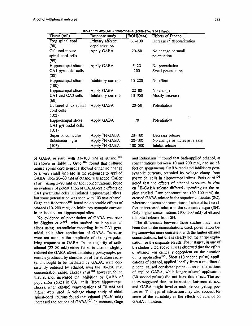
Table 1: In vitro GABA transmission (acute effects of ethanol). Tissue (ref.) Response study [EtOH](mM) Effects of Ethanol Frog spinal cord Primary afferent 33-100 Increase in depolarization (98) Cultured mouse spinal cord cells (9% Hippocampal slices CA1 pyrimidal cells (58) Hippocampal slices (100) Hippocampal slices CA1 and CA3 cells (60) Cultured chick spinal cord cells (102) Hippocampal slices CA1 pyrimidal cells (101) Superior colliculus Substantia nigra
depolarization Apply GABA
Apply GABA
Inhibitory currents
Apply GABA Inhibitory currents
Apply GABA
Apply GABA
Apply 3H-GABA Apply 3H-GABA
20-80
5-20 100
IO-200
22-88 10-350
20-50
70
20-100 20-100
No change or small potentiation
No potentiation Small potentiation
No effect
No change Mostly decrease
Potentiation
Potentiation
Decrease release No change or increase release
(103) Apply 3H-GABA loo-500 Inhibit release
of GABA in vitro with 33-100 mM of ethanolto’ as shown in Table 1. Gru01’~~ found that cultured mouse spinal cord neurons showed either no change or a very small increase in the responses to applied GABA when 2O80 mM of ethanol was added. Carlen et a16’ using 5-20 mM ethanol concentrations, found no evidence of potentiation of GABA-ergic effects on CA1 pyramidal cells in isolated hippocampal slices, but some potentiation was seen with 100 mM ethanol. Gage and Robertson lo3 found no detectable effects of ethanol (lo-200 mM) on inhibitory synaptic currents in an isolated rat hippocampal slice.
No evidence of potentiation of GABA was seen by Siggins et a163, who studied rat hippocampal slices using intracellular recording from CA1 pyra- midal cells after application of GABA. Increases were not seen in the amplitude of the hyperpolar- izing responses to GABA. In the majority of cells, ethanol (22-80 mM) either failed to alter or slightly reduced the GABA effect. Inhibitory postsynaptic po- tentials produced by stimulation of the stratum radia- turn, thought to be mediated by GABA, were con- sistently reduced by ethanol, over the 10-350 mM concentration range. Takada et al ‘04 however, found that ethanol increased the inhibition by GABA of population spikes in CA1 cells (from hippocampal slices), when ethanol concentrations of 70 mM and higher were used. A voltage clamp study of chick spinal-cord neurons found that ethanol (20-50 mM) increased the actions of GABAto5. In contrast, Gage
and Robertson103 found that bath-applied ethanol, at concentrations between 10 and 200 mM, had no ef- fect on spontaneous GABA-mediated inhibitory post- synaptic currents, recorded by voltage clamp from pyramidal cells in hippocampal slices. Peris et allM noted that the effects of ethanol exposure in vim on 3H-GABA release differed depending on the re- gion studied. Low concentrations (20-100 mM) de- creased GABA release in the superior colliculus (SC), whereas the same concentrations of ethanol had no ef- fect or increased release in the substantia nigra (SN). Only higher concentrations (lOO-500 mM) of ethanol inhibited release from SN.
The differences between these studies may have been due to the concentrations used, potentiation be- ing somewhat more consistent with the higher ethanol concentrations, but this is clearly not the entire expla- nation for the disparate results. For instance, in one of the studies cited above, it was observed that the effect of ethanol was critically dependent on the duration of its applicationro5. Short (10 second pulse) appli- cations of ethanol, applied locally from a multibarrel pipette, caused consistent potentiation of the actions of applied GABA, while longer ethanol application (30 second pulses) did not have this effect. The au- thors suggested that the interaction between ethanol and GABA might involve multiple competing pro- cesses. This type of interaction may also account for some of the variability in the effects of ethanol on GABA inhibition.
264 C.Y.K. Tan & D.F. Weaver
Tissue (ref.) Cerebral cortex
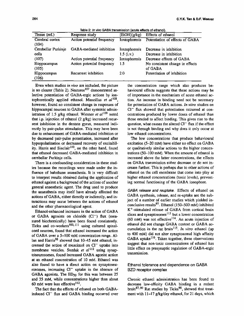
Table 2: In viw GABA transmission (acute effects of ethanol). Response study [EtOH](g/kg) Effects of ethanol Action potential frequency Iontophoresis Potentiation of effects of GABA
ww Cerebelliu Purkinje GABA-mediated inhibition Iontophoresis Decrease in inhibition cells 1.5 (i.v.) Decrease in inhibition (107) Action potential frequency Iontophoresis Decrease effects of GABA Hoppocampus Action potential frequency 1.5 No consistent change in effects (105) of GABA Hippocampus Recurrent inhibition 2.0 Potentiation of inhibition WW
Even when studies in vivu are included, the picture is no clearer (Table 2). Nestoros’07 demonstrated se- lective potent&ion of GABA-ergic actions by ion- tophoretically applied ethanol. Mantillas et al to8, however, found no consistent change in responses of hippocampal neurons to GABA after systemic admin- istration of 1.5 g/kg ethanol. Wiesner et al to9 noted that i.p. injection of ethanol (2 g/kg) increased recur- rent inhibition in the dentate gyrus, measured indi- rectly by pair-pulse stimulation. This may have been due to enhancement of GABA-mediated inhibition or by decreased pair-pulse potentiation, increased after hyperpolarization or decreased recovery of excitabil- ity. Harris and Sinclair”“, on the other hand, found that ethanol decreased GABA-mediated inhibition in cerebellar Purkinje cells.
There is a confounding consideration in these stud- ies because the recordings were made under the in- fluence of halothane anaesthesia. It is very difficult to interpret results obtained during the application of ethanol against a background of the actions of another general anaesthetic agent. The drug used to produce the anaesthesia may itself have already affected the actions of GABA, either directly or indirectly, and in- teractions may occur between the actions of ethanol and the other pharmacological agent.
Ethanol-enhanced increases in the action of GABA or GABA agonists on chloride (Cl-) flux (mea- sured biochemically) have been found consistently. ‘licku and co-workerslOO* ’ 1 ’ using cultured spinal- cord neurons, found that ethanol increased the action of GABA over a 5-100 mM concentration range. Al- lan and Hanisg8 showed that IO-45 mM ethanol, in- creased the action of muscimol on Cl- uptake into membrane vesicles. Suzdak et ul”* using synap- toneurosomes, found increased GABA agonist action at an ethanol concentration of 10 mM. Ethanol was also found to have a direct action on synaptoneu- rosomes, increasing Cl- uptake in the absence of GABA agonists. The EDso for this was between 25 and 35 mM, while concentrations higher than about 60 mM were less effective”*.
The fact that the effects of ethanol on both GABA- induced Cl- flux and GABA binding occurred over
the concentration range which also produces be- havioural effects suggests that these actions may be of importance in the mechanism of acute ethanol ac- tion. An increase in binding need not be necessary for potentiation of GABA actions. In vitro studies on Cl- flux showed that potentiation occurred at con- centrations produced by lower doses of ethanol than those needed to affect binding. This gives rise to the question, what causes the altered Cl- flux if the effect is not through binding and why does it only occur at low ethanol concentrations.
The low concentrations that produce behavioural excitation (5-20 mM) have either no effect on GABA or qualitatively similar actions to the higher concen- trations (50-100 mM). When the amount of ethanol is increased above the latter concentrations, the effects on GABA transmission either decrease or do not in- crease further. This is perhaps due to other actions of ethanol on the cell membrane that come into play at higher ethanol concentrations (toxic levels), prevent- ing normal functioning of the GABA ionophore.
GABA release and reuptake. Effects of ethanol on GABA synthesis, release, and re-uptake are the sub- ject of a number of earlier studies which yielded in- conclusive resultsg7. Ethanol (150-500 mM) inhibited K+-stimulated release of GABA from cortical brain slices and synaptosomes ‘I3 but a lower concentration (60 mM) was not effective’ 14. An acute injection of ethanol did not change GABA content or GABA ac- cumulation in the rat brain’ 15. In vitro ethanol (up to 400 mM) did not alter synaptosomal high affinity GABA uptake’16. Taken together, these observations suggest that non-toxic concentrations of ethanol has little effect on presynaptic regulation of GABA-ergic transmission.
Ethanol tolerance and dependence on GABA BZD receptor complex
Chronic ethanol administration has been found to decrease low-affinity GABA binding in a rodent brain87*90 Rat studies by Tickugo, showed that treat- ment with 1 l-17 g/kg/day ethanol, for 21 days, which

Alcohol withdrawal seizures
was sufficient to cause AWS, decreased the affinity of the low-affinity GABA binding site. This change was not seen after shorter treatment duration, which did not result in behavioural withdrawal, and was found only over the period during which audiogenic seizure activity was seen. Rastogi et al ’ I7 studied the effects of chronic ethanol treatment on GABA stim- ulation of [35S]-TBPS binding, a liquid that binds at the picrotoxin site, and GABA stimulation of [3H]- flunitrazepam binding, and found that these interac- tions were unchanged. These allosteric effects were considered to reflect the coupling between GABA re- ceptors and other sites on the GABA-Cl- ionophore complex’ 17.
Hunt and Dalton* ‘* did not find changes in GABA binding while Harris et al ‘I9 found GABA binding to be increased after chronic ethanol administration. The difference in results may be methodological. The lat- ter study was carried out on brain tissue from minia- ture swine, and there may be species differences. Binding at the BZD receptor is either decreased’20q t*’ or unchanged’*** ‘23 after chronic ethanol treatment. Deitrich et aI*’ reported that GABA agonists and BZD agonists potentiated the effect of ethanol (poten- tiate GABA-ergic responses), whereas BZD antago- nists blocked the potentiating effect of BZD agonists but did not affect the depressant effects of ethanol. These results suggested that ethanol can potentiate GABA-ergic effects but that it does not do so via a BZD-like action on the receptor-channel complex*‘.
Allan and Harris’* found that chronic ethanol treat- ment abolished the potentiation of muscimol-related Cl- uptake into microsacs seen with acute ethanol (10-45 mM) (prepared from mouse cerebellar tis- sue). Morrow et al 124 demonstrated tolerance action of ethanol on Cl- flux into synapneurosomes after chronic treatment with ethanol in viva inhalation for 14 days. In the latter study, potentiation by ethanol of GABA-agonist stimulated Cl- uptake was lost com- pletely after chronic treatment. Takada et al ‘04 stud- ied the effects of ethanol by extracellular recordings on hippocampal slices prepared from rats 4 days af- ter cessation of chronic ethanol treatment (2 g/kg, twice daily for 30 days). These authors104 found that inhibition by exogenous GABA resulted population spikes in the CA1 area of the hippocampal occurred to the same extent after chronic treatment as in con- trol slices. The potentiation of this effect by ethanol at concentrations above 70 mM, seen in control prepa- rations, was lost after chronic treatment. Whittington and Little125 described the patterns of hyperexcitabil- ity that were seen in extracellular recording from iso- lated hippocampal slices, prepared immediately on withdrawal from chronic ethanol treatment. Their re- sults suggested that several different mechanisms may underlie the withdrawal hyperexcitability, including a
decrease in GABA-mediated inhibition produced by down regulation of GABA after chronic ethanol ad- ministration. In addition, chronic ethanol treatment has been reported to decrease the behavioural effects of GABA agonists, suggesting either down-regulation of GABA receptors or alterations in coupling’26*127. However, there are other studies that do not show behavioural adaptations to ethanol.
As mentioned before, GABA agonists, such as muscimol, BZD and THIP, and agents that increase GABA concentrations such as sodium valproate and vigabatrin, have been found to decrease AWS128* I*‘. BZD agonists have been suggested to be the drugs of choice in treating withdrawal in alcoholics, but they do not prevent the dependence, they merely decrease AWS130. In addition, BZDs are anticonvulsants effec- tive against a wide variety of in vitro seizure models (other than those purely due to blockage of GABA transmission), including NMDA’?‘, digoxin13*, pilo- carpine133, and nicotine-induced seizures134. There- fore direct relevance to specific mechanism of AWS is still uncertain.
Chronic ethanol treatment has been reported to de- crease the number and affinity of GABA receptor sites. Can these changes be responsible for the AWS? Careful and detailed studies by Tickugo showed that there was a temporal correlation between binding changes and the appearance of withdrawal behaviour. Decreases in GABA binding can be responsible for tolerance to those actions of ethanol that are pro- duced by potentiation of GABA transmission. How- ever Harris et ~1”’ found no change in the number of GABA receptors after chronic ethanol intake. As mentioned before, the functional importance of the high affinity GABA site is uncertain, as it will be sat- urated at the normal GABA concentrations achieved in the synaptic cleft.
Summary of ethanol-GABA interactions
The hypothesis that ethanol potentiates GABA-ergic transmission is attractive and is quite consistent with many behavioural and neurochemical results. How- ever, there are many electrophysiological observa- tions that do not support this hypothesis. Attempt- ing to resolve the apparent differences between the effects of ethanol on GABA-ergic responses in var- ious preparations and laboratories may be prema- ture, because currently available studies do not clearly show which experimental variables are important and which are not*O. However, several hypotheses can be proposed that may prove important in integrat- ing these findings. First, several studies indicate that only some components of the GABA response are affected. This suggests that with certain methods of
C.Y.K. Tan 81 D.F. Weaver
ethanol application, differences will not be observed because the transient component cannot be easily de- tected. Second, it is possible that the effect of ethanol is region specific within the brain.
Another difference may relate to recent findings that there are multiple forms of the subunits for the re- cepto?** 135, which underlie the heterogeneity among the GABAA receptor subtypes. In addition, it is possi- ble that these subunits may have different sensitivities to the effects of ethanol, whilst some subunit(s) might not even be affected by ethanol. For example, Waf- ford and Whiting136 find that the y2L subunit of the GABAA receptor is minimally present in hippocam- pal membranes. This then raises the possibility that the y2L subunit may play an important role in AWS, whereas other subunits present in hippocampal mem- branes are not sensitive to ethanol. Further investiga- tions on the homogeneous y2L subunit will support this hypothesis. Unfortunately, no such investigation has been reported so far.
In general, brain regions in which GABA modu- lation by ethanol is typically not observed, such as the hippocampus, may have a receptor variant that is not sensitive to ethanol, whereas a receptor subtype that is affected will be expected in regions such as the cortex and possibly cerebellum, in which mod- ulation is observed. This type of heterogeneity may exist even among cells from the same brain area. For example, using cultured spinal cord neurons, Study and Barker’37 found that, although all of the cells tested respond to GABA, the GABA response was potentiated by diazepam in 82% of the cells, by pen- tobarbital in 85% of the cells, and by both drugs in only 53% of the cells.
In summary, both behavioural and neurochemical results support the MLPI mechanism which acute ad- ministration of ethanol potentiates GABA-ergic trans- mission. After chronic administration of ethanol, sub- sequent adaptation by down regulating the GABA- ergic transmission occurs. A sudden withdrawal of ethanol then leads to hyperexcitation, and results in AWS.
EFFECTS OF ETHANOL ON GLUTAMATE RECEPTORS
Glutamate (Glu) is one of the primary neurotrans- mitters mediating excitatory neurotransmission in the CNS by means of ligand-gated cation channels’. Ma- jor excitatory neuronal systems in the brain, including the cortical pyramidal cells, primary sensory affer- ems, cerebellar granule cells, and the ascending exci- tatory pathways, use Glu or related excitatory amino acids as their neurotransmitter138.
Glutamate receptors are widely distributed
throughout the CNS. There are four subtypes of Glu receptors, designed as NMDA, kainate, cr-amino-3-hydroxy-5-methyl-isoxazole-4-propionic acid (AMPA), and quisqualate’38. These can be clas- sified into two major families on the basis of their mechanisms of transduction: (1) ionotropic recep- tors, which are coupled to a cation ion channel, and (2) metabotropic receptors, which are coupled to inositol 1,4,5triphosphate turnover mediated by G protein. The Glu-gated ionophores are the receptor family that show the best correlation with the diverse ethanol effects on the CNS. These ion channels can be further subclassified, according to their respec- tive high affinity ligands, into NMDA, kainate, and AMPA subtypes.
The NMDA receptor is activated by Glu or NMDA, and channel opening mediates an increase in perme- ability to Na+, K+, and Ca*+. The activity of the NMDA receptor complex is subjected to modula- tion by a number of agents, including Mg2+, which causes a direct voltage-dependent blockage of the ion channel, Zn2+, which decreases channel function in a voltage-independent manner at a site peripheral to the channel, and glycine (Gly), which modulates NMDA receptor activity allosterically through a strychnine- insensitive Gly recognition site and has been sug- gested as a co-agonist 139 Within the channel there is . also a site that binds phencyclidine and MK-801 to produce a non-competitive inhibition140. Additional modulation is produced by a site that mediates the NMDA-enhancing effects of the polyamines14’. The NMDA receptors also play a major role in synap- tic plasticity (e.g. long-term potentiation) and neuro- toxicity (e.g. seizures and ischemic damage to neu- rons)142. It is the influx of Ca2+ which is believed to contribute to the involvement of the NMDA re- ceptor to synaptic plasticity and development. How- ever, when the receptor is excessively stimulated, the influx of Ca2+ can lead to seizure activity and cell death14’. There is evidence that alcohol affects glu- tamatergic transmission in three ways: by interfering with fast excitatory neurotransmission, by promoting excitatoxicity (persistent or excessive activation of Glu receptors), and by impairing neurodevelopment’.
Neurochemical effects of ethanol on glutamatergic transmission
During acute exposure, ethanol produces diverse ef- fects on neurotransmission. Ethanol inhibits the re- lease of serotonin, dopamine, norepinephrine, Glu, Asp, and GABA 143. These effects are suggested to be indirectly mediated through the NMDA recep- tor’. Ethanol acts like an NMDA antagonist, inhibit- ing NMDA-stimulated Ca influxlti. Gulya et al 145
Alcohol withdrawal seizures 267
found that 100 mM of ethanol inhibited the non- equilibrium binding of [3H]MK-801, which is an in- dex of NMDA receptor-channel opening. This inhi- bition can be reversed in a time- and concentration- dependent manner by the addition of Gly. In addi- tion, Gonzales and Woodward found that the in- hibition was also brain regional-dependent. In sub- sequent studies, Woodward et a114’, using rat stri- atal slices, showed that the ability of Gly to an- tagonize the ethanol-induced inhibition of NMDA- stimulated endogenous dopamine release was appar- ent at 25 mM, but not at ethanol concentrations of 50 mM and higher. This result suggested that even within brain regions, ethanol might possess multiple actions that cause a change in NMDA-stimulated pro- cesses.
Acute ethanol administration has no effect on Glu release, uptake, or tissue concentration in the fore- brain. However, it causes a significant decrease in Asp and Glu concentrations in midbrain and brain- stem, and decreases Glu tissue concentration (but not release) in the hippocampust4*, in agreement with the notion of regional brain dependence. Iorio et a114’ found that tolerance to some of these effects do not appear to develop, as there is no change in the ability of ethanol to inhibit the NMDA response in cerebel- lar granule cells after chronic exposure of neurons to ethanol.
When experimental animals were subjected to chronic ethanol administration, Michaelis et al Iso found that the number of Glu receptor binding sites increased in synaptosomal membranes. Gulya et al4 and Grant et a115’ found that chronic ingestion of ethanol (7% ethanol for 7 days) produced increases in MK-801 binding sites by approximately 40-50%, a marker for NMDA receptors, in cerebral cortex, shiatum, thalamus, and hippocampus. and that these increases lasted between 10 and 24 hours.
The increase in the number of MK-801 binding sites is probably a compensatory mechanism (up- regulation of NMDA receptors) for overcoming the ethanol-mediated inhibition of NMDA/Glu neuro- transmission. This inhibitory effect of ethanol has been observed in cultured cerebellar granule cells’52 and hippocampal cells ’ ’ , as well as in slices of rat cortex’52 and striatum 153. Thus, it is not surprising that NMDA receptors in these brain regions are up- regulated after chronic ethanol ingestion. However, it remains to be determined whether the increase in the number of MK-801 binding sites reflects an increase in function of NMDA receptors.
Several investigators have shown that both con- trol and ethanol-treated cells are protected against neurotoxicity if the cells are exposed to Glu in the presence of high concentrations of the NMDA recep- tor antagonists CGS-1975514t (a competitive antag-
onist of Glu), 5,7-dichlorokynurenate*4* (a Gly-site antagonist) or ADCI15’ (an ion channel antagonist). Snell et al ‘54 reported that chronic ethanol exposure did not affect the apparent affinities and densities of NMDA-specific [3H]Glu, [3H]CGS-19755, [3H]Gly or [3H]MK-801 binding sites. This result suggests that the inhibition of function is not mediated by di- rect competition of ethanol with ligand binding at any of these sites, which further suggests a specific modu- lation of ethanol on the NMDA channel ionophore in- stead. McGowan etaf I48 found that intracellular Ca*+ concentration is significantly enhanced after chronic in vitro exposure to ethanol. The enhancement is con- sistent with an increased number of NMDA receptors, through which Ca*+ can enter neurons. The authors further reported that during chronic dependent states, Glu release is decreased; conversely, uptake and tis- sue concentration of Glu are increased. However, dur- ing the ethanol withdrawal state, Glu synaptic release, uptake, and tissue concentration are increased. Re- gional variations were observed, by Keller et a1155, with regard to these effects and the degree of effects.
In addition, Hoffman170 reports that acute (2 hour, 200 PM) or chronic (3 days, 50 PM) exposure of cerebellar granule cells to ganglioside GMt pro- tects control and ethanol-treated cells against Glu neurotoxicity. However, while the acute ganglioside GM1 treatment does not interfere with initial re- sponse to Glu (increase in intracellular Ca*+), this response is ‘down-regulated’ after chronic ganglio- side GM1 treatment. Furthermore, chronic ganglio- side GM1 treatment during ethanol exposure has the potential to prevent the ethanol-induced up-regulation of NMDA receptors that underlies AWS and the in- creased susceptibility to excitotoxicity, which further prevent withdrawal-induced neuronal damage”‘.
Electrophysiological effects of ethanol on glutamatergic transmission
A predominant electrophysiological effect of ethanol is to reduce excitatory glutamatergic synaptic trans- mission156. Lovinger ef al ’ found that ethanol in- hibits the electrical current generated by NMDA re- ceptor activation, and that this inhibition is concentra- tion dependent. Concentrations of ethanol associated with intoxication in vivo correlate with those caus- ing inhibition of NMDA currents. Simson et a115’ reported that ethanol produces a dose-dependent in- hibition of the ability of NMDA to activate medial septal neurons in vivo. In addition, ethanol also po- tently inhibits NMDA-evoked neuronal activity in a current-dependent manner in the inferior colliculus and hippocampus. Clearly, the ability of ethanol to in- hibit NMDA-evoked neuronal activity varies region-
C.Y.K. Tan & D.F. Weaver
ally. For example, Simson et al ‘58 found that ethanol fails to inhibit NMDA-evoked activity in the lateral septum. The complex regulatory responses of sev- eral NMDA subunits may explain the variation in the sensitivity of different neurons to ethanol as with GABA subunits to ethanol, or may be due to region- specific differences in inhibition. Molecular cloning of Glu receptor subunits and the functional expres- sion of different combinations of Glu receptor sub- units indicated that the NMDARl and NMDAR2C channels are slightly less sensitive to ethanol inhi- bition than the other heterometric channels’5g. Sim- ilarly, Lovinger160 discovered that some forms of non-NMDA ionotropic Glu receptors have relatively high ethanol sensitivity, which appears to differ from that of endogenous AMPA or kainate receptors. In addition, Ishii et ~1’~’ reported that the NMDARI mRNA is expressed throughout diverse brain regions, whereas NMDAR2 mRNA is distinctly distributed in different brain regions.
Pathophysiological studies
Lustig et al 16* demonstrated that acute ethanol ad- ministration protects against Glu-induced excitotoxic degeneration of cortical neurons. Acutely, neuropro- tection by ethanol is accompanied by a decrease in the NMDA-evoked elevation of free intracellu- lar Ca*+. Because of its effect in attenuating glu- tamatergic transmission, acute ethanol administra- tion protects against NMDA-induced convulsions. Also, MK-801 and ethanol have synergistic anticon- vulsant effects163. However, chronic attenuation of Glu neurotransmission by ethanol results in com- pensatory up-regulation of NMDA receptors. As a consequence, ethanol withdrawal should be expected to be associated with increased excitatory neuro- transmission. Consistent with this deduction, a non- selective Glu antagonist, glutamic acid diethyl ester, attenuates the seizures associated with withdrawal of ethanol after chronic ingestionl”. Several investiga- tors’54* ‘63 found that NMDA exacerbates, while MK- 801 and other NMDA receptor antagonists decrease, the occurrence and severity of AWS. During with- drawal, Valverius ef al ‘65 reported that mice have 56-7096 more MK-801 binding sites. Ethanol-naive mice prone to withdrawal seizures have been found to have more MK-801 binding sites in the hippocampus.
Chronic exposure to ethanol can sensitize neurons to the excitotoxic effect of NMDA receptor activa- tion because of the up-regulation of the NMDA re- ceptors166* 16’. Of 11 studies addressing potential al- terations in NMDA-receptor-mediated neurotransmis- sion with ethanol dependence, 10 studies of rodents indicated that long-term exposure to ethanol causes
up-regulation of NMDA receptor density or its Ca*+- mediated events’49.151.154.165-167
Excitatory amino acids, including Glu and Asp, exert trophic influences on neuronal differentiation and are thus important for neurodevelopment’68. They regulate the formation of neuronal circuitry and synaptic plasticity. Activation of NMDA receptors enhances synaptogenesis in the hippocampus’6g. Glu exposure accelerates the maturation of synaptic pro- files. Exposure to excitatory amino-acid receptor an- tagonists may disrupt the maturational processes me- diated by excitatory amino acids and interfere with normal neurodevelopment.
Summary of ethanol-glutamate interactions
Overall, the results indicate a significant role for in- creased NMDA receptor function in both AWS and in the neuronal damage that is associated with chronic ethanol exposure and withdrawal. The increase in NMDA receptor function observed in ethanol-treated animals and cells may represent an adaptive response to the initial inhibition of NMDA receptor function by ethanol. The presence of ethanol in the cells is expected to protect the cells from the consequences of NMDA receptor up-regulation, which will appear after chronic treatment of ethanol, and result in AWS after ethanol is eliminated or removed. This further supports the MLPI mechanism in which acute ethanol treatment alters neuronal membrane lipids, resulting in an inhibition of glutamatergic transmission. Sub- sequent adaptation of this system (by up-regulation of NMDA receptors) occur after chronic treatment of ethanol. A sudden withdrawal of ethanol then causes hyperexcitation which leads to AWS.
As for neuronal damage, it is likely that the re- peated up-regulation of NMDA receptors is a key fac- tor. However, the question of how acute ethanol ex- posure results in decreased NMDA-stimulated Ca*+ influx which leads to an adaptive response still re- mains unanswered. Various reports support the con- tention that ethanol withdrawal hyperexcitability can be treated with drugs that are antagonists at the NMDA receptor. In addition, chronic ganglioside GM1 treatment during ethanol exposure has the po- tential to prevent the ethanol-induced up-regulation of NMDA receptors and thus AWS, which further prevents withdrawal-induced neuronal damage.
CONCLUSION
Ethanol produces alterations in neuronal membrane function, ranging from the activities of membrane- bound enzymes to the activities of ion channels and
Alcohol withdrawal seizures
neurotransmitter receptors. However, the extent to which these alterations affect membrane properties remains incompletely elucidated. More recently, it has been noted that certain membrane-bound pro- teins, such as GABAA receptors and NMDA recep- tors, are affected by low concentrations of ethanol (less than 50 mM), whereas other membrane-bound proteins, such as voltage-dependent Ca*+ channels, are affected only at higher concentrations of ethanol. These differential responses to low and high concen- trations of ethanol do not necessarily preclude mem- brane fluidization as the underlying mechanism. The bilayers of biomembranes consist of heterogeneous types of phospholipids, and different phospholipids have different sensitivities to the fluidizing effects of ethanol. Therefore, it is possible that the respon- siveness of a membrane-bound protein to ethanol de- pends upon the type of lipids present in the membrane microenvironment.
Studies have shown that membrane fluidity is un- likely to affect the activity of certain membrane- bound proteins directly, in particular those that are subjected to complex biochemical regulation, such as receptor gated ion channels. Rather, the alteration of membrane fluidity may perturb regulatory events that in turn affect the activity of these membrane-bound proteins or the membrane surface potential. These ef- fects can then affect the GABAA receptors, NMDA receptors and/or voltage-dependent Ca*+ channels synergistically or in any combination. The effects in- clude an increase of GABAA function, and a decrease of both NMDA action and voltage-dependent Ca*+ channel function, which result in different changes depending on brain region and/or cellular ethanol sen- sitivity. Subsequent adaptation of these cells, results in increased NMDA receptors and voltage-dependent Ca channels (up-regulation), and decreased GABAA function (down- regulation). A sudden withdrawal of ethanol then causes hyperexcitation which results in AWS. This hypothesis (MLPI mechanism) explains why there is not a strong consensus for a single mech- anism among the enormous number of studies that have been performed; none the less, these studies do show that each mechanism plays an important role in AWS.
Nevertheless, this hypothesis, as well as all the studies that have been presented in this review, do not explain how membrane fluidity actually causes changes in GABA and NMDA receptors, and cal- cium channels. As previously noted. ethanol has the potential to disrupt the surface membrane potential. This then raises questions: Can the alteration of sur- face membrane potential affect ion channels (i.e. ion flux)? How significant will changes of ion flux af- fect the activity of these membrane-bound proteins? Is the alteration ion channel specific? To answer these
269
questions, further experimental investigations are re- quired, such as studies on surface membrane po- tentials with voltage-dependent Ca*+ flux current, GABAA-activated Cl- current and ion current acti- vated by NMDA.
ACKNOWLEDGEMENTS
C.T. acknowledges a Natural Sciences and Engineer- ing Research Council (NSERC) of Canada Scholar- ship. D.F.W. acknowledges an Ontario Ministry of Health Career Scientist Award. This work supported in part by operating grants from the Medical Research Council (MRC) of Canada and NSERC.
REFERENCES
1.
2.
3.
4.
5.
6.
7.
8.
9.
10.
Il.
12.
13.
Tsai, G.. Gastfriend. D.R. and Coyle. J.T. The glutamatergic basis of human alcoholism. American Journal of Psychiarry 1995; 152: 332. Mattson, R.H. Aclohol-related seizures. In: Alcohol and Seizures (Eds R.J. Porter, R.H. Mattson, J.A. Cramer and 1. Diamond). Philadelphia, F.A. Davis Company, 1990: pp. 143-147. Victor, M. Alcohol withdrawal seizures: an overview. In: Alcohol and Seizures fE.ds R.J. Porter, R.H. Mattson, J.A. Cramer and 1. Diamond). Philadephia. F.A. Davis Company, 1990: pp. 148-161. Gulya. K., Grant, K.A.. Valverius. P.. Hoffman, P.L. and Tabakoff, B. Brain regional specificity and time-course of changes in the NMDA receptor-ionophore complex during ethanol withdrawal. Brain Research 1991; 547: 129-134. Tabakoff. B. and Rothstein. J.D. Biology of tolerance and dependence. In: Medical and Social Aspects of Alcohol Abuse (Eds B. Tabakoff, P.B. Sutker and C.L. Randall). New York, Plenum Press, 1983. Goldstein, D.B. and Chin, J.H. Interaction of ethanol with biological membrane. Federation Proceedings 198 1; 40: 1073-1076. Ho, C.. Williams, B.W.. Kelly, M.B. and Stubbs. C.D. Chronic ethanol intoxicaiton induces adaptive changes at the membrane protein/lipid interface. Eiochimica et Biophysics Acta 1994; 1189: 135-142. Lovinger. D.M.. White, G. and Weight, F.F. Ethanol inhibits NMDA-activated ion current in hippocampal neurons. Sci- ence 1989; 243: 1721-1724. Aguayo. L.G. Ethanol potentiates the GABAA-activated chloride current in mouse hippocampal and cortical neurons. European Journal of Pharmacology 1990; 187: 127-130. Harris, R.A. and Hood, W.F. Inhibition of synaptosomal calcium uptake by ethanol. Journal of Pharmacology and Erperimenral Therapeutics 1980; 213: 562-568. Grant. K.A.. Valverius. P., Hudspith. M. and Tabakoff. B. Ethanol withdrawal seizures and the NMDA receptor com- plex. European Journal of Pharmacology 1990; 176: 289- 296. Durand. D. and Carlen. P.L. Decreased neuronal inhibition in vitro after long-term administration of ethanol. Science 1984; 224: 1359-1361. Rogers, C.J. and Hunter, B.E. Chronic ethanol treatment reduces inhibition in CA1 of the rat hippocampus. Brain Research Bulletin 1992; 28: 587-592.

270 C.Y.K. Tan & D.F. Weaver
14. Dolin, ST. and Little, H.J. Are changes in neuronal calcium channels involved in e&UIOl tolerance. Journ of~hanna- cology and Experimental Therapeutics 1989; 250: 985-991.
15. Dolin. S.T.. Little, H.J.. Hudspith. M.. Littleton. J. and Pag- onis. C. Increased DHP sensitive calcium channels in rat brain may underly ethanol physical dependence. Neurophar- macofogy 1987; 26: 275-279.
16. Twombley, D.A., Herman. M.D., Kye. C.H. and Narahashi, T. Ethanol effects on two types of voltage-activated cal- cium channels. Journal of Pharmacology and Experimenral Therapeutics 1990; 254: 1029-1037.
17. Johansson. A., Keightley. CA.. Smith, G.A. and Metcalfe, LC. Cholesterol in sarcoplasmic reticulum and the physio- logical significance of membrane fluidity. Biochemical Jour- MI 1981; 196: 505.
18. Mouritsen, O.G. and Bloom, M. Mattress model of lipid pro- tein interactions in membranes. Biophysical Journal 1984; 46: 141.
19. Sperotto, M.M. and Mouritsen, O.G. Dependence of lipid membrane phase transition temperature on the mismatch of protein and lipid hydrophobic thickness. Biophysicul Jour- of 1988; 16: i-10.
20. Deitrich, R.A., Dunwiddie, T.V., Han-is, A.R. and Erwin, V.G. Mechanism of action of ethanol; initial CNS actions. Pharmacological Reviews 1989; 41: 489-537.
21. Harris, R.A.. Burnett, R., McQuilkin, S., Mcclard. A. and Simon, F.R. Effects of ethanol on membrane order: fluores- cence studies. Annals of rhe New York Academy of Sciences 1987; 492: 125.
22. Goldstein, D.B. Ethanol-induced adaptation in biological membranes. In: Alcohol and Seizures (Eds R.J. Porter, R.H. Mattson, J.A. Cramer and 1. Diamond). Philadelphia. F.A. Davis Company, 1990.
23. Chin, J.H. and Goldstein, D.B. Drug tolerance in biomem- branes: a spin label study of the effects of ethanol. Science 1977; 196: 684-685.
24. Hunt, W.A. Alcohol and biological membranes. In: The Guilford Alcohol Studies (Eds H. Blank and D. Goodwin). The Guilford Press, 1985.
25. Goldstein. D.B., Chin, J.H. and Lyon, R.C. Ethanol dis- ordering of spin-labeled mouse brain membranes: correla- tion with genetically determined ethanol sensitivity of mice. Proceedings of rhe National Academy of Science 1982; 79: 4231-4233.
26. Harris, R.A. and Bnmo, P. Effects of ethanol and other intoxicant-anesthetics on voltage-dependent sodium chan- nels of brain synaptosomes. Journal of Pharmacology and Experimental Therapeutics 1985; 232: 401-406.
27. Hitzemann, R.J. and Harris, R.A. Developmental changes in synaptic membrane fluidity: a comparison of 1.6-diphenyl- 1.3,5-hexatriene-DPH and I-[4-trimethylaminophenyl]-6- phenyl-1.3,5-hexatriene TMA-DPH. Brain Research 1984; 14: 113-120.
28. Lyon, R. and Goldstein, D.B. Changes in the synaptic order associated with chronic ethanol treatment in mice. Molecu- lar Pharmacology 1983; 23: 86-91.
29. Harris, R.A., Baxter, D.M.. Mitchell, M.A. and Hitzemann, R.J. Physical properties and lipid composition of brain mem- branes from ethanol tolerant-dependent mice. Molecular Pharmacology 1984; 25: 401-409.
30. Chin, J.H. and Goldstein, D.B. Membrane-disordering ac- tion of ethanol: variation with membrane cholesterol content and depth of the spin label study of the effects of ethanol. Molecular Pharmacology 198 1; 19: 425-43 1.
31. Hitzemann, R.J.. Schueler. NE, Graham, B.C. and Kreish- man, G.P. Ethanol-induced changes in neuronal membrane order: an NMR study. Biochimica et Biophysics Acta 1986; 859: 189-197.
32. Kreishman. G.P., Graham, B.C., Schueler. H. and Hitze- mann. R.J. On the use of NMR to study the mechanism(s)
of ethanol action. Alcohol Drug Research 1985; 86: l-13. 33. Kreishman, G.P.. Graham, B.C. and Hitzemann, R.J. Deter-
mination of ethanol partition coefficients to the interior and the surface of dipalmityl-phosphatidylcholine diposomes us- ing deuterium NMR. Biochemical and Biophysical Research Communicarions 1985; 130: 301-305.
34. Goldstein, D.B. and Zacchelein. R. Time course of func- tional tolerance produced in mice by inhalation of ethanol. Journal of Pharmacology and Experimental Therapeutics 1983; 227: 150-153.
35. Rottenberg. H.. Wong. A. and Rubin. E. Tolerance and cross-tolerance in chronic alcoholics: reduced membrane binding of ethanol and other drugs. Science 1981; 213: 583- 584.
36. Kelly, MS.. Waring, A.J.. Rottenberg, H. and Rubin, E. Effects of chronic ethanol consumption on the partition of lipophilic compounds into erythrocyte membranes. Labora- tory Investigation 1984; 50: 174-183.
37. Chin, J.H. and Goldstein, D.B. Cholesterol blocks the dis- ordering effects of ethanol in biomembranes. Lipidr 1984; 19: 929-935.
38. Droitte. P., L.amboeuf, Y. and Saint-Blanquat. G. Lipid com- position of the synaptosome and erythrocyte membranes during chronic ethanol treatment and withdrawal in the rat. Biochemical Pharamacology 1984; 33: 615-624.
39. Littleton. J.M. and John, G. Synaptosomal membrane lipids of mice during continuous exposure to ethanol. Journal of Pharmacy and Pharmacology 1977; 29: 579-580.
40. Rovinski, B. and Hosein, E.A. Adaptive changes in lipid composition of rat liver plasma membrane during postnatal development following material ethanol ingestion. Biochim- ica et Siophysica Acra 1983; 735: 407-417.
41. Ingram, L.O.. Ley. K.D. and Hoffmann. EM. Drug-induced changes in lipid composition of E. coli and of mammalian cells in culture: ethanol, pentobarbital and chlorpromazine. Life Science 1978; 22: 489.
42. Smith, T.L., Vickers. A.E.. Brendel. K. and Gerhart, M.J. Effects of ethanol diets on cholesterol content and phospho- lipid acyl composition of rat hepatocytes. Lipids 1982; 17: 124-128.
43. Ailing, C.. Becker. W., Jones, A.W. and Anggard, E. Bf- fects of chronic ethanol treatment on lipid composition and postaglandins in rats fed essential fatty acid deficient diets. Alcoholism: Clinical and Experimental Research 1984; 8: 238-242.
44. Smith, T.L. and Gerhart. M.J. Alterations in brain lipid com- position of mice made physically dependent to ethanol. Life Science 1982; 31: 1419-1425.
45. Crews, F.R.. Majchrowicz, E. and Meek, R. Changes in cor- tical synaptosomal plasma membrane fluidity and composi- tion in ethanol-dependent rats. Psychopharmacology 1983; 81: 208-213.
46. Ailing. C., Gustavsson. L., Mansson. J.E. et al. Phos- phatidylethanol formation in rat organs after ethanol treat- ment. Biochimica er Biophysics Acra 1984; 793: 119-122.
47. Omodeo-Sale, F., Lindi. C.. Palestini, P. and Masserini. M. Role of phosphatidylethanol in membranes: effects on membrane fluidity, tolerance to ethanol, and activity of membrane-bound enzymes. Biochemisrry 1991; 30: 2477- 2482.
48. France, R.. Canela, E. and Bozal, J. Heterogeneous localiza- tion of some purine enzymes in subcellular fractions of rat brain and cerebellum. Neurochemical Research 1986; 11: 423-435.
49. Iqbal. 2. and Sze, P.Y. Correlation between [‘=Il calo- modulin binding and lipid fluidity in synaptic plasma mem- branes: effects of ethanol and other short chain alcohols. Molecular Brain Research 1994; 27: 333-336.
50. Little, H.J. Possible mechanisms that may underlie the be- havioural effects of ethanol. Progress in Neurobiology 1991;
Alcohol withdrawal seizures 271
36: 171-194. 51. Nowycky. M.C.. Fox. A.P. and Tsien. R.W. Three types
of neuronai calcium channel with different calcium agonist sensitivity. Nature 1985; 316: 440-443.
52. Akaike. N., Kostyuk. P.G. and Gsipchuk, Y.V. DHP sensi- tive low-threshold calcium channels in isolated rat hypotha- lamic neurons. Journal of Physiology 1989; 412: 181-195.
53. Scott, R.H. and Dolphin, A.C. Activation of a G protein pro- motes agonist responses to calcium channel ligands. Nature 1987; 330: 760-762.
54. Harris. R.A. Ethanol and pentobarbitrd inhibition of intrasy- naptosomal sequestration of calcium. Biochemical Pharma- cology 1981; 30: 3209-3215.
55. Ross, D.H. Chronic ethanol administration inhibits calmodulin-dependent calcium uptake in synaptosomal membranes. Pharmacology, Biochemistry and Behavior 1986; 24: 1659-1664.
56. Shah, J. and Pant, H.C. Spontaneous calcium release in- duced by ethanol in the isolated rat brain microsomes. Brain Research 1988; 474: 94-99.
57. Biaustein, M.P. and Ector, AC. Barbiturate inhibition of calcium uptake by depolarized nerve terminals in vitro. Molecular Pharmacology 1975; 11: 369-378.
58. Friedman, M.B.. Erickson, C.K. and Leslie, SW. Effects of acute and chronic ethanol administration on whole mouse brain synaptosomal calcium influx. Biochemical Pharma- cology 1980; 29: 1903.
59. Stokes, J.A. and Harris, R.A. Alcohols and synaptosomal calcium transpott. Molecular Pharmacology 1982; 99-104.
60. Pozos. R.S. and Oakes. S.G. The effect of ethanol on the electrophysiology of calcium channels. Recent Devel- opments in Alcoholism 1987; 5: 327-345.
61. Carlen. P.L., Gumvich, N. and Durand. D. Ethanol in low doses augments calcium-mediated mechanisms measured intracelllularly in hippocampal neurons. Science 1982; 215: 306309.
62. Lynch, M.A. and Littleton, J.M. Possible association of al- cohol tolerance with increased synaptic calcium sensitivity. Nature 1983; 303: 175.
63. Siggins. G.R.. Pittman, Q.J. and French, E.D. Effects of ethanol on CA1 and CA3 pyramidal cells in hippocam- pal slice preparation: an intracellular study. Brain Research 1987; 414: 22-34.
64. Leslie, S.W.. Barl, E., Chandker. J. and Farrar, R.P. Inhibi- tion of fast and slow phase depolarisation dependent synap- tosomal calcium uptake by ethanol. Journal of Pharmacol- ogy and Experimental Therapeutics 1983; 225: 571-575.
65. Daniell. L.C. and Harris, R.A. Effects of chronic ethanol treatment and selective breeding for hypnotic sensitivity of ethanol on intracellular ionized calcium concentrations in synaptosomes. Alcoholism: Clinical and Experimental Re- search 1988; 12: 179-183
66. Messing, R.O.. Carpenter, C.L., Diamond, I. and Greenberg, D.A. Ethanol regulates calcium channels in clonal neural cells. Proceedings of the National Academy of Science 1986; 83: 6213-6215.
67. Greenberg. D.A., Carpenter, CL. and Messing, R.O. Ethanol induced component of calcium uptake in PC12 cells is sensitive to calcium channel modulating drugs. Brain Re- search 1987; 410: 143-146.
68. Skattebol, A. and Rubin, R.A. Effects of ethanol on cal- cium uptake in synaptosomes and PC12 cells. Biochemical Pharmacology 1987; 36: 2227-2229.
69. Hirming. L.D.. Fox, A.P., McCleskey. E.W. et al. Domi- nant role of N-type calcium channels in evoked release of nonrepinephrine from sympathetic neurons. Science 1988; 239: 57-60.
70. Greenberg, P.A. and Cooper, E.C. Effects of ethanol on [3H]-nitrendipine binding to calcium channels in brain membranes. Alcoholism: Clinical and Experimental Re-
search 1984, 8: 568-571. 71. Harris. R.A., Jones, S.B.. Bruno, P. and Byhind, D.B. Ef-
fects of DHP derivatives and anticonvulsant drugs on [3H]- nitrendipine binding and calcium and sodium fluxes in brain. Biochemical Pharmacology 1985; 34: 2187-2191.
72. Rius. R.A.. Bergamaschi, S., DiFonso. F., Govoni, I., Tra- bucci. M. and Rossi. F. Acute ethanol effects on calcium antagonist binding in rat brain. Brain Research 1987; 402: 359-361.
73. Bean, B.P. Nitrendipine block of cardiac calcium channels: high affinity binding to the inactivated state. Proceedings of the National Academy of Science 1984; 81: 6388-6392.
74. Dolin, S.J., Patch, T., Siarey. R.J., Whittington. M.A. and Little, H.J. Evidence for the involvement of DHP-sensitive calcium channels in ethanol dependence. British Journal of Pharmacology 1988: 95: 877.
75. Guppy, L.J. and Littleton. J.M. Increased [3H]-DHP binding sites in brain, heart and smooth muscle of ethanol dependent rats. British Journal of Pharmacology 1987; 92: 662.
76. Littleton, J.M., Little. H.J. and Whittington, M.A. Effects of DHP calcium channel antagonists in ethanol withdrawal; doses required, stereospecificity and actions of BAY K- 8644. Psychopharmacology 1990; 100: 387-392.
77. Whittington, M.A. and Little, H.J. Nltrendipine prevents the ethanol withdrawal syndrome, when administered chroni- cally with ethanol prior to withdrawal. British Journal of Pharmacology 1988; 94: 885.
78. Wu. P.H.. Fan, T. and Naranjo, CA. Increase in the brain re- gional depolarisation dependent calcium uptake in rats pre- ferring ethanol. Pharmacology, Biochemistry and Behavior 1987; 27: 355-357.
79. Harper, J.C.. Bemnan. C.H. and Littleton. J.M. Genetic up- regulation of calcium channels in a cellular model of ethanol dependence. Neumpharmacology 1989: 28: 1299-1302.
80. Huang. G.J. and McArdle. J.J. Chronic ingestion of ethanol 1 increases the number of calcium channels of hippocampal neurons of long-sleep but short-sleep mice. Brain Research 1993;615: 328-330.
81. Littleton, J.M. Calcium channel activity in alcohol depen- dency and withdrawal seizures. In: Alcohol and Seizures (Eds R.J. Porter, R.H. Mattson, J.A. Cramer and I. Dia- mond). Philadelphia, F.A. Davis Company, 1990: pp. 51- 59.
82. Little, H.J., Dolin. S.J. and Halsey, M.J. Calcium channel antagonists decrease the ethanol withdrawal syndrome. Life Science 1986; 39: 2059-2065.
83. Panxa, G.. Grebb. J.A.. Sanna, E., Wright, A.G. and Han- bauer. L. Evidence for down-regulation of [3H]-nitrendipine recognition sites in mouse brain after long term treatment with nifedipine or verapamil. Neumpharmacology 1985; 24: 1113-1117.
84. Little, H.J. and Dolin, S.J. Lack of tolerance to ethanol after concurrent administration of nitrendipine. British Journal of Pharmacology 1987; 92: 606.
85. Kendall, D.A. and Nahotski, S.R. DHP calcium channel activators and antagonists influence depolarization-evoked inositol phospholipid hydrolysis in brain. European Journal of Pharmacology 1985; 115: 31-36.
86. Dolin. S.J.. Hunter, A.B.. Halsey, M.J. and Little, H.J. Anti- convulsant profile of the DHP calcium channel antagonists, nitrendipine and nimodipine. European Journal of Pharma- cology 1988; 152: 19-27.
87. ‘licku. M.K.. Burch. T.P. and Davis, W.C. The interac- tions of ethanol with the benzodiazepine-GABA receptor- ionophote complex. Pharmacology, Biochemistry and Be- havior 1983; 18: 15-18.
88. Pritchett, D.. Schofield, P.R., Sontheimr, H., Ymer. S.. Ket- termann. S. and Seeburg. P. GABA receptor cDHAs ex- pressed in transfected ceils and studied by patch-clamp and binding assay. Society for Neuroscience Abstract 1988; 14:
272 C.Y.K. Tan & D.F. Weaver
641. 89. Nut, D.J.. Cowen. P.J.. Batts. C.C., Gmhame. D.G. and
Green, A.R. Repeated administration of subconvulsive doses of GABA antagonist drugs I. Effect on seizure thresh- old (kindling). Psychopharmacology 1982; 76: 84-87.
90. Ticku. M.K. The effect of acute and chronic ethanol admin- istration and its withdrawal on GABA receptor in rat brain. British Journal of Pharmacology 1980; 70: 403-410.
91. Davis, W.C. and Ticku, M.K. Ethanol enhances [3H]- diazepam binding of the benzodiazepine-GABA receptor ionophote complex. Molecular Pharmacology 198 I ; 20: 287-294.
92. Allan. A.M. and Harris, R.A. Involvement neuronal chloride channels in ethanol intoxication, tolerance, and dependence. In: Recent I)evelopment in Alcoholism (Ed. M. Galanter). New York, Plenum Publishing Corporation. 1987; 5: 313- 325.
93. Thyagarrjan. R. and Ticku, M.K. The effect of in vitro and in vivo ethanol administration on [35S]-r- butylbicyclophosphorothionate binding in C57 mice. Brain Research Bulletin 1985; 15: 343-345.
94. Liljequist. S.. Culp. S. and Tabakoff. B. Effect of ethanol on the binding of [35S]-r-buty1bicyclophosphorothionate to mouse brain membrane. Life Science 1986; 38: 1931-1939.
95. Sanna, E.. Concas. A., Serra, M. and Biggio, G. In vivo administration of ethanol enhances function of the GABA dependent chloride channel in the rat cerebral cortex. Jour- nal of Neurochemistry 1990; 63: 696-698.
96. Frye, D. GABA changes in alcohol withdrawal. In: AIcohol and Seizures (Eds R.J. Porter, R.H. Mattson, J.A. Cramer and 1. Diamond). Philadelphia, F.A. Davis Company, 1990; pp. 87-102.
97. Hunt, W. The effect of ethanol on GABA-ergic transmis- sion. Neuroscience and Biobehavioral Reviews 1983; 7: 87- 95.
98. Allan, A. and Harris, R. Acute and chronic ethanol treat- ments alter GABA receptor-operated chloride channels. Pharmacology, Biochemistry and Behavior 1987; 27: 665- 670.
99. Suzdak. P.. Schwartz, R.D., Skolnick. P. and Paul. SM. Alcohols stimulate GABA receptor-mediated chloride up- take in brain vesicles: correlation with intoxication potency. Brain Research 1988; 440: 340-345.
100. Ticku, M., Lowrimore, P. and Lehouliier, P. Ethanol en- hances GABA-induced chloride influx in primary spinal cord cultured neurons. Brain Research Bulletin 1986; 17: 123-126.
101. Davidoff. R.A. Alcohol and presynaptic inhibition in an iso- lated spinal cord preparation. Archives of Neurology 1973; 2a60-63.
102. Gruoal. D.L. Ethanol alters synaptic activity in cultured spinal cord neurons. Bruin Research 1982; 243: 25-33.
103. Gage, P.W. and Robertson, B. Prolongation of inhibitory postsynaptic currents by pentobarbitone. halothane and ke- tamine in CA1 pyramidal cells in rat hippocampus. British Journal of Pharmacology 1985; 85: 675-68 1.
104. Takada. R.. Saito. K.. Matsura, H. and Inoki, R. Effect of ethanol on hippocampal receptors in the rat brain. Alco- hlism 1989; 6: 115-119.
105. Celentaoo, J.J.. Gibbs, T.T. and Farb, D.H. Ethanol potenti- ates GABA- and glycine-induced chloride currents in chick spinal cord neurons. Brain Research 1988; 455: 377-380.
106. Peris, J.. Hardee, M.C., Burry, J. and Thompson, M.P. Se- lective changes in GABAergic transmission in substantia nigra and superior colliculus caused by ethanol and ethanol withdrawal. Alcoholism 1992; 16: 311-319.
107. Nestoros. J.N. Ethanol specifically potentiates GABA- mediated neurotransmission in jeline cerrebral cortex. Sci- ence 1980; 209: 709-710.
108. Mantillas, J.R., S&gins, G.R. and Bloom, F.E. Systemic
ethanol: selective enhancement of responses to acetylcholine and somatostatin in hippocampus. Science 1986; 231: l61- 163.
109. Wiesner, J.B.. Henriksen, S.J. and Bloom. F.E. Ethanol en- hances recurrent inhibition in the dentate gyrus of the hip- pocampus. Neuroscience Letter 1987; 79: 169-173.
110. Harris, D.P. and Sinclair, J.G. Ethanol-GABA interactions at the rat Purkinje cell. General Pharmacology 1984; 15: 449454.
I II. Mehta. A.K. and licku. M.K. Ethanol potentiation of GABAergic transmission cultured spinal cord neurons GABAA-gated chloride channels. Journal of Pharmacology and Erperimenlal Therapeutics 1988; 246: 558-564.
112. Sudzak, P.D.. Schwartz, R.D. and Skolnick. P. Ethanol stimulates GABA receptor-mediated chloride transport in rat brain synaptoneurosomes. Proceedings of the National Academy of Science 1986; 83: 4071-4075.
113. Howerton, T.C. and Collins, A.C. Ethanol-induced inhibi- tion of GABA release from LS and SS mouse brain slice. Alcoholism 1984; 1: 471-477.
114. Murphy, J.M., Cunningham, SD. and McBridge. W.J. Ef- fects of 250 mg ethanol on monoamine and amino acid re- lease from rat striatal slices. Brain Research Bulletin 1985; 14: 439-442.
115. Frye, G.D. and Fincher, AS. Effect of ethanol on GABA- induced GABA accumulation in the substatia nigra and on synaptosome GABA content in six rat brain region. Brain Research 1988: 449: 71-79.
116. Mrak, R.E. and North, P.E. Ethanol inhibition of synap- tosomal high affinity choline uptake. European Journal of Pharmacology 1988; 151: 51-58.
117. Rastogi, S.K., Thyagarajan. R.. Clothier, J. and licku. M.K. Distribution and possible metabolic function of class III al- cohol dehydrogenase in the human brain. Neuropharmncol- ogy 1986; 25: 1179-1184.
118. Hunt, W.A. and Dalton, T.K. Neurotransmitter receptor binding in various brain regions in ethanol dependent rats. Pharmacology, Biochemistry and Behavior 1981; 14: 733- 739.
119. Harris, R.A., Fenner. D.. Feller. D. er al. Neurochemical effects of long-term ingestion of ethanol by Sinclair (S-l) swine. Pharmacology, Biochemistry and Behavior 1983; 18: 363-367.
120. Volicer, L. and Biagione. T.M. Effect of ethano1 adminis- tration and withdrawal on benzodiazepine receptor binding in the rat brain. Neuropharmncology 1982; 21: 283-286.
121. Kochman. R.L., Hirch. J.D. and Clay, G.A. Changes in [3H]-diazepam receptor binding after subacute ethanol ad- ministration. Research Communication in Substances of Abuse 1981; 2: 135-144.
122. Schoemaker, H.. Smith, T.L. and Yamamura, H.I. Effects of chronic ethanol consumption on central peripheral type ben- zodiazepine binding sites in the mid brain. Brain Research 1983;258: 347-350.
123. Hemmingsen. R.. Braestmp, C. Nielson, M. and Barry, D.I. The benzadiazepine GABA receptor complex during severe ethanol intoxication and withdrawal in the rat. Acra Psychi- atrica Scandinavica 1982; 65: 120-126.
124. Morrow, A.L.. Suzdak. P.D., Karanian. I.W. and Paul, S.M. Chronic ethanol administration alters GABA. pentobarbital and ethanol mediated chloride uptake in cerebral cortical synaptoneurosomes. Journal of Pharmacology and Experi- mental Therapeuncs 1988; 246: 158-164.
125. Whittington. M.A. and Little, H.J. Patterns of changes in field potentials in the isolated hippocampal slice on with- drawal from chronic ethanol treatment of mice in vivo. Brain Research 1990; 523: 237-244.
126. Tabemer. P.V. and Unwin, J.W. Behavioural effects of mus- cimol, amphetamine and chlorpromazine on ethanol tolerant mice. Brirish Journal of Pharmacology 198 I; 74: 276.

Alcohol withdrawal seizures 273
127. Martz, A., Deitrich, R.A. and Harris, R.A. Behavioural ev- 146. Gonzales. R.A. and Woodward, J.J. Ethanol inhibits NMDA idence for the involvement of GABA in the actions of stimulated [3H)-nompinephrine release from rat cortical ethanol. European Journal of Pharmacology 1983; 89: 53- slices. Journal of Pharmacology and Experimental Thera- 62. peutics 1990; 252: 1138-l 144.
128. Frye, G.D., McGowan, J. and Breese. G.R. Differential sen- sitivity of ethanol withdrawal signs in the rat to GABA- mimetics: blockade of audiogenic seizures but not forelimb tremors. Journal of Pharmacology and Experimental Ther- apeutics 1983; 226: 720-726.
147. Woodward. J.J.. Brown, L. and Gonzales, R.A. Modula- tion of ethanol-induced inhibitor of NMDA stimulated neu- rotransmitter release by glycine. Alcoholism Supplement I 1991; 177-180.
129. Gonzales. L.P. and Hettinger, M.K. Intranigral muscimol suppresses ethanol withdrawal seizures. Brain Research 1984; 298: 163-166.
148. McGowan. T.J.. Frye, G.D. and Breese, G.B. Evidence for site specific ethanol actions in the CNS. Alcohol and Drug Research 1986; 6: 423429.
130. Nutt. D.J.. Adinoff, B. and Linnolia, M. Benzodiazepines in the treatment of alcoholism. Recent Devefopmenrs in Alco- holism 1989; 7: 283-313.
131. Moreau. J.L.. Pieri. L. and Prud’Hon. B. Convulsions in- duced by centrally administered NMDA in mice: effects of NMDA antagonists, benzodiazepine, minor tranquilisera and anticonvulsants. Brirish Journal of Pharmacology 1989; 98: 1050-1054.
149. Iorio. K.R.. Reinlib. L.. Tabakoff. B. and Hoffman, P.L. Chronic exposure to cerebellar granule cells to ethanol results in increased NMDA receptor function. Molecular Pharmacology 1992; 41: 1142-l 148.
150. Michaelis. E.K., Mulvaney. M.J. and Freed, W.J. Effects of acute and chronic ethanol intake on synaptosomal glu- tamate binding activity. Biochemical Pharmacology 1978; 27: 1685-1691.
132. Kulkkarm. S.K. and Mehta, A.K. Possible mechanism of digoxin-induced convulsions. Psychopharmacology 1983; 79: 287-289.
133. Truski, W.A.. Cavalheiro. E.A. and Bortolotto. Z.A. Seizures produced by pilocarpine in mice: a behavioural electroincephalographic and morphological analysis. Brain Research 1984; 321: 237-253.
151. Grant, K.A., Snell. L.D.. Rogawski. M.A.. Thurkauf. A. and Tabakoff. B. Journal of Pharmacology and Experimental Therapeutics 1992; 260: 1017-1022.
152. Hoffman, P.L.. Rabe. C.S.. Moses, F. and Tabakoff. B. NMDA receptors and ethanol: inhibition of calcium flux and cyclic GMP production. Journal of Neurochemistry 1989; 52: 1937-1940.
134. Sofia, R.D.. Solomon. T.A. and Barry, H. Anticonvulsant activity of delta 9-tetrahydrocannabo compared with three other drugs. European Journal of Pharmacology 1976; 7- 16.
153. Gothert, M. and Fink, K. Inhibition of NMDA and L- glutamate-induced and acetylcholine release in the rat brain by ethanol. Naunyn-Schmiedeberg ‘s Archives Pharmacology 1989; 340: 516-521.
135. Pritchett, D.B.. Sontheimer, H.. Shivers, B.D. er al. Im- portance of a novel GABAA receptor subunit for benza- diazepine pharmacology. Nature 1989; 338: 582-585.
154. Snell. L.D.. Tabakoff. B. and Hoffman, P.L. Radioligand binding to the NMDA receptor/ionophore complex: alter- ations bv ethanol in virm and bv chronic in vivo ethanol ingestioi. Brain Research 1993; &Z: 9 l-98.
136.
137.
138.
139.
140.
141.
142.
143.
144.
145.
Waff&d. K.A. and Whittington, P.T. Ethanol potentiation of GABAA receptors requires phosphorylation of the altema- tively spliced variant of the gamma 2 subunit. FEES Lerrers 1992; 313: 113-I 17. Study, R.E. and Barker, J.L. Diazepam and pentobarbitol: fluctuation analysis reveals different mechanisms for po- tentiation of GABA response in cultured central neurons. Proceedings of the National Academy of Science 198 1; 78: 7180-7184. Cotman, C.W. and Monaghan. D.T. Chemistry and anatomy of excitatory amino acid system. In: Psychopharmacology (Ed. H.Y. Meltzer). New York, Raven Press, 1987. Kleckner. N.W. and Dingledine, R. Requirement for glycine in activation of NMDA-receptors expressed in xenopus oocytes. Science 1988; 241: 835-837. Javitt, D.C. and Zukin. S.R. Bioexponential kinetics of [3H] MK-801 binding: evidence for access to closed and open NMDA receptor channels. Molecular Pharmacology 1989; 35: 387-393. Collingridge. G.L. and Lester. R.A. Excitatory amino acid receptors in the vertebrate CNS. Pharmacology Reviews 1989; 40: 143-210. Mayer, M.L. and Westbrook. G.L. The physiology of exci- tatory amino acids in the vertebrate CNS. Progress in Neu- robiology 1987; 28: 197-276. Martin, D. and Swartzwelder. H.S. Ethanol inhibits release of excitatory amino acids from slices of hippocampal area CA 1. European Journal of Pharmacology 1992; 219: 469- 472. Carboni. S., Isola, R., Gessa, G.L. and Rossetti, Z.L. Ethanol prevents the glutamate release induced by NMDA in the rat striatum. Neuroscience Letter 1993; 152: 133-136. Gulya, K., Grant, K.A.. Valverius. P.. Hoffman, P.L. and Tabakoff. B. Brain regional specificity and time course of changes in the NMDA receptor-ionophore complex during ethanol withdrawal. Brain Research 1991; 547: 129-134.
155.
156.
157.
158.
159.
160.
161.
162.
163.
164.
Keller, F.. Cummins. J.T. and Hungen, K.V. Regional ef- fects of ethanol on glutamate level, uptake and release in slice and synaptosome preparations from rat brain. Sub- srance Alcohol Acrions/Uisuse 1983; 4: 383-392. Nie. Z.. Yuan. X.. Madamba. S.G. and S&ins. G.R. Ethanol decrease glutamate@ synaptic transmission in rat nucleus accumbens in vitro: naloxone reversal. Journal of Pharma- cology and Experimental Therapeutics 1993; 266: 1705- 1712. Simson, P.E., Criswell, H.E. and Johnson, K.G. Ethanol in- hibits NMDA-evoked electrophysiological activity in vivo. Journal of Pharmacology and Experimental Therapeutics 1991; 257: 225-231. Simson. P.E.. Criswell, H.E. and Breese, G.R. Inhibition of NMDA evoked electrophysiological activity by ethanol in selected brain regions: evidence for ethanol-sensitive and ethanol-insensitive NMDA-evoked responses. Brain Re- search 1993; 607: 9-16. Kunner, T.. Schoepfer, R. and Korpi, E.R. Ethanol inhibits glutamate-induced currents in herometric NMDA receptors subtypes. NeumReport 1993; 5: 297-300. Lovinger, D.M. High ethanol sensitivity of recombinant AMPA-type glutamate receptors expressed in mammalian cells. Neumscience Letter 1993; 159: 83-87. Ishi. T.. Moriyoshi. K.. Sugihara. H. er al. Molecular char- acterization of the family of the NMDA receptor subunits. Journal of Biology and Chemistry 1993; 268: 2836-2843. Lustig, H.S.. Chan. 1. and Greenberg, D.A. Ethanol inhibits excitotoxicity in cerebral cortical cultures. Neuroscience Letter 1992; 135: 259-261. Sharma. A.C.. Thorat. S.N.. Nayar, U. and Kulkami. S.K. Dizoulpine ketamine and ethanol reverse NMDA-induced BEG changes and convulsions in rats and mice. Indian Jour- nal of Physiology and Pharmacology 1991; 35: 11 l-l 16. Freed, W.J. and Michaelis. E.K. Glutamic acid and ethanol dependence. Pharmacology, Biochemistry and Behavior
274 C.Y.K. Tan 81 D.F. Weaver
1978; 8: 509-514. 165. Valverius. P.. Crabbe, J.C., Hoffman, P.L. and Tabakoff, B.
NMDA receptors in mice bred to be prone or resistant to ethanol withdrawal seizures. Europeun Journ~f ofPharma- cology 1990; 184: 185-189.
166. Chandler, L.J., Newsom, H.. Sumners. C. and Crews. F. Chronic ethanol exposure potentiates NMDA excitotoxic- ity in cerebral cortical neurons. JOWMI of Neurochemistry 1993; 60: 1578-1581.
167. lorio. K.R.. Tabakoff. B. and Hoffman, P.L. Glutamate- induced neurotoxicity is increased in cerebellar granule cells exposed chronically to ethanol. European Journal of Phur- macology 1993; 2r18: 209-212.
168. Savage, D.D.. Monfano, C.Y., Otero, M.A. and Paxton, L.L. Prenatal ethanol exposure decreases hippocampal NMDA- sensitive [3HJ-glutamate binding site density in 45 day old rats. Alcoholism 1991; 8: 193-201.
169. Queen, S.A., Sanchez. CF.. Lopez, S.R.. Paxton. L.L. and Savage, D.D. Dose and age dependent effects of prena- tal ethanol on hippocampal metabotropic-glutame receptor- stimulated phosphoinositide hydrolysis. Alcoholism: Clini- cal and Experimenral Research 1993; 17: 887-893.
170. Hoffman, P.L. Glutamate receptors in alcohol withdrawal- induced neurotoxicity. Metabolic Brain Disease 1995; 10: 73-79.