monocyte migration through the alveolar epithelial barrier
TRANSCRIPT

of April 8, 2018.This information is current as
Mechanisms and Impact of ChemokinesEpithelial Barrier: Adhesion Molecule Monocyte Migration Through the Alveolar
Grimminger, Werner Seeger and Jürgen LohmeyerKatja Leissner, Ulrich Maus, Konstantin Mayer, Friedrich Simone Rosseau, Jochen Selhorst, Kristina Wiechmann,
http://www.jimmunol.org/content/164/1/427doi: 10.4049/jimmunol.164.1.427
2000; 164:427-435; ;J Immunol
Referenceshttp://www.jimmunol.org/content/164/1/427.full#ref-list-1
, 21 of which you can access for free at: cites 57 articlesThis article
average*
4 weeks from acceptance to publicationFast Publication! •
Every submission reviewed by practicing scientistsNo Triage! •
from submission to initial decisionRapid Reviews! 30 days* •
Submit online. ?The JIWhy
Subscriptionhttp://jimmunol.org/subscription
is online at: The Journal of ImmunologyInformation about subscribing to
Permissionshttp://www.aai.org/About/Publications/JI/copyright.htmlSubmit copyright permission requests at:
Email Alertshttp://jimmunol.org/alertsReceive free email-alerts when new articles cite this article. Sign up at:
Print ISSN: 0022-1767 Online ISSN: 1550-6606. Immunologists All rights reserved.Copyright © 2000 by The American Association of1451 Rockville Pike, Suite 650, Rockville, MD 20852The American Association of Immunologists, Inc.,
is published twice each month byThe Journal of Immunology
by guest on April 8, 2018
http://ww
w.jim
munol.org/
Dow
nloaded from
by guest on April 8, 2018
http://ww
w.jim
munol.org/
Dow
nloaded from
Monocyte Migration Through the Alveolar Epithelial Barrier:Adhesion Molecule Mechanisms and Impact of Chemokines1
Simone Rosseau,2 Jochen Selhorst, Kristina Wiechmann, Katja Leissner, Ulrich Maus,Konstantin Mayer, Friedrich Grimminger, Werner Seeger, and Jurgen Lohmeyer
Alveolar monocyte influx requires adherence and transmigration through the vascular endothelium, extracellular matrix, andalveolar epithelium. For investigating the monocyte migratory process across the epithelial barrier, we employed both the A549cell line and isolated human alveolar epithelial cells. Under baseline conditions, spontaneous bidirectional transepithelial monocytemigration was noted, which was dose-dependently increased in the presence of the monocyte chemoattractant protein-1. TNF-astimulation of the alveolar epithelium provoked the polarized apical secretion of monocyte chemoattractant protein-1 andRANTES and up-regulation of ICAM-1 and VCAM-1 expression, accompanied by markedly enhanced transepithelial monocytetraffic in the basal-to-apical direction. Multiple adhesive interactions were noted to contribute to the enhanced monocyte trafficacross the TNF-a-stimulated alveolar epithelium: these included theb2 integrins CD11a, CD11b, CD11c/CD18, theb1 integrinsvery late Ag (VLA)-4, -5, and -6, and the integrin-associated protein CD47 on monocytes, as well as ICAM-1, VCAM-1, CD47, andmatrix components on the epithelial side. In contrast, spontaneous monocyte migration through unstimulated epithelium de-pended predominantly on CD11b/CD18 and CD47, with some additional contribution of VLA-4, -5, and -6. In summary, unliketransendothelial monocyte traffic, for which b1 and b2 integrins are alternative mechanisms, monocyte migration across thealveolar epithelium largely depends on CD11b/CD18 and CD47 but required the additional engagement of theb1 integrins foroptimal migration. In response to inflammatory challenge, the alveolar epithelium orchestrates enhanced monocyte traffic to theapical side by polarized chemokine secretion and up-regulation of ICAM-1 and VCAM-1. The Journal of Immunology,2000, 164:427–435.
T he recruitment of leukocytes is one of the fundamentalmechanisms involved in inflammatory processes, andmonocyte emigration into the alveolar compartment is a
prominent feature of acute and chronic inflammatory lung injury(1–4). Monocytes are potent effector cells that modulate the in-flammatory process through the release of cytokines, growth fac-tors, oxygen radicals, and proteases (5). They were suggested tocontribute to the early events of lung injury in the acute respiratorydistress syndrome but also to the late fibroproliferative phase ofthis prototype inflammatory disease (6, 7). The process of alveolarmonocyte recruitment requires the leukocytes to adhere and to mi-grate through the vascular endothelium, the extracellular matrix ofendothelial and epithelial cells, and finally the alveolar epithelialbarrier. The mechanisms of transendothelial migration have beeninvestigated extensively. They comprise the sequential interactionof monocyte selectins,b2 (CD11/CD18) andb1 integrins (very lateAg (VLA)-4 and VLA-5),3 and platelet-endothelial cell adhesion
molecule-1 (PECAM-1) with endothelial selectins, ICAM-1,VCAM-1, and PECAM-1 (8–10). However, in the pulmonary mi-crocirculation, monocytes may also useb1 and b2 integrin-inde-pendent pathways during emigration from the vasculature (11).
Much less is known about the mechanisms of monocyte migra-tion into the alveolar compartment once the cells have traversedthe vascular endothelium. In a rat model, intratracheal applicationof endotoxin induced a pronounced monocyte influx into the lungs,with the vast majority of monocytes being recovered from the lungparenchyma, whereas only few cells were found in the bronchoal-veolar lavage (11). These findings suggested a differential regula-tion of transendothelial migration into the interstitial space vs em-igration into the alveolar compartment in the course of theinflammatory process. Thus, alveolar epithelial cells might play animportant role in regulating the expansion of the alveolar mono-cyte pool. In this context, it is of major interest that the epithelialcells release monocyte chemotactic activity (12, 13), such as thechemokines monocyte chemoattractant protein-1 (MCP-1) andRANTES, that may be significantly up-regulated in the presence ofproinflammatory cytokines (14).
Epithelium-leukocyte interactions are partly mediated by carbo-hydrates (15, 16), but adhesion of lymphocytes and neutrophilgranulocytes to epithelial ICAM-1 has also been reported (17, 18).ICAM-1 is expressed on type I and type II pneumocytes (19–22)and is up-regulated in the presence of proinflammatory cytokines(23, 24). Other candidates for epithelial-monocyte interaction arethe integrin-associated protein CD47 located at the basolateral sur-face of epithelial cells, which was shown to play an important rolein the migratory process of neutrophils through intestinal epithelialcells (25), and the VLA-4 ligand VCAM-1, which has been de-tected on bronchial (17) and renal epithelium (26, 27). Epithelialcells probably regulate directional leukocyte traffic by differential
Department of Internal Medicine, Justus-Liebig-University, Giessen, Germany
Received for publication July 20, 1999. Accepted for publication October 13, 1999.
The costs of publication of this article were defrayed in part by the payment of pagecharges. This article must therefore be hereby markedadvertisementin accordancewith 18 U.S.C. Section 1734 solely to indicate this fact.1 This work was supported by the Deutsche Forschungsgemeinschaft, Sonderforschungs-bereich 547 “Kardiopulmonales Gefaßystem.”2 Address correspondence and reprint requests to Dr. Simone Rosseau, Department ofInternal Medicine, Justus-Liebig-University, Klinikstrasse 36, 35385 Giessen, Ger-many. E-mail address: [email protected] Abbreviations used in this paper: VLA, very late Ag; PECAM, platelet-endothelialcell adhesion molecule; MCP, monocyte chemoattractant protein; MIP, macrophage-inflammatory protein; HAEpC, human alveolar epithelial cell; TI-HAEpC, type IHAEpC; TII-HAEpC, type II HAEpC; RPMI-FCS, RPMI 1640 containing 10% heat-inactivated FCS.
Copyright © 2000 by The American Association of Immunologists 0022-1767/00/$02.00
by guest on April 8, 2018
http://ww
w.jim
munol.org/
Dow
nloaded from
expression of these adhesion molecules, compartmentalized che-mokine secretion (28, 29), and deposition of extracellular matrixproteins (30, 31), which might modulate leukocyte integrin func-tion, supporting either adhesion or migration of the cells.
In the present study, we analyzed the transmigration process ofmonocytes through the alveolar epithelial cell line A549 and iso-lated human alveolar epithelial cells (HAEpC) with type I pneu-mocyte characteristics. The transepithelial migration was quanti-fied in both the apical-to-basal and the basal-to-apical direction inthe absence or presence of chemoattractants and proinflammatorystimuli. Moreover, the impact of polarized chemokine secretionand the participation of monocyte and epithelial cell adhesion mol-ecules was investigated.
Materials and MethodsMonoclonal Abs
The following adhesion-blocking murine mAbs against human Ags wereused: R1/1 (anti-ICAM-1 (CD54); Bender MedSystems, Vienna, Austria),1G11 (anti-VCAM-1 (CD106); Coulter-Immunotech, Marseille, France),CIKM1 (anti-CD47; PharMingen, San Diego, CA), GI18 (anti-PECAMdomains 11 2 (CD31); generously provided by S. Santoso, Giessen, Ger-many), W6/32 (anti-HLA class I; a gift from A. Ziegler, Berlin, Germany),clone 38 (anti-CD11a; R&D Systems, Wiesbaden, Germany), clone 44(anti-CD11b, R&D Systems), CBR-p150/4G1 (anti-CD11c; Bender Med-Systems), MEM 48 (anti-CD18; R&D Systems), HP2/1 (anti-a4-chain ofVLA-4 integrin (CD49d); Serotec, Oxford, U.K.), SAM-1 (anti-a5-chain ofVLA-5 (CD49e); Serotec), GoH3 (anti-a6-chain of VLA-6 (CD49f);Coulter-Immunotech); Dreg 56 (anti-L-selectin (CD62L); PharMingen).Neutralizing murine Abs against human MCP-1, RANTES and macroph-age-inflammatory protein-1a (MIP-1a) were obtained from R&D Systems.
Culture of A549 cells
A total of 5 3 104 A549 cells (cell line with alveolar type II epithelial cellcharacteristics; CCL 185; American Type Culture Collection, Manassas,VA) were seeded on the upper or lower side of polycarbonate filter inserts(5 mm pore size; diameter, 6.5 mm; Costar, Cambridge, MA) and culturedin complete Ham’s F-12 medium (Life Technologies, Eggenstein, Ger-many) containing 10% FCS (Life Technologies), 4 mML-glutamine (LifeTechnologies), and penicillin-streptomycin-amphotericin B solution (LifeTechnologies). Cells became confluent after 7 days and formed a mono-layer with permeability,2% when tested by125I-HSA (Amersham,Braunschweig, Germany) diffusion as described for endothelial cells (32).
Isolation of HAEpC
Type II HAEpC were isolated as described previously, with some modi-fications (19, 20, 33, 34). Human lung tissue was obtained from lobectomyspecimens distal from tumors (Departments of Pathology and Surgery, Jus-tus-Liebig-University, Giessen, Germany). This was approved by the localethics committee of the Justus-Liebig-University. The lung tissue wasminced and washed extensively in HEPES-buffered saline and was subse-quently digested by the use of Dispase II (2.5 mg/ml; Boehringer Mann-heim, Mannheim, Germany) in the presence of 2 mM calcium and 1.3 mMmagnesium for 60 min at 37°C under continuous rotation. A cell-rich sus-pension was obtained by sequential filtration through sterilized 100-mmpore size, 60-mm pore size, and 20-mm pore size meshes (Millipore, Es-chborn, Germany). Type II pneumocytes were separated by ficoll (FicollPaque, Amersham Pharmacia Biotech, Uppsala, Sweden) density centrif-ugation (12003 g, 15 min, 21°C), followed by depleting the interfacialcells of contaminating leukocytes by anti-CD45 magnetic beads (Coulter-Immunotech). The isolated cells were composed of 88–95% epithelial cells(flow cytometric analysis of the epithelial cell-specific Ag HEA-125; anti-HEA-125; Camon, Wiesbaden, Germany) and 5–12% alveolar macro-phages and lymphocytes (light scatter characteristics and expression ofCD45; HI30; PharMingen). Because intracellular alkaline phosphatase ac-tivity is specific for type II cells in the lung (35, 36), alkaline phosphatasecytochemistry was performed on cytospin preparations and revealed 96–99% type II pneumocytes in the epithelial cell population. The freshlyisolated HAEpC are termed TII-HAEpC (type II HAEpC).
Culture of HAEpC
A total of 5 3 105 human type II pneumocytes were seeded on the upperor lower side of human type IV collagen (Sigma, Munich, Germany)-coat-ed (34) polycarbonate filter inserts (5-mm pore size; diameter, 6.5 mm;
Costar) and cultured in complete Ham’s F-12 medium containingD-valine(Life Technologies) instead ofL-valine to prevent growth of fibroblasts(37). Medium was changed every 2 days, and when the epithelial cellsreached confluence after 6–7 days, the medium was changed to completeHam’s F-12 medium containingL-valine, and cells were cultured for fur-ther 2 days. HAEpC exhibited no or only little proliferation in tissue cultureas confirmed by nuclear Ki-67 staining (,0.5%; Ref. 38), and they pro-gressively lost their type II cell characteristics and underwent differentia-tion into type I alveolar epithelial cells (loss of intracellular alkaline phos-phatase activity; up-regulation of ICAM-1 expression and down-regulationof HLA-DR expression as shown in Fig. 6; Refs. 39–41). Based on thesefindings, HAEpC that were cultured for 7–9 days are termed TI-HAEpC(type I HAEpC). TI-HAEpC formed tight monolayers, with a125I-humanserum albumin permeability, 0.5%.
Monocyte isolation and labeling
Human monocytes (from buffy coats of healthy blood donors, approved bythe local ethics committee) were isolated using a combination of ficolldensity gradient centrifugation (8003 g, 30 min, 21°C) and counterflowcentrifugal elutriation (Beckmann J2-21 M/E centrifuge with JE-B6 elu-triator rotor, standard 5-ml elutriation chamber; Beckman Instruments,Palo Alto, CA). Cell counts were determined by hemocytometer counts oftrypan blue-stained aliquots. The monocyte fraction consisted of 93–97%monocytes, 3–7% lymphocytes, 0–1% granulocytes, and essentially noplatelets, and cell viability always ranged.95% as determined by Pap-penheim-stained cytospin preparations and trypan blue dye exclusion, re-spectively. The isolated monocytes were radiolabeled with 5mCi 111In (10mCi/ml 111InCl; Amersham) tropolon (Fluka, Neu-Ulm, Germany) as pre-viously described (42). Labeled monocytes were suspended at a density of1 3 107 cells/ml in RPMI 1640 (Life Technologies) containing 10% heat-inactivated FCS (RPMI-FCS).
Monocyte migration across A549 and human alveolarepithelium barriers
A549 or TI-HAEpC monolayers on the inserts and the lower compartmentsof the transwell chambers were washed twice with complete HAM’s F12medium and incubated for 24 h in fresh medium or were stimulated for 4,12, or 24 h by the addition of 10 ng/ml TNF-a (sp. act. 1.13 106 U/mgprotein; R&D Systems) to the medium. After this incubation, the filterswere washed trice on the lower and upper surfaces with RPMI-FCS andtransferred to new, clean wells of 24-well low-cluster plates (Costar). Tothese wells, 500ml of RPMI-FCS were added, and before immersion of theinserts, 100 ml of the monocyte suspension containing 13 106
111In-labeled cells were added above the filter units. Monocytes were al-lowed to transmigrate the epithelium barrier for 120 min at 37°C and 5%CO2 in absence or presence of 1, 10, or 100 ng/ml MCP-1 (sp. act. 2.53105 U/mg protein; R&D Systems) in the lower compartment of the trans-well chamber. The migration was stopped by washing the upper compart-ment with RPMI-FCS, and the undersurface was rinsed into the lowercompartment and was swabbed with a cotton swab soaked in ice-cold PBS/EDTA solution (43). The monocytes in the lower compartment were lysedby the addition of 0.5% Triton X-100 (Sigma), and lysed cells combinedwith the respective cotton swab were counted in a gamma counter to de-termine the number of migrated cells. The number of monocytes that mi-grated through the epithelial barrier was expressed as percentage of lysedcells plus swab counts in relation to counts initially added to the uppercompartment.
In some experiments, saturating amounts of neutralizing murine Absagainst human MCP-1, RANTES, or MIP-1a were added to the epithelialcells before the addition of monocytes. To investigate the role of adhesionmolecules in monocyte transepithelial migration,111In-labeled monocyteswere treated with saturating amounts of adhesion blocking mAbs (30–50mg/ml) for 30 min at room temperature and epithelial cells were incubatedwith mAbs (40–80mg/ml) for 60 min at 37°C. FcIgG receptors wereblocked by preincubation with human Igs (10 mg/ml; Octagam; Octa-pharma, Langenfeld, Germany). Monocytes and epithelial cells werewashed twice in RPMI-FCS to remove unbound mAbs before the migra-tion assay.
Monocyte-specific chemokine secretion by alveolar epithelialcells
A total of 53 104 A549 or 53 105 HAEpC were seeded on the upper (forquantification of basolateral chemokine secretion) or the lower side (forquantification of apical chemokine secretion) of polycarbonate filter insertsand cultured for 7 days. Epithelial cell monolayers on the inserts of the
428 MONOCYTE MIGRATION THROUGH ALVEOLAR EPITHELIUM
by guest on April 8, 2018
http://ww
w.jim
munol.org/
Dow
nloaded from
transwell chambers were washed twice with complete Ham’s F-12 me-dium, and 100ml of fresh medium was added to the upper and 500ml tothe lower compartment of the transwell chamber, respectively. Cells wereincubated for 24 h or stimulated for 4, 12, and 24 h by the addition of 10ng/ml TNF-a to the medium. After this incubation, the medium from thelower compartment was collected, centrifuged, and stored at280°C untilchemokine measurement.
The apical and basolateral secretion of the chemokines MCP-1 andRANTES was quantified by ELISA technique. Maxisorp microtiter plates(Nunc, Wiebaden, Germany) were coated overnight at 4°C with polyclonalgoat Abs to human MCP-1 and RANTES (R&D Systems) followed bythree washing steps with PBS containing 0.05% Tween 20 (Sigma). Then,50ml of cell culture samples was dispensed into the wells and incubated for2 h at room temperature. After washing, application of a monoclonalmouse Ab directed against MCP-1 or RANTES (R&D Systems) was fol-lowed by sequential incubation with a biotinylated donkey anti-mouse IgAb (Dianova, Hamburg, Germany), avidin and biotinylated HRP (Di-anova), and the substrate 2,29-azinobis(3-ethylbenzthiazoline-6-sulfonicacid) (Dianova). Serial dilutions of human recombinant MCP-1 orRANTES (R&D Systems) provided a standard curve for each individualELISA.
Expression of adhesion molecules by A549 and HAEpC
A total of 53 105 A549 cells were cultured in six-well tissue culture platesfor 7 days, and cells were washed twice and incubated for 24 h in freshmedium or were stimulated by the addition of 10 ng/ml TNF-a. HAEpCwere cultured for 7 days on type IV collagen-coated six-well tissue cultureplates before TNF-a stimulation. The medium was removed and cells weredetached by a short incubation in trypsin solution (Life Technologies).Immunofluorescence labeling of cultured A549 and TI-HAEpC as well asfreshly isolated TII-HAEpC was performed by incubation with mousemAbs directed against VCAM-1, ICAM-1, MHC class II (HLA-DR; Bec-ton Dickinson, Heidelberg, Germany), CD47, PECAM-1, E-selectin(BBIG-E1 (CD62E); R&D Systems); P-selectin 9E1 (CD62P); R&D Sys-tems), or isotype controls (Dianova), and PE-labeled F(ab9)2 of an anti-mouse Igs Ab (Dianova).
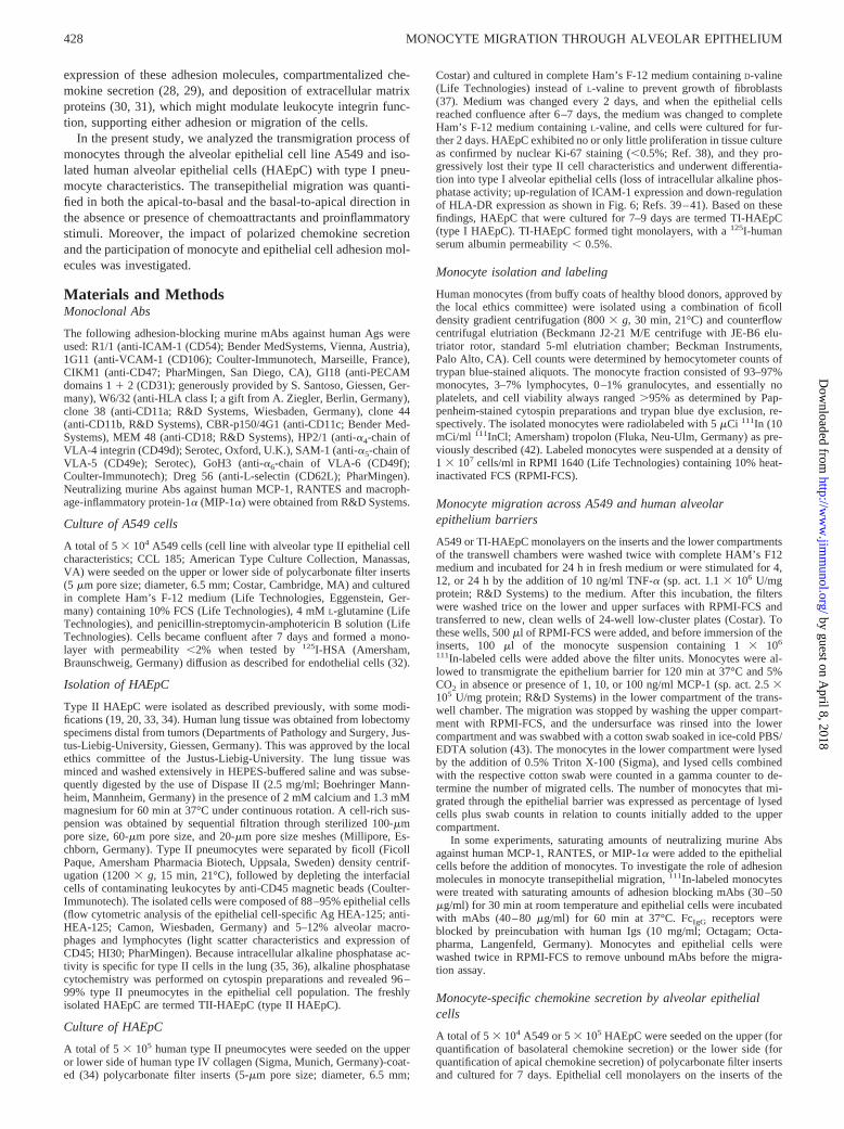
For analysis of VCAM-1 gene expression by TII-HAEpC, TI-HAEpC,as well as TNF-a stimulated HUVEC and TI-HAEpC, total cellular RNAof these cells was isolated using the acid guanidinium thiocyanate-phenol-chloroform method (44). The mRNA was reverse transcribed in a Gene-Amp PCR System 2400 (Perkin-Elmer, Norwalk, CA), and the PCR wasperformed with first-strand DNA using intron-spanning specific primers forb-actin (59-AAAGAACCTGTACGCCAACACAGTGCTGTCT-39, 59-CGTCATACTCCTGCTTGCTGATCCACATCTG-39; Stratagene, Hei-delberg, Germany) and VCAM-1 (59-GAAGATGGTCGTGATCCTTG-39,59-GGACTTCCTGTCTGCATCCT-39; Stratagene). Aliquots of RT-PCRproducts were electrophoresed through 1.8% (w/v) Nusieve/agarose gelsand stained with ethidium bromide for;2 h at 75V.
Statistitcal analysis
Data were expressed as mean6 SEM. For analyzing statistical difference,two-tailed Student’st test for unpaired samples was performed. After Bon-ferroni’s correction, statistically significant differences were defined as val-ues ofp , 0.05.
ResultsMonocyte migration through alveolar epithelial cells
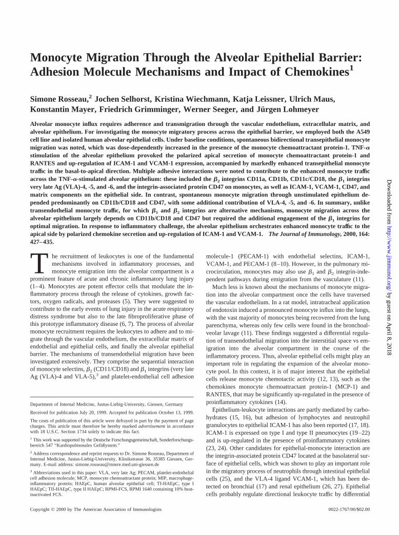
A significant spontaneous migration of monocytes through A549and TI-HAEpC monolayers from the apical to the basal (epitheli-um seeded on the upper side of transwell filter inserts) as well asfrom the basolateral to the apical surface (cells seeded on the lowerside of filter inserts) of epithelial cells was noted. The migratoryresponse was markedly and dose-dependently enhanced in thepresence of MCP-1 admixed beneath the epithelium barrier (Fig.1). Notably, spontaneous transepithelial migration through TI-HA-EpC was significantly higher when monocytes migrated from thebasolateral to the apical compartment of the epithelium, whereasthe enhanced monocyte migration in the presence of MCP-1 dis-played no significant differences when both directions were com-pared (Fig. 1). Stimulation of epithelial monolayers with TNF-atime-dependently up-regulated monocyte transepithelial migration(Fig. 1), with significant predominance of the basal-to-apical di-rection (Fig. 1). Monocyte migration across the epithelial barrier
was confirmed by cell counting in the lower compartments oftranswell chambers and by flow cytometric analysis of migratedleukocytes (data not given in detail).
Spontaneous and TNF-a-induced migration through TI-HAEpCwas significantly inhibited in the presence of neutralizing Absagainst the epithelium-derived monocyte-specific chemokinesMCP-1 and RANTES (Fig. 2), whereas a neutralizing Ab againstMIP-1a had no effect (Fig. 2). Monocyte migration across un-stimulated A549 cells was slightly but not significantly inhibitedby anti-RANTES (14.46 8.6% inhibition; n 5 5), and anti-MCP-1 and anti-MIP-1a had no effect. Like with TI-HAEpC, themigration across TNF-a-stimulated A549 cells was markedly in-hibited by anti-RANTES (38.66 9.1% inhibition; n 5 5, p ,0.01) and anti-MCP-1 (53.56 5.9% inhibition;n 5 5, p , 0.005),and anti-MIP-1a had no effect.
Secretion of chemokines by alveolar epithelial cells
Because larger numbers of monocytes transmigrated the TNF-a-stimulated epithelium in the basal-to-apical compared with the api-cal-to-basal direction, and migration was inhibited in the presenceof neutralizing Abs against MCP-1 and RANTES, we hypothe-sized a polarized secretion of MCP-1 and RANTES by alveolarepithelial cells. Therefore, the release of both chemokines was an-alyzed separately in the apical and the basolateral compartment ofthe epithelial barrier. TI-HAEpC secreted RANTES and MCP-1under baseline conditions (Fig. 2), whereas resting A549 cells re-leased small amounts of RANTES in both directions without sig-nificant difference (apical 656 17 pg/ml, basolateral 436 29pg/ml; p 5 0.18,n 5 5) but no MCP-1 (apical and basolateral,5pg/ml;n 5 5). After stimulation with TNF-a, chemokine secretionwas time-dependently increased in both cell types (shown for HA-EpC in Fig. 2). The release of MCP-1 and RANTES into the apical
FIGURE 1. Left,Dose-dependent influence of MCP-1 on monocyte mi-gration through A549 (upper panel) or TI-HAEpC monolayers (alveolarepithelium;lower panel). Monocytes were allowed to transmigrate the ep-ithelium in the basal-to-apical (M) or the apical-to-basal direction (f) for120 min in absence or presence of 1, 10, or 100 ng/ml MCP-1 beneath theepithelium. Right, Time-dependent influence of TNF-a stimulation ofA549 cells (upper panel) and TI-HAEpC (alveolar epithelium;lowerpanel) on the migratory response of monocytes. A549 and TI-HAEpC weresham-incubated (control) or stimulated by the addition of 10 ng/ml TNF-afor 4, 12, or 24 h. Monocytes were allowed to transmigrate the epitheliumin the basal-to-apical (M) or the apical-to-basal (f) direction for 120 min.All data are presented as percent migrating monocytes of 13 106 mono-cytes added to the transwell filter inserts (mean6 SEM, n 5 10 each;p, p , 0.01 compared with the respective apical-to-basal migratoryresponse).
429The Journal of Immunology
by guest on April 8, 2018
http://ww
w.jim
munol.org/
Dow
nloaded from

compartment of TI-HAEpC markedly surpassed the basolateral se-cretion (Fig. 2) under both baseline (MCP-1; Fig. 2) and inflam-matory conditions (MCP-1 and RANTES; Fig. 2). Within 24 h,TNF-a-stimulated A549 cells secreted 8926 175 pg/ml MCP-1into the apical and 2256 89 pg/ml into the basolateral compart-ment (p , 0.01, n 5 5), and they secreted 5926 108 pg/mlRANTES into the apical and 2366 71 pg/ml into the basolateralcompartment (p , 0.01,n 5 5). In addition, the apical and baso-lateral chemokine secretion of A549 cells seeded on the upper sideof transwell filter inserts was analyzed in the same culture. NativeA549 cells did not secrete MCP-1 (,5 pg,n 5 5) within 24 h, butthey released 416 13 pg RANTES into the apical and 216 12 pginto the basolateral compartment without statistical significant dif-ference (absolute amounts;p 5 0.29, n 5 5). TNF-a-stimulatedA549 cells released 2476 29 pg RANTES into the upper and1186 31 pg RANTES into the lower compartment (p , 0.01,n 5
5), and they secreted 3886 68 pg MCP-1 into the apical and1136 48 pg MCP-1 into the basolateral compartment (p , 0.01,n 5 5).
Expression of adhesion molecules by alveolar epithelial cells
It has been reported previously, that ICAM-1 is constitutively ex-pressed on type II pneumocytes and expression increases upondifferentiation into type I cells (19–21). Indeed, ICAM-1 wasnoted to be expressed at low levels on unstimulated A549 cells andfreshly isolated TII-HAEpC, and expression increased on culturedTI-HAEpC. TNF-a stimulation of A549 cells, and to a minor ex-tent of cultured TI-HAEpC, increased the expression of ICAM-1(Fig. 3). The integrin-associated protein CD47 was expressed onunstimuated and TNF-a-stimulated A549 and TI-HAEpC with nosignificant differences, as well as on freshly isolated type II HA-EpC (Fig. 3). HLA-DR was expressed at high levels on freshlyisolated TII-HAEpC and expression decreased during cell cultureand differentiation into TI-HAEpC, with no difference in absenceor presence of TNF-a (Fig. 3). The expression of VCAM-1 byepithelial cells was reported for bronchial and renal epithelium (17,26, 27), whereas contradictory results were reported for alveolarepithelial cells (19, 20). Our results indicated expression ofVCAM-1 on A549 and cultured TI-HAEpC, but VCAM-1 expres-sion was not detected on freshly isolated TII-HAEpC (Fig. 3).Stimulation of A549 and TI-HAEpC with TNF-a up-regulated theexpression of VCAM-1 by both cell types (Fig. 3). In line withflow cytometric results, RT-PCR revealed basal VCAM-1 geneexpression by cultured TI-HAEpC, which was markedly up-regu-lated by TNF-a (Fig. 4). VCAM-1 gene expression was not de-tected in freshly isolated TII-HAEpC (Fig. 4). Native TII-HAEpCas well as native or TNF-a-stimulated TI-HAEpC and A549 cellsdid not express PECAM-1, E-selectin, or P-selectin.
Role of adhesion molecules in transepithelial migration ofmonocytes
Under baseline conditions, the transepithelial migration of mono-cytes through A549 monolayers from the basolateral to the apical
FIGURE 2. Upper panel,Influence of neutralizing Abs against MCP-1,RANTES, and MIP-1a on monocyte migration through native (baseline;f) or TNF-a-stimulated (M) TI-HAEpC monolayers. TI-HAEpC weresham-incubated or stimulated by the addition of 10 ng/ml TNF-a for 24 h.Monocytes were allowed to transmigrate the epithelium in the presence ofsaturating amounts of neutralizing Abs in the basal-to-apical direction for120 min. Data are presented as percent inhibition of monocyte migration inthe absence of neutralizing Abs calculated from each individual experiment(mean6 SEM). In the absence of neutralizing Abs, baseline migration was23.66 3.2% and TNF-a-induced migration was 42.96 5.2% of 13 106
monocytes added to the transwell filter inserts in these experiments (n 5 8each;p, p , 0.005 compared with migration in the absence of anti-MCP-1or -RANTES). Middle and lower panel, Secretion of MCP-1 (middlepanel) and RANTES (lower panel) into the apical (M) or basolateral (f)compartment of TI-HAEpC. TI-HAEpC on the lower (apical secretion) orupper side (basolateral secretion) of transwell filter inserts were sham-incubated (basal) or stimulated by the addition of 10 ng/ml TNF-a for 4,12, or 24 h. MCP-1 and RANTES secreted into the transwell chamberswere quantified by ELISA technique. Data are presented as mean6 SEM(n 5 6 each;p, p , 0.01 compared with basolateral secretion).
FIGURE 3. Expression of adhesion molecules by A549 cells (native),TII-HAEpC (TII native), and TI-HAEpC (TI native), as well as TNF-a-stimulated A549 cells (TNF-a) and TI-HAEpC (TI TNF-a). A549 cells orHAEpC were sham-incubated or stimulated by the addition of 10 ng/mlTNF-a for 24 h. Immunofluorescence was performed by the use of mouseanti-human ICAM-1, VCAM-1, CD47, HLA DR mAbs (black histo-grams), or isotype controls (gray histograms), and a PE-labeled anti-mouseIg Ab. Cells were analyzed on a FACScan cytometer (Becton Dickinson).Each histogram of this representative experiment represents 10,000 events(ordinate; abscissa5 fluorescence intensity;n 5 5).
430 MONOCYTE MIGRATION THROUGH ALVEOLAR EPITHELIUM
by guest on April 8, 2018
http://ww
w.jim
munol.org/
Dow
nloaded from
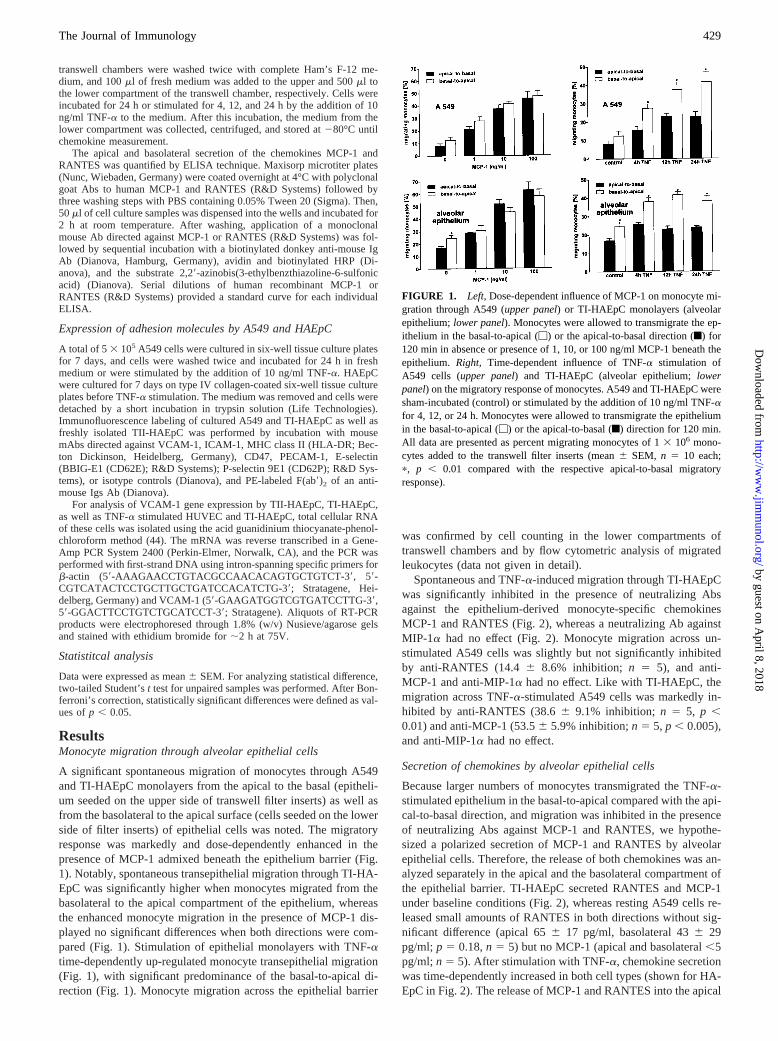
surface depended mainly on theb2 integrin CD11b/CD18 and theintegrin-associated protein CD47, whereas mAbs against CD11aand CD11c only marginally inhibited migration (Fig. 5). CD47 isexpressed on monocytes as well as on epithelial cells (Fig. 4), andboth blocking CD47 on monocytes (M-CD47; Fig. 5) or epithelialcells (Ep-CD47; Fig. 5) virtually completely inhibited the migra-tion response. However, blocking theb2 integrin ligand ICAM-1,which is constitutively expressed on epithelial cells (Fig. 4) did notinhibit monocyte migration (Fig. 5), suggesting the presence ofalternative ligands for CD11b/CD18 on the basolateral surface ofalveolar epithelium. Theb1-integrins VLA-4, -5, and -6 on mono-cytes were also involved in the process of monocyte transepithelialmigration, as suggested by the inhibitory effect of the correspond-ing Abs. Extracellular matrix proteins such as fibronectin and lami-nin, secreted by epithelial cells in culture (30, 31), are known to actas ligands for these monocyteb1 integrins (45). Anti-VLA-4 in-hibited monocyte migration by;26%, and anti-VLA-5 andVLA-6 inhibited migration by;50%. The combination of anti-VLA-4, -5, and -6 resulted in;75% inhibition (Fig. 5). Althoughthe VLA-4 ligand VCAM-1 was expressed by A549 cells, incu-bation of unstimulated A549 with a blocking mAb againstVCAM-1 did not significantly inhibit the transepithelial migrationof monocytes (Fig. 5). Incubation of epithelial cells with a mAbagainst MHC class I also had no effect on monocyte migration.Blocking L-selectin (mAb Dreg 56) or domains 1 and 2 of PE-CAM-1 (mAb GI18), which reportedly mediated monocyte trans-endothelial migration (46), also did not influence the monocytemigration process through the A549 barrier (Fig. 5).
In the presence of 10 ng/ml MCP-1 beneath the A549 barrier,the transmigration process again depended on CD11b/CD18 andCD47, but the contribution of CD11a, CD11c, and VLA-4 wassignificantly increased (Fig. 6). Moreover, blocking ICAM-1 orVCAM-1 under these conditions markedly reduced the transepi-thelial migration of monocytes (Fig. 6). Anti-PECAM-1 and anti-L-selectin did not inhibit monocyte migration in the presence ofMCP-1 (Fig. 6).
TNF-a stimulation of A549 cells further increased ICAM-1 andVCAM-1 dependency of the transepithelial monocyte migration(Fig. 7). In line with this finding, the engagement of the ICAM-1ligand CD11a and the VCAM-1 ligand VLA-4 was also enhanced.Anti-CD11c also markedly inhibited TNF-a-induced monocyte re-cruitment, but the participation of CD11c did not significantly dif-fer from MCP-1-induced monocyte migration through unstimu-lated epithelium. The inhibitory capacity of anti-VLA-6 was alsoincreased when epithelial cells were stimulated with TNF-a,whereas VLA-5-dependent migration was not significantly differ-ent from migration through unstimulated A549 in absence or pres-ence of MCP-1 (Fig. 7). Anti-PECAM-1 and anti-L-selectin didnot inhibit monocyte migration through TNF-a-stimulated epithe-lial cells (Fig. 7).
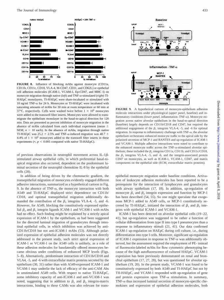
In accordance with the data in A549 cells, the migration ofmonocytes through unstimulated TI-HAEpC was mainly depen-dent on CD11b/CD18 and the integrin-associated protein CD47 onmonocytes and epithelial cells (Fig. 8). However, in contrast toA549 cells, CD11a, CD11c, and VLA-4 on monocytes, as well asICAM-1 and VCAM-1 on TI-HAEpC, were significantly involvedin the spontaneous transmigration process (Fig. 8). Stimulation ofTI-HAEpC with TNF-a further increased the migration depen-dency on monocyte CD11a and VLA-4, and the epithelial ligandsICAM-1 and VCAM-1 (Fig. 8). In line with the findings in A549cells, mAbs directed against PECAM-1 and L-selectin had no ef-fect on the monocyte migratory response (Fig. 8).
DiscussionIn the present study, we investigated monocyte migration acrossthe alveolar epithelium barrier, employing both the A549 cell line
FIGURE 4. Analysis of VCAM-1 gene expression by freshly isolatedTII-HAEpC, cultured TI-HAEpC (TI-HAEpC), as well as TNF-a-stimu-lated TI-HAEpC (TI-HAEpC 4, 12, 24 h TNF) and HUVEC (HUVEC 4 hTNF). A representative experiment is given (n 5 5).
FIGURE 5. Influence of blocking mAbs against monocyte (CD11a,CD11b, CD11c, CD18, VLA-4, VLA-5, VLA-6, M-CD47, CD31, andCD62L) or epithelial cell adhesion molecules (ICAM-1, VCAM-1, Ep-CD47, and MHC I) on monocyte migration through native A549 epithe-lium barriers. Monocytes or A549 cells were incubated with saturatingamounts of mAbs for 30 min at room temperature or 60 min at 37°C,respectively. Cells were washed twice before 13 106 monocytes wereadded to the transwell filter inserts. Monocytes were allowed to transmi-grate the A549 monolayer for 120 min in the basal-to-apical direction. Dataare presented as percent inhibition of monocyte migration in the absence ofmAbs (11.36 1.8% of 13 106 monocytes added to the inserts) calculatedfrom each individual experiment (mean6 SEM; n 5 10 each).
431The Journal of Immunology
by guest on April 8, 2018
http://ww
w.jim
munol.org/
Dow
nloaded from

and isolated HAEpC differentiated to possess type I pneumocytecharacteristics in vitro. Spontaneous bidirectional monocyte mi-gration was noted, which was increased several fold by MCP-1.TNF-a challenge of the alveolar epithelial cells provoked strongpolarized luminal secretion of MCP-1 and RANTES, in compan-ion with markedly enhanced transepithelial monocyte migration inthe basal-to-apical direction. Assessment of epithelial adhesionmolecule expression and employment of various specific Abs sug-gested multiple adhesive interactions to underlie the enhancedtransepithelial monocyte traffic under these conditions. These in-cluded theb2 integrins CD11a, CD11b, CD11c/CD18, theb1 in-tegrins VLA-4, -5, and -6, and the integrin-associated Ag CD47 onthe monocyte side, as well as ICAM-1, VCAM-1, CD47, and pre-sumably matrix components on the epithelial side. Spontaneousmonocyte migration in the absence of inflammatory challenge, incontrast, appears to largely rely on CD11b/CD18 and CD47.
Marked spontaneous monocyte migration from both the baso-lateral to the apical as well from the apical to the basal compart-ment of the epithelium was noted in the absence of exogenouschemokine admixture. This observation is well in line with the factthat monocytes as the precursor cells of alveolar macrophages en-ter the alveoli under physiological conditions in vivo (47), butalveolar macrophages may also traverse the epithelium from thealveolar to the submucosal side when migrating into pulmonarylymph nodes (48). In the presence of MCP-1, this bidirectionalmonocyte migration was greatly enhanced, which supports the pre-viously established role of this agent as a potent monocyte che-mokine (6, 49, 50).
When mimicking inflammatory conditions by TNF-a stimula-tion of epithelial cells, marked enhancement of transepithelialmonocyte traffic was again noted, but then with predominant mi-gration from the basal to the apical compartment. This observationfurther supports the view that the basolateral and the apical cellmembranes of epithelial cells have different functions (51). Whenanalyzing this directed migration in more detail, it was found to belargely ascribed to polarized chemokine secretion with establish-ment of a chemotactic gradient by the epithelial cells under TNF-achallenge, as has been described for rat alveolar epithelium (52).First, MCP-1 and RANTES were both noted to be preferentiallyreleased from the epithelium into the apical compartment underthese conditions. Second, neutralizing Abs against MCP-1 andRANTES inhibited the TNF-a-induced enhancement of the mi-gratory response to the apical surface. And third, the TI-HAEpC,but not the A549 cells, displayed some polarized MCP-1 secretionalso under baseline conditions, concomitant with the finding ofsome preferential basal-to-apical monocyte migration through thenonstimulated TI-HAEpC but not the A549 barrier. Among thetwo agents shown to be responsible for the currently noted che-motactic gradient, MCP-1 may thus be even more relevant for thedirected transepithelial monocyte traffic than RANTES. It mayalso be more relevant than TGF-b and leukotriene B4, whichdisplayed monocyte chemotactic activity and were constitutivelysecreted by A549 cells (12). The present findings are reminiscent
FIGURE 6. Influence of blocking mAbs against monocyte (CD11a,CD11b, CD11c, CD18, VLA-4, VLA-5, VLA-6, M-CD47, CD31, andCD62L) or epithelial cell adhesion molecules (ICAM-1, VCAM-1, Ep-CD47, and MHC I) on monocyte migration through native A549 cellmonolayers in the presence of 10 ng/ml MCP-1 beneath the epitheliumbarrier. Monocytes or A549 cells were incubated with saturating amountsof mAbs for 30 min at room temperature or 60 min at 37°C, respectively.Cells were washed twice before 13 106 monocytes were added to thetranswell filter inserts. Monocytes were allowed to transmigrate the A549monolayer in the basal-to-apical direction for 120 min. Data are presentedas percent inhibition of monocyte migration in the absence of mAbs (4564.5% of 1 3 106 monocytes added to the inserts) calculated from eachindividual experiment (mean6 SEM;n 5 10 each;p, p , 0.005 comparedwith native A549 cells in Fig. 5).
FIGURE 7. Influence of blocking mAbs against monocyte (CD11a,CD11b, CD11c, CD18, VLA-4, VLA-5, VLA-6, M-CD47, CD31, andCD62L) or epithelial cell adhesion molecules (ICAM-1, VCAM-1, Ep-CD47, and MHC I) on monocyte migration through TNF-a-stimulatedA549 cell monolayers. A549 cells were stimulated with 10 ng/ml TNF-afor 24 h. Monocytes or A549 cells were incubated with saturating amountsof mAbs for 30 min at room temperature or 60 min at 37°C, respectively.Cells were washed twice before 13 106 monocytes were added to thetranswell filter inserts. Monocytes were allowed to transmigrate the A549monolayer in the basal-to-apical direction for 120 min. Data are presentedas percent inhibition of monocyte migration in the absence of mAbs(39.36 2.8% of 13 106 monocytes added to the inserts) calculated fromeach individual experiment (mean6 SEM; n 5 10 each;p, p , 0.002compared with native A549 cells in Fig. 5;C, p , 0.005 compared withnative A549 cells in the presence of MCP-1 in Fig. 6).
432 MONOCYTE MIGRATION THROUGH ALVEOLAR EPITHELIUM
by guest on April 8, 2018
http://ww
w.jim
munol.org/
Dow
nloaded from
of previous observations in neutrophil movement across IL-1b-stimulated airway epithelial cells, in which preferential basal-to-apical migration also occurred, dependent on the predominant lu-minal secretion of the neutrophil chemokine IL-8 by the epithelialcells (28).
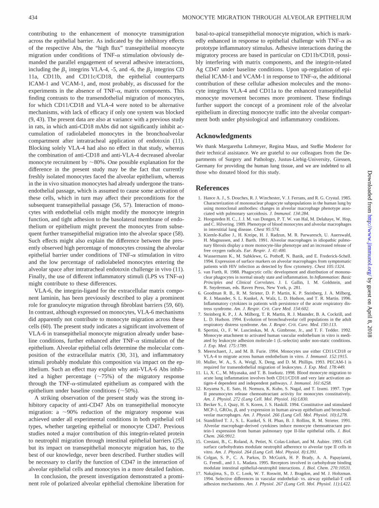
In addition of being driven by the chemotactic gradient, thetransepithelial migration of monocytes evidently engaged differentadhesive interactions, summarized as a hypothetical cartoon in Fig.9. In the absence of TNF-a, the monocyte interaction with bothA549 and TI-HAEpC displayed strict dependence on CD11b/CD18, and optimal transepithelial migration additionally de-manded the contribution of theb1 integrins VLA-4, -5, and -6.However, for A549, blocking the constitutively expressed epithe-lial b2 andb1 integrin ligands ICAM-1 and VCAM-1 with mAbshad no effect. Such finding might be explained by a strictly apicalexpression of ICAM-1 by the epithelium, as had been suggestedfor the directed luminal migration of granulocytes through intes-tinal epithelial cells, in which inhibition was achieved by anti-CD11b/CD18 but not anti-ICAM-1 mAbs (53). Although polar-ized expression of epithelial adhesion molecules was not directlyaddressed in the present study, such strict apical expression ofICAM-1 or VCAM-1 on the A549 cells is unlikely, as a role ofthese adhesion molecules for basolaterally offered monocytes be-came obvious under conditions of enhanced MCP levels (Figs.5–8). Alternatively, predominant interaction of CD11b/CD18 andVLA4, -5, and -6 with extracellular matrix proteins secreted by theepithelium (30, 31) rather than with A549-expressed ICAM-1 andVCAM-1 may underlie the lack of efficacy of the anti-CAM Absin unstimulated A549 cells. With respect to native TI-HAEpC,some inhibitory capacity of anti-ICAM-1 and anti-VCAM-1 wasnoted, suggesting that in addition tob1 and b2 integrin-matrixinteractions, binding to these CAMs was also relevant for trans-
epithelial monocyte migration under baseline conditions. Activa-tion of leukocyte adhesion molecules has been reported to be aprerequisite for the interaction of lymphocytes and granulocyteswith airway epithelium (17, 18). In addition, up-regulation ofmonocyteb1 andb2 integrin expression and function by MCP-1has been described (54, 55). So one might speculate that exoge-nous MCP-1 added to A549 cells, or MCP-1 constitutively se-creted by TI-HAEpC, initiated the interaction ofb1 and b2 inte-grins with epithelial ICAM-1 and VCAM-1.
ICAM-1 has been detected on alveolar epithelial cells (19–22,41), but up-regulation was suggested to be rather a function ofcellular differentiation from type II into type I pneumocytes than aresponse to inflammatory stimuli (21, 41). Our data confirmedICAM-1 up-regulation on HAEpC during cell culture, i.e., duringdifferentiation into type I cells. Moreover, significant up-regulationof ICAM-1 expression in response to TNF-a was additionally ob-served, but the assessment required the employment of PE- insteadof fluorescein-labeled mAbs for flow cytometric phenotyping be-cause of the high autofluorescence of cultured HAEpC. VCAM-1expression has been previously demonstrated on renal and bron-chial epithelium (17, 27, 28), but was questioned for alveolar ep-ithelium (19, 20). In the present study, VCAM-1 was found to beconstitutively expressed by both A549 and TI-HAEpC but not byTII-HAEpC, and VCAM-1 responded with up-regulation of geneand protein expression upon TNF-a stimulation. In summary,TNF-a thus increased luminal secretion of monocyte-specific che-mokines and expression of epithelial adhesion molecules, both
FIGURE 8. Influence of blocking mAbs against monocyte (CD11a,CD11b, CD11c, CD18, VLA-4, M-CD47, CD31, and CD62L) or epithelialcell adhesion molecules (ICAM-1, VCAM-1, Ep-CD47, and MHC I) onmonocyte migration through native (left) and TNF-a-stimulated (right) TI-HAEpC monolayers. TI-HAEpC were sham-incubated or stimulated with10 ng/ml TNF-a for 24 h. Monocytes or TI-HAEpC were incubated withsaturating amounts of mAbs for 30 min at room temperature or 60 min at37°C, respectively. Cells were washed twice before 13 106 monocyteswere added to the transwell filter inserts. Monocytes were allowed to trans-migrate the epithelium monolayer in the basal-to-apical direction for 120min. Data are presented as percent inhibition of monocyte migration in theabsence of mAbs calculated from each individual experiment (mean6SEM; n 5 10 each). In the absence of mAbs, migration through nativeTI-HAEpC was 25.26 2.5% and TNF-a-induced migration was 40.764.4% of 13 106 monocytes added to the transwell filter inserts in theseexperiments (p,p , 0.005 compared with native TI-HAEpC).
FIGURE 9. A hypothetical cartoon of monocyte-epithelium adhesionmolecule interactions under physiological (upper panel,baseline) and in-flammatory conditions (lower panel,inflammation–TNF-a). Monocyte mi-gration across native alveolar epithelium in the basal-to-apical direction(baseline) largely depends on CD11b/CD18 and CD47, but required theadditional engagement of theb1 integrins VLA-4, -5, and -6 for optimalmigration. In response to inflammatory challenge with TNF-a, the alveolarepithelium orchestrates enhanced monocyte traffic to the apical side by thepolarized secretion of MCP-1 and RANTES and up-regulation of ICAM-1and VCAM-1. Multiple adhesive interactions were noted to contribute tothe enhanced monocyte traffic across the TNF-a-stimulated alveolar epi-thelium; these included theb2 integrins CD11a, CD11b, and CD11c/CD18,the b1 integrins VLA-4, -5, and -6, and the integrin-associated proteinCD47 on monocytes, as well as ICAM-1, VCAM-1, CD47, and matrixcomponents on the epithelial side (ECM, extracellular matrix proteins).
433The Journal of Immunology
by guest on April 8, 2018
http://ww
w.jim
munol.org/
Dow
nloaded from
contributing to the enhancement of monocyte transmigrationacross the epithelial barrier. As indicated by the inhibitory effectsof the respective Abs, the “high flux” transepithelial monocytemigration under conditions of TNF-a stimulation obviously de-manded the parallel engagement of several adhesive interactions,including theb1 integrins VLA-4, -5, and -6, theb2 integrins CD11a, CD11b, and CD11c/CD18, the epithelial counterpartsICAM-1 and VCAM-1, and, most probably, as discussed for theexperiments in the absence of TNF-a, matrix components. Thisfinding contrasts to the transendothelial migration of monocytes,for which CD11/CD18 and VLA-4 were noted to be alternativemechanisms, with lack of efficacy if only one system was blocked(9, 43). The present data are also at variance with a previous studyin rats, in which anti-CD18 mAbs did not significantly inhibit ac-cumulation of radiolabeled monocytes in the bronchoalveolarcompartment after intratracheal application of endotoxin (11).Blocking solely VLA-4 had also no effect in that study, whereasthe combination of anti-CD18 and anti-VLA-4 decreased alveolarmonocyte recruitment by;80%. One possible explanation for thedifference in the present study may be the fact that currentlyfreshly isolated monocytes faced the alveolar epithelium, whereasin the in vivo situation monocytes had already undergone the trans-endothelial passage, which is assumed to cause some activation ofthese cells, which in turn may affect their preconditions for thesubsequent transepithelial passage (56, 57). Interaction of mono-cytes with endothelial cells might modify the monocyte integrinfunction, and tight adhesion to the basolateral membrane of endo-thelium or epithelium might prevent the monocytes from subse-quent further transepithelial migration into the alveolar space (58).Such effects might also explain the difference between the pres-ently observed high percentage of monocytes crossing the alveolarepithelial barrier under conditions of TNF-a stimulation in vitroand the low percentage of radiolabeled monocytes entering thealveolar space after intratracheal endotoxin challenge in vivo (11).Finally, the use of different inflammatory stimuli (LPS vs TNF-a)might contribute to these differences.
VLA-6, the integrin-ligand for the extracellular matrix compo-nent laminin, has been previously described to play a prominentrole for granulocyte migration through fibroblast barriers (59, 60).In contrast, although expressed on monocytes, VLA-6 mechanismsdid apparently not contribute to monocyte migration across thesecells (60). The present study indicates a significant involvement ofVLA-6 in transepithelial monocyte migration already under base-line conditions, further enhanced after TNF-a stimulation of theepithelium. Alveolar epithelial cells determine the molecular com-position of the extracellular matrix (30, 31), and inflammatorystimuli probably modulate this composition via impact on the ep-ithelium. Such an effect may explain why anti-VLA-6 Abs inhib-ited a higher percentage (;75%) of the migratory responsethrough the TNF-a-stimulated epithelium as compared with theepithelium under baseline conditions (;50%).
A striking observation of the present study was the strong in-hibitory capacity of anti-CD47 Abs on transepithelial monocytemigration: a ;90% reduction of the migratory response wasachieved under all experimental conditions in both epithelial celltypes, whether targeting epithelial or monocyte CD47. Previousstudies noted a major contribution of this integrin-related proteinto neutrophil migration through intestinal epithelial barriers (25),but its impact on transepithelial monocyte migration has, to thebest of our knowledge, never been described. Further studies willbe necessary to clarify the function of CD47 in the interaction ofalveolar epithelial cells and monocytes in a more detailed fashion.
In conclusion, the present investigation demonstrated a promi-nent role of polarized alveolar epithelial chemokine liberation for
basal-to-apical transepithelial monocyte migration, which is mark-edly enhanced in response to epithelial challenge with TNF-a asprototype inflammatory stimulus. Adhesive interactions during themigratory process are based in particular on CD11b/CD18, possi-bly interfering with matrix components, and the integrin-relatedAg CD47 under baseline conditions. Upon up-regulation of epi-thelial ICAM-1 and VCAM-1 in response to TNF-a, the additionalcontribution of these cellular adhesion molecules and the mono-cyte integrins VLA-4 and CD11a to the enhanced transepithelialmonocyte movement becomes more prominent. These findingsfurther support the concept of a prominent role of the alveolarepithelium in directing monocyte traffic into the alveolar compart-ment both under physiological and inflammatory conditions.
AcknowledgmentsWe thank Margaretha Lohmeyer, Regina Maus, and Steffie Moderer fortheir technical assistance. We are grateful to our colleagues from the De-partments of Surgery and Pathology, Justus-Liebig-University, Giessen,Germany for providing the human lung tissue, and we are indebted to allthose who donated blood for this study.
References1. Hance A. J., S. Douches, R. J. Winchester, V. J. Ferrans, and R. G. Crystal. 1985.
Characterization of mononuclear phagocyte subpopulations in the human lung byusing monoclonal antibodies: changes in alveolar macrophage phenotype asso-ciated with pulmonary sarcoidosis.J. Immunol. 134:284.
2. Hoogsteden H. C., J. J. M. van Dongen, P. T. W. van Hal, M. Delahaye, W. Hop,and C. Hilvering. 1989. Phenotype of blood monocytes and alveolar macrophagesin interstitial lung disease.Chest 95:574.
3. Kiemle-Kallee J., H. Kreipe, H. J. Radzun, M. R. Parwaresch, U. Auerswald,H. Magnussen, and J. Barth. 1991. Alveolar macrophages in idiopathic pulmo-nary fibrosis display a more monocyte-like phenotype and an increased release offree oxygen radicals.Eur. Respir. J. 41:400.
4. Wassermann K., M. Subklewe, G. Pothoff, N. Banik, and E. Frederick-Schell.1994. Expression of surface markers on alveolar macrophages from symptomaticpatients with HIV infection as detected by flow cytometry.Chest 105:1324.
5. van Furth, R. 1988. Phagocytic cells: development and distribution of mononu-clear phagocytes in normal steady state and inflammation. InInflammation: BasicPrinciples and Clinical Correlates. J. I. Gallin, I. M. Goldstein, andR. Snyderman, eds. Raven Press, New York, p. 281.
6. Goodman R. B., R. M. Strieter, D. P. Martin, K. P. Steinberg, J. A. Milberg,R. J. Maunder, S. L. Kunkel, A. Walz, L. D. Hudson, and T. R. Martin. 1996.Inflammatory cytokines in patients with persistence of the acute respiratory dis-tress syndrome.Am. J. Respir. Crit. Care Med. 154:602.
7. Steinberg K. P., J. A. Milberg, T. R. Martin, R. J. Maunder, B. A. Cockrill, andL. D. Hudson. 1994. Evolution of bronchoalveolar cell populations in the adultrespiratory distress syndrome.Am. J. Respir. Crit. Care. Med. 150:113.
8. Spertini, O., F. W. Luscinskas, M. A. Gimbrone, Jr., and T. F. Tedder. 1992.Monocyte attachment to activated human vascular endothelium in vitro is medi-ated by leukocyte adhesion molecule-1 (L-selectin) under non-static conditions.J. Exp. Med. 175:1789.
9. Meerschaert, J., and M. B. Furie. 1994. Monocytes use either CD11/CD18 orVLA-4 to migrate across human endothelium in vitro.J. Immunol. 152:1915.
10. Muller, W. A., S. A. Weigl, X. Deng, and D. M. Phillips. 1993. PECAM-1 isrequired for transendothelial migration of leukocytes.J. Exp. Med. 178:449.
11. Li, X. C., M. Miyasaka, and T. B. Issekutz. 1998. Blood monocyte migration toacute lung inflammation involves both CD11/CD18 and very late activation an-tigen-4 dependent and independent pathways.J. Immunol. 161:6258.
12. Koyama S., E. Sato, H. Nomura, K. Kubo, S. Nagal, and T. Izumi. 1997. TypeII pneumocytes release chemoattractant activity for monocytes constitutively.Am. J. Physiol. 272 (Lung Cell. Mol. Physiol. 16):L830.
13. Becker S., J. Quay, H. S. Koren, J. S. Haskill. 1994. Constitutive and stimulatedMCP-1, GROa, b, andg expression in human airway epithelium and bronchoal-veolar macrophages.Am. J. Physiol. 266 (Lung Cell. Mol. Physiol. 10):L278.
14. Standiford T. J., S. L. Kunkel, S. H. Phan, B. J. Rollins, R. M. Strieter. 1991.Alveolar macrophage-derived cytokines induce monocyte chemoattractant pro-tein-1 expression from human pulmonary type II-like epithelial cells.J. Biol.Chem. 266:9912.
15. Crestani, B., C. Roland, A. Petiet, N. Colas-Linhart, and M. Aubier. 1993. Cellsurface carbohydrates modulate neutrophil adherence to alveolar type II cells invitro. Am. J. Physiol. 264 (Lung Cell. Mol. Physiol. 8):L391.
16. Colgan, S. P., C. A. Parkos, D. McGuirk, H. P. Brady, A. A. Papayianni,G. Frendl., and J. L. Madara. 1995. Receptors involved in carbohydrate bindingmodulate intestinal epithelial-neutrophil interactions.J. Biol. Chem. 270:10531.
17. Nakajima, S., D. C. Look, W. T. Roswitt, M. J. Bragdon, and M. J. Holtzman.1994. Selective differences in vascular endothelial- vs. airway epithelial-T celladhesion mechanisms.Am. J. Physiol. 267 (Lung Cell. Mol. Physiol. 11):L422.
434 MONOCYTE MIGRATION THROUGH ALVEOLAR EPITHELIUM
by guest on April 8, 2018
http://ww
w.jim
munol.org/
Dow
nloaded from
18. Look, D. C., B. T. Keller, S. R. Rapp, and M. J. Holtzman. 1992. Selectiveinduction of intercellular adhesion molecule-1 by interferon-g in human airwayepithelial cells.Am. J. Physiol 263 (Lung Cell. Mol. Physiol. 7):L79.
19. Cunningham, A. C., and J. A. Kirby. 1995. Regulation and function of adhesionmolecule expression by human alveolar epithelial cells.Immunology 86:279.
20. Cunningham, A. C., D. S. Milne, J. Wilkes, J. H. Dark, T. D. Tetley, andJ. A. Kirby. 1994. Constitutive expression of MHC and adhesion molecules byalveolar epithelial cells (type II pneumocytes) isolated from human lung andcomparison with immunocytochemical findings.J. Cell. Sci. 107:443.
21. Barton, W. W., S. Wilcoxen, P. J. Christensen, and R. Paine. 1995. Disparatecytokine regulation of ICAM-1 in rat alveolar epithelial cells and pulmonaryendothelial cells in vitro.Am. J. Physiol. 269 (Lung Cell. Mol. Physiol. 13):L127.
22. Guzman, J., T. Izumi, S. Nagai, and U. Costabel. 1994. ICAM-1 and integrinexpression on isolated human alveolar type II pneumocytes.Eur. Respir. J.7:736.
23. Burke-Gaffney, A., and P. G. Hellewell. 1996. Tumor necrosis factor-a-inducedICAM-1 expression in human vascular endothelial and lung epithelial cells: mod-ulation by tyrosine kinase inhibitors.Br. J. Pharmacol. 119:1149.
24. Burns, A. B., F. Takei, and C. M. Doershuk. 1994. Quantitation of ICAM-1expression in mouse lung during pneumonia.J. Immunol. 153:3189.
25. Parkos, C. A., S. P. Colgan, T. W. Liang, A. Nusrat, A. E. Bacarra, D. K. Carnes,and J. L. Madara. 1996. CD47 mediates post-adhesive events required for neu-trophil migration across polarized intestinal epithelia.J. Cell Biol. 132:437.
26. Garner, C. M., G. M. Richards, D. Adu, A. A. Pall, C. M. Taylor, N. T. Richards,and J. Michael. 1994. Intercellular adhesion molecule-1 (ICAM-1) and vascularcell adhesion molecule-1 (VCAM-1) expression and function on cultured humanglomerular epithelial cells.Clin. Exp. Immunol. 95:322.
27. Oertli, B., B. Beck-Schimmer, X. Fan, and R. P. Wuthrich. 1998. Mechanisms ofhyaluronan-induced up-regulaton of ICAM-1 and VCAM-1 expression by murinekidney tubular epithelial cells: hyaluronan triggers cell adhesion molecule ex-pression through a mechanism involving activation of nuclear factor-kB and ac-tivating protein-1.J. Immunol. 161:3431.
28. Liu, L., F. P. J. Mul, R. Lutter, D. Roos, and E. F. Knol. 1996. Transmigrationof human neutrophils across airway epithelial cell monolayers is preferentially inthe physiologic basolateral-to-apical direction.Am. J. Respir. Cell Mol. Biol.15:771.
29. Carolan, E. J., D. A. Mower, and T. B. Casale. 1997. Cytokine-induced neutro-phil transepithelial migration is dependent upon epithelial orientation.Am. J. Respir. Cell Mol. Biol. 17:727.
30. Dunsmore, S. E., C. Martinez-Williams, R. A. Goodman, and E. Rannels. 1995.Composition of extracellular matrix of type II pulmonary epithelial cells in pri-mary culture.Am. J. Physiol. 269 (Lung Cell. Mol. Physiol. 13):L754.
31. Dunsmore, S. E., C. Martinez-Williams, R. A. Goodman, and E. Rannels. 1995.Turnover of fibronectin and laminin by alveolar epithelial cells.Am. J. Physiol.269 (Lung Cell. Mol. Physiol. 13):L76.
32. Issekutz, T. B., and N. Lopez. 1993. Endotoxin activation of endothelium forpolymorphonuclear leukocyte transendothelial migration and modulation by in-terferon-g. Immunology 79:600.
33. Dobbs, L. G. 1990. Isolation and culture of alveolar type II cells.Am. J. Physiol.258 (Lung Cell. Mol. Physiol. 2):L134.
34. Papadopoulos, T., L. Ionescu, J. Dammrich, H. Toomes, and H. K. Muller-Hermelink. 1990. Type I and type IV collagen promote adherence and spreadingof human type II pneumocytes in vitro.Lab. Invest. 5:562.
35. Miller, B. E., R. E. Chapin, K. E. Pinkerton, L. B. Gilmore, R. R. Maronpot, andG. E. R. Hook. 1987. Quantitation of silica-induced type II cell hyperplasia byusing alkaline phosphatase histochemistry in glycol methacrylate embedded lung.Exp. Lung. Res. 12:135.
36. Edelson, J. D., J. M. Shannon, and R. J. Mason. 1988. Alkaline phosphatase: amarker of alveolar type II cell differentiation.Am. Rev. Respir. Dis. 138:1268.
37. Magee, J. C., A. E. Stone, K. T. Oldham, and K. S. Guice. 1994. Isolation,culture, and characterization of rat lung microvascular endothelial cells.Am. J. Physiol. 267 (Lung Cell. Mol. Physiol 11):L433.
38. Falini, B., S. Canino, S. Sacchi, C. Ciani, M. F. Martelli, J. Gerdes, H. Stein,S. Pileri, M. Gobbi, M. Fagioli, O. Minelli, and L. Flenghi. 1987. Immunocyto-chemical evaluation of the percentage of proliferating cells in pathological bonemarrow and peripheral blood samples with the Ki-67 and anti-bromo-deoxyuridine monoclonal antibodies.Br. J. Haematol. 69:311.
39. Danto, S. I., S. M. Zabski, and E. D. Crandall. 1992. Reactivity of alveolarepithelial cells in primary culture with type I cell monoclonal antibodies.Am. J. Respir. Cell Mol. Biol. 6:296.
40. Borok, Z., A. Hami, S. I. Danto, S. M. Zabski, and E. D. Candall. 1995. Rat seruminhibits progression of alveolar epithelial cells toward the type I cell phenotypein vitro. Am. J. Respir. Cell Mol. Biol. 12:50.
41. Rochat, T. R. J. M. Casale, and G. W. Hunninghake. 1988. Characterization oftype II alveolar epithelial cells by flow cytometry and fluorescent markers.J. Lab.Clin. Med. 112:418.
42. Doherty, D. E., C. Haslett, M. G. Tonnesen, and P. M. Henson. 1987. Humanmonocyte adherence: a primary effect of chemotactic factors on the monocyte tostimulate adherence to human endothelium.J. Immunol. 138:1762.
43. Shang, X. Z., B. J. Lang, and A. C. Issekutz. 1998. Adhesion molecule mecha-nisms mediating monocyte migration through synovial fibroblast and endothe-lium barriers: role for CD11/CD18, very late antigen-4 (CD49d/CD29), very lateantigen-5 (CD49e/CD29), and vascular cell adhesion molecule-1 (CD106).J. Im-munol. 160:467.
44. Chomczynski P., and N. Sacchi. 1987. Single-step method of RNA isolation byacid guanidinium thiocyanat-phenol-chloroform extraction.Anal. Biochem. 162:156.
45. Hemler, M. E. 1990. VLA proteins in the integrin family: structures, functions,and their role on leukocytes.Annu. Rev. Immunol. 8:365.
46. Liao, F., H. K. Huynh, A. Eiroa, T. Greene, E. Polizzi, and W. A. Muler. 1995.Migration of monocytes across endothelium and passage through extracellularmatrix involve separate molecular domains of PECAM-1.J. Exp. Med. 182:1337.
47. Blusse van Oud Ablas, A., and R. van Furth. 1979. Origin, kinetics and charac-teristics of pulmonary macrophages in the normal steady state.J. Exp. Med.149:1504.
48. Lauweryins, J. M., and J. H. Baert. 1977. Alveolar clearance and the role ofpulmonary lymphatics.Am. Rev. Respir. Dis. 115:625.
49. Car B. D., F. Meloni, M. Luisetti, G. Semenzato, G. Gialdroni-Grassi, andA. Walz. 1994. Elevated IL-8 and MCP-1 in the bronchoalveolar lavage fluid ofpatients with idiopathic pulmonary fibrosis and pulmonary sarcoidosis.Am. J. Respir. Crit. Care Med. 149:655.
50. Sugiyama Y., T. Kasahara, N. Mukaida, K. Matsushima, S. Kitamura. 1995.Chemokines in bronchoalveolar lavage fluid in summer-type hypersensitivitypneumonitis.Eur. Respir. J. 8:1084.
51. Widdicombe, J. H. 1994. Ion and fluid transport by airway epithelium. InAirwaySecretion. T. Takishima and S. Shimura, eds. Marcel Dekker, New York, p. 399.
52. Paine, R. 3d, M. W. Rolfe, T. J. Standiford, M. D. Burdick, B. J. Rollins, andR. M. Strieter. 1993. MCP-1 expression by rat type II alveolar epithelial cells inprimary culture.J. Immunol. 150:4561.
53. Parkos, C. A., S. P. Colgan, M. S. Diamond, A. Nusrat, T. W. Liang,T. A. Springer, and J. L. Madara. 1996. Expression and polarization of intercel-lular adhesion molecule-1 on human intestinal epithelia: consequences forCD11b/CD18-mediated interactions with neutrophils.Mol. Med. 2:489.
54. Jiang, Y., J. F. Zhu, F. W. Luscinskas, and D. T. Graves. 1994. MCP-1-stimulatedmonocyte attachment to laminin is mediated byb2-integins.Am. J. Physiol. 267(Cell. Physiol. 36):C1112.
55. Vaddi K, and R. C. Newton. 1994. Regulation of monocyte integrin expressionby b-family chemokines.J. Immunol. 153:4721.
56. By Siu, K. L., D. T. Golenbrock, P. M. Sass, A. Maskati, H. Xu, andR. L. Silverstein. 1997. Engagement of the Lewis X antigen CD15 results inmonocyte activation.Blood 89:307.
57. Rosales, C., and R. L. Juliano. 1995. Signal transduction by cell adhesion recep-tors in leukocytes.J. Leukocyte Biol. 57:189.
58. Weber, C. R. Alon, B. Moser, and T.A. Springer. 1996. Sequential regulation ofa4b1 anda5b1 integrin avidity by CC chemokines in monocytes: implications fortransendothelial chemotaxis.J. Cell Biol. 134: 1063.
59. Gao, J. X., J. Wilkins, and A. C. Issekutz. 1995. Migration of human polymor-phonuclear leukocytes through a synovial fibroblast barrier is mediated by bothb2 (CD11/CD18) integrins and theb1 (CD29) integrins VLA-5 and VLA-6.Cell.Immunol. 163:178.
60. Shang, X. Z., and A. C. Issekutz. 1997.b2 (CD18) andb1 (CD29) integrinmechanisms in migration of human polymorphonuclear leukocytes and mono-cytes through lung fibroblast barriers: shared and distinct mechanisms.Immunol-ogy 92:527.
435The Journal of Immunology
by guest on April 8, 2018
http://ww
w.jim
munol.org/
Dow
nloaded from