monocyte subsets and their role in tumor...
TRANSCRIPT

3
Monocyte Subsets and
Their Role in Tumor Progression
Andrea I. Doseff* and Arti Parihar
Department of Internal Medicine, Division of Pulmonary, Allergy,
Critical Care and Sleep, Department of Molecular Genetics, The Heart and Lung Research Institute, The Ohio State University, Columbus, OH,
USA
1. Introduction
Monocytes are essential components of the innate immune system responsible for
phagocytosis of pathogens, dead cells, and anti-tumor activities. These cells are involved in
a remarkably diverse array of homeostatic processes ranging from host defense to tissue
turnover and are emerging as key players in the pathophysiology of several diseases
including atherosclerosis, arthritis, obesity, autoimmunity, and cancer. These mononuclear
blood cells respond to “self” and “non-self” stimulatory signals by mediating immune
responses, controlling inflammatory cytokines, and accumulating at sites of “danger”. Thus,
monocytes play a critical role in the protection against invaders. In the case of invader
“tumors”, monocyte accumulation has been shown to promote neoangiogenesis and tumor
progression. This paradoxical role of monocytes in normal and tumor development may
result in the polarized expression of either pro- or anti-tumor functions. Recognition of
diverse monocyte subsets helps explain the plethora of functions attributed to monocytes in
acute and chronic inflammatory diseases. Microenvironmental signals, to which monocytes
are exposed, play a key role in the setting of their phenotype selectively tuning their
functions. In the tumor microenvironment, recruitment of selected monocyte subsets and
the inhibition of the apoptotic program promote increase numbers of macrophages in the
tumor. A complex network of differentiation factors and inflammatory stimuli determine
monocyte life span by blocking the apoptotic pathway and activating a myriad of survival
pathways. The present chapter will discuss molecular changes that dictate the fate and
behavior of monocyte subsets contributing to tumor biology. In addition, we will discuss the
antagonistic and synergistic interplay of transcriptional and posttranscriptional regulatory
networks that contribute to the specification of monocytic cell fate and their contribution in
tumor progression. The recognition of these molecular networks will furnish strategies to
decrease monocytic cell recruitment, survival at the tumor sites, and facilitate monocytic
“re-education” programs reestablishing their normal anti-tumor function helping to define
novel therapeutic strategy against cancer and other inflammatory diseases.
* Corresponding Author
www.intechopen.com

Tumor Microenvironment and Myelomonocytic Cells
44
2. Monocyte subsets: Molecular and functional heterogeneity
Heterogeneous populations of monocytes originate from common myeloid precursor cells
and are responsible for controlling inflammation, tissue repair, stimulating angiogenesis,
tumor progression, and growth. These populations are dynamic, capable to adapt their
identity, fate, and immunological function in response to environmental cues. Monocytes
help to neutralize self or non-self danger signals through innate mechanisms. In this
defensive reaction, the blood and lymphatic vascular system are essential partners. In
addition, recruitment of monocytic cells leads to new blood vessel formation or
angiogenesis, a central feature of tumor growth. Monocytes act as defenders, secreting
cytokines and in turn modulate other cells of the innate and adaptive immune system trying
to combat the tumor. Interestingly, cues from the tumor microenvironment promote
changes in monocytic cells fate and immune function leading to changes in myelogenous
populations that ensure tumor growth and metastasis, in a co-adaptation process yet not
well understood.
The tumor microenvironment is a complex milieu composed of cellular and noncellular
(matrix) components. Myeloid cells, among others monocytes/macrophages, constitute
about 50% of the infiltrating cells (Pollard, 2004). The origins and identities of tumor
infiltrating myeloid cells have been recently uncovered as technical advances helped
identify these heterogeneous subsets.
Circulating human and mouse monocytes are broadly classified on the basis of surface
receptor markers and biological responses into three subtypes. Based on the recently
approved nomenclature by the Nomenclature Committee of the International Union of
Immunological Societies (Ziegler-Heitbrock et al., 2010) human monocyte subtypes are
categorized into “classical” expressing high levels of CD14 and lacking CD16 expression
(CD14++CD16- or CD16-) constituting 90% of the population and the remaining 10%
expressing CD14 and CD16 or FcIII receptor (CD14+CD16+). The latest group is further
subdivided based on the level of CD14 and CD16 expression into “intermediate”
(CD14++CD16+ or CD16+) and “non-classical” (CD14+CD16++ or CD16++) (Parihar et al., 2010;
Ziegler-Heitbrock et al., 2010) (Fig. 1). The mouse circulating monocyte counterparts are
classified into three types based on the expression of the surface glycoprotein Ly6C and the
transmembrane sialoglycoprotein CD43. These are “classical: Ly6C++CD43+, “intermediate”:
Ly6C++CD43++ and “non-classical”: Ly6C+CD43++ monocytes (Ziegler-Heitbrock et al., 2010).
Ly6C is part of the epitope of granulocyte differentiation antigen-1 (Gr-1), also recognized
by Gr-1 antibodies (Hanninen et al., 1997). Hence, Ly6C++ (Ly6Chigh) monocytes are Gr-1+
and Ly6C+ (Ly6Clow) are Gr-1- (Geissmann et al., 2010). Classical mouse monocytes (Ly6Chigh
CD43+) express CCR2+ and CD62L+ and low levels of the chemokine receptor CX3CR1low,
whereas non-classical (LY6ClowCD43++) are CX3CR1high but CCR2−CD62L− (Auffray et al.,
2009). Recently, closer association has been shown between the human classical,
intermediate, and mouse Ly6Chigh, irrespective of CD16 expression while the Ly6Clow
correspond to non-classical CD14lowCD16++ monocytes (Cros et al., 2010) (Fig. 1).
Several types of myeloid cells are important components of the tumor stroma contributing
to diverse tumor-promoting activities including Tumor-Infiltrating Monocytes (TIM) and
Tumor-Associated Macrophages (TAM) (Hanahan & Weinberg, 2011) (Fig. 1). Circulating
www.intechopen.com
Monocyte Subsets and Their Role in Tumor Progression
45
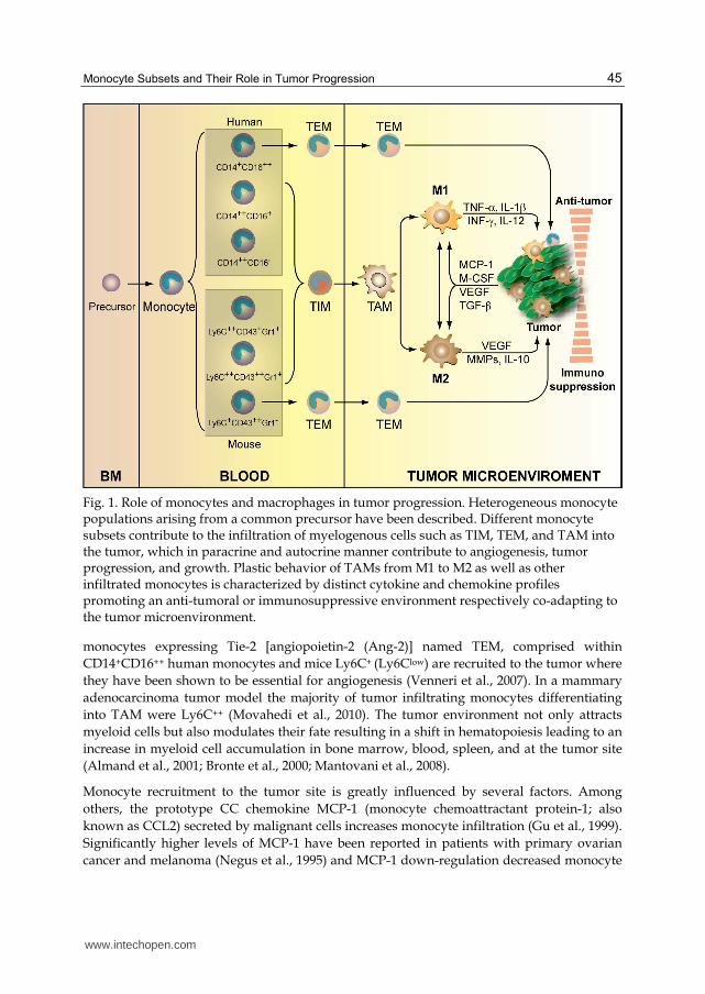
Fig. 1. Role of monocytes and macrophages in tumor progression. Heterogeneous monocyte populations arising from a common precursor have been described. Different monocyte subsets contribute to the infiltration of myelogenous cells such as TIM, TEM, and TAM into the tumor, which in paracrine and autocrine manner contribute to angiogenesis, tumor progression, and growth. Plastic behavior of TAMs from M1 to M2 as well as other infiltrated monocytes is characterized by distinct cytokine and chemokine profiles promoting an anti-tumoral or immunosuppressive environment respectively co-adapting to the tumor microenvironment.
monocytes expressing Tie-2 [angiopoietin-2 (Ang-2)] named TEM, comprised within
CD14+CD16++ human monocytes and mice Ly6C+ (Ly6Clow) are recruited to the tumor where
they have been shown to be essential for angiogenesis (Venneri et al., 2007). In a mammary
adenocarcinoma tumor model the majority of tumor infiltrating monocytes differentiating
into TAM were Ly6C++ (Movahedi et al., 2010). The tumor environment not only attracts
myeloid cells but also modulates their fate resulting in a shift in hematopoiesis leading to an
increase in myeloid cell accumulation in bone marrow, blood, spleen, and at the tumor site
(Almand et al., 2001; Bronte et al., 2000; Mantovani et al., 2008).
Monocyte recruitment to the tumor site is greatly influenced by several factors. Among
others, the prototype CC chemokine MCP-1 (monocyte chemoattractant protein-1; also
known as CCL2) secreted by malignant cells increases monocyte infiltration (Gu et al., 1999).
Significantly higher levels of MCP-1 have been reported in patients with primary ovarian
cancer and melanoma (Negus et al., 1995) and MCP-1 down-regulation decreased monocyte
www.intechopen.com

Tumor Microenvironment and Myelomonocytic Cells
46
migration in ovarian cancer (Negus et al., 1998). Other chemokines and growth factors such
as placental growth factor (PGF), TGF-┚, PGE-2, CCL3 (MIP-1┙), CCL4 (MIP-1┚), RANTES
(also known as CCL-5), and M-CSF, are found at high levels in tumors and contribute to the
recruitment, survival, and differentiation of monocytes into the tumor (Fig. 1). High M-CSF
expression is related with TAMs accumulation in breast carcinomas (Tang et al., 1992).
Vascular endothelial growth factor (VEGF) is also involved in monocyte recruitment into
tumors (Dineen et al., 2008; Murdoch et al., 2008). Elevated levels of RANTES were reported
in ovarian cancer and breast cancer contributed to cancer progression and monocytes
recruitment (Azenshtein et al., 2002; Negus et al., 1997).
Thus, secreted factors by tumor-infiltrating monocytes and tumor cells help modulate tumor growth and monocyte infiltration and fate determination contributing to tumor growth and progression.
3. Monocytic transcriptomes
In recent years the improvement in cell isolation techniques has allowed great advances in the understanding of transcriptomes in circulating monocytes helping identify unique signatures for different monocyte subsets and share remarkably transcriptional similarity (Ancuta et al., 2009; Mobley et al., 2007; Zhao et al., 2009). Genome wide studies on monocytes transcriptomes suggest that both CD16- and CD16+ monocytes share a common precursor (Ancuta et al., 2009). Despite the high similarity on expression profiles, differences relate to genes corresponding to cell-cell adhesion, trafficking, inflammation and immune responses, cell cycle, signal trasnduction and proliferation. Strict statistical analysis indicated as few as ~ 60 differentially expressed genes between monocyte subsets (Ancuta et al., 2009).
Classical monocytes (CD14+CD16-) have higher expression of genes involved in adhesion such as CCR2, CD62L and CD11b. In addition, the genes related to inflammation, angiogenesis and wound healing are also highly expressed in classical monocytes, revealing its implication in tissue repair (Wong et al., 2011). These classical monocytes also express higher level of CD14 and the cytokine IL-8 (Mobley et al., 2007). In these monocytes all phagocytosis enhancing proteins were highly expressed including levels of CD93, the receptor for complement component C1q1 (C1q1R), a component of a larger receptor complex for C1q complement factor and mannose-binding lectin (MBL2) (Ancuta et al., 2009). Furthermore, classical monocytes also showed high level expression of proinflammatory molecules such as S100A12, S100A9, S1008, and were among the top 50 most highly expressed genes (Wong et al., 2011).
CD16+ subsets possess a more differentiated profile with increased expression of macrophage and dendritic cell-like genes, probably indicating a more advanced stage of differentiation of these cells (Ancuta et al., 2009). These cells express high levels of genes involved in antigen processing, antimicrobial activity and host defense. High expression of genes encoding defensins, lysosomal proteases including cathepsins and elastase have been found (Mobley et al., 2007). Intermediate monocytes (CD14++ CD16+) showed enrichment for genes under major histocompatibility complex (MHC) class II processing and presenting, these genes mainly includes CD47, HLA-DO and CD40. MARCO (macrophage receptor with collagenous structure) is also one of the highly expressed gene
www.intechopen.com

Monocyte Subsets and Their Role in Tumor Progression
47
in the intermediate subset of monocytes (Wong et al., 2011). CD16+ monocytes express
higher levels of TNF- and chemokine receptor CX3CR1, CX3CR2 and colony-stimulating factor 1 receptor (CSF1R), the receptor for macrophage colony-stimulating factor (M-CSF) than classical CD16- monocytes (Ancuta et al., 2009; Wong et al., 2011; Zhao et al., 2009). Proteomic analysis also showed small differences with only 235 proteins differentially expressed between the monocyte subsets (Martinez, 2009). In addition to CD16, higher expression of the hematopoietic cell kinase (HCK) and the tyrosine protein kinase (LYN) were observed (Zhao et al., 2009). Interestingly the nonclassical monocytes (CD16++) showed the gene enrichment in the category of cytoskeleton rearrangement and phagocytosis. Genes related to phagocytosis such as LYN, HCK, ITAM of FCRs, C1QA, C1QB and also SLAN were found highly expressed in nonclassical monocytes (Wong et al., 2011). In pathological conditions, monocyte functions are finely tuned by the microenvironment. In this regard, hypoxic conditions found in tumors affect gene expression. Transcriptome analysis of hypoxic primary human monocytes revealed modulation of a significant cluster of genes with immunological relevance (Bosco et al., 2006). These included scavenger receptors: CD163, also found highly upregulated in CD16- subsets as well as MARCO , stabilin-1 (STAB1), macrophage scavenger receptor-1 (MSR1) (Mobley et al., 2007). Toll like receptor-7 (TLR7), immunoregulatory, costimulatory, and adhesion molecules: CD32, CD64, CD69, CD89, leukocyte membrane Ag (CMRF-35H), integrin ┚-5 (ITGB5), chemokines/cytokines receptors (CCL23, CCL15, CCL8, CCR1, CCR2, RDC1, IL-23A, IL-6ST) were also highly expressed under hypoxia (Bosco et al., 2006). Hypoxia also controlled gene expression of chemokine receptors including CXCL1, CXCL8, CCL3, CXCR4 (Murdoch et al., 2004).
Transcriptome studies are unraveling the complexities of the monocyte populations and provide evidence of the specialized function of specific subsets. High expression of IL-8 and adhesion molecules in CD16- subsets seem to support the role as inflammatory, capable to leave the circulation and infiltrate. In addition, high expression of M-CSF in CD16+ support their role in driving differentiation of CD16-, a more immature pool that could replenish macropahges at inflammatory sites. Studies in gene expression profiles in human monocyte subpopulations as part of tumor biology remain scarce and future studies in this area will thereby help to understand their specific roles as well as define future approaches to re-educate monocytes in the tumor microenvironment.
4. Role of monocyte subsets in tumor progression
4.1 Monocyte deactivation
It has been widely accepted that cancer progression is an inherently proinflammatory process that involves the activation of the innate and adaptive immune system. During tumorigenesis, monocytes are destined to give anti-tumor response of the host and act both as cells presenting tumor-associated antigens to tumor-infiltrating lymphocytes and as cytotoxic effector cells (Mytar et al., 2003). However, cancer cells have developed mechanisms that inhibit immune surveillance (Pardoll, 2003), characterized by the impaired
ability of monocytes to produce IFN-, TNF-┙, IL-12, while enhance IL-10 secretion. IL-10 is immunosuppressive, tumor promoting, and inhibits the production of IL-2, IFN┛, IL-12, TNF-┙, resulting in a reduced Th1 response (Sica et al., 2006). Different studies have reported increased IL-10 serum levels in patients with melanoma and other solid tumors
www.intechopen.com

Tumor Microenvironment and Myelomonocytic Cells
48
(Fortis et al., 1996; Sato et al., 1996). IL-12 plays central role in activating anti-tumor
immunity by stimulating the production of IFN- and TNF-┙ necessary for cytotoxic effects. In many cancers, for instance in colorectal cancer reduced production of IL-12 was accompanied by increased production of IL-10 (O'Hara et al., 1998). Deactivation of monocytes can be reversed with Bovis bacillus Calmette-Guerin (BCG), the prototype immunomodulator, which inhibits IL-10 production, thus reversing monocyte deactivation (Baran et al., 2004).
Deactivation of TIM is also mediated by other mechanisms. Hyaluronan (HA), an important
tumor microenvironment matrix structure, produced by tumor cells, is now emerging as a
key factor in monocyte deactivation and tumor progression (Mytar et al., 2003; Toole, 2004).
Ligation of CD44 by HA is a proinflammatory event that regulates monocyte adhesion and
cytokine production and was found to stimulate the expression of IRAK (IL-1 receptor-
associated kinase)-M. High levels of IRAK-M were reported in patients of chronic myeloid
leukemia and metastasis (del Fresno et al., 2005). Co-culture experiments of CD14+
monocytes with a variety of tumor cells show that tumor cells express high level of
prostaglandin (PG) that also contributes to the downregulation of TNF-┙, IL-12, IRAK-1
interrupting the inflammatory response against cancer progression. These findings strongly
suggest that the functional activity of monocytes is adversely modified by the local tumor
microenvironment. Notably, the deactivation mechanisms of monocyte in cancer may not
just be limited to tumor microenvironment but also players in other inflammatory diseases
such as atherosclerosis, arthritis, and obesity.
4.2 Angiogenesis
Once the tumor cells escape recognition and destruction by infiltrating monocyte, these
infiltrating cells participate in tumor growth by promoting angiogenesis (Lin et al., 2001),
an essential process in the tumor progression and growth. High numbers of human
vascular leukocytes found in human ovarian cancer have been suggested to form
neovessels in mouse xenotransplantations (Conejo-Garcia et al., 2005). Gr-1+ monocytes
promote angiogenesis via paracrine mechanisms (Yang et al., 2004). Vascular leukocytes, a
subset of CD11C+ MHC-II+ dendritic-cell precursors expressing endothelial vascular
markers VE-cadherin, CD34, and CD146 also contribute to tumor angiogenesis (Conejo-
Garcia et al., 2005; Yang et al., 2004). Circulating TEM derived from non-classical
monocytes contributes to tumor angiogenesis, migrating towards Ang-2, released at high
levels by activated endothelial cells and angiogenic vessels at tumor sites (Venneri et al.,
2007). Ang-2 also inhibits TNF-┙ release (Lewis et al., 2007; Murdoch et al., 2007),
normally responsible for promoting apoptosis of both tumor and endothelial cells
(Petrache et al., 2001). Hence, Ang-2-mediated down-regulation of TNF-┙ may increase
metastasis and angiogenesis contributing to tumor growth. The molecular basis of factors
that render proangiogenic activity in TEM are still not well understood. But elimination of
TEMs in mouse glioma models was shown to reduce tumor growth and vascularity (De
Palma et al., 2005), supporting the role of TEM in tumor blood vessels formation. In
human tumor models, CD14+ TIM monocytes can develop endothelial phenotype
(Schmeisser et al., 2001) and actively participate in neovascularization during tumor
growth (Urbich et al., 2003).
www.intechopen.com

Monocyte Subsets and Their Role in Tumor Progression
49
Recent studies indicated that CCL2 synthesized by metastatic tumor cells and by the target-site tissue stroma is critical for the recruitment of CCR2-expressing monocyte subsets. Activation of the CCL2-CCR2 signaling axis promotes extravasation and recruitment of inflammatory monocyte subsets into metastatic tumor sites and helps promote differentiation of these monoyctes into non-proliferating TAMs (Qian et al., 2011). Transcriptome studies in both resident and inflammatory monocytes show higher expression of VegfA, a potent angiogenic factor in inflammatory monocytes CD14+CD16- subsets, recruited in large numbers to metastatic areas (Qian et al., 2011). Future comprehensive trsancriptome analysis in purified monocyte populations under pathophysiological conditions should be helpful to gain a further understanding of the functional roles of different monocyte populations in tumor progression.
5. Regulation of monocyte subsets
5.1 Regulation of survival and cell death of monocytes in tumors
A complex network of survival and apoptotic pathways determines monocyte fate. Several
kinases, transcription factors and anti- and pro-apoptotic proteins play key roles in
determining monocyte survival and cell death. All circulating monocyte subsets have very
short life span of just few days (Fahy et al., 1999; Zeigler et al., 2003). Inflammatory and
differentiation stimuli are known to halt apoptosis and inducing prolonged monocyte
survival. Among others M-CSF, TNF-, and IL-1 promote survival by deactivating the
apoptotic program lead by caspases (Fahy et al., 1999; Goyal et al., 2002; Kelley et al., 1999).
The tumor microenvironment characterized by alterations in cytokine, chemokine, and
growth factor expression contributes to mediate prolonged monocyte survival (Zou, 2005).
In fact, increase levels of TGF-┚, IL-10, and VEGF alter TIM and TEM from anti-tumoral to
immunosuppressor and proangiogenic in part by switching their cytokine and growth
factors expressions (Whiteside, 2008) (Fig. 1).
Among the signaling molecules involved in activation and survival, the phosphatidylinositol 3-kinase (PI3K)-AKT axis has a central role in regulating a multitude of essential myelogenous events, such as differentiation, phagocytosis, oxidative burst, and TLR-mediated responses (Franke et al., 1997; Parihar et al., 2010). PI3K activation was also shown to direct infiltration (Funamoto et al., 2002; Wang et al., 2002). M-CSF-induced activation of AKT through caspase-9 phosphorylation inhibits apoptosis (Kelley et al., 1999), promoting prolonged survival and sustaining the differentiation program (Gonzalez-Mejia & Doseff, 2009). In addition, PI3K activates protein kinase C (PKC) in monocytes (Herrera-Velit et al., 1997) leading to the induction of the activated mitogen-activated protein kinase (MAPK) pathway (Rao, 2001), including the extracellular signal-regulated kinase (ERK), the c-JNK and p38 which are hubs to multiple networks of survival (Parihar et al., 2010). Pro-inflammatory mediators induce the PI3K/AKT and MAPK/ERK pathways that in turn are responsible in determining the balance of inflammatory versus anti-inflammatory cytokines.
In the hypoxic regions of a tumor, activation of PI3K/AKT leads to HIF (hypoxia inducing
factor)-1 activity which in turn regulates tumor growth, angiogenesis, metastasis, and
monocytes/macrophages recruitment (Semenza, 2003). While the role of HIF-1 in different
monocyte subsets and macrophages is not fully understood, increased HIF-1 expression in
TAMs was found to contribute to tumor angiogenesis and invasiveness (Werno et al., 2010).
www.intechopen.com

Tumor Microenvironment and Myelomonocytic Cells
50
The transcription factor nuclear factor-κB (NF-κB), plays a critical role in tumor biology (Karin & Greten, 2005). While in other cell types NF-κB has various tumor-promoting functions, in monocytes the activation of NF-κB results in the release of cytokines, such as TNF-┙ and IL-6 which not only trigger prosurvival signals in tumor cells but also support growth and progression, and importantly sustain a dysregulated immune function (Hagemann et al., 2009; Pikarsky et al., 2004). Activation of NF-κB leads to increase expression of angiogenic factors such as VEGF, CXCL1 and CXCL8 and anti-apoptotic molecules including the inhibitor of apoptotic proteins (IAPs), and bcl-2 (Richmond, 2002). The relevance of the NF-κB signaling pathway is illustrated in a mouse model of colitis associated cancer, where deletion of IKK-┚ reduced the production of tumor promoting paracrine factors and subsequently decreased carcinoma growth (Greten et al., 2004). In hepatocellular carcinomas the NF-κB activating kinase IKK-┚ suppresses early chemically-induced liver tumorigenesis by inhibiting hepatocyte death and compensatory proliferation. This anti-tumorigenic activity of hepatocyte IKK-┚ was suggested to be due to the induction of NF-κB-dependent pro-survival and anti-oxidant genes (He et al., 2010). Deletion of IKK-┚ in myeloid cells resulted in a significant decrease in tumor size and diminished the expression of proinflammatory cytokines that may serve as tumor growth factors (Yoshimura, 2006).
Caspases, the cysteine proteases central to programmed cell death, including “activators” (caspases-1, 2, 8, 9, and 10) and ‘executioners’ (caspases-3, 6, and 7) play fundamental roles in myelogenous cells through the activation of the extrinsic and intrinsic apoptotic cascade (Riedl & Shi, 2004). Fas receptor-induced apoptosis has a key role in monocyte biology as both homozygous FasR deficient (lpr/lpr) and heterozygous FasR and Fas ligand deficient (lpr/gld) mice have increased numbers of inflammatory and resident monocytes resulting in splenomegaly, lymphadenopathy, and accumulation of tissue macrophages (Ashkenazi & Dixit, 1998; Brown et al., 2004). Cytokines, CCL2 and IL-6 abundant in the tumor microenvironment have been shown to inhibit caspase-8 and promote enhanced autophagic activity to protect the monocytes recruited to the tumor and, at the same time, induce their differentiation toward M2-type macrophages (Roca et al., 2009). Futhermore, M-CSF induces caspase-9 inhibition leading to reduce apoptosis (Kelley et al., 1999). Several other inhibitors of the apoptotic pathways including members of the Bcl-2 family have been characterized (Youle & Strasser, 2008) and XIAP also play a crucial role in cell survival and tumor development. XIAP directly inhibits activator caspase-9 and also executioner caspases in cancer cells and increase expression of XIAP has been reported in monocyte differentiation (Sasaki et al., 2000). However, the role of XIAP in different monocyte subsets has not been studied. Our studies identified the small heat shock protein (Hsp27) as a direct inhibitor of caspase-3 in monocytes. Notably a significant increase in Hsp27 expression was found to be required during monocyte-macrophage differentiation (Voss et al., 2007). Hsp27 is highly expressed in several tumors and high levels of Hsp27 in plasma have been associated with risk of lung cancer (Wang et al., 2010). Hsp27 was found to have functions in IL-1-induced cell signaling and pro-inflammatory gene expression suggesting its ability to modulate immunity (Alford et al., 2007). Whether the expression of apoptosis inhibitors such as Hsp27 is altered in TIM and TAMs have yet to be studied. These findings suggest that anti-apoptotic factors in plasma can switch monocyte life span contributing to their accumulation into tumors. It is possible to hypothesize that monocyte re-education programs could target molecular networks involved in shifting survival and cell death programs as well as immunoparalysis in monocytes.
www.intechopen.com

Monocyte Subsets and Their Role in Tumor Progression
51
5.2 Epigenetics and microRNAs regulation in monocytes and macrophages during tumor progression
During hematopoiesis gradual changes in gene expression orchestrate lineage-specification.
The myelomonocytic lineage originates from a ganulocyte-erythrocyte-megakaryocyte-
macrophage colony-forming unit (GEMM-CFU) that promotes formation of the GM-CFU or
ganulocyte-macrophage colony-forming unit. GM-CFU under the control of G-MCSF, M-
CSF and IL-3 regulate the differentiation of this progenitor to a monocyte precursor (M-
CFU) that becomes a promonocyte in the bone marrow. Differentiation of precursor cells is
controlled by transcription factors that regulate differentation and survival. A combination
of transcription factors including GATA-2, GATA-1, SCL, and members of the homeobox
proteins (HOXB) control monocyte survival. Repression of GATA-1, SCL, and c-Myc
expression allows monocytic differentiation (Valledor et al., 1998). Earliest stages of myeloid
lineage specification involve the activity of Runt-related transcription factor 1 (RUNX1). One
of the main targets of RUNX1 is PU.1, a member of the ETS (E-twenty six) family
transcription factor (Olson et al., 1995). PU.1 is key in differentiation by controlling the
expression of M-CSF and GM-CSF receptors. In addition, PU.1 regulates expression of FC receptors involved in phagocytosis. Thus, PU.1 has a critical role in monocytic
differentiation by regulating expression of molecules essential for differentiation and
function of monocytic lineages. Intermediate stages of differentiation are regulated among
others by transcription factor CCAAT enhancer-binding protein (C/EBP) members, c-Myc
and HOXB7, the latest induced by GM-CSF (Friedman, 2002; Yeamans et al., 2007). In
addition, c-Jun dimmers (AP-1) and STAT members contribute to the induction of
monocytic genes (Friedman, 2002). STAT3 is one of the important transcription factors that
play an essential role in cell survival, proliferation, and differentiation. Classical monocytes,
express high levels of AP-1-axis regulated genes, and it has been suggested that this gene
repertoire may be responsible for the plastic behavior of this monocyte subset to recognize
self and non-self stimuli (Wong et al., 2011). In non-classical subsets transcription factors
controlling apoptosis, differentiation, and proliferation were highly expressed, among them
E2F1, ETS1, and FOXO1, well known to regulate proliferation. Intermediate monocyte
subsets showed reciprocal increases in transcription factors found in both classical and non-
classical subsets (Wong et al., 2011).
Epigenetic changes regulated by histone-modifying enzymes such as histone acetyltransferases (HATs), histone deacetylases (HDAC), and methylases provide additional regulatory mechanisms for monocytic gene expression. Abnormal activity of these enzymes leads to changes in gene expression affecting differentiation and apoptosis and causing
neoplasia and other diseases (Haberland et al., 2009). Acetylation of NF-B induces modifications in a temporal manner leading to recruitment of other co-activator and re-modeling complexes and the induction of inflammatory gene expression (Ito et al., 2000; Lee et al., 2006). In monocytes, histone acetylation of the TNF-┙ promoter has been shown to be developmentally regulated and is required for TNF-┙ expression during acute inflammation (Lee et al., 2003). Changes in acetylation of the Decoy Receptor 3 (DcR3) promoter, a member of the TNF receptor superfamily, has been reported in tumors affecting expression of MHC class II (MHC-II)-dependent antigen presentation (Chang et al., 2008). Recent studies found distinct DNA methylation profiles in CD34+ hematopoietic progenitor cells and differentiated myeloid cells with pronounced DNA hypomethylation in monocytes (Bocker et al., 2011).
www.intechopen.com

Tumor Microenvironment and Myelomonocytic Cells
52
Interestingly, age-related methylation changes in CD34+ cells were found. Older progenitor cells showed a bimodal pattern with hypomethylation of differentiation associated genes and de novo methylation events resembling epigenetic mutations, thus providing an important insight into the methylation dynamics during differentiation and suggest that epigenetic changes contribute to hematopoietic progenitor cell aging (Bocker et al., 2011). Induction of inflammatory genes IL-6, IL-8 and IL-12 were found to depend on HAT/HDAC activity (Lu et al., 2005; Schmeck et al., 2008). Treatment of monocytic leukemia cell lines and patient samples with demethylating and HDAC inhibitors induced reversion to gene profiles found in normal subjects, highlighting the role of chromatin remodeling in monocyte behavior (Serrano et al., 2008). Treatment of macrophages with broad-spectrum HDACs inhibitors showed anti- and pro-inflammatory effects, HDACs suppressed LPS-induced expression of the pro-inflammatory MCP-3, and IL-12 but amplified the expression of the pro-atherogenic factors Cox-2 (Halili et al., 2010). Dietary compounds with HDAC inhibitory activities, including garcinol, curcumin, and anacardic acid, modulate epigenetic status and are being investigated as potential anti-cancer agents (Bolden et al., 2006; Inoue et al., 2004). It will be of interest to evaluate how these therapies influence monocytes epigenomes. Future studies to evaluate the epigenetic dynamics of monocyte subsets will be of great value to further understand their unique functional contributions.
MicroRNAs (miRNAs) are small non-coding RNAs emerging as new post-trancriptional
regulators and have been found to contribute in several monocyte functions. The overall
relevance of miRNA in hematopoiesis has been discussed in detail elsewhere (Baltimore et al.,
2008). Based on the epidemiological studies inflammation contributes to 25% of all cancers by
increasing cancer risk and cancer development (Mantovani et al., 2008). Several miRNAs were
found to be elevated in inflammation and cancer. In particular, miRNA-155, miRNA-125b, and
miRNA-21 have emerged as important miRNAs regulating immune responses. MiRNA-155 is
elevated in leukemia and lymphoma and transgenic mice overexpressing miRNA-155 in B
cells, develop B-cell leukemia and sustained expression of miRNA-155 in hematopoietic stem
cells causes myeloproliferative disorders (O'Connell et al., 2008). MiRNA-155 targets among
others suppressors of cytokine signaling (SOCS1) and SH2-domain-containing inositol-5-
phosphatase 1 (SHIP-1), both negative regulators of TLR signaling in monocyte/macrophage
inflammatory response. Recent studies showed that tumor environment causes a sustained
reduction of miRNA-155 in monocytes/macrophages, which in turn activates the C/EBP┚ (He
et al., 2009). C/EBP┚, a member of C/EBP family of leucine zipper transcription factors, plays
pivotal roles in coordinating the expression of a wide variety of genes that control immune
responses including COX-2 (Li et al., 2007). C/EBP┚-deficient mice exhibit defects in
macrophage activation and differentiation (He et al., 2009). Monocytes exposed to tumor
microenvironment showed C/EBP┚ expression inversely correlated with miR-155 expression
and it was found that miRNA-155 could suppress the C/EBP┚. Furthermore, over-expression
of miRNA-155 significantly attenuated the cytokine production in tumor-activated monocytes
(He et al., 2009). Expression of miRNA-146 affects downstream TLR signaling molecules such
IRAK1 and 2 or TNF receptor-associated factor (TRAF) 6, all involved in the activation of the
NF-B axis (O'Connell et al., 2010). MiRNA-21 and miRNA-125b are also elevated in
inflammatory conditions and cancer (Esquela-Kerscher & Slack, 2006).
Recent studies highlighted different miRNAs profile in circulating monocytes when
compared with dendritic cells or macrophages (Tserel et al., 2011). However, it is presently
www.intechopen.com

Monocyte Subsets and Their Role in Tumor Progression
53
unknown whether miRNAs expression is altered in different monocytes subsets in normal
conditions or in tumorigenesis.
6. Molecular pathways involved in monocyte fate and re-education programming
Reprogramming implies the conversion of a fully differentiated cell type into another cell type without pluripotent intermediate and generally achieved by overexpressing key transcription-factors (Zhou & Melton, 2008). Recent studies have reported that various cells including fibroblasts can be reprogrammed into blood-cell progenitors (Szabo et al., 2010), neurons (Vierbuchen et al., 2010), and cardiomyocytes (Ieda et al., 2010), demonstrating the ample applicability of this approach for therapeutic uses.
Myelomonocytic cells re-education programs have been described. TAMs have been
suggested to be programmed to specific subtypes such as M1 and M2 upon arrival to the
tumor microenvironment. It has been shown that administration of GM-CSF in murine
breast cancer models induces soluble VEGFR-1 resulting in the suppression of VEGF and
angiogenesis (Eubank et al., 2009). Cytokine-dependent reprogramming using IL-12, which
impacts innate and adaptive immune systems, has proven the most interesting (Trinchieri,
1995). IL-12 in its soluble or lipid-encapsulated forms injected into tumor-bearing mice
resulted in a strong cytotoxic anti-tumor response (Hill et al., 2002), suggesting its capability
to restore normal immune functions. Inflammatory monocytes expressing high levels of the
chemokine receptor CCR2 but not CD14-CD16+ were found in increased numbers in several
chronic inflammatory conditions including atherosclerosis and asthma (Parihar et al., 2010).
Recent studies showed that administration of lipid nanoparticles containing a CCR2-
silencing short interfering RNA in mice, prevents monocyte accumulation at inflammatory
sites (Leuschner et al., 2011). Ectopic expression of PU.1 in lymphocytes and neural stem
cells induced transdifferentiation to the myeloid lineage with functional chemotactic and
immune functions characteristic of monocytes (Forsberg et al., 2010; Laiosa et al., 2006).
Transcriptome analysis showed that PU. 1 expression affects chromatin remodeling leading
to epigenetic changes that ensure macrophage specification (Ostuni & Natoli, 2011). These
monocytes may serve as vehicles to modulate microenvironments with dysregulated
immunity such as found in the tumor. Hence, alteration of epigenetic dynamics may also be
a potential approach to alter monocyte re-programming. It is recognized that macrophages
adapt in response to the microenvironment (referred to other Chapters in this book). Part of
this adaptation is based on changes in their transcriptomes (Lawrence & Natoli, 2011).
However, the molecular mechanisms determining macrophage genetic adaptations remain
mostly unknown. In the case of monocyte subsets similar studies have not yet been
conducted. Thus, dissecting the genomic determinants in normal and pathophysiological
conditions of functional distinct monocyte subsets and TEMs will provide possibilities to re-
educate these cells towards an anti-tumor phenotype, re-establishing apoptotic programs or
halting their extravasation activities.
7. Conclusion
Tumor progression is marked by dynamic changes of the tumor microenvironment from
early neoplastic events to advanced tumor stages. Recruitment of circulating monocytes to
www.intechopen.com

Tumor Microenvironment and Myelomonocytic Cells
54
specific tumor sites contributes to progressive modulation of signaling molecules such as
chemokines, cytokines, growth factors, and transcription factors. Specific contributions of
different monocyte subsets to the tumor associated myelogenous populations of TEM, TIM,
and TAMs are starting to emerge. Advances in understanding the molecular networks
regulating myelomonocytic cells functions and fate provide opportunities to implement re-
education programs to rehabilitate normal anti-tumor monocyte behavior. These strategies
should help limiting myelomonocytic cell survival, halting recruitment to tumor sites, and
increasing cytotoxic functions providing novel approaches for cancer treatment. Advances
in this area will not be limited to tumor biology but will also impact our understanding of
other chronic inflammatory diseases.
8. Acknowledgements
Work in Dr. Doseff’s lab is supported by grant NIH (RO1-HL075040) and NSF-MCB
(0542244). We thank Dr. Oliver Voss for help with some illustrations and Mrs. Malavez for
comments. We apologize to those of our colleagues who made important contributions but
which were omitted due to space limitations.
9. References
Alford, K. A.; Glennie, S.; Turrell, B. R.; Rawlinson, L.; Saklatvala, J. & Dean, J. L. (2007).
Heat shock protein 27 functions in inflammatory gene expression and transforming
growth factor-beta-activated kinase-1 (TAK1)-mediated signaling. J Biol Chem.
282(9):6232-6241.
Almand, B.; Clark, J. I.; Nikitina, E.; van Beynen, J.; English, N. R.; Knight, S. C.; Carbone, D.
P. & Gabrilovich, D. I. (2001). Increased production of immature myeloid cells in
cancer patients: a mechanism of immunosuppression in cancer. J Immunol.
166(1):678-689.
Ancuta, P.; Liu, K. Y.; Misra, V.; Wacleche, V. S.; Gosselin, A.; Zhou, X. & Gabuzda, D.
(2009). Transcriptional profiling reveals developmental relationship and distinct
biological functions of CD16+ and CD16- monocyte subsets. BMC Genomics. 10(403.
Ashkenazi, A. & Dixit, V. M. (1998). Death receptors: signaling and modulation. Science.
281(5381):1305-1308.
Auffray, C.; Sieweke, M. H. & Geissmann, F. (2009). Blood monocytes: development,
heterogeneity, and relationship with dendritic cells. Annu Rev Immunol. 27(669-692.
Azenshtein, E.; Luboshits, G.; Shina, S.; Neumark, E.; Shahbazian, D.; Weil, M.; Wigler, N.;
Keydar, I. & Ben-Baruch, A. (2002). The CC chemokine RANTES in breast
carcinoma progression: regulation of expression and potential mechanisms of
promalignant activity. Cancer Res. 62(4):1093-1102.
Baltimore, D.; Boldin, M. P.; O'Connell, R. M.; Rao, D. S. & Taganov, K. D. (2008).
MicroRNAs: new regulators of immune cell development and function. Nat
Immunol. 9(8):839-845.
Baran, J.; Baj-Krzyworzeka, M.; Weglarczyk, K.; Ruggiero, I. & Zembala, M. (2004).
Modulation of monocyte-tumour cell interactions by Mycobacterium vaccae. Cancer
Immunol Immunother. 53(12):1127-1134.
www.intechopen.com

Monocyte Subsets and Their Role in Tumor Progression
55
Bocker, M. T.; Hellwig, I.; Breiling, A.; Eckstein, V.; Ho, A. D. & Lyko, F. (2011). Genome-wide promoter DNA methylation dynamics of human hematopoietic progenitor cells during differentiation and aging. Blood. 117(19):e182-189.
Bolden, J. E.; Peart, M. J. & Johnstone, R. W. (2006). Anticancer activities of histone deacetylase inhibitors. Nat Rev Drug Discov. 5(9):769-784.
Bosco, M. C.; Puppo, M.; Santangelo, C.; Anfosso, L.; Pfeffer, U.; Fardin, P.; Battaglia, F. & Varesio, L. (2006). Hypoxia modifies the transcriptome of primary human monocytes: modulation of novel immune-related genes and identification of CC-chemokine ligand 20 as a new hypoxia-inducible gene. J Immunol. 177(3):1941-1955.
Bronte, V.; Apolloni, E.; Cabrelle, A.; Ronca, R.; Serafini, P.; Zamboni, P.; Restifo, N. P. & Zanovello, P. (2000). Identification of a CD11b(+)/Gr-1(+)/CD31(+) myeloid progenitor capable of activating or suppressing CD8(+) T cells. Blood. 96(12):3838-3846.
Brown, N. J.; Hutcheson, J.; Bickel, E.; Scatizzi, J. C.; Albee, L. D.; Haines, G. K., 3rd; Eslick, J.; Bradley, K.; Taricone, E. & Perlman, H. (2004). Fas death receptor signaling represses monocyte numbers and macrophage activation in vivo. J Immunol. 173(12):7584-7593.
Chang, Y. C.; Chen, T. C.; Lee, C. T.; Yang, C. Y.; Wang, H. W.; Wang, C. C. & Hsieh, S. L. (2008). Epigenetic control of MHC class II expression in tumor-associated macrophages by decoy receptor 3. Blood. 111(10):5054-5063.
Conejo-Garcia, J. R.; Buckanovich, R. J.; Benencia, F.; Courreges, M. C.; Rubin, S. C.; Carroll, R. G. & Coukos, G. (2005). Vascular leukocytes contribute to tumor vascularization. Blood. 105(2):679-681.
Cros, J.; Cagnard, N.; Woollard, K.; Patey, N.; Zhang, S. Y.; Senechal, B.; Puel, A.; Biswas, S. K.; Moshous, D.; Picard, C.; Jais, J. P.; D'Cruz, D.; Casanova, J. L.; Trouillet, C. & Geissmann, F. (2010). Human CD14dim monocytes patrol and sense nucleic acids and viruses via TLR7 and TLR8 receptors. Immunity. 33(3):375-386.
De Palma, M.; Venneri, M. A.; Galli, R.; Sergi Sergi, L.; Politi, L. S.; Sampaolesi, M. & Naldini, L. (2005). Tie2 identifies a hematopoietic lineage of proangiogenic monocytes required for tumor vessel formation and a mesenchymal population of pericyte progenitors. Cancer Cell. 8(3):211-226.
del Fresno, C.; Otero, K.; Gomez-Garcia, L.; Gonzalez-Leon, M. C.; Soler-Ranger, L.; Fuentes-Prior, P.; Escoll, P.; Baos, R.; Caveda, L.; Garcia, F.; Arnalich, F. & Lopez-Collazo, E. (2005). Tumor cells deactivate human monocytes by up-regulating IL-1 receptor associated kinase-M expression via CD44 and TLR4. J Immunol. 174(5):3032-3040.
Dineen, S. P.; Lynn, K. D.; Holloway, S. E.; Miller, A. F.; Sullivan, J. P.; Shames, D. S.; Beck, A. W.; Barnett, C. C.; Fleming, J. B. & Brekken, R. A. (2008). Vascular endothelial growth factor receptor 2 mediates macrophage infiltration into orthotopic pancreatic tumors in mice. Cancer Res. 68(11):4340-4346.
Esquela-Kerscher, A. & Slack, F. J. (2006). Oncomirs - microRNAs with a role in cancer. Nat Rev Cancer. 6(4):259-269.
Eubank, T. D.; Roberts, R. D.; Khan, M.; Curry, J. M.; Nuovo, G. J.; Kuppusamy, P. & Marsh, C. B. (2009). Granulocyte macrophage colony-stimulating factor inhibits breast cancer growth and metastasis by invoking an anti-angiogenic program in tumor-educated macrophages. Cancer Res. 69(5):2133-2140.
www.intechopen.com

Tumor Microenvironment and Myelomonocytic Cells
56
Fahy, R. J.; Doseff, A. I. & Wewers, M. D. (1999). Spontaneous human monocyte apoptosis utilizes a caspase-3-dependent pathway that is blocked by endotoxin and is independent of caspase-1. J Immunol. 163(4):1755-1762.
Forsberg, M.; Carlen, M.; Meletis, K.; Yeung, M. S.; Barnabe-Heider, F.; Persson, M. A.; Aarum, J. & Frisen, J. (2010). Efficient reprogramming of adult neural stem cells to monocytes by ectopic expression of a single gene. Proc Natl Acad Sci U S A. 107(33):14657-14661.
Fortis, C.; Foppoli, M.; Gianotti, L.; Galli, L.; Citterio, G.; Consogno, G.; Gentilini, O. & Braga, M. (1996). Increased interleukin-10 serum levels in patients with solid tumours. Cancer Lett. 104(1):1-5.
Franke, T. F.; Kaplan, D. R.; Cantley, L. C. & Toker, A. (1997). Direct regulation of the Akt proto-oncogene product by phosphatidylinositol-3,4-bisphosphate. Science. 275(5300):665-668.
Friedman, A. D. (2002). Transcriptional regulation of granulocyte and monocyte development. Oncogene. 21(21):3377-3390.
Funamoto, S.; Meili, R.; Lee, S.; Parry, L. & Firtel, R. A. (2002). Spatial and temporal regulation of 3-phosphoinositides by PI 3-kinase and PTEN mediates chemotaxis. Cell. 109(5):611-623.
Geissmann, F.; Manz, M. G.; Jung, S.; Sieweke, M. H.; Merad, M. & Ley, K. (2010). Development of monocytes, macrophages, and dendritic cells. Science. 327(5966):656-661.
Gonzalez-Mejia, M. E. & Doseff, A. I. (2009). Regulation of monocytes and macrophages cell fate. Front Biosci. 14(2413-2431.
Goyal, A.; Wang, Y.; Graham, M. M.; Doseff, A. I.; Bhatt, N. Y. & Marsh, C. B. (2002). Monocyte survival factors induce Akt activation and suppress caspase-3. Am J Respir Cell Mol Biol. 26(2):224-230.
Greten, F. R.; Eckmann, L.; Greten, T. F.; Park, J. M.; Li, Z. W.; Egan, L. J.; Kagnoff, M. F. & Karin, M. (2004). IKK┚ links inflammation and tumorigenesis in a mouse model of colitis-associated cancer. Cell. 118(3):285-296.
Gu, L.; Tseng, S. C. & Rollins, B. J. (1999). Monocyte chemoattractant protein-1. Chem Immunol. 72(7-29.
Haberland, M.; Montgomery, R. L. & Olson, E. N. (2009). The many roles of histone deacetylases in development and physiology: implications for disease and therapy. Nat Rev Genet. 10(1):32-42.
Hagemann, T.; Biswas, S. K.; Lawrence, T.; Sica, A. & Lewis, C. E. (2009). Regulation of macrophage function in tumors: the multifaceted role of NF-κB. Blood. 113(14):3139-3146.
Halili, M. A.; Andrews, M. R.; Labzin, L. I.; Schroder, K.; Matthias, G.; Cao, C.; Lovelace, E.; Reid, R. C.; Le, G. T.; Hume, D. A.; Irvine, K. M.; Matthias, P.; Fairlie, D. P. & Sweet, M. J. (2010). Differential effects of selective HDAC inhibitors on macrophage inflammatory responses to the Toll-like receptor 4 agonist LPS. J Leukoc Biol. 87(6):1103-1114.
Hanahan, D. & Weinberg, R. A. (2011). Hallmarks of cancer: the next generation. Cell. 144(5):646-674.
www.intechopen.com

Monocyte Subsets and Their Role in Tumor Progression
57
Hanninen, A.; Jaakkola, I.; Salmi, M.; Simell, O. & Jalkanen, S. (1997). Ly-6C regulates endothelial adhesion and homing of CD8(+) T cells by activating integrin-dependent adhesion pathways. Proc Natl Acad Sci U S A. 94(13):6898-6903.
He, G.; Yu, G. Y.; Temkin, V.; Ogata, H.; Kuntzen, C.; Sakurai, T.; Sieghart, W.; Peck-Radosavljevic, M.; Leffert, H. L. & Karin, M. (2010). Hepatocyte IKK┚/NF-κB inhibits tumor promotion and progression by preventing oxidative stress-driven STAT3 activation. Cancer Cell. 17(3):286-297.
He, M.; Xu, Z.; Ding, T.; Kuang, D. M. & Zheng, L. (2009). MicroRNA-155 regulates inflammatory cytokine production in tumor-associated macrophages via targeting C/EBP┚. Cell Mol Immunol. 6(5):343-352.
Herrera-Velit, P.; Knutson, K. L. & Reiner, N. E. (1997). Phosphatidylinositol 3-kinase-dependent activation of protein kinase C-ζ in bacterial lipopolysaccharide-treated human monocytes. J Biol Chem. 272(26):16445-16452.
Hill, H. C.; Conway, T. F., Jr.; Sabel, M. S.; Jong, Y. S.; Mathiowitz, E.; Bankert, R. B. & Egilmez, N. K. (2002). Cancer immunotherapy with interleukin 12 and granulocyte-macrophage colony-stimulating factor-encapsulated microspheres: coinduction of innate and adaptive antitumor immunity and cure of disseminated disease. Cancer Res. 62(24):7254-7263.
Ieda, M.; Fu, J. D.; Delgado-Olguin, P.; Vedantham, V.; Hayashi, Y.; Bruneau, B. G. & Srivastava, D. (2010). Direct reprogramming of fibroblasts into functional cardiomyocytes by defined factors. Cell. 142(3):375-386.
Inoue, S.; MacFarlane, M.; Harper, N.; Wheat, L. M.; Dyer, M. J. & Cohen, G. M. (2004). Histone deacetylase inhibitors potentiate TNF-related apoptosis-inducing ligand (TRAIL)-induced apoptosis in lymphoid malignancies. Cell Death Differ. 11 Suppl 2(S193-206.
Ito, K.; Barnes, P. J. & Adcock, I. M. (2000). Glucocorticoid receptor recruitment of histone deacetylase 2 inhibits interleukin-1beta-induced histone H4 acetylation on lysines 8 and 12. Mol Cell Biol. 20(18):6891-6903.
Karin, M. & Greten, F. R. (2005). NF-κB: linking inflammation and immunity to cancer development and progression. Nat Rev Immunol. 5(10):749-759.
Kelley, T. W.; Graham, M. M.; Doseff, A. I.; Pomerantz, R. W.; Lau, S. M.; Ostrowski, M. C.; Franke, T. F. & Marsh, C. B. (1999). Macrophage colony-stimulating factor promotes cell survival through Akt/protein kinase B. J Biol Chem. 274(37):26393-26398.
Laiosa, C. V.; Stadtfeld, M.; Xie, H.; de Andres-Aguayo, L. & Graf, T. (2006). Reprogramming of committed T cell progenitors to macrophages and dendritic
cells by C/EBP and PU.1 transcription factors. Immunity. 25(5):731-744. Lawrence, T. & Natoli, G. (2011). Transcriptional regulation of macrophage polarization:
enabling diversity with identity. Nat Rev Immunol. 11(11):750-761. Lee, J. Y.; Kim, N. A.; Sanford, A. & Sullivan, K. E. (2003). Histone acetylation and chromatin
conformation are regulated separately at the TNF- promoter in monocytes and macrophages. J Leukoc Biol. 73(6):862-871.
Lee, K. Y.; Ito, K.; Hayashi, R.; Jazrawi, E. P.; Barnes, P. J. & Adcock, I. M. (2006). NF-κB and activator protein 1 response elements and the role of histone modifications in IL-1┚-induced TGF-┚1 gene transcription. J Immunol. 176(1):603-615.
www.intechopen.com

Tumor Microenvironment and Myelomonocytic Cells
58
Leuschner, F.; Dutta, P.; Gorbatov, R.; Novobrantseva, T. I.; Donahoe, J. S.; Courties, G.; Lee, K. M.; Kim, J. I.; Markmann, J. F.; Marinelli, B.; Panizzi, P.; Lee, W. W.; Iwamoto, Y.; Milstein, S.; Epstein-Barash, H.; Cantley, W.; Wong, J.; Cortez-Retamozo, V.; Newton, A.; Love, K.; Libby, P.; Pittet, M. J.; Swirski, F. K.; Koteliansky, V.; Langer, R.; Weissleder, R.; Anderson, D. G. & Nahrendorf, M. (2011). Therapeutic siRNA silencing in inflammatory monocytes in mice. Nat Biotechnol. 29(11):1005-1010.
Lewis, C. E.; De Palma, M. & Naldini, L. (2007). Tie2-expressing monocytes and tumor angiogenesis: regulation by hypoxia and angiopoietin-2. Cancer Res. 67(18):8429-8432.
Li, H.; Gade, P.; Xiao, W. & Kalvakolanu, D. V. (2007). The interferon signaling network and transcription factor C/EBP-┚. Cell Mol Immunol. 4(6):407-418.
Lin, E. Y.; Nguyen, A. V.; Russell, R. G. & Pollard, J. W. (2001). Colony-stimulating factor 1 promotes progression of mammary tumors to malignancy. J Exp Med. 193(6):727-740.
Lu, J.; Sun, H.; Wang, X.; Liu, C.; Xu, X.; Li, F. & Huang, B. (2005). Interleukin-12 p40 promoter activity is regulated by the reversible acetylation mediated by HDAC1 and p300. Cytokine. 31(1):46-51.
Mantovani, A.; Allavena, P.; Sica, A. & Balkwill, F. (2008). Cancer-related inflammation. Nature. 454(7203):436-444.
Martinez, F. O. (2009). The transcriptome of human monocyte subsets begins to emerge. J Biol. 8(11):99.
Mobley, J. L.; Leininger, M.; Madore, S.; Baginski, T. J. & Renkiewicz, R. (2007). Genetic evidence of a functional monocyte dichotomy. Inflammation. 30(6):189-197.
Movahedi, K.; Laoui, D.; Gysemans, C.; Baeten, M.; Stange, G.; Van den Bossche, J.; Mack, M.; Pipeleers, D.; In't Veld, P.; De Baetselier, P. & Van Ginderachter, J. A. (2010). Different tumor microenvironments contain functionally distinct subsets of macrophages derived from Ly6C(high) monocytes. Cancer Res. 70(14):5728-5739.
Murdoch, C.; Giannoudis, A. & Lewis, C. E. (2004). Mechanisms regulating the recruitment of macrophages into hypoxic areas of tumors and other ischemic tissues. Blood. 104(8):2224-2234.
Murdoch, C.; Tazzyman, S.; Webster, S. & Lewis, C. E. (2007). Expression of Tie-2 by human monocytes and their responses to angiopoietin-2. J Immunol. 178(11):7405-7411.
Murdoch, C.; Muthana, M.; Coffelt, S. B. & Lewis, C. E. (2008). The role of myeloid cells in the promotion of tumour angiogenesis. Nat Rev Cancer. 8(8):618-631.
Mytar, B.; Woloszyn, M.; Szatanek, R.; Baj-Krzyworzeka, M.; Siedlar, M.; Ruggiero, I.; Wieckiewicz, J. & Zembala, M. (2003). Tumor cell-induced deactivation of human monocytes. J Leukoc Biol. 74(6):1094-1101.
Negus, R. P.; Stamp, G. W.; Relf, M. G.; Burke, F.; Malik, S. T.; Bernasconi, S.; Allavena, P.; Sozzani, S.; Mantovani, A. & Balkwill, F. R. (1995). The detection and localization of monocyte chemoattractant protein-1 (MCP-1) in human ovarian cancer. J Clin Invest. 95(5):2391-2396.
Negus, R. P.; Stamp, G. W.; Hadley, J. & Balkwill, F. R. (1997). Quantitative assessment of the leukocyte infiltrate in ovarian cancer and its relationship to the expression of C-C chemokines. Am J Pathol. 150(5):1723-1734.
www.intechopen.com

Monocyte Subsets and Their Role in Tumor Progression
59
Negus, R. P.; Turner, L.; Burke, F. & Balkwill, F. R. (1998). Hypoxia down-regulates MCP-1 expression: implications for macrophage distribution in tumors. J Leukoc Biol. 63(6):758-765.
O'Connell, R. M.; Rao, D. S.; Chaudhuri, A. A.; Boldin, M. P.; Taganov, K. D.; Nicoll, J.; Paquette, R. L. & Baltimore, D. (2008). Sustained expression of microRNA-155 in hematopoietic stem cells causes a myeloproliferative disorder. J Exp Med. 205(3):585-594.
O'Connell, R. M.; Rao, D. S.; Chaudhuri, A. A. & Baltimore, D. (2010). Physiological and pathological roles for microRNAs in the immune system. Nat Rev Immunol. 10(2):111-122.
O'Hara, R. J.; Greenman, J.; MacDonald, A. W.; Gaskell, K. M.; Topping, K. P.; Duthie, G. S.; Kerin, M. J.; Lee, P. W. & Monson, J. R. (1998). Advanced colorectal cancer is associated with impaired interleukin 12 and enhanced interleukin 10 production. Clin Cancer Res. 4(8):1943-1948.
Olson, M. C.; Scott, E. W.; Hack, A. A.; Su, G. H.; Tenen, D. G.; Singh, H. & Simon, M. C. (1995). PU. 1 is not essential for early myeloid gene expression but is required for terminal myeloid differentiation. Immunity. 3(6):703-714.
Ostuni, R. & Natoli, G. (2011). Transcriptional control of macrophage diversity and specialization. Eur J Immunol. 41(9):2486-2490.
Pardoll, D. (2003). Does the immune system see tumors as foreign or self? Annu Rev Immunol. 21(807-839.
Parihar, A.; Eubank, T. D. & Doseff, A. I. (2010). Monocytes and macrophages regulate immunity through dynamic networks of survival and cell death. J Innate Immun. 2(3):204-215.
Petrache, I.; Verin, A. D.; Crow, M. T.; Birukova, A.; Liu, F. & Garcia, J. G. (2001). Differential
effect of MLC kinase in TNF--induced endothelial cell apoptosis and barrier dysfunction. Am J Physiol Lung Cell Mol Physiol. 280(6):L1168-1178.
Pikarsky, E.; Porat, R. M.; Stein, I.; Abramovitch, R.; Amit, S.; Kasem, S.; Gutkovich-Pyest, E.; Urieli-Shoval, S.; Galun, E. & Ben-Neriah, Y. (2004). NF-κB functions as a tumour promoter in inflammation-associated cancer. Nature. 431(7007):461-466.
Pollard, J. W. (2004). Tumour-educated macrophages promote tumour progression and metastasis. Nat Rev Cancer. 4(1):71-78.
Qian, B. Z.; Li, J.; Zhang, H.; Kitamura, T.; Zhang, J.; Campion, L. R.; Kaiser, E. A.; Snyder, L. A. & Pollard, J. W. (2011). CCL2 recruits inflammatory monocytes to facilitate breast-tumour metastasis. Nature. 475(7355):222-225.
Rao, K. M. (2001). MAP kinase activation in macrophages. J Leukoc Biol. 69(1):3-10. Richmond, A. (2002). Nf-κB, chemokine gene transcription and tumour growth. Nat Rev
Immunol. 2(9):664-674. Riedl, S. J. & Shi, Y. (2004). Molecular mechanisms of caspase regulation during apoptosis.
Nat Rev Mol Cell Biol. 5(11):897-907. Roca, H.; Varsos, Z. S.; Sud, S.; Craig, M. J.; Ying, C. & Pienta, K. J. (2009). CCL2 and
interleukin-6 promote survival of human CD11b+ peripheral blood mononuclear cells and induce M2-type macrophage polarization. J Biol Chem. 284(49):34342-34354.
www.intechopen.com

Tumor Microenvironment and Myelomonocytic Cells
60
Sasaki, H.; Sheng, Y.; Kotsuji, F. & Tsang, B. K. (2000). Down-regulation of X-linked inhibitor of apoptosis protein induces apoptosis in chemoresistant human ovarian cancer cells. Cancer Res. 60(20):5659-5666.
Sato, T.; McCue, P.; Masuoka, K.; Salwen, S.; Lattime, E. C.; Mastrangelo, M. J. & Berd, D. (1996). Interleukin 10 production by human melanoma. Clin Cancer Res. 2(8):1383-1390.
Schmeck, B.; Lorenz, J.; N'Guessan P, D.; Opitz, B.; van Laak, V.; Zahlten, J.; Slevogt, H.; Witzenrath, M.; Flieger, A.; Suttorp, N. & Hippenstiel, S. (2008). Histone acetylation and flagellin are essential for Legionella pneumophila-induced cytokine expression. J Immunol. 181(2):940-947.
Schmeisser, A.; Garlichs, C. D.; Zhang, H.; Eskafi, S.; Graffy, C.; Ludwig, J.; Strasser, R. H. & Daniel, W. G. (2001). Monocytes coexpress endothelial and macrophagocytic lineage markers and form cord-like structures in Matrigel under angiogenic conditions. Cardiovasc Res. 49(3):671-680.
Semenza, G. L. (2003). Targeting HIF-1 for cancer therapy. Nat Rev Cancer. 3(10):721-732. Serrano, E.; Carnicer, M. J.; Lasa, A.; Orantes, V.; Pena, J.; Brunet, S.; Aventin, A.; Sierra, J.
& Nomdedeu, J. F. (2008). Epigenetic-based treatments emphasize the biologic differences of core-binding factor acute myeloid leukemias. Leuk Res. 32(6):944-953.
Sica, A.; Schioppa, T.; Mantovani, A. & Allavena, P. (2006). Tumour-associated macrophages are a distinct M2 polarised population promoting tumour progression: potential targets of anti-cancer therapy. Eur J Cancer. 42(6):717-727.
Szabo, E.; Rampalli, S.; Risueno, R. M.; Schnerch, A.; Mitchell, R.; Fiebig-Comyn, A.; Levadoux-Martin, M. & Bhatia, M. (2010). Direct conversion of human fibroblasts to multilineage blood progenitors. Nature. 468(7323):521-526.
Tang, R.; Beuvon, F.; Ojeda, M.; Mosseri, V.; Pouillart, P. & Scholl, S. (1992). M-CSF (monocyte colony stimulating factor) and M-CSF receptor expression by breast tumour cells: M-CSF mediated recruitment of tumour infiltrating monocytes? J Cell Biochem. 50(4):350-356.
Toole, B. P. (2004). Hyaluronan: from extracellular glue to pericellular cue. Nat Rev Cancer. 4(7):528-539.
Trinchieri, G. (1995). Interleukin-12: a proinflammatory cytokine with immunoregulatory functions that bridge innate resistance and antigen-specific adaptive immunity. Annu Rev Immunol. 13(251-276.
Tserel, L.; Runnel, T.; Kisand, K.; Pihlap, M.; Bakhoff, L.; Kolde, R.; Peterson, H.; Vilo, J.; Peterson, P. & Rebane, A. (2011). MicroRNA expression profiles of human blood monocyte-derived dendritic cells and macrophages reveal miR-511 as putative positive regulator of Toll-like receptor 4. J Biol Chem. 286(30):26487-26495.
Urbich, C.; Heeschen, C.; Aicher, A.; Dernbach, E.; Zeiher, A. M. & Dimmeler, S. (2003). Relevance of monocytic features for neovascularization capacity of circulating endothelial progenitor cells. Circulation. 108(20):2511-2516.
Valledor, A. F.; Borras, F. E.; Cullell-Young, M. & Celada, A. (1998). Transcription factors that regulate monocyte/macrophage differentiation. J Leukoc Biol. 63(4):405-417.
Venneri, M. A.; De Palma, M.; Ponzoni, M.; Pucci, F.; Scielzo, C.; Zonari, E.; Mazzieri, R.; Doglioni, C. & Naldini, L. (2007). Identification of proangiogenic TIE2-expressing
www.intechopen.com

Monocyte Subsets and Their Role in Tumor Progression
61
monocytes (TEMs) in human peripheral blood and cancer. Blood. 109(12):5276-5285.
Vierbuchen, T.; Ostermeier, A.; Pang, Z. P.; Kokubu, Y.; Sudhof, T. C. & Wernig, M. (2010). Direct conversion of fibroblasts to functional neurons by defined factors. Nature. 463(7284):1035-1041.
Voss, O. H.; Batra, S.; Kolattukudy, S. J.; Gonzalez-Mejia, M. E.; Smith, J. B. & Doseff, A. I. (2007). Binding of caspase-3 prodomain to heat shock protein 27 regulates monocyte apoptosis by inhibiting caspase-3 proteolytic activation. J Biol Chem. 282(34):25088-25099.
Wang, F.; Herzmark, P.; Weiner, O. D.; Srinivasan, S.; Servant, G. & Bourne, H. R. (2002). Lipid products of PI(3)Ks maintain persistent cell polarity and directed motility in neutrophils. Nat Cell Biol. 4(7):513-518.
Wang, H.; Xing, J.; Wang, F.; Han, W.; Ren, H.; Wu, T. & Chen, W. (2010). Expression of Hsp27 and Hsp70 in lymphocytes and plasma in healthy workers and coal miners with lung cancer. J Huazhong Univ Sci Technolog [Med Sci]. 30(4):415-420.
Werno, C.; Menrad, H.; Weigert, A.; Dehne, N.; Goerdt, S.; Schledzewski, K.; Kzhyshkowska, J. & Brune, B. (2010). Knockout of HIF-1alpha in tumor-associated macrophages enhances M2 polarization and attenuates their pro-angiogenic responses. Carcinogenesis. 31(10):1863-1872.
Whiteside, T. L. (2008). The tumor microenvironment and its role in promoting tumor growth. Oncogene. 27(45):5904-5912.
Wong, K. L.; Tai, J. J.; Wong, W. C.; Han, H.; Sem, X.; Yeap, W. H.; Kourilsky, P. & Wong, S. C. (2011). Gene expression profiling reveals the defining features of the classical, intermediate, and nonclassical human monocyte subsets. Blood. 118(5):e16-31.
Yang, L.; DeBusk, L. M.; Fukuda, K.; Fingleton, B.; Green-Jarvis, B.; Shyr, Y.; Matrisian, L. M.; Carbone, D. P. & Lin, P. C. (2004). Expansion of myeloid immune suppressor Gr+CD11b+ cells in tumor-bearing host directly promotes tumor angiogenesis. Cancer Cell. 6(4):409-421.
Yeamans, C.; Wang, D.; Paz-Priel, I.; Torbett, B. E.; Tenen, D. G. & Friedman, A. D. (2007).
C/EBP binds and activates the PU.1 distal enhancer to induce monocyte lineage commitment. Blood. 110(9):3136-3142.
Yoshimura, A. (2006). Signal transduction of inflammatory cytokines and tumor development. Cancer Sci. 97(6):439-447.
Youle, R. J. & Strasser, A. (2008). The BCL-2 protein family: opposing activities that mediate cell death. Nat Rev Mol Cell Biol. 9(1):47-59.
Zeigler, M. M.; Doseff, A. I.; Galloway, M. F.; Opalek, J. M.; Nowicki, P. T.; Zweier, J. L.; Sen, C. K. & Marsh, C. B. (2003). Presentation of nitric oxide regulates monocyte survival through effects on caspase-9 and caspase-3 activation. J Biol Chem. 278(15):12894-12902.
Zhao, C.; Zhang, H.; Wong, W. C.; Sem, X.; Han, H.; Ong, S. M.; Tan, Y. C.; Yeap, W. H.; Gan, C. S.; Ng, K. Q.; Koh, M. B.; Kourilsky, P.; Sze, S. K. & Wong, S. C. (2009). Identification of novel functional differences in monocyte subsets using proteomic and transcriptomic methods. J Proteome Res. 8(8):4028-4038.
Zhou, Q. & Melton, D. A. (2008). Extreme makeover: converting one cell into another. Cell Stem Cell. 3(4):382-388.
www.intechopen.com

Tumor Microenvironment and Myelomonocytic Cells
62
Ziegler-Heitbrock, L.; Ancuta, P.; Crowe, S.; Dalod, M.; Grau, V.; Hart, D. N.; Leenen, P. J.; Liu, Y. J.; MacPherson, G.; Randolph, G. J.; Scherberich, J.; Schmitz, J.; Shortman, K.; Sozzani, S.; Strobl, H.; Zembala, M.; Austyn, J. M. & Lutz, M. B. (2010). Nomenclature of monocytes and dendritic cells in blood. Blood. 116(16):e74-80.
Zou, W. (2005). Immunosuppressive networks in the tumour environment and their therapeutic relevance. Nat Rev Cancer. 5(4):263-274.
www.intechopen.com

Tumor Microenvironment and Myelomonocytic CellsEdited by Dr. Subhra Biswas
ISBN 978-953-51-0439-1Hard cover, 298 pagesPublisher InTechPublished online 30, March, 2012Published in print edition March, 2012
InTech EuropeUniversity Campus STeP Ri Slavka Krautzeka 83/A 51000 Rijeka, Croatia Phone: +385 (51) 770 447 Fax: +385 (51) 686 166www.intechopen.com
InTech ChinaUnit 405, Office Block, Hotel Equatorial Shanghai No.65, Yan An Road (West), Shanghai, 200040, China
Phone: +86-21-62489820 Fax: +86-21-62489821
Tumor microenvironment represents an extremely dynamic niche shaped by the interplay of different cell types(e.g. tumor cells, stromal cells), their soluble products (e.g.cytokines, chemokines and growth factors) andvaried physico-chemical conditions (e.g low oxygen concentration or hypoxia). Recent studies have identifiedmyelomonocytic cells as key players in regulating the tumor microenvironment and hence, tumor progressionin a variety of cancers. In view of these findings, the present book attemps to provide a comprehensiveaccount of the diversity of tumor microenvironment across different cancers and how myelomonocytic cellshave taken the center-stage in regulating this niche to direct cancer progression. A better understanding of themyelomonocytic cells and the mechanisms by which they regulate cancer progression will open new vistas incancer therapeutics.
How to referenceIn order to correctly reference this scholarly work, feel free to copy and paste the following:
Andrea Doseff and Arti Parihar (2012). Monocyte Subsets and Their Role in Tumor Progression, TumorMicroenvironment and Myelomonocytic Cells, Dr. Subhra Biswas (Ed.), ISBN: 978-953-51-0439-1, InTech,Available from: http://www.intechopen.com/books/tumor-microenvironment-and-myelomonocytic-cells/monocyte-subsets-and-their-role-in-tumor-progression

© 2012 The Author(s). Licensee IntechOpen. This is an open access articledistributed under the terms of the Creative Commons Attribution 3.0License, which permits unrestricted use, distribution, and reproduction inany medium, provided the original work is properly cited.