more effective nanomedicines through particle...
TRANSCRIPT

Nanomedicine
More Effective Nanomedicines through Particle Design
Jin Wang , James D. Byrne , Mary E. Napier , and Joseph M. DeSimone*
Dedicated to Dr. Mirkin
© 2011 Wilesmall 2011, 7, No. 14, 1919–1931
1. Introduction ..........................................1920
2. Cellular Entry Pathways for Particles ..............................................1920
3. The Impact of Particle Design on Cellular Internalization .....................................1922
4. The Impact of Particle Design on Biodistribu-tion and Pharmacokinetics ...................1925
5. Conclusion ..........................................1928
From the Contents
Nanomedicine is an emerging fi eld that applies concepts in nanotechnology to develop novel diagnostics and therapies. Physical and chemical properties of particles, including size, shape, modulus, surface charge and surface chemistry, play an important role in determining particle–cell interactions, cellular traffi cking mechanisms, biodistribution, and pharmacokinetics. This discussion focuses on both nanoparticles and microparticles since microparticles can also provide many insights for the development of drug carriers and possess advantages over nanoparticles in certain applications. This review covers recent major advancement in the nanomedicine fi eld and also highlights studies using the PRINT technology.
1919y-VCH Verlag GmbH & Co. KGaA, Weinheim wileyonlinelibrary.com
J. Wang et al.
1920
reviews
Dr. J. Wang , J. D. Byrne , Dr. M. E. Napier , Prof. J. M. DeSimone Department of ChemistryCarolina Center of Cancer Nanotechnology ExcellenceLineberger Comprehensive Cancer CenterUniversity of North CarolinaChapel Hill, NC 27599E-mail: [email protected]
Prof. J. M. DeSimone Department PharmacologyInstitute for Advanced MaterialsInstitute for NanomedicineUniversity of North CarolinaChapel Hill, NC 27599
Prof. J. M. DeSimone Department of Chemical and Biomolecular EngineeringNorth Carolina State UniversityRaleigh, NC 27695
Prof. J. M. DeSimone Sloan-Kettering Institute for Cancer ResearchMemorial, Sloan-Kettering Cancer CenterNew York, NY 10021
DOI: 10.1002/smll.201100442
1. Introduction
Targeted drug delivery is one of the greatest challenges
in medicine. Great strides have been made in the design
and implementation of drugs and drug carriers over the last
50 years. [ 1–4 ] Nanomedicine, an emerging fi eld that applies
concepts in nanotechnology to the development of novel diag-
nostics and therapeutics, is shifting the paradigm in the drug
delivery of small-molecule drugs and biologics. [ 1 , 5,6 ] Compared
to small-molecule therapeutics, nanomedicines can enhance
drug accumulation in the site of interest while averting many of
the side effects common to small-molecule drugs. For example,
small-molecule anticancer drugs are systemically distributed
and preferentially kill rapidly proliferating cells (i.e., cancer
cells). There are signifi cant side effects of these drugs, such as
immunosuppression, hair loss, nausea, and vomiting. [ 7–9 ] Nano-
particles can encapsulate or confi ne small-molecule anticancer
drugs to avoid interaction with healthy cells and preferentially
accumulate in the tumor through the enhanced permeability
and retention (EPR) effect (c.f. Section 4.1). [ 5 , 10 ] Furthermore,
nanocarriers play a vital role in the development of macro-
molecular therapeutics, such as nucleic acids (e.g., siRNA,
antisense RNA) and proteins, which are diffi cult to deliver
intracellularly. [ 11 ] Nanocarriers are also capable of removing
disease-related harmful materials such as molecular sponges,
low-density lipoproteins (LDLs), and cholesterols. [ 12,13 ]
Researchers use a wide range of fabrication techniques
in order to produce nanomedicines. The two major nanofab-
rication techniques are bottom-up fabrication techniques and
top-down fabrication techniques. Bottom-up fabrication tech-
niques involve self-assembly of molecules and atoms to create
particles, and top-down fabrication techniques generate par-
ticles through the processing of bulk materials. [ 14 ] The self-
assembly of amphiphlic molecules forming liposomes, micelles,
and polymersomes are examples of bottom-up fabrication
techniques. [ 15,16 ] The self-assembled nature of these parti-
cles produces spherical shapes and a large size distribution.
Milling and grinding of bulk materials are examples of top-
down fabrication techniques. In order to produce shape-spe-
cifi c particles on the micro- and nanometer scales, a number
of advanced top-down particle fabrication techniques have
been developed, such as hard-template methods, microfl uid-
ics-based methods, particle-stretching methods, and photo-
and e-beam lithography. [ 17 ] Recently, our group developed a
top-down fabrication technique termed particle replication
in nonwetting templates (PRINT) technology, which enables
independent control over particle size, shape, modulus, surface
chemistry, and composition ( Figure 1 ). PRINT also provides a
convenient approach for systematically tailoring the chemical
composition of nanoparticles without changing the size, shape,
and dynamics of the particle, a problem that often plagues
many other particle fabrication technologies, especially those
derived from self-assembly approaches when one adjusts the
chemical composition. [ 17–28 ] A wide range of materials can be
used for PRINT particle fabrication, including biocompatible/
biodegradable polymers, inorganic materials, and biologics.
There are a number of excellent comprehensive reviews
summarizing the many exciting advancements in the nano-
medicine fi eld. [ 1 , 19 , 29–44 ] This review will focus on the effect of
www.small-journal.com © 2011 Wiley-VCH Verlag Gm
particle physical and chemical properties on their interactions
with cells in vitro and their pharmacokinetics and dynamics
in vivo. We will cover both nanoparticles and microparticles,
as microparticles also provide good insight into the develop-
ment and application of drug carriers. [ 45–50 ]
2. Cellular Entry Pathways for Particles
Particles and macromolecules can be taken up into cells
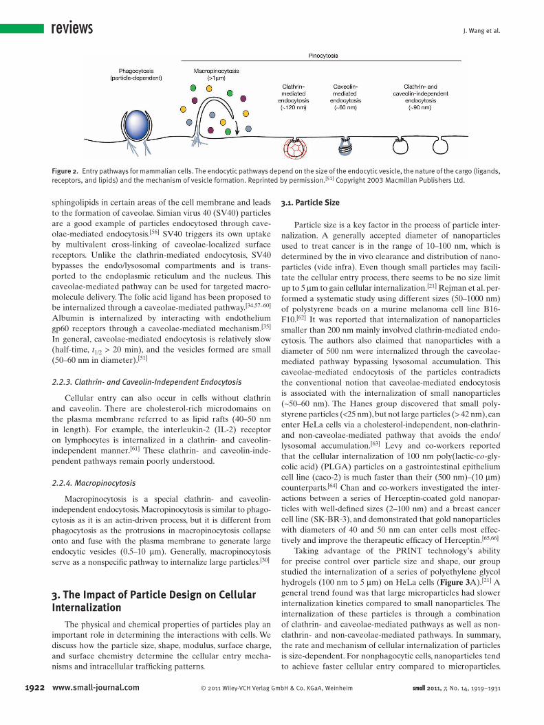
through a process called endocytosis . [ 51 ] There are two major
mechanisms of endocytosis ( Figure 2 ), which are phagocy-tosis (cell eating, the uptake process of large particles) and
pinocytosis (cell drinking, the uptake process for small par-
ticles, fl uid, and solutes). Phagocytosis primarily occurs in
macrophages and polymorphonuclear neutrophils (PMNs).
In contrast, pinocytosis occurs in all types of cells through
at least four distinct mechanisms: macropinocytosis, clathrin-
mediated endocytosis (CME), caveolae-mediated endocy-
tosis, and clathrin- and caveolae-independent endocytosis. All
of these cellular entry processes are highly regulated in order
to precisely control cellular responses to the environment. [ 51 ]
2.1. Phagocytosis
Macrophages and PMNs are professional “eaters” that
are capable of clearing foreign materials, pathogens, including
bacteria or yeasts, and cellular debris. A specifi c signaling cas-
cade triggers the assembly of actin, a globular 42-kDa pro-
tein, and the formation of cell-surface protrusions that engulf
the foreign material or cellular debris. [ 52,53 ] Macrophages can
engulf particles as large as 10 μ m in diameter and are one of
the major barriers that limit the effective delivery of particles
to the site of disease. In this review, we will mainly focus on
the detailed mechanisms of pinocytosis, which are more rel-
evant to the cellular internalization of nanomedicines.
bH & Co. KGaA, Weinheim small 2011, 7, No. 14, 1919–1931

More Effective Nanomedicines through Particle Design
Joseph DeSimone is the Chancellor’s
Eminent Professor of Chemistry at the
University of North Carolina at Chapel Hill,
William R. Kenan Jr. Professor of Chemical
Engineering at North Carolina State Univer-
sity, and an Adjunct Member at Memorial
Sloan-Kettering Cancer Center in New York.
DeSimone has published over 270 scientifi c
articles and has over 115 issued patents. In
2005 DeSimone was elected into the National
Academy of Engineering and the American
Academy of Arts and Sciences. He has
received over 40 major awards and recogni-
tions including the 2010 AAAS Mentor
Award, the 2009 NIH Director's Pioneer Award, and the 2008 Lemelson-MIT Prize.
2.2. Pinocytosis
Pinocytosis, or fl uid-phase uptake, is the most common
form of endocytosis and occurs in all cell types. [ 51 ] Solutes
can nonspecifi cally adsorb to the cell membrane to achieve
internalization (adsorptive pinocytosis). When solutes are
captured by specifi c high-affi nity receptors, they can be inter-
nalized through a receptor-mediated endocytosis (RME),
which is the most effi cient pinocytic pathway. Overall, the
pinocytotic pathway is determined by the surface interaction
of the particle and the cell.
2.2.1. Clathrin-Mediated Endocytosis (CME)
Clathrin-mediated endocytosis requires the formation of
coated pits by the assembly of clathrin, a protein that forms
a triskelion shape composed of three clathrin heavy chains
and three light chains. Coated pits develop into vesicles upon
endocytosis. Two major examples of CME are the internali-
zation of cholesterol-rich low-density lipoprotein (LDL)
particles through the LDL receptor and the internalization
of iron-loaded transferrin (Tf) through transferrin receptors
(TfR). It is worth noting that even though both LDL and Tf
are internalized through CME, their intracellular destinies
are distinct; LDL is delivered to lysosomes for degradation,
and Tf is recycled back to the cell surface through a recycling
mechanism. The fi nal destination of an endocytic vesicle is
not only determined by the internalization mechanism, but
also by receptor signaling. As such, the intracellular traf-
fi cking of nanoparticles conjugated with targeting ligands
© 2011 Wiley-VCH Verlag Gmbsmall 2011, 7, No. 14, 1919–1931
Figure 1 . Shape-specifi c PRINT particles. All particles were fabricated with
will not always be the same as the traffi cking pathway for
the free ligand. [ 26 , 54,55 ] Detailed examples will be discussed in
the next section. Vesicles formed by CME have an average
size of 120 nm and are typically directed towards the lyso-
some for degradation of their cargo. [ 51 ] Thus, for particles
internalized by CME, endosomal escape is necessary for
successful delivery of macromolecules.
2.2.2. Caveolae-Mediated Endocytosis
Caveolae are small fl ask-shaped (50–60 nm) invagina-
tions of the plasma membrane on many cell types, particu-
larly endothelial cells. [ 51 ] The structure of caveolae is created
by caveolin, a dimeric protein that binds cholesterol and
1921H & Co. KGaA, Weinheim www.small-journal.com
polyethylene glycol unless otherwise specifi ed.
J. Wang et al.
1922
reviews
Figure 2 . Entry pathways for mammalian cells. The endocytic pathways depend on the size of the endocytic vesicle, the nature of the cargo (ligands, receptors, and lipids) and the mechanism of vesicle formation. Reprinted by permission. [ 51 ] Copyright 2003 Macmillan Publishers Ltd.
sphingolipids in certain areas of the cell membrane and leads
to the formation of caveolae. Simian virus 40 (SV40) particles
are a good example of particles endocytosed through cave-
olae-mediated endocytosis. [ 56 ] SV40 triggers its own uptake
by multivalent cross-linking of caveolae-localized surface
receptors. Unlike the clathrin-mediated endocytosis, SV40
bypasses the endo/lysosomal compartments and is trans-
ported to the endoplasmic reticulum and the nucleus. This
caveolae-mediated pathway can be used for targeted macro-
molecule delivery. The folic acid ligand has been proposed to
be internalized through a caveolae-mediated pathway. [ 34 , 57–60 ]
Albumin is internalized by interacting with endothelium
gp60 receptors through a caveolae-mediated mechanism. [ 35 ]
In general, caveolae-mediated endocytosis is relatively slow
(half-time, t 1/2 > 20 min), and the vesicles formed are small
(50–60 nm in diameter). [ 51 ]
2.2.3. Clathrin- and Caveolin-Independent Endocytosis
Cellular entry can also occur in cells without clathrin
and caveolin. There are cholesterol-rich microdomains on
the plasma membrane referred to as lipid rafts (40–50 nm
in length). For example, the interleukin-2 (IL-2) receptor
on lymphocytes is internalized in a clathrin- and caveolin-
independent manner. [ 61 ] These clathrin- and caveolin-inde-
pendent pathways remain poorly understood.
2.2.4. Macropinocytosis
Macropinocytosis is a special clathrin- and caveolin-
independent endocytosis. Macropinocytosis is similar to phago-
cytosis as it is an actin-driven process, but it is different from
phagocytosis as the protrusions in macropinocytosis collapse
onto and fuse with the plasma membrane to generate large
endocytic vesicles (0.5–10 μ m). Generally, macropinocytosis
serve as a nonspecifi c pathway to internalize large particles. [ 30 ]
3. The Impact of Particle Design on Cellular Internalization
The physical and chemical properties of particles play an
important role in determining the interactions with cells. We
discuss how the particle size, shape, modulus, surface charge,
and surface chemistry determine the cellular entry mecha-
nisms and intracellular traffi cking patterns.
www.small-journal.com © 2011 Wiley-VCH Verlag Gm
3.1. Particle Size
Particle size is a key factor in the process of particle inter-
nalization. A generally accepted diameter of nanoparticles
used to treat cancer is in the range of 10–100 nm, which is
determined by the in vivo clearance and distribution of nano-
particles (vide infra). Even though small particles may facili-
tate the cellular entry process, there seems to be no size limit
up to 5 μ m to gain cellular internalization. [ 21 ] Rejman et al. per-
formed a systematic study using different sizes (50–1000 nm)
of polystyrene beads on a murine melanoma cell line B16-
F10. [ 62 ] It was reported that internalization of nanoparticles
smaller than 200 nm mainly involved clathrin-mediated endo-
cytosis. The authors also claimed that nanoparticles with a
diameter of 500 nm were internalized through the caveolae-
mediated pathway bypassing lysosomal accumulation. This
caveolae-mediated endocytosis of the particles contradicts
the conventional notion that caveolae-mediated endocytosis
is associated with the internalization of small nanoparticles
( ∼ 50–60 nm). The Hanes group discovered that small poly-
styrene particles ( < 25 nm), but not large particles ( > 42 nm), can
enter HeLa cells via a cholesterol-independent, non-clathrin-
and non-caveolae-mediated pathway that avoids the endo/
lysosomal accumulation. [ 63 ] Levy and co-workers reported
that the cellular internalization of 100 nm poly(lactic- co -gly-
colic acid) (PLGA) particles on a gastrointestinal epithelium
cell line (caco-2) is much faster than their (500 nm)–(10 μ m)
counterparts. [ 64 ] Chan and co-workers investigated the inter-
actions between a series of Herceptin-coated gold nanopar-
ticles with well-defi ned sizes (2–100 nm) and a breast cancer
cell line (SK-BR-3), and demonstrated that gold nanoparticles
with diameters of 40 and 50 nm can enter cells most effec-
tively and improve the therapeutic effi cacy of Herceptin. [ 65,66 ]
Taking advantage of the PRINT technology’s ability
for precise control over particle size and shape, our group
studied the internalization of a series of polyethylene glycol
hydrogels (100 nm to 5 μ m) on HeLa cells ( Figure 3 A). [ 21 ] A
general trend found was that large microparticles had slower
internalization kinetics compared to small nanoparticles. The
internalization of these particles is through a combination
of clathrin- and caveolae-mediated pathways as well as non-
clathrin- and non-caveolae-mediated pathways. In summary,
the rate and mechanism of cellular internalization of particles
is size-dependent. For nonphagocytic cells, nanoparticles tend
to achieve faster cellular entry compared to microparticles.
bH & Co. KGaA, Weinheim small 2011, 7, No. 14, 1919–1931

More Effective Nanomedicines through Particle Design
Figure 3 . a) Internalization profi le of PRINT particles with HeLa cells over a 4-h incubation period at 37 ° C. Legend depicts the particle diameter per particle volume. b) Probing the mechanisms of cellular internalization by using inhibitors of endocytosis. HeLa cells were incubated with the indicated inhibitors in the graph as outlined in the experimental methods. Percent internalization was normalized to particle internalization in the absence of inhibitors. Reproduced with permission. [ 21 ] Copyright 2008 National Academy of Science, USA.
Very small nanoparticles in the low tens of nanometer range
may utilize a non-clathrin- and non-caveolae-mediated
pathway avoiding endo/lysosomal degradation, which has sig-
nifi cant implications for delivery of macromolecules.
3.2. Particle Shape
As the majority of particles used for drug delivery are
fabricated using the bottom-up fabrication strategy and tend
to be spherical, there are a limited number studies evaluating
the relationship between particle shape and cellular inter-
nalization. The Mitragotri group fabricated a series of poly-
styrene particles with distinct size and shape by stretching
methods and discovered that particle shape, not size, plays a
key role in macrophage phagocytosis. [ 67–69 ] The tangent angles
of the particle surface at the point making initial contact with
macrophages need to be smaller than 45 ° to allow for particle
internalization. [ 67 ] If the contact angle is larger than 45 ° , the
cell can spread on the particle surface but cannot internalize
it. Taking advantage of these results, the same group devel-
oped a shape-switching PLGA particle to control the particle
phagocytosis by macrophages. [ 70 ] Ferrari and co-worker pre-
dicted that prolate ellipsoids are the most effectively attached
to macrophages but are the least effectively internalized. [ 71 ]
The groups of Mitragotri and Smith used prolate ellipsoids,
oblate ellipsoids, and spheres to study their particle shape
interactions with macrophages and demonstrated Ferrari's
prediction experimentally. [ 72 ]
© 2011 Wiley-VCH Verlag Gmbsmall 2011, 7, No. 14, 1919–1931
Some very interesting observations have been made using
PRINT particles by studying the internalization of a series
of polyethylene-glycol-based hydrogels (100 nm to 5 μ m) on
HeLa cells (Figure 3 A). [ 21 ] The internalization of the rodlike,
high-aspect-ratio (AR) nanoparticles (depth, d = 150 nm;
height, h = 450 nm; volume = 0.00795 μ m 3 ) occurs much
more rapidly and effectively than the cylindrical counterparts
( d = 200 nm, h = 200 nm, volume = 0.00628 μ m 3 ), even though
they have very similar volume, indicating the particle shape
plays a great role in the internalization process. To further
elucidate the cellular internalization mechanisms, the endo-
cytosis of three types of particles, 150 nm (AR = 3), 200 nm
(AR = 1), and 1 μ m (AR = 1), was investigated using dif-
ferent biochemical inhibitors of energy-dependent processes,
clathrin-mediated endocytosis, caveolae-mediated endocy-
tosis, and macropinocytosis (Figure 3 B). These particles were
internalized through an energy-dependent pathway based
on a NaN 3 inhibition experiment. In the presence of three
inhibitors for clathrin-mediated endocytosis, Dynasore, gen-
istein, and chlorpromazine, both 150 nm and 200 nm particles
showed a marked decrease in cell uptake; the internalization
of the 1 μ m particles was only signifi cantly affected by chlo-
rpromazine. It is clear that the nanoparticles, at least in part,
enter cells through clathrin-mediated pathway. But the cell
entry mechanism cannot be delineated for 1 μ m particles.
In the presence of two inhibitors for caveolae-mediated
endocytosis, genistein and β -cyclodextrin, the internaliza-
tion was retarded for the small nanoparticles but not the
large microparticles. Since caveolae have a cavity of ∼ 60 nm,
1923H & Co. KGaA, Weinheim www.small-journal.com

J. Wang et al.
192
reviews
microparticle internalization through a caveolae-mediatedpathway was not expected. In addition, inhibition of > 95%
particle internalization was not found for any inhibitor, indi-
cating the possiblity for non-clathrin- and non-caveolae-me-
diated pathways for internalization.
The Chan group reported that rodlike gold nanoparticles
(14 × 40 nm and 14 × 74 nm) entered cells less effectively com-
pared to their spherical counterparts (74 nm in diameter). [ 65 ]
But it is worth noting that the surface of the gold nano-
rods was stabilized with cetyl trimethylammonium bromide
(CTAB), which is different from the citric-acid-coated spher-
ical gold nanoparticles. The difference in surface chemistry
may explain the disparity in particle internalization. [ 65 ] In a
follow-up study by the same group, transferrin-coated gold
nanorods was also internalized more slowly than transferrin-
coated spherical gold nanoparticles. [ 73 ] The studies by the
DeSimone and Chan groups present contradictory results.
The studies of the impact of particle shape and cell interac-
tions are still in their infancy, and more systems need to be
explored.
3.3. Particle Modulus
The modulus of particles is another important parameter
to induce or prevent particle internalization. [ 40 ] Macrophages
are trained to clear bacteria and other pathogens, which usu-
ally have very rigid cell walls. Beningo and Wang reported
that soft polyacrylamide beads (1–6 μ m) frustrate the actin
fi lament formation by macrophages and subsequently pre-
vent phagocytosis, whereas their stiff counterparts can be
readily internalized. [ 74 ] However, Lee and co-workers showed
that rigid liposomes can decrease complement activation and
reduce subsequent macrophage uptake. [ 75 ] In a recent study
by Merkel et al., very soft red blood cell mimics can circulate
several days, a 30-fold increase of elimination half-life com-
pared to their rigid counterparts. [ 28 ] But both the soft and
rigid particles showed very minimal uptake on human umbil-
ical vein endothelial cells (HUVEC), probably due to their
large sizes. No conclusive relationship has emerged between
the particle modulus and cellular internalization process.
3.4. Particle Surface Charge
In general, nanoparticles with positively charged surfaces
can be effectively internalized by cells through electrostatic
interactions with the negatively charged cell plasma membrane
(adsorptive endocytosis, possibly involving CME). There are
numerous examples of positively charged particles for cellular
internalization, such as poly( l -lysine) (PLL)-modifi ed PLGA,
chitosan, and searylamine-coated PEG- co -PLA (poly(ethylene
glycol)- co -poly(lactic acid)). [ 38 , 76,77 ] The DeSimone group has
also reported that by keeping the size and shape of particles
constant, positively charged nanoparticles were internalized
in 84% of cells after 1-h incubation, whereas the negatively
charged counterparts were not internalized ( < 5%). [ 21 ]
On the other hand, negatively charged nanoparticles, such
as DOXIL and micelles are more likely to take advantage
4 www.small-journal.com © 2011 Wiley-VCH Verlag Gm
of caveolae-mediated pathways. [ 78 ] It also has been reported
that negatively charged PLGA nanoparticles ( ∼ 100 nm) can
enter cells through caveolae-independent pathways. [ 79–81 ] The
Mirkin group successfully demonstrated the use of 13 nm gold
nanoparticles for gene delivery. [ 82–84 ] The gold nanoparticles
were negatively charged, but they were effectively internalized
and delivered the nucleic acids to the cytosol and nucleus.
3.5. Particle Surface Chemistry
Surface modifi cation of particles is an area of intense
investigation for tissue- and cell-specifi c delivery. A number
of targeting strategies and surface chemistry have been
developed. [ 41 ] A general targeting strategy takes advantage
of the over-expression of certain cell surface receptors. Nano-
particles conjugated with molecules that can specifi cally bind
to the receptors are expected to boost the particle avidity (i.e.,
multivalent affi nity) to cells. Transferrin receptors are overex-
pressed on the majority of cancer cells and are widely used
as a cancer cell target. [ 85 ] Transferrin and transferrin receptor
antibodies have also been used for site-specifi c drug delivery
for various systems, including protein/toxin conjugates,
poly mer/drug conjugates, modifi ed viral vectors, liposomes/
polyplexes, and nanoparticles. [ 5 , 86,87 ] Two types of liposomal
formulations for targeted drug/gene delivery, MBP-426 and
SGT-53, that are currently under phase I clinical trials, uti-
lize transferrin and an anti-transferrin receptor single-chain
antibody fragment as targeting moieties, respectively. [ 5 ]
The Davis group developed the most successful targeted
delivery system to date for small interfering RNA (siRNA),
transferrin-conjugated cyclodextrin polymer-based nano-
particles (NPs) (CALAA-01), which is undergoing a phase
I clinical trial. [ 11 , 88–93 ] US Food and Drug Admnistration
(FDA)-approved monoclonal antibodies have also been used
as targeting ligands, such as Herceptin and Rituxan. [ 66 , 94,95 ]
The Schnitzer group reported that aminopeptidase-
P-antibody-conjugated gold nanoparticles can be trans-
ported into endothelium cells through a caveolae-mediate
mechanism. [ 35 , 96–99 ] Small peptides have also been explored
as targeting ligands, such as the RGD peptide (arginine–
glycine–aspartic acid). [ 100,101 ] Aptamers, short single-stranded
nucleic acids, are a class of new targeting ligands, which can
bind any target, including proteins and small molecules, with
very high affi nity. [ 102–104 ] The Langer and Farokhzad groups
developed nanoparticle–aptamer bioconjugates for cancer
targeting. [ 105–110 ] Small-molecule ligands can also be used as
targeting moieties. Pioneered by the Low group, folic acid has
been widely used as a targeting ligand for drug conjugates,
liposomes, and nanoparticles. [ 34 ] Sigma receptors ( σ 1, σ 2) have
been targeted by Huang and colleagues using anisamide as a
high-affi nity sigma-receptor ligand. [ 111–118 ] Liposomes conju-
gated with anisamide can substantially improve the delivery
of chemotherapeutics and siRNA to tumors. Mukherjee et al.
reported that haloperidol, another sigma-receptor ligand, can
increase DNA delivery effi ciency by tenfold to breast carci-
noma cells. [ 119 ]
Not only boosting avidity to cells, multivalent lig-
ands on nanoparticles can also have an impact on cell
bH & Co. KGaA, Weinheim small 2011, 7, No. 14, 1919–1931
More Effective Nanomedicines through Particle Design
biology that cannot be achieved by the monovalent form of
ligands. [ 26 , 66 , 120–122 ] Our group recently discovered that the
multivalent presentation of transferrin, the fourth most abun-
dant serum protein in humans, on nanoparticles can transform
this benign protein into a potential “drug-free” chemotherapy
against a B-cell lymphoma. [ 26 ] Kopecek and co-workers also
took advantage of the multivalent ligands to cross-link CD20
receptors to induce apoptosis in B cells. [ 123 ] Multivalent pres-
entation of certain monoclonal antibodies on nanoparticles
has been reported to enhance the therapeutic effi cacy of the
antibody. [ 66 , 95 ] Huang et al. fabricated dinitrophenyl-conjugated
gold nanoparticles with well-controlled ligand density and
showed that these multivalent nanoparticles can regulate sig-
naling in mast cells as a function of particle size and surface
ligand density. [ 124 ] Multivalent nanoparticles can also inhibit
human immunodefi ciency virus (HIV) fusion to human T
cells and kill multidrug-resistant bacteria, where the monova-
lent ligands did not show any biological activity. [ 121,122 ]
It is worth noting that the cellular traffi cking mechanisms for
multivalent-ligand-conjugated nanoparticles may be different
from the monovalent ligand. Endothelial cells express intercel-
lular adhesion molecule 1 (ICAM-1), but they do not internalize
ICAM-1 antibodies. However, anti-ICAM-1-antibody-coated
nanoparticles can be readily taken up by endothelial cells
through a unique cell adhesion molecule (CAM)-mediated
endocytosis, which is different from clathrin-, caveolae-, macropi-
nocytosis- and phagocytosis-mediated pathways. [ 125 ] Mukherjee
and co-workers also demonstrated that upon multivalent pres-
entation on nanoparticles, the patterning and dynamics of anti-
EGFR (epidermal growth factor receptor) antibody cetuximab
is distinct from its monovalent form. [ 126 ] Iversen and colleagues
showed that transferring-conjugated quantum dots were inter-
nalized through clathrin-mediated endocytosis, but the exocy-
tosis pathway of free transferrin was blocked. [ 55 ]
4. The Impact of Particle Design on Biodistribution and Pharmacokinetics
The size, shape, modulus, and surface chemistry of par-
ticles also play an important role in the biodistribution and
pharmacokinetics. Fundamental in vivo studies regarding
particle design enable the engineering of better medicines for
the treatment of a variety of diseases. There remains much to
be elucidated, but the impact of particle design has proven to
be important for drug delivery.
4.1. Particle Size
Particle size is an important component in the design of
long-circulating particle systems. As discussed previously,
the generally accepted diameter of nanomedicine for cancer
is in the range of 10–100 nm. The lower limit is determined
by the sieving coeffi cients for the glomerular capillary wall
in the kidney to avoid renal fi ltration. [ 127 ] Particles of sizes
ranging from 10 nm to 15 μ m have different pharmacokinetic
parameters and biodistribution. Under normal homeostatic
conditions, large particles are mechanically fi ltered through
© 2011 Wiley-VCH Verlag Gmsmall 2011, 7, No. 14, 1919–1931
the spleen and liver by the reticuloendothelial system (RES).
The RES, one of the body’s defense and fi ltration mecha-
nisms, functions to remove old or irregular red and white
blood cells, as well as opsonized constituents and large foreign
objects. Fenestrations in the spleen are typically 200–500 nm
in width, and thus, particles larger than 200 nm must com-
pensate by deformability. [ 42 ] For certain diseases, such as
cancer, size of particles can play a large role in accumula-
tion of the particles for therapeutic and diagnostic purposes.
Solid tumors typically present with aberrant angiogenic vas-
culature that enables passive accumulation of particles. In a
human colon adenocarcinoma xenograft model, the cutoff
size of the vascular pores was determined to be 400–600 nm
in diameter. [ 128 ] Characteristic solid tumors have higher inter-
stitial pressure in the center of the tumor compared to the
periphery. An outward convective fl ow reduces drug diffusion
to the center of the tumor, and particles and drugs that gain
interstitial access have higher retention times than in normal
tissues. This aberrant vasculature and higher interstitial fl uid
pressure create an enhanced permeability and retention of
the nanoparticles. Particles that are smaller than the fenestra-
tion can gain access and be retained in the tumor. [ 41,42 , 129 ]
4.2. Particle Shape
The importance of particle shape has derived the mor-
phological adaptation of pathogens in nature. The shape
of the pathogen serves a critical biological function, where
infectious and metastatic agents with different shapes, sizes,
and moduli can cope with and adapt to external conditions.
Examples in nature are infl uenza, ebola, and fi loviruses—
these are viruses with morphologies that have been subject to
the selective forces of survival and proliferation. [ 130 ]
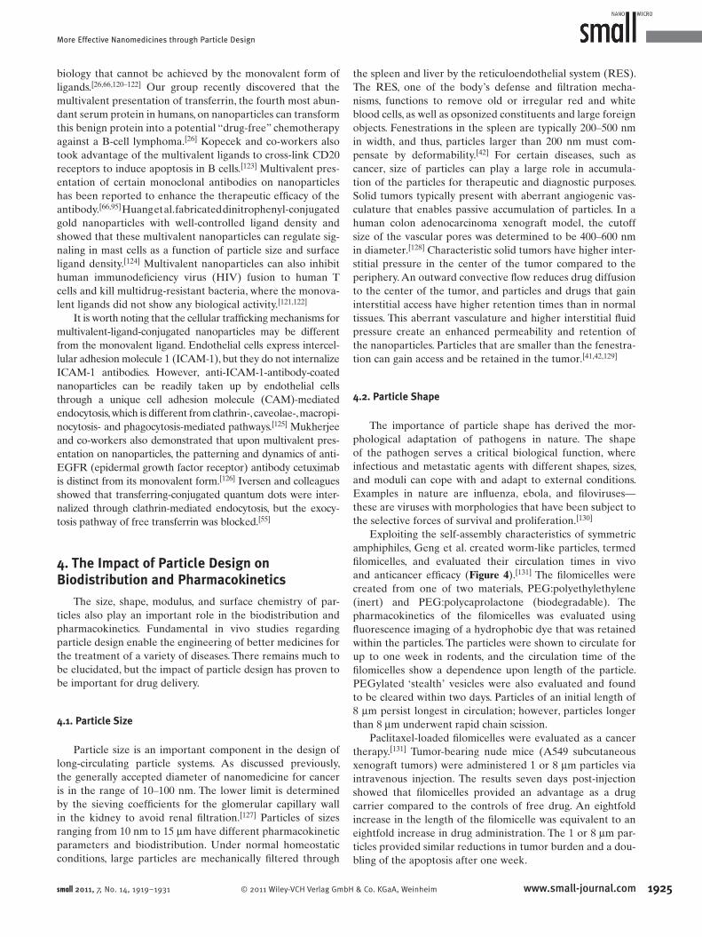
Exploiting the self-assembly characteristics of symmetric
amphiphiles, Geng et al. created worm-like particles, termed
fi lomicelles, and evaluated their circulation times in vivo
and anticancer effi cacy ( Figure 4 ). [ 131 ] The fi lomicelles were
created from one of two materials, PEG:polyethylethylene
(inert) and PEG:polycaprolactone (biodegradable). The
pharmacokinetics of the fi lomicelles was evaluated using
fl uorescence imaging of a hydrophobic dye that was retained
within the particles. The particles were shown to circulate for
up to one week in rodents, and the circulation time of the
fi lomicelles show a dependence upon length of the particle.
PEGylated ‘stealth’ vesicles were also evaluated and found
to be cleared within two days. Particles of an initial length of
8 μ m persist longest in circulation; however, particles longer
than 8 μ m underwent rapid chain scission.
Paclitaxel-loaded fi lomicelles were evaluated as a cancer
therapy. [ 131 ] Tumor-bearing nude mice (A549 subcutaneous
xenograft tumors) were administered 1 or 8 μ m particles via
intravenous injection. The results seven days post-injection
showed that fi lomicelles provided an advantage as a drug
carrier compared to the controls of free drug. An eightfold
increase in the length of the fi lomicelle was equivalent to an
eightfold increase in drug administration. The 1 or 8 μ m par-
ticles provided similar reductions in tumor burden and a dou-
bling of the apoptosis after one week.
1925bH & Co. KGaA, Weinheim www.small-journal.com
J. Wang et al.
1926
reviews
Figure 4 . Filomicelles and their persistent circulation. a) Filomicelles are self-assembled from diblock copolymers: yellow/green in cross-section indicates hydrophobic polymer, orange/blue is hydrophilic, and aqua is water. Electron microscopy demonstrates the nanometre-scale diameter of the fi lomicelles, and fl uorescence microscopy shows a single fi lomicelle. Distributions of fi lomicelle length are shown for two samples. b) Injection of fl uorescent fi lomicelles into rodents, followed by fl uorescent imaging of blood samples showed that fi lomicelles circulated in vivo for up to one week. c) Relative numbers of fi lomicelles in the circulation show that inert fi lomicelles (of OE70) persist when compared with stealth polymersomes and l-phage. d) Degradable fi lomicelles (of OCL3) also persist, and fi lomicelles with longer initial lengths ( L o ) circulate longer up to a limiting length. The error bars in c and d show the standard deviation for four or more animals. Reprinted by permission. [ 131 ] Copyright 2007 Macmillan Publishers Ltd.
Furthermore, the circulation times and endothelial cell
targeting of intravenously injected ICAM-1-targeted elliptical
disks (0.1 μ m × 1 μ m × 3 μ m) and ICAM-1-targeted spheres
(0.1, 1, 5, and 10 μ m) were evaluated in C57Bl/6 mice. [ 54 ] The
0.1 μ m ICAM-1-targeted spheres were rapidly cleared from
the blood post-injected, with 16.2 ± 2.7% of the injected dose
(% ID) and 5.2 ± 0.5% ID remaining in the blood 1 and 30 min
post-injection, respectively. Hepatic uptake of these carriers
was substantial at 43.7 ± 4.5% of the ID per gram of organ
tissue. Pulmonary uptake was signifi cant compared to the
nontargeted control spheres, showing 114.7 ± 11.1% ID/g
versus 10.2 ± 3.9% ID/g, respectively. The larger ICAM-
1-targeted spheres were cleared faster than the 0.1 μ m
ICAM-1-targeted spheres. The ICAM-1-targeted elliptical
disks remained in circulation longer than the spheres, with
25.5 ± 2.8% ID and 20.9 ± 1.6% ID remained in the blood at
www.small-journal.com © 2011 Wiley-VCH Verlag Gm
1 and 30 minutes post-injection, respectively. There was much
lower hepatic uptake (15.7 ± 2.3% ID/g) and signifi cant pul-
monary uptake (186.2 ± 15.4% ID/g). Thus, the elliptical disks
showed greater pulmonary targeting compared to spherical
particles. [ 54 ]
4.3. Particle Modulus
A difference between the moduli of metastatic cancer
cells and nonmetastatic cancer cells has been discovered, and
this difference in modulus has been thought to be a major
mechanism enabling these cells to relocate and take hold in
other parts of the body. [ 132,133 ]
Modulus was evaluated using red blood cell mimic
(RBCM) hydroxyethyl acrylate (HEA) hydrogel PRINT par-
bH & Co. KGaA, Weinheim small 2011, 7, No. 14, 1919–1931
More Effective Nanomedicines through Particle Design
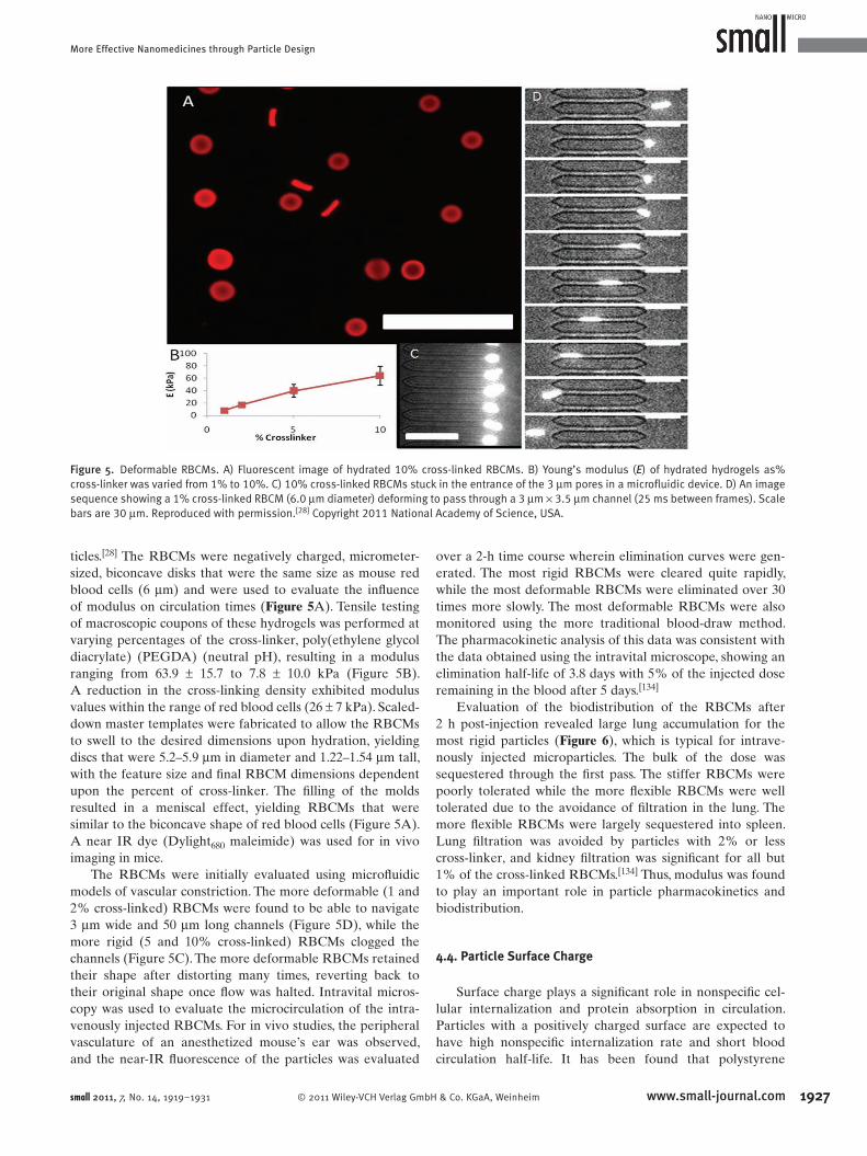
Figure 5 . Deformable RBCMs. A) Fluorescent image of hydrated 10% cross-linked RBCMs. B) Young’s modulus ( E ) of hydrated hydrogels as% cross-linker was varied from 1% to 10%. C) 10% cross-linked RBCMs stuck in the entrance of the 3 μ m pores in a microfl uidic device. D) An image sequence showing a 1% cross-linked RBCM (6.0 μ m diameter) deforming to pass through a 3 μ m × 3.5 μ m channel (25 ms between frames). Scale bars are 30 μ m. Reproduced with permission. [ 28 ] Copyright 2011 National Academy of Science, USA.
ticles. [ 28 ] The RBCMs were negatively charged, micrometer-
sized, biconcave disks that were the same size as mouse red
blood cells (6 μ m) and were used to evaluate the infl uence
of modulus on circulation times ( Figure 5 A). Tensile testing
of macroscopic coupons of these hydrogels was performed at
varying percentages of the cross-linker, poly(ethylene glycol
diacrylate) (PEGDA) (neutral pH), resulting in a modulus
ranging from 63.9 ± 15.7 to 7.8 ± 10.0 kPa (Figure 5 B).
A reduction in the cross-linking density exhibited modulus
values within the range of red blood cells (26 ± 7 kPa). Scaled-
down master templates were fabricated to allow the RBCMs
to swell to the desired dimensions upon hydration, yielding
discs that were 5.2–5.9 μ m in diameter and 1.22–1.54 μ m tall,
with the feature size and fi nal RBCM dimensions dependent
upon the percent of cross-linker. The fi lling of the molds
resulted in a meniscal effect, yielding RBCMs that were
similar to the biconcave shape of red blood cells (Figure 5 A).
A near IR dye (Dylight 680 maleimide) was used for in vivo
imaging in mice.
The RBCMs were initially evaluated using microfl uidic
models of vascular constriction. The more deformable (1 and
2% cross-linked) RBCMs were found to be able to navigate
3 μ m wide and 50 μ m long channels (Figure 5 D), while the
more rigid (5 and 10% cross-linked) RBCMs clogged the
channels (Figure 5 C). The more deformable RBCMs retained
their shape after distorting many times, reverting back to
their original shape once fl ow was halted. Intravital micros-
copy was used to evaluate the microcirculation of the intra-
venously injected RBCMs. For in vivo studies, the peripheral
vasculature of an anesthetized mouse’s ear was observed,
and the near-IR fl uorescence of the particles was evaluated
© 2011 Wiley-VCH Verlag Gmsmall 2011, 7, No. 14, 1919–1931
over a 2-h time course wherein elimination curves were gen-
erated. The most rigid RBCMs were cleared quite rapidly,
while the most deformable RBCMs were eliminated over 30
times more slowly. The most deformable RBCMs were also
monitored using the more traditional blood-draw method.
The pharmacokinetic analysis of this data was consistent with
the data obtained using the intravital microscope, showing an
elimination half-life of 3.8 days with 5% of the injected dose
remaining in the blood after 5 days. [ 134 ]
Evaluation of the biodistribution of the RBCMs after
2 h post-injection revealed large lung accumulation for the
most rigid particles ( Figure 6 ), which is typical for intrave-
nously injected microparticles. The bulk of the dose was
sequestered through the fi rst pass. The stiffer RBCMs were
poorly tolerated while the more fl exible RBCMs were well
tolerated due to the avoidance of fi ltration in the lung. The
more fl exible RBCMs were largely sequestered into spleen.
Lung fi ltration was avoided by particles with 2% or less
cross-linker, and kidney fi ltration was signifi cant for all but
1% of the cross-linked RBCMs. [ 134 ] Thus, modulus was found
to play an important role in particle pharmacokinetics and
biodistribution.
4.4. Particle Surface Charge
Surface charge plays a signifi cant role in nonspecifi c cel-
lular internalization and protein absorption in circulation.
Particles with a positively charged surface are expected to
have high nonspecifi c internalization rate and short blood
circulation half-life. It has been found that polystyrene
1927bH & Co. KGaA, Weinheim www.small-journal.com

J. Wang et al.
1928
reviews
Figure 6 . Tissue distribution of RBCMs. A) Distribution of RBCMs into various tissues 2 h postdosing by percent recovered dose normalized for tissue weight. B) Lung tissue from a mouse dosed with 10% cross-linked RBCMs. Particles are shown in red, with cell nuclei in purple and cytosleton (F-actin) stained green. C) Lung tissue from a mouse dosed with 1% cross-linked RBCMs, with tissue stained as in B. Very few RBCMs were in this tissue compared to mice dosed with more rigid particles, consistent with the tissue distribution data. Scale bars are 50 μ m. Reproduced with permission. [ 28 ] Copyright 2011 National Academy of Science, USA.
microparticles with primary amine surface functionalities
undergo substantially more phagocytosis compared to the
same particles with carboxyl, sulfate, and hydroxyl groups. [ 135 ]
Yamamoto et al. evaluated surface charge on poly(ethylene
glycol)–poly( d , l -lactide) block copolymer micelles using
neutral (tyrosine) and negative (tyrosine–glutamine) func-
tionalities. No difference was found between blood clearance
kinetics, but the negatively-charged micelles displayed a 10 ×
lower accumulation in liver and spleen 4 h post-intravenous
administration. [ 136 ] He et al. evaluated negatively charged
(rhodamine-B-labeled carboxymethyl chitosan grafted) nano-
particles and positively charged (chitosan-hydrochloride-grafted
nanoparticles) for cellular uptake and biodistribution. [ 137 ] Par-
ticles with a surface charge below 15 mV showed a reduction
in phagocytic uptake by murine macrophages, and the particles
with lower surface charges promoted longer blood residence
time and higher accumulation in the tumors of subcutaneous
xenograft H-22 tumor-bearing mice.
4.5. Particle Surface Chemistry
The surface properties of a particle can affect its interac-
tions with molecules, cells, or physiological systems in the
body and impart a variety of desirable characteristics for the
www.small-journal.com © 2011 Wiley-VCH Verlag GmbH & Co. KGaA, Weinh
delivery of therapeutics and diagnostics. For
example, the chemical conjugation of PEG
groups onto the surface of particles has
enabled stealthlike properties when in cir-
culation. The PEG groups have the ability
to reduce nonspecifi c uptake by cells, to
diminish adsorption by proteins and other
biomolecules in the serum, and to elude
phagocytosis by macrophages, all of which
contribute to longer circulation time. [ 138 ]
Another desirable characteristic for parti-
cles in biomedical applications is the ability
to selectively interact with specifi c cell
types such as cancer cells. This selectivity
can ensure that an effective amount of the
therapeutic payload will be delivered to the
target cells of interest, thereby minimizing
the potential side effects that accompany
intravenous chemotherapy. Li and Huang
have proposed that ideal particle delivery
systems transport to the target site at > 10%
injected dose in 4 h and are cleared from
circulation between 4 and 10 h for safety.
PEG density plays an important role, and
for the treatment of cancer, it has been pro-
posed that high-density and sheddable PEG
chains are the key for tumor targeting. [ 129 ]
Surface modifi cation with targeting
moieties is advantageous for cellular
endocytosis after particle accumulation
in the site of interest. Weissleder et al.
used a combinatorial approach to fi nd tar-
geting moieties for cells of interest. This
approach involved the creation of 146 iron
oxide nanoparticles decorated with different synthetic small
molecules. [ 139 ] The library created was tested for specifi city for
endothelial cells (HUVEC), activated human macrophages
(U937), and pancreatic cancer cells (PaCa-2), and small mol-
ecules were delineated for their specifi city to each system.
Fourteen candidates showed signifi cant uptake in the pan-
creatic cancer cells, and from these fourteen, two candidates
exhibited high pancreatic cancer cell uptake and low macro-
phage/endothelial cell uptake. The two candidates were isatoic
anhydride (designated 261-15-28) and 5-chloro-isatoic anhy-
dride (261-14-17). Fluorescently labeled candidates (CLIO-
isatoic-Cy5.5) were intravenously injected into subcutaneous
xenograft mice and were compared with a control group
CLIO-NH 2 -Cy3.5 using fl uorescence imaging (target-to-back-
ground ratios (TBR) of 1.62 versus 0.16, statistical p value <
0.0001). The results proved an increase in pancreatic cancer
detection in the mouse model using the targeting moiety. [ 139 ]
5. Conclusion
In summary, the following physical parameters for effec-
tive nanomedicines are recommended:
• Size: Particles in the range of 10–100 nm can avoid
renal fi ltration and are small enough to effi ciently
eim small 2011, 7, No. 14, 1919–1931

More Effective Nanomedicines through Particle Design
[ 1 ] K. Riehemann , S. Schneider , T. Luger , B. Godin , M. Ferrari , H. Fuchs , Angew. Chem. Int. Ed. 2009 , 48 , 872 .
[ 2 ] R. Singh , J. Lillard , Exp. Molecular Pathol. 2009 , 86 , 215 . [ 3 ] P. Debbage , Curr. Pharm. Des. 2009 , 15 , 153 . [ 4 ] M. Muthu , S. Singh , Nanomedicine 2009 , 4 , 105 . [ 5 ] J. R. Heath , M. E. Davis , Annu. Rev. Med. 2008 , 59 , 251 . [ 6 ] N. Doshi , S. Mitragotri , Adv. Funct. Mater. 2009 , 19 , 3843 . [ 7 ] C. L. W. Chaffer , A. Robert , Science 2011 , 331 , 1559 . [ 8 ] E. Marshall , Science 2011 , 331 , 1540 . [ 9 ] M. R. Stratton , Science 2011 , 331 , 1553 . [ 10 ] M. E. Davis , Z. G. Chen , D. M. Shin , Nat. Rev. Drug Discov. 2008 ,
7 , 771 . [ 11 ] M. E. Davis , Mol. Pharm. 2009 , 6 , 659 . [ 12 ] C. S. Thaxton , W. L. Daniel , D. A. Giljohann , A. D. Thomas ,
C. A. Mirkin , J. Am Chem. Soc. 2009 , 131 , 1384 . [ 13 ] A. J. Luthi , P. C. Patel , C. H. Ko , R. K. Mutharasan , C. A. Mirkin ,
C. S. Thaxton , Trends Mol. Med. 2010 , 16 , 553 . [ 14 ] H. Nalwa , Nanostructured Materials and Nanotechnology ,
Academic Press , San Diego, CA, USA 2002 . [ 15 ] D. E. Discher , F. Ahmed , Annu. Rev. Biomed. Eng. 2006 , 8 , 323 . [ 16 ] M. L. Immordino , F. Dosio , L. Cattel , Int. J. Nanomed. 2006 , 1 , 297 .
accumulate in tumors through the EPR effect. How-
ever, microparticles may be better suited for vascular
targeting than nanoparticles.
• Shape: Certain shape specifi c delivery systems have
demonstrated advantages over spherical systems.
Filamentous particles can help evade macrophage up-
take, and shape-specifi c microparticles can enhance
vascular adhesion.
• Modulus: Soft particles can navigate through pores
smaller than the size of particles and achieve extended
circulation.
• Surface charge and surface chemistry: Neutral
PEGylated particles are ideal to avoid macrophage
uptake. Introducing targeting ligands can also poten-
tially enhance cancer cell uptake.
Future studies in this fi eld should focus on creating more
effective nanomedicines by designing particles to target cer-
tain organs, control particle intracellular traffi cking pathways
and deliver cargo to intracellular organelles.
Acknowledgements
This work was supported in part by the STC Program of the National Science Foundation (CHE-9876674), National Institutes of Health Program Project Grant PO1-GM059299, National Institutes of Health Grants U54-CA119343 (the Carolina Center of Cancer Nano technology Excellence) and R01-EB009565, Prostate Cancer Foundation, University of North Carolina Cancer Research Fund, the Chancellor's Eminent Professorship and William R. Kenan Professorship at the University of North Carolina at Chapel Hill, and a sponsored research agreement with Liquidia Technologies.
This Review is part of the Special Issue dedicated to Chad Mirkin celebrating 20 years of infl uential research at Northwestern University.
© 2011 Wiley-VCH Verlag Gmbsmall 2011, 7, No. 14, 1919–1931
[ 17 ] T. J. Merkel , K. P. Herlihy , J. Nunes , R. M. Orgel , J. P. Rolland , J. M. DeSimone , Langmuir 2010 , 26 , 13086 .
[ 18 ] J. Rolland , B. Maynor , L. Euliss , A. Exner , G. Denison , J. DeSimone , J. Am. Chem. Soc. 2005 , 127 , 10096 .
[ 19 ] L. Euliss , J. DuPont , S. Gratton , J. DeSimone , Chem. Soc. Rev. 2006 , 35 , 1095 .
[ 20 ] S. E. Gratton , M. E. Napier , P. A. Ropp , S. Tian , J. M. DeSimone , Pharmaceutical Res. 2008 , 25 , 2845 .
[ 21 ] S. E. A. Gratton , P. A. Ropp , P. D. Pohlhaus , J. C. Luft , V. J. Madden , M. E. Napier , J. M. DeSimone , Proc. Natl. Acad. Sci. USA 2008 , 105 , 11613 .
[ 22 ] J. Kelly , J. DeSimone , J. Am. Chem. Soc. 2008 , 130 , 5438 . [ 23 ] H. Zhang , J. Nunes , S. Gratton , K. Herlihy , P. Pohlhaus ,
J. DeSimone , New J. Phys. 2009 , 11 , 075018 . [ 24 ] W. Jeong , M. E. Napier , J. M. DeSimone , Nanomedicine (Lond)
2010 , 5 , 633 . [ 25 ] M. C. Parrott , J. C. Luft , J. D. Byrne , J. H. Fain , M. E. Napier ,
J. M. Desimone , J. Am Chem. Soc. 2010 , 132 , 17928 . [ 26 ] J. Wang , S. Tian , R. A. Petros , M. E. Napier , J. M. Desimone , J. Am
Chem. Soc. 2010 , 132 , 11306 . [ 27 ] E. M. Enlow , J. C. Luft , M. E. Napier , J. M. Desimone , Nano Lett.
2011 , 11 , 808 . [ 28 ] T. J. Merkel , S. W. Jones , K. P. Herlihy , F. R. Kersey , A. R. Shields ,
M. Napier , J. C. Luft , H. Wu , W. C. Zamboni , A. Z. Wang , J. E. Bear , J. M. DeSimone , Proc. Natl. Acad. Sci. USA 2011 , 108 , 586 .
[ 29 ] A. Verma , F. Stellacci , Small 2010 , 6 , 12 . [ 30 ] G. Sahay , D. Y. Alakhova , A. V. Kabanov , J. Control. Release 2010 ,
145 , 182 . [ 31 ] S. Park , K. Hamad-Schifferli , Curr. Opin. Chem. Biol. 2010 , 14 ,
616 . [ 32 ] H. Hillaireau , P. Couvreur , Cell Mol. Life Sci. 2009 , 66 , 2873 . [ 33 ] L. M. Bareford , P. W. Swaan , Adv. Drug Deliv. Rev. 2007 , 59 ,
748 . [ 34 ] Y. Lu , P. S. Low , Adv. Drug Deliv. Rev. 2002 , 54 , 675 . [ 35 ] J. E. Schnitzer , Adv. Drug Deliv. Rev. 2001 , 49 , 265 . [ 36 ] D. E. Discher , A. Eisenberg , Science 2002 , 297 , 967 . [ 37 ] J. W. Yoo , E. Chambers , S. Mitragotri , Curr. Pharm. Des. 2010 ,
16 , 2298 . [ 38 ] O. Harush-Frenkel , Y. Altschuler , S. Benita , Crit. Rev. Ther. Drug.
Carrier. Syst. 2008 , 25 , 485 . [ 39 ] N. Rosi , C. Mirkin , Chem. Rev. 2005 , 105 , 1547 . [ 40 ] A. V. Kabanov , S. V. Vinogradov , Angew. Chem. Int. Ed. Engl.
2009 , 48 , 5418 . [ 41 ] J. D. Byrne , T. Betancourt , L. Brannon-Peppas , Adv. Drug Deliv.
Rev. 2008 , 60 , 1615 . [ 42 ] R. A. Petros , J. M. DeSimone , Nat. Rev. Drug Discov. 2010 , 9 ,
615 . [ 43 ] W. Sanhai , J. Sakamoto , R. Canady , M. Ferrari , Nat. Nanotechnol.
2008 , 3 , 242 . [ 44 ] L. Y. Chou , K. Ming , W. C. Chan , Chem. Soc. Rev. 2011 , 40 , 233 . [ 45 ] P. Decuzzi , M. Ferrari , Biomaterials 2008 , 29 , 377 . [ 46 ] S.-Y. Lee , M. Ferrari , P. Decuzzi , J. Biomechanics 2009 , 42 ,
1885 . [ 47 ] S.-Y. Lee , M. Ferrari , P. Decuzzi , Nanotechnology 2009 , 20 ,
495101 . [ 48 ] R. E. Serda , J. Gu , R. C. Bhavane , X. Liu , C. Chiappini , P. Decuzzi ,
M. Ferrari , Biomaterials 2009 , 30 , 2440 . [ 49 ] P. Decuzzi , B. Godin , T. Tanaka , S. Y. Lee , C. Chiappini , X. Liu ,
M. Ferrari , J. Control. Release 2010 , 141 , 320 . [ 50 ] P. Decuzzi , M. Ferrari , Biomaterials 2006 , 27 , 5307 . [ 51 ] S. D. Conner , S. L. Schmid , Nature 2003 , 422 , 37 . [ 52 ] A. Aderem , D. M. Underhill , Annu. Rev. Immunol. 1999 , 17 ,
593 . [ 53 ] G. Chimini , P. Chavrier , Nat. Cell Biol. 2000 , 2 , E191 . [ 54 ] S. Muro , C. Garnacho , J. A. Champion , J. Leferovich , C. Gajewski ,
E. H. Schuchman , S. Mitragotri , V. R. Muzykantov , Mol. Ther. 2008 , 16 , 1450 .
1929H & Co. KGaA, Weinheim www.small-journal.com
J. Wang et al.
193
reviews
[ 55 ] C. Tekle , B. Deurs , K. Sandvig , T. G. Iversen , Nano Lett. 2008 , 8 ,1858 . [ 56 ] L. Pelkmans , D. Puntener , A. Helenius , Science 2002 , 296 , 535 . [ 57 ] K. G. Rothberg , Y. S. Ying , J. F. Kolhouse , B. A. Kamen ,
R. G. Anderson , J. Cell Biol. 1990 , 110 , 637 . [ 58 ] S. Mayor , K. G. Rothberg , F. R. Maxfi eld , Science 1994 , 264 ,
1948 . [ 59 ] M. Wu , J. Fan , W. Gunning , M. Ratnam , J. Membr. Biol. 1997 ,
159 , 137 . [ 60 ] R. Varma , S. Mayor , Nature 1998 , 394 , 798 . [ 61 ] C. Lamaze , A. Dujeancourt , T. Baba , C. G. Lo , A. Benmerah ,
A. Dautry-Varsat , Mol. Cell 2001 , 7 , 661 . [ 62 ] J. Rejman , V. Oberle , I. S. Zuhorn , D. Hoekstra , Biochem. J. 2004 ,
377 , 159 . [ 63 ] S. K. Lai , K. Hida , S. T. Man , C. Chen , C. Machamer , T. A. Schroer ,
J. Hanes , Biomaterials 2007 , 28 , 2876 . [ 64 ] M. P. Desai , V. Labhasetwar , E. Walter , R. J. Levy , G. L. Amidon ,
Pharm Res 1997 , 14 , 1568 . [ 65 ] B. D. Chithrani , A. A. Ghazani , W. C. Chan , Nano Lett. 2006 , 6 ,
662 . [ 66 ] W. Jiang , B. Y. Kim , J. T. Rutka , W. C. Chan , Nat. Nanotechnol.
2008 , 3 , 145 . [ 67 ] J. A. Champion , S. Mitragotri , Proc. Natl. Acad. Sci. USA 2006 ,
103 , 4930 . [ 68 ] N. Doshi , S. Mitragotri , PLoS One 2010 , 5 , e10051 . [ 69 ] J. A. Champion , Y. K. Katare , S. Mitragotri , Proc. Natl. Acad. Sci.
USA 2007 , 104 , 11901 . [ 70 ] J. W. Yoo , S. Mitragotri , Proc. Natl. Acad. Sci. USA 2010 , 107 ,
11205 . [ 71 ] P. Decuzzi , M. Ferrari , Biophys. J. 2008 , 94 , 3790 . [ 72 ] G. Sharma , D. T. Valenta , Y. Altman , S. Harvey , H. Xie ,
S. Mitragotri , J. W. Smith , J. Control. Release 2010 , 147 , 408 .
[ 73 ] B. D. Chithrani , W. C. Chan , Nano Lett. 2007 , 7 , 1542 . [ 74 ] K. A. Beningo , Y. L. Wang , J. Cell Sci. 2002 , 115 , 849 . [ 75 ] T. M. Allen , G. A. Austin , A. Chonn , L. Lin , K. C. Lee , Biochim.
Biophys. Acta 1991 , 1061 , 56 . [ 76 ] O. Harush-Frenkel , N. Debotton , S. Benita , Y. Altschuler , Bio-
chem. Biophys. Res. Commun. 2007 , 353 , 26 . [ 77 ] O. Harush-Frenkel , E. Rozentur , S. Benita , Y. Altschuler , Biomac-
romolecules 2008 , 9 , 435 . [ 78 ] G. Sahay , J. O. Kim , A. V. Kabanov , T. K. Bronich , Biomaterials
2010 , 31 , 923 . [ 79 ] J. Panyam , V. Labhasetwar , Pharm. Res. 2003 , 20 , 212 . [ 80 ] J. Panyam , S. K. Sahoo , S. Prabha , T. Bargar , V. Labhasetwar , Int.
J. Pharm. 2003 , 262 , 1 . [ 81 ] M. G. Qaddoumi , H. Ueda , J. Yang , J. Davda , V. Labhasetwar ,
V. H. Lee , Pharm. Res. 2004 , 21 , 641 . [ 82 ] N. Rosi , D. Giljohann , C. Thaxton , A. Lytton-Jean , M. Han ,
C. Mirkin , Science 2006 , 312 , 1027 . [ 83 ] D. Giljohann , D. Seferos , A. Prigodich , P. Patel , C. Mirkin , J. Am.
Chem. Soc. 2009 , 131 , 2072 . [ 84 ] D. Zheng , D. S. Seferos , D. A. Giljohann , P. C. Patel , C. A. Mirkin ,
Nano Lett. 2009 , 9 , 3258 . [ 85 ] H. Li , Z. M. Qian , Med. Res. Rev. 2002 , 22 , 225 . [ 86 ] T. R. Daniels , T. Delgado , G. Helguera , M. L. Penichet , Clin.
Immunol. 2006 , 121 , 159 . [ 87 ] T. R. Daniels , T. Delgado , J. A. Rodriguez , G. Helguera ,
M. L. Penichet , Clin. Immunol. 2006 , 121 , 144 . [ 88 ] N. C. Bellocq , S. H. Pun , G. S. Jensen , M. E. Davis , Bioconjugate
Chem. 2003 , 14 , 1122 . [ 89 ] S. Hu-Lieskovan , J. D. Heidel , D. W. Bartlett , M. E. Davis ,
T. J. Triche , Cancer Res. 2005 , 65 , 8984 . [ 90 ] D. Bartlett , M. Davis , Bioconjugate Chem. 2007 , 18 , 456 . [ 91 ] J. D. Heidel , Z. Yu , J. Y. Liu , S. M. Rele , Y. Liang , R. K. Zeidan ,
D. J. Kornbrust , M. E. Davis , Proc. Natl. Acad. Sci. USA 2007 , 104 , 5715 .
0 www.small-journal.com © 2011 Wiley-VCH Verlag Gm
[ 92 ] C. H. J. Choi , C. A. Alabi , P. Webster , M. E. Davis , Proc. Natl. Acad. Sci. USA 2010 , 107 , 1235 .
[ 93 ] M. E. Davis , J. E. Zuckerman , C. H. Choi , D. Seligson , A. Tolcher , C. A. Alabi , Y. Yen , J. D. Heidel , A. Ribas , Nature 2010 , 464 , 1067 .
[ 94 ] O. Ishida , K. Maruyama , H. Tanahashi , M. Iwatsuru , K. Sasaki , M. Eriguchi , H. Yanagie , Pharm. Res. 2001 , 18 , 1042 .
[ 95 ] G. N. Chiu , L. A. Edwards , A. I. Kapanen , M. M. Malinen , W. H. Dragowska , C. Warburton , G. G. Chikh , K. Y. Fang , S. Tan , J. Sy , C. Tucker , D. N. Waterhouse , R. Klasa , M. B. Bally , Mol. Cancer Ther. 2007 , 6 , 844 .
[ 96 ] D. P. McIntosh , X. Y. Tan , P. Oh , J. E. Schnitzer , Proc. Natl. Acad. Sci. USA 2002 , 99 , 1996 .
[ 97 ] P. Oh , Y. Li , J. Yu , E. Durr , K. M. Krasinska , L. A. Carver , J. E. Testa , J. E. Schnitzer , Nature 2004 , 429 , 629 .
[ 98 ] P. Oh , P. Borgstrom , H. Witkiewicz , Y. Li , B. J. Borgstrom , A. Chrastina , K. Iwata , K. R. Zinn , R. Baldwin , J. E. Testa , J. E. Schnitzer , Nat. Biotechnol. 2007 , 25 , 327 .
[ 99 ] P. Valadon , B. Darsow , T. N. Buss , M. Czarny , N. M. Griffi n , H. N. Nguyen , P. Oh , P. Borgstrom , A. Chrastina , J. E. Schnitzer , J. Biol. Chem. 2010 , 285 , 713 .
[ 100 ] E. Garanger , D. Boturyn , P. Dumy , Anticancer Agents Med. Chem. 2007 , 7 , 552 .
[ 101 ] G. Ferro-Flores , M. Ramirez Fde , L. Melendez-Alafort , C. L. Santos-Cuevas , Mini Rev. Med. Chem. 2010 , 10 , 87 .
[ 102 ] S. Jayasena , Clin. Chem. 1999 , 45 , 1628 . [ 103 ] M. Golden , B. Collins , M. Willis , T. Koch , J. Biotechnol. 2000 , 81 ,
167 . [ 104 ] D. Bunka , P. Stockley , Nat. Rev. Microbiol. 2006 , 4 , 588 . [ 105 ] O. C. Farokhzad , S. Jon , A. Khademhosseini , T. N. Tran ,
D. A. Lavan , R. Langer , Cancer Res. 2004 , 64 , 7668 . [ 106 ] O. C. Farokhzad , J. Cheng , B. A. Teply , I. Sherifi , S. Jon , P. W. Kantoff ,
J. P. Richie , R. Langer , Proc. Natl. Acad. Sci. USA 2006 , 103 , 6315 .
[ 107 ] O. C. Farokhzad , J. M. Karp , R. Langer , Expert Opin. Drug Deliv. 2006 , 3 , 311 .
[ 108 ] S. Dhar , F. X. Gu , R. Langer , O. C. Farokhzad , S. J. Lippard , Proc. Natl. Acad. Sci. USA 2008 , 105 , 17356 .
[ 109 ] F. Gu , L. Zhang , B. A. Teply , N. Mann , A. Wang , A. F. Radovic-Moreno , R. Langer , O. C. Farokhzad , Proc. Natl. Acad. Sci. USA 2008 , 105 , 2586 .
[ 110 ] N. Kolishetti , S. Dhar , P. M. Valencia , L. Q. Lin , R. Karnik , S. J. Lippard , R. Langer , O. C. Farokhzad , Proc. Natl. Acad. Sci. USA 2010 , 107 , 17939 .
[ 111 ] S. D. Li , L. Huang , Mol. Pharm. 2006 , 3 , 579 . [ 112 ] S. Chono , S. D. Li , C. C. Conwell , L. Huang , J. Control. Release
2008 , 131 , 64 . [ 113 ] S. D. Li , Y. C. Chen , M. J. Hackett , L. Huang , Mol. Ther. 2008 , 16 ,
163 . [ 114 ] S. D. Li , S. Chono , L. Huang , Mol. Ther. 2008 , 16 , 942 . [ 115 ] S. D. Li , S. Chono , L. Huang , J. Control. Release 2008 , 126 , 77 . [ 116 ] Y. Chen , J. Sen , S. R. Bathula , Q. Yang , R. Fittipaldi , L. Huang ,
Mol. Pharm. 2009 , 6 , 696 . [ 117 ] Y. Chen , S. R. Bathula , Q. Yang , L. Huang , J. Invest. Dermatol.
2010 , 130 , 2790 . [ 118 ] O. Nakagawa , X. Ming , L. Huang , R. L. Juliano , J. Am Chem. Soc.
2010 , 132 , 8848 . [ 119 ] A. Mukherjee , T.K. Prasad , N. M. Rao , R. Banerjee , J. Biol. Chem.
2005 , 280 , 15619 . [ 120 ] L. L. Kiessling , J. E. Gestwicki , L. E. Strong , Angew. Chem. Int. Ed.
Engl. 2006 , 45 , 2348 . [ 121 ] Y. Zhao , Y. Tian , Y. Cui , W. Liu , W. Ma , X. Jiang , J. Am Chem. Soc.
2010 , 132 , 1 2349 . [ 122 ] M. Bowman , T. Ballard , C. Ackerson , D. Feldheim , D. Margolis ,
C. Melander , J. Am. Chem. Soc. 2008 , 130 , 6896 . [ 123 ] K. Wu , J. Liu , R. N. Johnson , J. Yang , J. Kopecek , Angew. Chem.
Int. Ed. Engl. 2010 , 49 , 1451 .
bH & Co. KGaA, Weinheim small 2011, 7, No. 14, 1919–1931
More Effective Nanomedicines through Particle Design
[ 124 ] Y. F. Huang , H. Liu , X. Xiong , Y. Chen , W. Tan , J. Am. Chem. Soc. 2009 , 131 , 17328 .
[ 125 ] S. Muro , C. Garnacho , J. A. Champion , J. Leferovich , C. Gajewski , E. H. Schuchman , S. Mitragotri , V. R. Muzykantov , Mol. Ther. 2008 , 16 , 1450 .
[ 126 ] S. Bhattacharyya , R. Bhattacharya , S. Curley , M. A. McNiven , P. Mukherjee , Proc. Natl. Acad. Sci. USA 2010 , 107 , 14541 .
[ 127 ] D. Venturoli , B. Rippe , Am. J. Physiol. Renal Physiol. 2005 , 288 , F605 . [ 128 ] F. Yuan , M. Dellian , D. Fukumura , M. Leunig , D. A. Berk ,
V. P. Torchilin , R. K. Jain , Cancer Res. 1995 , 55 , 3752 . [ 129 ] S. D. Li , L. Huang , J. Control. Release 2010 , 145 , 178 . [ 130 ] K. D. Young , Curr. Opin. Microbiol. 2007 , 10 , 596 . [ 131 ] Y. Geng , P. Dalhaimer , S. Cai , R. Tsai , M. Tewari , T. Minko ,
D. E. Discher , Nat. Nanotechnol. 2007 , 2 , 249 . [ 132 ] J. Guck , S. Schinkinger , B. Lincoln , F. Wottawah , S. Ebert ,
M. Romeyke , D. Lenz , H. M. Erickson , R. Ananthakrishnan , D. Mitchell , J. Kas , S. Ulvick , C. Bilby , Biophys. J. 2005 , 88 , 3689 .
© 2011 Wiley-VCH Verlag Gmbsmall 2011, 7, No. 14, 1919–1931
[ 133 ] G. Y. Lee , C. T. Lim , Trends Biotechnol. 2007 , 25 , 111 .
[ 134 ] T. J. Merkel , S. W. Jones , K. P. Herlihy , F. R. Kersey , A. R. Shields , M. Napier , J. C. Luft , H. Wu , W. C. Zamboni , A. Z. Wang , J. E. Bear , J. M. DeSimone , Proc. Natl. Acad. Sci. USA 2010 .
[ 135 ] F. Alexis , E. Pridgen , L. K. Molnar , O. C. Farokhzad , Mol. Pharm. 2008 , 5 , 505 .
[ 136 ] Y. Yamamoto , Y. Nagasaki , Y. Kato , Y. Sugiyama , K. Kataoka , J. Control. Release 2001 , 77 , 27 .
[ 137 ] C. He , Y. Hu , L. Yin , C. Tang , C. Yin , Biomaterials 2010 , 31 , 3657 .
[ 138 ] C. Fang , B. Shi , Y. Y. Pei , M. H. Hong , J. Wu , H. Z. Chen , Eur. J. Pharm. Sci. 2006 , 27 , 27 .
[ 139 ] R. Weissleder , K. Kelly , E. Y. Sun , T. Shtatland , L. Josephson , Nat. Biotechnol. 2005 , 23 , 1418 .
Received: March 8, 2011Published online: June 22, 2011
1931H & Co. KGaA, Weinheim www.small-journal.com