morphology and function of the feeding apparatus of the ...glauder/reprints_unzipped/...journal of...
TRANSCRIPT
JOURNAL OF MORPHOLOGY 187:81-108 (1986)
Morphology and Function of the Feeding Apparatus of the Lungfish , Lepidosiren paradoxa (Di pnoi)
W. E. BEMIS AND GEORGE V. LAUDER Department of Anatomy, University of Chicago, Chicago, Illinois
ABSTRACT The feeding mechanism of the South American lungfish, Lep idosiren paradoxa retains many primitive teleostome characteristics. In partic- ular, the process of initial prey capture shares four salient functional features with other primitive vertebrates: 1) prey capture by suction feeding, 2) cranial elevation at the cranio-vertebral joint during the mouth opening phase of the strike, 3) the hyoid apparatus plays a major role in mediating expansion of the oral cavity and is one biomechanical pathway involved in depressing the mandible, and 4) peak hyoid excursion occurs after maximum gape is achieved.
Lepidosiren also possesses four key morphological and functional specializa- tions of the feeding mechanism: 1) tooth plates, 2) an enlarged cranial rib serving as a site for the origin of muscles depressing the hyoid apparatus, 3) a depressor mandibulae muscle, apparently not homologous to that of amphibi- ans, and 4) a complex sequence of manipulation and chewing of prey in the oral cavity prior to swallowing. The depressor madibulae is always active during mouth opening, in contrast to some previous suggestions.
Chewing cycles include alternating adduction and transport phases. Between each adduction, food may be transported in or out of the buccal cavity to position it between the tooth plates. The depressor mandibulae muscle is active in a double-burst pattern during chewing, with the larger second burst serving to open the mouth during prey transport. Swallowing is characterized by prolonged activity in the hyoid constrictor musculature and the geniotho- racicus.
Lepidosiren uses hydraulic transport achieved by movements of the hyoid apparatus to position prey within the oral cavity. This function is analagous to that of the tongue in many tetrapods.
A key event in the evolution of land verte- brates from fishes was the transition from aquatic suction feeding to terrestrial feeding with the tongue and jaws. This transition involved numerous major changes in the feeding mechanism, such as the evolution of novel mechanisms for acquiring food, han- dling it in the oral cavity, and swallowing the processed food (Bramble and Wake, '85).
Although much has been written about the fish--amphibian transition, little information exists on the feeding mechanics of the Dipnoi or lungfshes, one of the most important liv- ing groups relevant to the origin of terres- trial feeding (Thomson, '69). The phylogen- etic position of lungfishes makes this group critical to our understanding of vertebrate evolution. Whether lungfishes are regarded as the sister group of tetrapods (Rosen et al., '81) or whether they are considered to be a
more distant outgroup (Schultze and Trueb, '81), lungfshes are the closest living out- group of tetrapods, Latimeria probably being more distantly related to tetrapods than pre- viously supposed (Miles, '77; Wiley, '79; Ro- sen et al., '81). As such, dipnoans are in a unique position to offer insight into the evo- lution of tetrapod feeding systems. At the same time, members of the radiation of lung- fishes, distinct from its first appearance in the lower Devonian, have diverged exten- sively from the common primitive body plan that lungfishes and tetrapods shared. The extant lungfishes are in some respects poor models of phylogenetically primitive lung-
Dr. Bemis' present address is Department of Zoology, Univer-
Dr. Lauder's present address is Department of Developmental sity of Massachusetts, Amherst, MA 01003-0027.
and Cell Biology, University of California, Irvine CA 92717.
0 1986 ALAN R. LISS, INC.
82 W.E. BEMIS AND G.V. LAUDER
fishes. We know this, because their excellent fossil record documents extensive morpholog- ical change during their evolution (Bemis, '84b). For example, early in their history, lungfishes exhibited much evolutionary div- ersification and specialization in the feeding apparatus, and two distinct groups-one with specialized tooth plates and the other show- ing periodic growth and shedding of denti- cles-can be recognized (Campbell and Bar- wick, '83).
In this paper, we provide the first cinema- tographic and electromyographic study of the feeding system of any dipnoan. The subject of our study is the South American lungfish, Lepidosiren paradoxa. Lepidosiren and the closely related African lungfishes, Protopte rus spp., are members of a clade that is dis- tinct in many respects from the Australian lungfish, Neoceratodus. Thomson ('69) pro- vided a general discussion of dipnoan food and feeding systems, and concluded that the manner of feeding in all three genera is sim- ilar. In the only detailed examination of the feeding mechanics of living lungfishes, Per- kins ('72) based conclusions about muscle function solely on dissections. Most research on the functional morphology of lungfishes has focused on respiration and circulation, such as the electromyography of respiration in Protopterus (McMahon, '691, cineradiogra- phy of air breathing in Lepidosiren (Bishop and Foxon, '68), and functional anatomy and physiology of lungfish hearts and circulatory systems (e.g., Johansen and Hanson, '68).
The major goal of our study was to describe and analyze the feeding of Lepidosiren and use this information to identify anatomical and functional features of the feeding system that are primitive to dipnoans. These primi- tive features are then compared to those previously identified in the feeding systems of other lower vertebrates (e.g., ray-finned fishes, coelacanths, and salamanders). Com- parison allows us to recognize which features of the vertebrate feeding mechanism are phylogenetically common to all of these lin- eages, which features are common to tetra- pods and lungfishes, and which are special- izations limited to the Dipnoi. A second goal was to provide a detailed functional analysis of the tooth plates, which are the major spe- cialization of the feeding mechanism of ex- tant lungfkhes. Unlike the vast majority of fishes, lungfishes process food items using a marginally placed dental apparatus, with movements that are broadly convergent to
mammalian mastication. A final goal of this paper will thus be to discuss some of these convergent features, particularly the lingual transport system typically used by tetrapods and the hydraulic transport system used by lungfkhes.
MATERIALS AND METHODS Animals and diet
Nine individual Lepidosiren paradoxa ob- tained from commercial dealers were avail- able for this study. The five specimens used for experiments ranged in size from 15 cm total length (TL) to 20 cm TL. Individuals were housed in 20-liter aquaria held at ap- proximately 26 degrees C. Animals were trained to feed readily on pieces of earth- worm (Lumbricus). A special training period to accustom the animals to feeding under the bright movie lights began one month prior to the start of filming. Individuals were starved for up to two weeks prior to either filming sessions or electromyographic studies. Large African lungfish, Protopterus aethwpicus, are known to feed extensively on molluscs, al- though smaller individuals consume a diver- sity of both hard and soft bodied inverte- brates (Corbet, '61). Sparse ecological data available for Lepidosiren is inconclusive con- cerning dietary specialization, though it seems likely that they are opportunistic om- nivores. Carter and Beadle ('30) maintained young specimens on a diet of earthworms, prepared (presumably shell-less) snails, and plant material. They considered plant mate- rial to be especially important, and stated that only a few potential prey items-worms, insects, and insect larvae-were available in the water where the fish were taken. On the other hand, Lankester (1896), citing the field observations of Bohls, stated that Lepidosi- ren feeds chiefly on the hard-shelled snail Ampullaria, and that plant material may be ingested by accident.
A natorny Gross morphological features of the feeding
apparatus (osteology, arthrology, and myol- ogy) were examined by dissection using a Zeiss IVb dissecting microscope. A camera lucida attachment was used to prepare the figures from dissected specimens and cleared and stained specimens. Three individuals were cleared and stained (for both cartilage and bone) following procedures modified from Wassersug ('76) and Dingerkus and Uhler ('77). In addition, prepared skeletons were
FEEDING MECHANICS OF LUNGFISH 83
used to examine osteological details. Both dry and cleared and stained material was also available for Protopterus.
The serially sectioned head of a single in- dividual prepared for an earlier study (Be- mis, ’84a) was available for analysis. Briefly, this specimen was fixed in 5% neutral buf€- ered formalin, embedded in low viscosity ni- trocellulose (type RS 1/2, Hercules Inc., Wilmington, Delaware), and serially cross sectioned at 30 pm. Most sections were stained with Ehrlich’s hematoxylin or Ver- hoeff s elastin, counterstained with picro- ponceau or PAS, following methods outlined in Humason (’72). These sections were stud- ied for details about the tooth plates, cranial bones, and muscles.
Cinematography and video Kodak 4X Reversal Film was used in a
Photosonics 16-1PL camera to record all phases of feeding. Filming rate varied from 200 frames per second (for suction feeding) to 100 frames per second (for extended slow chewing sequences). Three 600 Watt Smith- Victor filming lights provided illumination. We filmed more than 40 suction feeding se- quences. Nineteen of these sequences met the minimum requirements for further kine- matic analysis, in that the view was lateral throughout the sequence and the film was judged to be up to speed on the basis of tim- ing light marks on the edge of the film. Ten chewing sequences were filmed, and the two longest usable sequences, each representing an entire “bout” of activity (see below), were analyzed in detail.
Kinematic patterns were analyzed by pro- jecting every third frame of a sequence onto a Houston Instruments Hi-Pad digitizer and measuring a series of variables. Measuring every third frame provided more than ade- quate resolution of the extreme excursions, based on the frame-by-frame analysis of sev- eral test sequences. Variables measured in both the suction feeding and chewing se- quences were: hyoid depression, gape dis- tance, distance from the prey to the plane of the gape, and cranial elevation. Two addi- tional variables were measured in the chew- ing sequences: adductor bulge and “crush.” Adductor bulge is the perpendicular distance that the adductor mandibulae posterior mus- cle bulges above the surface of the head. The “crush” measurement is highly correlated with adductor bulge, but differs in that it incorporates information on the positions of
both the lower jaw and the hyoid apparatus. It was measured as the perpendicular dis- tance from a line drawn between the hyoid and the lower lip to the most indented point on the lower jaw between the two end points. Together, these two measurements provide a qualitative estimate of both the length and strength of the adduction phase of chewing.
To aid in interpreting electromyographic recordings, simultaneous video recordings were made of feeding behavior. A Panasonic video cassette recorder, two Panasonic video cameras, and a special effects generator per- mitted the recording of the animal’s behavior and the simultaneous electromyographic ac- tivity as displayed on the chart recorder. The time between frames on the videotape was .0333 sec, more than adequate for the analy- sis of prolonged chewing sequences. Overall, ten simultaneous video and electromyogra- phic sequences were analyzed, each includ- ing sustained chewing bouts; because each feeding sequence involves one prey-capture suction and a small number of constrictions, we recorded fewer successful simultaneous recordings of these events.
E lectromyography Eledromyograms were obtained using
standard procedures (see Lauder, ’83a,b). Briefly, the fish was anaesthetized with tri- caine methane sulfonate, aild fine wire steel alloy bipolar electrodes were implanted in as many as eight cranial muscles. We based our protocol for electrode placement on detailed study and measurement of dissected speci- mens. Owing to the small number of living specimens available, we did not kill individ- uals to check our electrode placements fol- lowing successful recordings. Consistency of electromyographic patterns from implanta- tion to implantation was accepted as evi- dence of consistent electrode placement.
Simultaneous signals from up to six mus- cles were amplified with Grass P511 J pream- plifiers (set for a 30 to 3000 Hz bandpass) and recorded on tape with a Bell and Howell 4020A FM tape recorder. Electromyograms were played back at one-eighth speed on a Gould 260 chart recorder, so that the effective frequency response of the myograms dis- played on the chart recorder was nearly 1,000 Hz.
Electromyographic patterns obtained dur- ing feeding were analyzed in several ways. First, the overall pattern of muscle activity was summarized using the method outlined
84 W.E. BEMIS AND G.V. LAUDER
in Lauder (‘83a). For each of the three behav- iorally discrete events discussed below, a summary diagram was constructed (e.g., Figs. 10, 13). This was done by choosing a reference muscle for each behavior, and us- ing the onset of activity in this muscle as the point from which the onset and offset times for activity in other muscles were measured. Our method for determining onset and offset times is somewhat subjective, particularly in the case of muscles such as the adductor mandibulae, in which a train of isolated spikes typically precedes the major continu- ous burst of activity (e.g., Figs. 12, 14). In such cases, we accepted the first spike of an evenly spaced train of spikes as the onset of a muscle’s activity period. A number of elec- tromyograms was measured and, for each muscle, the mean onset and offset times, the standard errors of these means, and the per- centage of experiments that the muscle was active were calculated. This information was combined into a summary diagram showing the overall pattern of activity for the be- havior.
We also analyzed the amplitude and spike patterns of electromyograms by playing the signals back from the tape recorder into a 12 bit DAS analog to digital converter. The tape recorder speed was slowed down to give an effective sampling frequency of 8,000 Hz. The digitized myograms were stored on an IBM XT computer where the signals were pro- cessed using the spike counting algorithms developed by Beach et al. (‘82) and Gorniak and Gans (‘80). Both digitized myograms and the derived spike and amplitude analyses were plotted using an HP 7470A plotter (1000 points per inch resolution; Fig. 14).
To be sure that the sample rate of the ana- log to digital conversion was adequate to rep- resent the information in the electromyo- grams, we sampled one channel of data at a rate of 31.4 KHz. A Fast Fourier Transform (FFT, calculated using the Cooley-Tukey al- gorithm) of these signals showed that vir- tually no power is present above 1000 Hz, and thus our sample rate of 8,000 Hz more than satisfies the Nyquist sample criterion.
Statistical analyses Statistical procedures used in this study
(analysis of variance, both nested and one- way, regression, paired t-tests) generally fol- low Sokal and Rohlf (‘81). One of the statisti- cal analyses in this paper, the nested analysis of variance (used to assess interindividual
differences, differences due to electrode im- plants and experimental days) follows proce- dures presented in Shaffer and Lauder (‘85a), where this approach is discussed further.
RESULTS Anatomy
Osteology and tooth plates Figure 1 illustrates the cranial bones and
cartilages of Lepidosiren. In comparison to the primitive condition for osteichthyans and amphibians, the skulls of living dipnoans show a reduction of many bony elements (Bridge, 1898; Miles, ’77). For example, the maxillary-premaxillary arcade, which is only debatably present in any dipnoan (Rosen et al., ’81; Campbell and Banvick, ’83), is ab- sent in all three living genera. Such extreme reduction and modification make it difficult to identify homologies of some cranial bones. Our terminology follows Bemis (‘84a).
The largest and most important dermal elements from the standpoint of the feeding mechanism are the pterygoid and prearticu- lar Fig. 1: PT, P). These bones support the massive, three-bladed tooth plates character- istic of lepidosirenids. The right and left prearticular bones are immovably attached to each other at the broad mandibular sym- physis; a similar symphysis binds the right and left pterygoids to each other. Because of the absence of the maxillary arcade and den- tary, the prearticular and pterygoid tooth plates topologically make up the margins of the jaws, even though they are located well behind the lips and the oral opening (Fig. 1). During feeding, the anatomical limitations of the jaw joint and symphysis restrict the motion of the lower jaw to an up and down hinge-like motion, which in turn, causes the tooth plates to occlude precisely (Bemis, ’84a). Anteriorly, a small pair of so-called vomerine teeth are embedded in cartilage beneath the dermal ethmoid (Fig. 1). The articulation of the dermal ethmoid with the supraorbital is not kinetic as believed by Berman (‘79). The fossa for the large adductor mandibulae mus- cles are bounded above by the supraorbital, on its median surf ce by the frontal bone and
neath the squamosal are the two small ele- ments of the oDercular series. a-eatlv reduced
braincase, and be B ow by the squamosal. Be-
from their coAdition in fossiydipnoans (Fig. 1: OP. SOP’).
The’ only ‘element of the visceral skeleton exhibiting any ossification is the ceratohyal (Fig. 1:CH). The ceratohyals are suspended

FEEDING MECHANICS OF LUNGFISH 85
Fig. 1. Lateral and ventral views of the cranial skel- eton of Lepidoscren paradoxa Note the vomerine tooth (V), and the pterygoid (PT) and prearticular (P) bones, which support the tooth plates. Also, note the relation- ship of the ceratohyal (CH) to the pectoral girdle (PG)
by hyoid suspensory ligaments from the ven- tral surface of the quadrate cartilage and squamosal bone. A second ligament, the mandibulohyoid ligament, ties each cerato- hyal to the lower jaw. Cranial ribs (Fig. 1:CR) extend ventro-posteriorly from the posterior
and cranial rib (CR). Other labeled elements are: AN, angular; DE, dermal ethmoid; EO, exoccipital; FR, fron- tal; MC, Meckel’s cartilage; N, notochord OP, opercu- lum; S, supraorbital; SOP, suboperculum; SQ, squamosal.
corners of the skull. Anterior-posterior mo- tion of the ribs is allowed by the well-devel- oped synovial articulations with the skull. The pectoral girdle is embedded in the mus- culature halfway between the cranial ribs and the ceratohyal.

86 W.E. BEMIS AND G.V. LAUDER
Fig. 2. Lateral and ventral views of the superficial cranial musculature of Lepidosiren paradona. AMa and AMP, adductor mandibulae anterior and posterior; DM, depressor mandibulae; EP, epaxialis; GTa and G O , gen- iothoracicus (anterior and posterior subdivisions are for
electromyographic purposes); IH, interhyoideus; IM, in- termandibularis; RAO, retractor anguli oris superfici- alis; RCa and RCp, rectus cervicis (anterior and posterior subdivisions are for electromyographic purposes).
Myology The major cranial muscles of Lepidosiren
are illustrated in Figure 2. Many authors have contributed to understanding the cra- nial musculature of dipnoans, including Owen(1839)' Bridge (1898), Luther ('13)' Edge- worth ('26, ,351, Fox ('651, and McMahon ('69). However, not all authors agree on the details of cranial myology in the group (e.g., Wiley, '79; Jollie, '82). For the present purpose of clarifying function, it is simplest to divide the musculature into five functional cate- gories based on origins and insertions, and then describe the individual muscles of each group. These groups are: 1) jaw closing; 2)
jaw opening; 3) jaw and hyoid compressing or constricting; 4) hyoid depressing; and 5) head raising.
There are four muscles in the first group (jaw closing muscles), and all are innervated by the trigeminal nerve: the adductor man- dibulae anterior and posterior, and the re- tractor anguli oris superficialis and pro- fundus (Fig. 2). The major jaw closing mus- cles are the pinnately fibered anterior and posterior divisions of the adductor mandibu- lae. Although these two portions are distinct, they arise developmentally from a common primordium (Edgeworth, '35)' and insert on the coronoid process of the lower jaw via a common tendon. Both portions of the adduc-

87 FEEDING MECHANICS OF LUNGFISH
tor mandibulae were studied electromyo- graphically.
The remaining two muscles of the first group, the lip retractors, originate from the fascia covering the posterior portion of the adductor mandibulae as well as the squamo- sal bone. They insert on the connective tissue of the upper lip. The superficial lip retractor was studied electromyographically.
The three muscles in the second group are all potential jaw-opening muscles: the geni- othoracicus, rectus cervicis, and depressor mandibulae. The geniothoracicus and rectus cervicis are hypobranchial muscles, while the depressor mandibulae is derived from the hyoid arch. The geniothoracicus originates on the fascia of the ventral trunk muscles and its straight, parallel fibers continue an- teriorly to the area beneath the hyoid (Fig. 2). The muscle becomes a thin tendinous sheet by the point of its insertion near the symphysis. For the purposes of electromyog- raphy, we distinguished an anterior portion at the hyoid and a posterior portion in the belly of the muscle. The large recti cervicis muscles are described under group 4 below. Although hyoid depression is the main func- tion of these muscles, they are potential jaw depressors due to the ligamentous connection between the hyoid and mandible.
The muscle we refer to here as the depres- sor mandibulae has been known by a variety of names, most commonly, retractor mandi- bulae (e.g., McMahon, ’69; Edgeworth, ’35). Thomson, (’69, pg. 98) refers to a postero- dorsal depressor mandibulae muscle of dip- noans, presumably the same muscle we des- ignate here. Our use of the name depressor mandibulae is intended to reflect the correct function of the muscle. The muscle originates from the surfaces of the opercular and subop- ercular bones and inserts via a tendon onto the posterior corner of the lower jaw. Due to the large diameter of the jaw joint, the mus- cle actually inserts quite far from the center of rotation of the joint. Thus, mechanically, the depressor mandibulae is well suited to depress the lower jaw. Functionally, the jaw joint cannot allow retraction. Electrodes were placed in the center of the muscle.
The two muscles of group three, the inter- mandibularis and interhyoideus, are derived from the mandibular and hyoid arch respec- tively. The muscles originate as thin sheets from the ventral borders of the lower jaw and suboperculum and meet in a tendinous sheet at the ventral midline. The posterior part of
the interhyoideus forms the muscle sur- rounding the opercular opening. The inter- mandibularis and interhyoideus muscles function as constrictors, acting to raise the hyoid apparatus or stiffen the opercular flap. Both were studied electromyographically.
In the fourth group is the major hyoid de- pressor, the rectus cervicis (= sternohyoi- deus). At its posterior end, the large paired rectus cervicis muscles are continuous with the ventral trunk musculature: some fibers originate from the cranial ribs, others at the pectoral girdle. The fibers of the recti cervicis run antero-medially to insert on the cerato- hyals. The primary action of these muscles is to depress the hyoid; however, hyoid depres- sion is coupled to lower jaw depression by the mandibulohyoid ligament. Anterior and pos- terior subdivisions of this muscle were rec- ognized for electromyographic study. Anter- ior electrodes were placed near the muscle’s insertion on the ceratohyal. Posterior elec- trodes were located in the main body of the muscle, ventral to the base of the pectoral fin.
The fifth group, the head-elevating mus- cles, consists of the epaxialis. The epaxial muscles continue from the trunk up over the posterior end of the adductor musculature, where they are attached both to the skull and to the fascia overlying the adductor mus- cles. Epaxial muscles were studied by plac- ing electrodes about 0.5 cm posterior to the skull.
Histology Parker (1892) is an excellent source on cra-
nial histology of Protopterus and Lepidosiren. In cross section, the oral cavity is shaped like an inverted “U” (Fig. 3A,B). The palatal mu- cosa is underlain by the parasphenoid bone, ethmoid cartilage, and dense connective tis- sue, and the sides of the cavity are supported by the ventral wings of the pterygoid bones.
Anteriorly, the mucosal surface of the tongue is supported by a stiff, chondroid con- nective tissue “pad” (Fig. 3A,B). The tongue pad continues posteriorly, along the lateral margins of the tongue, where it supports a prominent ridge on each side. Posteriorly, the mucosal surface of the tongue is sup- ported by a thin layer of connective tissue lying over the large recti cervicis muscles. There is no separate intrinsic musculature. The outer fibrous layer of the tongue pad is continuous with the periosteum of the cera- tohyal bones. Thus, the tongue cannot move
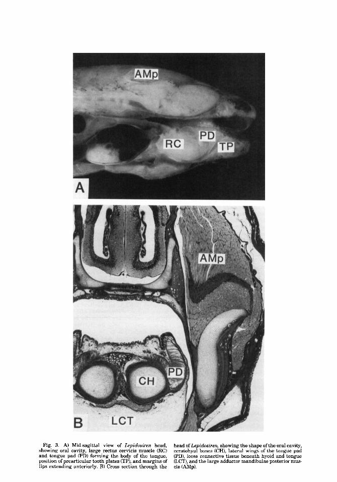
Fig. 3. A) Mid-sagittal view of Lepidosiren head, showing oral cavity, large rectus cervicis muscle (RC) and tongue pad (PD) forming the body of the tongue, position of prearticular tooth plates VP), and margins of lips extending anteriorly. B) Cross section through the
head of Lepidosiren, showing the shape of the oral cavity, ceratohyal bones (CH), lateral wings of the tongue pad (PD), loose connective tissue beneath hyoid and tongue GCT), and the large adductor mandibulae posterior mus- cle (AMP).

FEEDING MECHANICS OF LUNGFISH 89
independently of the hyoid apparatus. When the hyoid is in a raised position, the tongue nearly fills the oral cavity, so that there is little dead volume prior to mouth opening.
Strong ventral movements of the hyoid ap- paratus and its attached tongue occur during feeding and respiration. The sublingual re- gion is filled with a very loose connective tissue, providing an easily deformed cushion into which the hyoid apparatus can be moved (Fig. 3). Scalation of the skin in this region is suppressed, presumably facilitating disten- sion.
Anteriorly, the mouth is constricted to an approximately circular opening. The lips ex- tend out, away from the underlying bones. They are supported by various cartilaginous derivatives of the nasal capsule and Meckel's cartilage as well as abundant chondroid tis- sue. The margin of the upper lip curls lat- erally around the margin of the lower lip. The upper lip is supported by a rod of chon- droid tissue that contains elastic fibers (Bert- mar, '66, believed this rod to be homologous with an upper labial cartilage). The lower lip does not contain a comparable support bar, though its medial edge is well defined, with a slight curl that mates with the upper lip.
The tooth plates are surrounded by fleshy pads of tissue so that only the apical ridges of the tooth plates appear to project from the epithelium (Parker, 1892). Like the tongue pad, these tooth plate pads are supported by a core of chondroid connective tissue. The pads are easily deformed or moved when pressed upon. Thus, food items being chewed presumably contact more than just the ex- posed ridges of the tooth plates.
Overview of events during feeding The process of prey capture and swallowing
in lungfishes may be divided into four com- ponents: 1) approach to the prey; 2) the strike; 3) chewing; and 4) swallowing. The approach to prey located on the bottom often occurs with the head depressed ventrally and the anterior body arched. Prolonged searching may occur before prey are located, and dur- ing this time the head is usually maintained at an angle to the substrate.
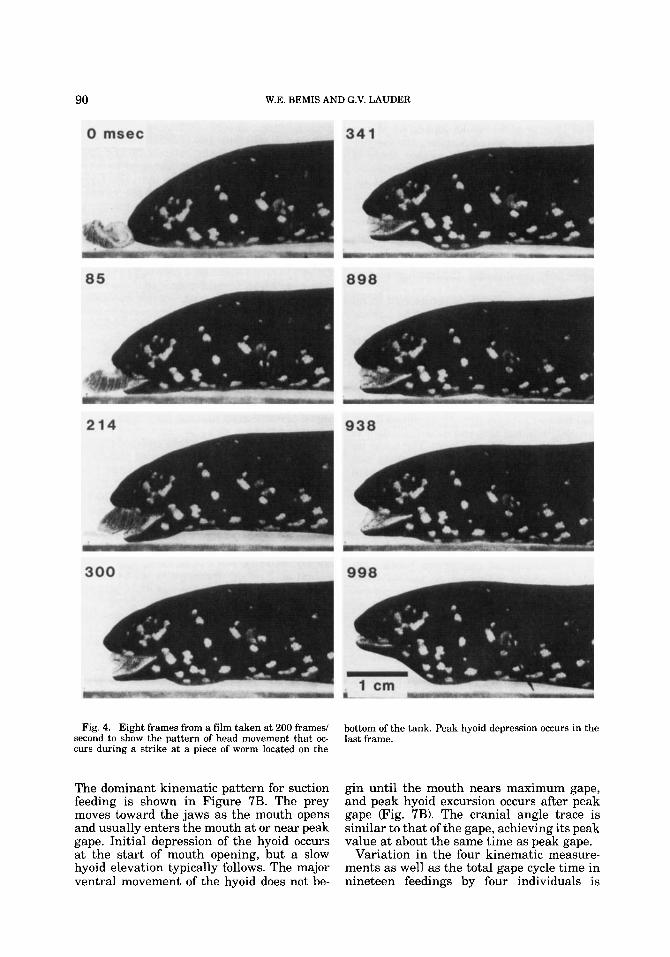
After a food item is located, the entire feed- ing sequence may last from 10 seconds to several minutes, with the average in this study being 36 seconds. Figure 5 summarizes the events and terminology used here to de- scribe feeding. The strike is usually initiated only when the prey is either touching the
snout or within several millimeters of it (Fig. 4: frame 1). Strikes are characterized by a relatively short duration (between 50 and 200 ms), during which the prey is engulfed and carried into the buccal cavity with the flow of water created by expansion of the mouth cavity (Fig. 4). There is usually a pause of from one to several seconds following the strike before chewing begins. This is shown in Figure 5 by the absence of any electromy- ographic activity immediately following the strike. Chewing of the prey between the tooth plates occurs in several stages that we refer to as chewing bouts, with successive bouts separated by pauses (Fig. 5). From one to five or more chewing bouts may occur during a feeding (mean = 2.5, based on 16 feedings).
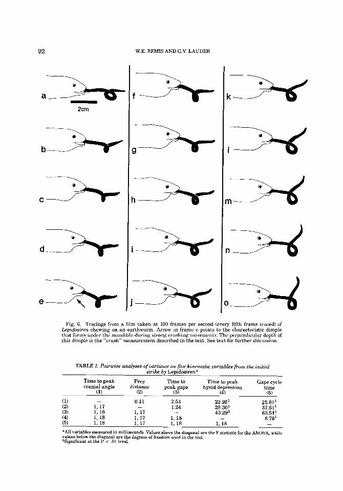
Each chewing bout consists of a series of chewing or adduction cycles (mean number of cycles per bout = 7.2, based on 40 bouts). Each chewing cycle in turn consists of a pe- riod of jaw adduction in which the tooth plates are adducted and the prey crushed (= adduction phase), and a period during which the prey is transported within the oral cavity and repositioned between the tooth plates (= transport phase). Alternation of these two phases produces the key characteristic of the chewing bouts, which is the movement of prey into and out of the oral cavity, with crushing of the prey occurring between each transport phase. Figure 6 illustrates a chew- ing sequence in which the prey is being transported into the mouth cavity (frames a, b, and c), crushed (frames d to k), and then again transported into the mouth (frames 1 to 0) before being crushed again.
After the final chewing bout has ended, the prey is located within the mouth cavity and swallowing occurs. Swallowing may be initi- ated by rapid hyoid depression that creates a strong flow of water through the oral cavity and moves the prey posteriorly to the pha- ryngeal region. Subsequent contraction of the hyoid musculature constricts the posterior part of the buccal cavity and forces the food into the esophagus. We term this behavior a constriction or buccal compression.
The strike: initial suction Photographs of a complete strike are given
in Figure 4. Considerable variation was found both within and between individuals in the time course of prey capture by suction feeding. Gape profiles, for example, can be either unimodal or have multiple peaks and can vary significantly in duration (Fig. 7A).
90 W.E. BEMIS AND G.V. LAUDER
Fig. 4. Eight frames from a film taken at 200 frames/ second to show the pattern of head movement that oc- curs during a strike at a piece of worm located on the
bottom of the tank. Peak hyoid depression occurs in the last frame.
The dominant kinematic pattern for suction feeding is shown in Figure 7B. The prey moves toward the jaws as the mouth opens and usually enters the mouth at or near peak gape. Initial depression of the hyoid occurs at the start of mouth opening, but a slow hyoid elevation typically follows. The major ventral movement of the hyoid does not be-
gin until the mouth nears maximum gape, and peak hyoid excursion occurs after peak gape (Fig. 7B). The cranial angle trace is similar to that of the gape, achieving its peak value at about the same time as peak gape.
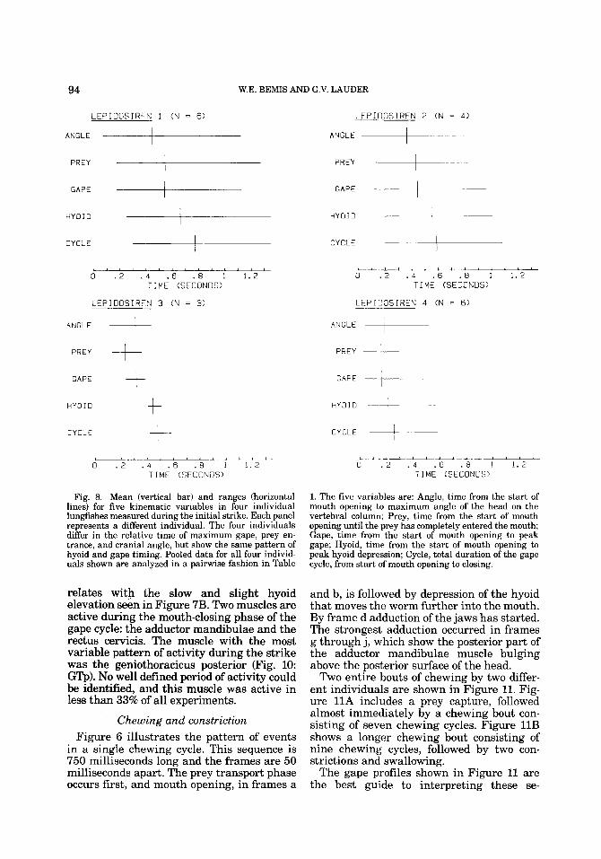
Variation in the four kinematic measure- ments as well as the total gape cycle time in nineteen feedings by four individuals is

FEEDING MECHANICS OF LUNGFISH 91
U I I I ! 1-
STRIKE CHEWING BOUT CHEWING BOUT CONSTRICT & SWALLOW
A 1
ADDUCTION CYCLE
DM Y 71
1 sec -
r I ' *+++ IH
Fig. 5. Five electromyograms recorded simultane- ously to show the behavioral events in a feeding se- quence and the terminology used in this paper. Calibration bars on left side of each trace = 200 micro- volts. Initial prey capture occurs at the strike, and is followed by a one second pause with no muscle activity.
shown in Figure 8, and the results of pair- wise analyses of variance (ANOVA) among these variables is given in Table 1. In all feedings, the hyoid reaches its maximum ventral excursion significantly after peak gape (Table 1). Cranial angle, however, can reach its peak either before or after gape (Fig. B), and the times to maximum gape and cranial angle are not significantly different (Table 1).
The electromyographic patterns during suction feeding differed greatly with the speed of the strike (Fig. 9). In slow strikes, only the depressor mandibulae and adductor mandibulae muscles showed activity, and there was a 100 to 200 ms delay between the offset of depressor activity and the onset of the adductor muscles (Fig. 9A). In the slowest strikes (gape cycles of a second or more), no activity was recorded from the adductor mandibulae muscles. When the strike was
Two chewing bouts then occur, with about a one second pause between bouts. Within a chewing bout are several adduction cycles (= chewing cycle), each consisting of an adduction phase and a transport phase. At the end of the last bout, two constrictions (= buccal compressions) occur.
more rapid, a decrease in time between the onset of the depressor mandibulae and the onset of the adductor was seen (Fig. 9B). Other muscles, such as the interhyoideus and the geniothoracicus become active also. The fastest strikes (gape cycle times of 40 ms) display considerable overlap between mouth opening and closing muscles (Fig. 9C): the jaw adductors and depressors become active within 20 ms of each other.
A summary pattern for N = 34 strikes is shown in Figure 10. The depressor mandibu- lae was active in all strikes as was the rectus cervicis posterior (first burst). In at least two- thirds of all feedings analyzed, the anterior rectus cervicis also was active with the onset of depressor activity. The epaxial and inter- hyoideus muscles become active, on the av- erage, during the first half of the activity period of the depressor mandibulae. The ac- tivity period of the interhyoideus muscle cor-
92 W.E. BEMIS AND G.V. LAUDER
2cm
b n C
e - i a k 3 I x m = n x
Fig. 6. Tracings from a film taken at 100 frames per second (every fifth frame traced) of Lepidosiren chewing on an earthworm. Arrow in frame e points to the characteristic dimple that forms under the mandible during strong crushing movements. The perpendicular depth of this dimple is the “crush” measurement described in the text. See text for further discussion.
TABLE 1. Pairwise analyses of variance on five kinematic variables from the initial strike by Lepidosiren*
Time to peak Prey Time to Time to peak Gape cycle cranial angle distance peak gape hyoid depression time
(1) (2) (3) (4) (5)
0.11 2.54 22.95’ 25.81‘ (1) - (2) 1, 17 - 1.24 28.30’ 37.611 (3) 1, 18 1, 17 - 43.29l 63.511 (4) 1, 18 1, 17 1, 18 - 8.79’ (5) 1, 18 1, 17 1, 18 1, 18 - *All variables measured in milliseconds. Values above the diagonal are the F statistic for the ANOVA, while values below the diagonal are the degrees of freedom used in the test. ‘Significant at the P < .01 level.

A
h E " W LL
0
h E " W L
0
h E " W
<
h f " W
<
B
,. E " W
c
,. f " + 0
> W
L
+8
,. E " K
W 0
E > 1
0 B 0 < z n
VARIATION IN GAPE PROFILES FOR LEPIDOSIREN 1
.27 :::: .12 0 .2 . 4 . 6 . B 1
.::;I
.25
. 1 0 .2 . 4 . 6 .8 1
GAPE, PREY DISTANCE, HYOID DEPRESSION. AN0 CRANIAL ANGLE IN LEPIDOSIREN 3
.65k I
-:::I . l s 0
.6 .45 .3 .15 0
2 I: 0 .15 TIME .3 (SECONOS) .45 . 6
Fig. 7. AIVariation in the profile of gape distance dur- ing four feedings by one individual. B)Four kinematic measurements plotted against time to show the kine- matic pattern during capture of a worm. Note that peak excursion of the hyoid is reached after peak gape, and that maximum cranial elevation is nearly coincident
with maximum gape. The four kinematic measurements are: Gape, the distance between the upper and lower jaw; Prey Dist, the distance from the prey to the plane of the gape; Hyoid Depr, the distance of hyoid depression; Cran Ang, the angle of the head with respect to the body.
94 W.E. BEMIS AND G.V. LAUDER
LEPIDOSIREN 1 (N = 6)
ANGLE I LEPIOOSIREN 2 (N = 4)
ANGLE +- PREY PREY
GAPE I GAPE -- HYOID +- HYOID
CYCLE CYCLE
0 . 2 . 4 . 6 .8 1 1.2 d ' . > .i ' . 6 ' . a i 1.2 TIME (SECONDS)
LEPIOOSIREN 3 (N = 3 )
ANGLE I
TI ME (SECONDS)
LEPIOOSIREN 4 (N = 6 )
ANGLE
PREY PREY
GAPE t GAPE
--I HYOID t HYOID
CYCLE CYCLE +- I
Fig. 8. Mean (vertical bar) and ranges (horizontal lines) for five kinematic variables in four individual lungfishes measured during the initial strike. Each panel represents a different individual. The four individuals differ in the relative time of maximum gape, prey en- trance, and cranial angle, but show the same pattern of hyoid and gape timing. Pooled data for all four individ- uals shown are analyzed in a pairwise fashion in Table
relates with the slow and slight hyoid elevation seen in Figure 7B. Two muscles are active during the mouth-closing phase of the gape cycle: the adductor mandibulae and the rectus cervicis. The muscle with the most variable pattern of activity during the strike was the geniothoracicus posterior (Fig. 10: GQ). No well defined period of activity could be identified, and this muscle was active in less than 33% of all experiments.
Chewing and constriction Figure 6 illustrates the pattern of events
in a single chewing cycle. This sequence is 750 milliseconds long and the frames are 50 milliseconds apart. The prey transport phase occurs first, and mouth opening, in frames a
L . 1 A i l - A . 2 L -
0 . 2 . 4 . 6 . 8 1 1.2 7 I ME (SECONDS)
1. The five variables are: Angle, time from the start of mouth opening to maximum angle of the head on the vertebral column; Prey, time from the start of mouth opening until the prey has completely entered the mouth; Gape, time from the start of mouth opening to peak gape; Hyoid, time from the start of mouth opening to peak hyoid depression; Cycle, total duration of the gape cycle, from start of mouth opening to closing.
and b, is followed by depression of the hyoid that moves the worm further into the mouth. By frame d adduction of the jaws has started. The strongest adduction occurred in frames g through j, which show the posterior part of the adductor mandibulae muscle bulging above the posterior surface of the head.
Two entire bouts of chewing by two differ- ent individuals are shown in Figure 11. Fig- ure 11A includes a prey capture, followed almost immediately by a chewing bout con- sisting of seven chewing cycles. Figure 11B shows a longer chewing bout consisting of nine chewing cycles, followed by two con- strictions and swallowing.
The gape profiles shown in Figure 11 are the best guide to interpreting these se-

A.
SL
OW
AM
P I
DM
I.
I.
IH
ST
RIK
E
4
100
mse
c -
RC
a
GT
a
GTP
6.
ME
DIU
M S
TR
IKE
100
maw
I
-
IM
GT
P
1 I
C.
FA
ST
ST
RIK
E
RA
O
I
2
Fig
. 9.
Dif
fere
nces
in E
MG
pat
tern
s for
dif
fere
nt st
rike
spe
eds.
A) S
low
stri
ke; B
) mod
erat
ely
rapi
d st
rike
; C) r
apid
stri
ke. I
n B
, che
win
g on
the
prey
beg
ins j
ust
afte
r th
e of
fset
of a
ctiv
ity
in
the
firs
t add
ucto
r m
andi
bula
e pos
teri
or m
uscl
e bu
rst.
EMG
cal
ibra
tion
bar
= 2
00 m
icro
volt
s.
96 W.E. BEMIS AND G.V. LAUDER
I
DM - EP ‘ * I
I n ‘ - I
-lB%z+ I I A M P
RCo - REP - GTP I+
I I 0 400 600 eon I000
T I M E (MILLISECONDS) 200
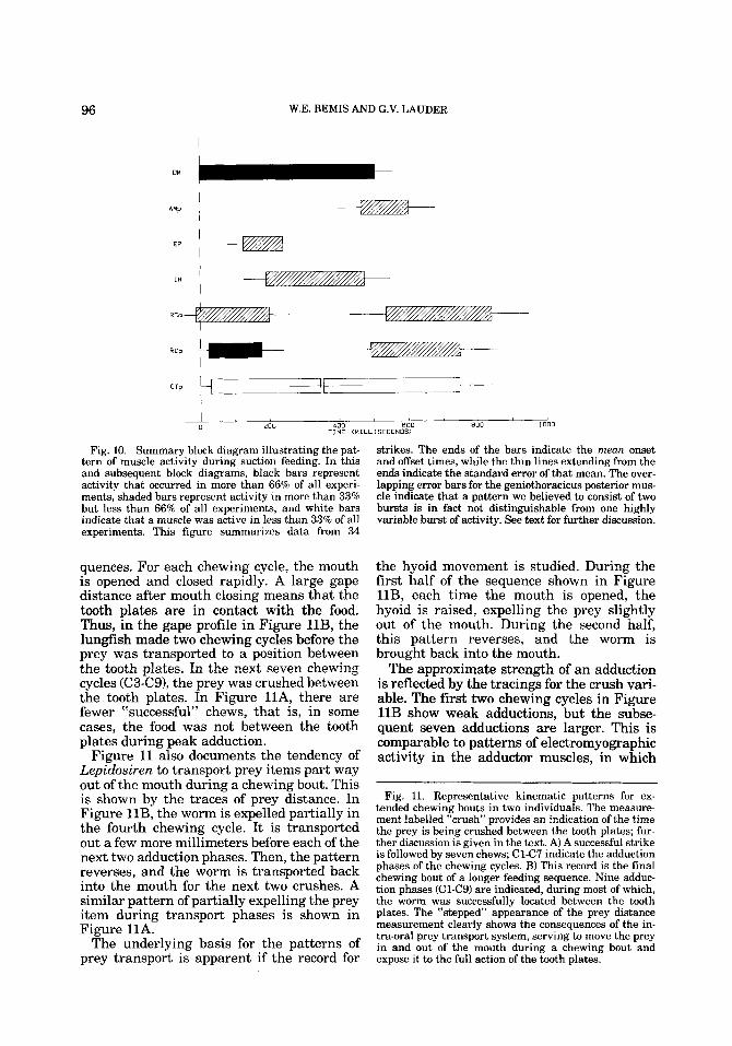
Fig. 10. Summary block diagram illustrating the pat- tern of muscle activity during suction feeding. In this and subsequent block diagrams, black bars represent activity that occurred in more than 66% of all experi- ments, shaded bars represent activity in more than 33% but less than 66% of all experiments, and white bars indicate that a muscle was active in less than 33% of all experiments. This figure summarizes data from 34
quences. For each chewing cycle, the mouth is opened and closed rapidly. A large gape distance after mouth closing means that the tooth plates are in contact with the food. Thus, in the gape profile in Figure 11B, the lungfish made two chewing cycles before the prey was transported to a position between the tooth plates. In the next seven chewing cycles (C3-C9), the prey was crushed between the tooth plates. In Figure l lA, there are fewer %uccessful” chews, that is, in some cases, the food was not between the tooth plates during peak adduction.
Figure 11 also documents the tendency of Lepidosiren to transport prey items part way out of the mouth during a chewing bout. This is shown by the traces of prey distance. In Figure 11B, the worm is expelled partially in the fourth chewing cycle. It is transported out a few more millimeters before each of the next two adduction phases. Then, the pattern reverses, and the worm is transported back into the mouth for the next two crushes. A similar pattern of partially expelling the prey item during transport phases is shown in Figure 11A.
The underlying basis for the patterns of prey transport is apparent if the record for
strikes. The ends of the bars indicate the mean onset and offset times, while the thin lines extending from the ends indicate the standard error of that mean. The over- lapping error bars for the geniothoracicus posterior mus- cle indicate that a pattern we believed to consist of two bursts is in fact not distinguishable from one highly variable burst of activity. See text for further discussion.
the hyoid movement is studied. During the first half of the sequence shown in Figure 11B, each time the mouth is opened, the hyoid is raised, expelling the prey slightly out of the mouth. During the second half, this pattern reverses, and the worm is brought back into the mouth.
The approximate strength of an addudion is reflected by the tracings for the crush vari- able. The first two chewing cycles in Figure 11B show weak adductions, but the subse- quent seven adductions are larger. This is comparable to patterns of electromyographic activity in the adductor muscles, in which
Fig. 11. Representative kinematic patterns for ex- tended chewing bouts in two individuals. The measure- ment labelled “crush” provides an indication of the time the prey is being crushed between the tooth plates; fur- ther discussion is given in the text. A) A successful strike is followed by seven chews; C1C7 indicate the adduction phases of the chewing cycles. B) This record is the final chewing bout of a longer feeding sequence. Nine adduc- tion phases (ClC9) are indicated, during most of which, the worm was successfully located between the tooth plates. The “stepped” appearance of the prey distance measurement clearly shows the consequences of the in- tra-oral prey transport system, serving to move the prey in and out of the mouth during a chewing bout and expose it to the full action of the tooth plates.

FEEDING MECHANICS OF LUNGFISH
1.2-
. 8 -
97
. 4 -
0 -
A
A E " w a L?
h E 0
b-
w
L3
> w (L: [L
h
E w
K
w 0
0
0 > H
h
E " 1 3 K U
6
A
0
W
v
a
A E " + ul 0 > !d n a
-
n E "
n a w
0
0
I
u
A
E " 1 3 L1: U
1 - ,
PREY CAPTURE AND CHEWING BY LEPIDOSIREN 1
c
I - L r 4 W , - , " " 0 .5 1 1.5 2 2. 5 3 3.5 4
.45[ I
0 .5 1 1.5 2 2 .5 3 3.5 4
t I
CHEWING AN0 SWALLOWING BY LEPIOOSIREN 2 L
CoNSTRIC CONSTRIC 2
. 4 - c3
.11 I 0 1.5 3 4.5 6 7. 5

98 W.E. BEMIS AND G.V. LAUDER
Q 0 [1:
c

AMP
AMo
RAO
EP
OM
I H
I M
RCo
RCP
GTO
GTP
FEEDING MECHANICS OF LUNGFISH
I-ADDUCTIDN PHASE- -TRANSPORT PHASE - 7+ - START OF NEXT CHEWING CYCLE
1 I -- I -- I I I *- 1 - - I 1 I I +zZBZ-+ZzmBm& I
- - - - - 0 200 400 600 800 1000
TIME (MILLISECONDS)
Fig. 13. Summary diagram of eledromyographic ac- tivity during a chewing cycle. Conventions as in Figure 10. This figure summarizes data from 89 chewing cycles. The adduction phase and the transport phase of chewing cycles are definable both kinematically and electromy- ographically. The adduction phase indicates crushing of
the amplitude of addudor bursts in a given chewing bout starts out low, and builds up to a much larger level as the chewing bout pro- gresses (e.g., Fig. 5).
The kinematics of constriction movements are shown at the end of the sequence in Fig- ure 11B. During constrictions, the mouth is kept tightly closed, the prey is located within the buccal cavity, and the hyoid is moved sharply down and then slowly elevated. In this sequence, there are two constrictions. During each, the amount of hyoid depression is steadily reduced. After these two constric- tions, swallowing occurred.
Figure 12 shows an electromyogram for a chewing bout consisting of eight chewing cycles. The start of this bout is indicated by slight activity in the depressor mandibulae muscle. This activity indicates mouth open- ing and the beginning of a transport phase. After two such transport phases, during which the food is positioned between the tooth plates, there is a small burst in the adductor muscles. The subsequent, much larger adductor bursts are correlated with crushing of the food. These larger adductor bursts occurred regularly, with a period of about one second. There is some tendency (described below) for this rhythm to speed up
99
food between the tooth plates, while the transport phase reflects intra-oral movement of food. The three adductor muscles (AMP, AMa, RAO) are active only during the adduction phase, while many muscles, including in par- ticular the depressor mandibulae (DM), have a double- burst pattern.
during a chewing bout. The depressor man- dibulae shows two periods of activity in each chewing cycle (Fig. 13: DM). A synchronous burst of activity in the depressor accompan- ies the adductor burst. The second, and usu- ally larger, period of activity in the depressor mandibulae occurs between successive ad- ductor bursts. This activity indicates mouth opening associated with the transport phase.
The summary block diagram (Fig. 13) for eledromyographic activity at all 11 record- ing sites shows an obvious break between adduction and transport phases. During the adduction phase, the three adductor muscles (AMP, AMa, RAO) are active. During trans- port, these muscles are silent. Many muscles exhibit two bursts of activity comparable to the synchronous and asynchronous bursts of the depressor mandibulae. However, it is usually the second burst that is the stronger of the two. The most consistently active mus- cle during transport is the depressor mandi- bulae, because mouth opening occurs in every transport phase. Several muscles act directly or indirectly on the hyoid, tending to depress or raise it. Those such as the recti cervicis tend to depress the hyoid, bring the food item farther into the mouth, and make the trans- port appear kinematically similar to initial
100 W.E. BEMIS AND G.V. LAUDER
4-
3 -
2 -
1 -
D.
UNSCALED EMG - ADDUCTOR MANDIBULAE POSTERIOR (AMP)
- MEAN SPIKE NUMBER * SPIKE AMPLITUDE - DEPRESSOR MANDIBULAE (N = 5)
L A -
c ' I I I
20
..?.., --*,,- . NUMBER * SPIKE AMPLITUDE - AMP (N = 5 ) 12
prey capture. The interhyoideus in particu- lar, but also the intermandibularis, tend to elevate the hyoid region, forcing the prey item forward to position it between the tooth plates.
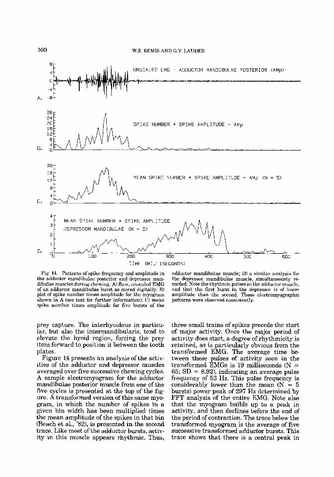
Figure 14 presents an analysis of the activ- ities of the adductor and depressor muscles averaged over five successive chewing cycles. A sample electromyogram for the adductor mandibulae posterior muscle from one of the five cycles is presented at the top of the fig- ure. A transformed version of this same myo- gram, in which the number of spikes in a given bin width has been multiplied times the mean amplitude of the spikes in that bin (Beach et al., '82), is presented in the second trace. Like most of the adductor bursts, activ- ity in this muscle appears rhythmic. Thus,
three small trains of spikes precede the start of major activity. Once the major period of activity does start, a degree of rhythmicity is retained, as is particularly obvious from the transformed EMG. The average time be- tween these pulses of activity seen in the transformed EMGs is 19 milliseconds (N = 65; SD = 8.92), indicating an average pulse frequency of 53 Hz. This pulse frequency is considerably lower than the mean (N = 5 bursts) power peak of 297 Hz determined by FFT analysis of the entire EMG. Note also that the myogram builds up to a peak in activity, and then declines before the end of the period of contraction. The trace below the transformed myogram is the average of five successive transformed adductor bursts. This trace shows that there is a central peak in

FEEDING MECHANICS OF LUNGFISH 101
TABLE 2. Three level nested ANOVA on the duration of electrical activity in the adductor mandibulae Dosterior muscle*
Source of Degrees of Sum of Mean variation freedom squares square F Significance
Among individuals 4 330,023 82,506 1.6 NS Among implants within indiv. 3 209,349 69,783 3.5 NS Among days within implants 7 136,871 19,553 6.0 P < ,001 Among bursts within days 74 240,800 3,254 Total 88 917.045
*Variance component (%I: Among individuals = 17.5; among implants = 30.8; among days = 24.1; and among bursts = 27.6.
activity for all five of these myograms. Fi- nally, the bottom trace in Figure 14 shows the corresponding pattern of activity in the depressor mandibulae for these five chewing cycles. There is no evidence of crosstalk in raw myograms of these two muscles, and we regularly recorded strong alternating activ- ity in both muscles (e.g., Fig. 5B). Note that the synchronous depressor bursts are not symmetrically located with respect to the ad- ductor bursts. Note also that the second pe- riod of activity in the depressor mandibulae is much larger than the synchronous burst. This is interpreted as the major burst of ac- tivity responsible for mouth opening. There is a one to one correlation between the tim- ing of the rhythmic pulses of activity in the two muscles (r = .99; N = 37).
Activity in the adductor mandibulae mus- cle shows no overall pattern during a com- plete feeding sequence, but there is a tendency for later bursts within a bout to occur more frequently (Fig. 15). These data were measured from electromyographic rec- ords of a long chewing sequence, consisting of a total of 46 chewing cycles in six separate chewing bouts. The burst period (length of time that the adductor mandibulae was ac- tive) and interburst period (length of time between successive bursts of adductor man- dibulae activity, unless this was the end of a chewing bout, in which case no measurement is reported) were measured in these 46 chew- ing cycles. The two period measurements are plotted against chewing cycle number (Fig. 15A,B), and against each other (Fig. 15C). These graphs suggest that there is neither a consistent change in the pattern of activity within a total feeding event (Fig. 15A,B), nor is there an obvious relationship between the length of time that the adductor muscle is active and the length of time that it is inac- tive (Fig. 15C). However, within each of the six chewing bouts, there is a significant re- lationship between the chewing cycle num-
ber within the bout and the length of time that the muscle is inactive. This is investi- gated further in Figure ED, which shows the burst and interburst periods plotted against cycle number within each of the six chewing bouts. The regression line through the points indicating the burst period is not significantly different from 0 (r = -.26; t = 1.59, NS). However, the slope of the line through the interburst periods is signifi- cantly different from 0 and is negative (r = -.71; t = 5.83, p < .001). Thus, during an average chewing bout, the later chewing cycles tend to be more rapid, due to the shorter interburst period.
We performed three-level nested analyses of variance on the periods of activity for sev- eral muscles to determine the extent of vari- ation and to evaluate the repeatability of muscle activity patterns. In Table 2, we re- port the results for the adductor mandibulae posterior, the key muscle used in chewing: the analyses of other muscles (e.g., depressor mandibulae) show comparable results. Activ- ity periods for 89 bursts of activity in the adductor mandibulae posterior were mea- sured. These bursts were grouped by experi- mental day within electrode implant and by implant within individual lungfkhes. Varia- tion within days accounted for nearly 28% of the total variance. Variation among days ac- counted for about one quarter of the total variance and was significant. Although the greatest percentage of variation (30.8%) was present among implants, this variation is not significant, and the relatively low variation (17.5%) among individuals was not signifi- cant. This analysis suggests t ha t 1) there was a good deal of repeatability from individ- ual to individual, and 2) that our electrode placements recorded consistent patterns of activity. The only significant variation is at the level of days, which presumably reflects small differences in the details of a particu- lar feeding event, such as the prey size.

102
. 2 5 -
. 4 5
W.E. BEMIS AND G.V. LAUDER
BOUT 4 ' BOUT 5 * * B~~~ 6 x BOUT 2
X I )
BOUT 1
* * x f * *
* * . 8 5 Y f X * *
* , I , , , * , I I I I I , I > , , ,
1.
1 .4
1.2-
1 -
. 8 -
. 6 -
. 4 -
. 2 -
1
6-
-
PERIOOS OF ADOUCTOR A C T I V I T Y AN0 I N A C T I V I T Y DURING A FEEDING EVENT . 3 5 , I
r = .06
* I
f f
* x
. 4 5 . 65 .85 1.05 1.25 1 .45 .05
INTERBURST TIME. SECONDS
PERIODS OF AOOUCTOR ACTIVITY AND INACTIVITY WITHIN BOUTS (N = 6 )
* +t *
INTERBURST r = -.71
* \ :
IME, SECONOS
*
*
* *
BURST TIME, SECONOS
e r = -.26 e c e
m c e
0 e m e e c
e e m f
0
01 0 1 2 3 4 5 6
CHEWING CYCLE NUMBER W I T H I N A B O U T
Fig. 15. Periods of adductor activity and inactivity. A) interburst duration, showing no relationship; D) burst Burst duration (time in seconds of adductor activity) in and interburst durations within single chewing bouts, the adductor mandibulae posterior muscle, plotted showing a significant relationship between the length of against chewing cycle number within an entire feeding the interburst and the number of the chewing cycle. sequence consisting of 46 chews; B) interburst durations Thus, later chews within a chewing bout tend to occur (time in seconds between successive adductor bursts) for closer together in time. these same 46 chews; C) burst duration as a function of

FEEDING MECHANICS OF LUNGFISH 103
GTo -7- I I
I I
GTP
0 400 600 BOO 1000 T I M E (MILLISECONOS>
200
Fig. 16. Summary diagram of the pattern of muscle activity during constriction (buccal compression) follow- ing chewing bouts. Conventions for this figure follow those of Figure 10. This figure summarizes data from 45 constrictions. All muscles recorded are active during
Figure 16 diagrams patterns of electrical activity during constriction. The most impor- tant muscle during constriction movements is the interhyoideus and it is used as the reference muscle for this diagram. These eight muscles all showed significantly over- lapping activity during constriction. Three of the muscles (the depressor mandibulae, in- termandibularis, and geniothoracicus poste- rior, Fig. 16: DM, IM, GTp), are active significantly later than the other muscles ex- amined. The last half of depressor mandibu- lae activity does not overlap activity in the adductor mandibulae.
DISCUSSION Behavioral patterns and functional
morphology We distinguish three basic behavioral pat-
terns in the feeding system of Lepidosiren. These are: 1) the strike or prey capture by suction; 2) chewing cycles, each consisting of an adduction phase and a transport phase; and 3) constriction or buccal compression. A given feeding event includes an initial strike, followed by a variable number of chewing cycles arranged into chewing bouts. Usually, each chewing bout is terminated by one or
constrictions, although at different amplitudes. Note the overlapping but not synchronous bursts in the adductor and depressor mandibulae, and the extended activity in the interhyoideus (IH), rectus cervicis (RCp), and geni- othoracicus (GTp).
more constriction events. When chewing is complete, a final series of constrictions forces the prey item into the esophagous, and swal- lowing ensues.
The mechanics of each of these phases are summarized in Figure 17. The white arrows represent the movements seen externally. The black arrows represent the approximate lines of action of the muscles involved, as well as an estimate of the relative amplitude of electromyographic activity. During suc- tions, the lower jaw is opened by jaw-depress- ing muscles, the depressor mandibulae and rectus cervicis, and the hyoid is depressed by the large recti cervicis. During the adduction phase of chewing, the jaw-closing muscles are strongly active, while most other compo- nents are weakly active. During the trans- port phase, the adductor muscles are inactive. The lower jaw is abducted by the depressor mandibulae, while the hyoid region is moved up or down through the action of the recti cervicis or interhyoideus muscles. During constriction (buccal compression), the mus- cles elevating the hyoid region, principally the interhyoideus muscle, are strongly ac- tive. Other muscles, notably the adductor mandibulae, are active to prevent food from escaping through the front of the mouth.

104 W.E. BEMIS AND G.V. LAUDER
A. SUCTION C. CHEWING - TRANSPORT
6. CHEWING - ADDUCTION D. CONSTRICTION
Fig. 17. Summary diagram showing the dominant features of the kinematic and electromyo- graphic patterns during four stages of feeding. White arrows indicate head movements visible externally, while black arrows indicate EMG activity. Width of the black arrows approximates the relative amplitude of EMG activity.
An especially noteworthy feature of muscle activity during chewing is the pattern of spike activity within adductor muscle bursts. During the adduction phase of chewing, the adductor mandibulae muscles consistently show rhythmic pulses of electrical activity so that each overall burst is composed of shorter, relatively high amplitude and high fre- quency activity followed by lower level adiv- ity (Fig. 14A,B). The result is a "pulsed" pattern with an average frequency of 53 Hz, which stands out sharply in contrast to the average peak power frequency of the myo- grams, 297 Hz. This pulsed pattern is suffi- ciently consistent that averaging several transformed bursts does not decrease its clar- ity (Fig. 140 During these adductor muscle bursts, the antagonistic depressor mandibu- lae muscle is also active. It also exhibits a rhythmic pattern of pulses, with a one to one correspondence to the adductor pulses.
A pulsed pattern of EMG activity during crushing may reflect biomechanical or phys- iological properties advantageous in crush- ing (Irish, '83). Gans et al. ('85) documented a pulsed pattern of adductor activity in
skinks feeding on snails. They suggest that because such a pattern produces graded force development at the tooth row, it may reduce shock impacts between opposing teeth. Irish ('83) reported that pulsed adductor activity occurred less frequently in the fish, Colos- soma, when fed on soft rather than hard food items. Lungfishes are widely regarded as du- rophagic, yet the earthworm prey used in this study scarcely qualify as hard. Never- theless, we did find pulsed activity during feeding on earthworms. It will be interesting to study the pulsed adductor burst patterns for lungfish feeding on hard prey items.
There are few relevant data on the physio- logical properties of fish muscles (e.g., Bone, '78; Johnston, '831, and the comparative data that do exist suggest that there can be impor- tant differences among taxa. Nevertheless, based on the data of Johnston ('80) €or skate and cod fin muscle fibers, our observed pulse frequency of 53 Hz indicates that the muscle probably is being stimulated at a rate suffi- cient to produce at least unfused tetanus (see also Gans and de Vree, '84). By stimulating the muscle at about 50 Hz, the tension devel-

FEEDING MECHANICS OF LUNGFISH 105
oped probably rises and falls, as in unfused tetanus. These and other ideas need further study before the functional significance of the pulsed EMG pattern in lungfish adductor muscles is fully understood.
A further noteworthy feature of the chew- ing phase is the consistent presence of elec- trical activity in antagonistic muscles. The best example of this is the antagonistic activ- ity in the depressor and adductor mandibu- lae muscles when prey are crushed (Fig. 14D). The first, or synchronous, burst of depressor mandibulae activity is a regular feature of our data, although it is usually of lower am- plitude than the second burst. Antagonistic muscle activity in vigorous movements is common in musculoskeletal systems (Bas- majian, ’74), and perhaps reflects the stabili- zation of joint articulations or increased control due to modulation of activity in antagonists.
The nested ANOVA performed on the burst duration in the adductor mandibulae pro- vides considerable insight into the experi- mental procedure and into the variability present in the feeding mechanism. By parti- tioning the variation in one variable (such as burst duration of a muscle) into three levels, variation among individuals, among elec- trode implants on the same individual on different days, and among recording sessions on different days with the same electrode implants, it is possible to assess the effect of repeated experiments on the same individu- als as well as to determine how much indi- viduals differ from each other. Our results (Table 2) show that although there is varia- tion in our data owing to different electrode implantations on the same individuals (many weeks apart), this variation is not statisti- cally significant. There is also no evidence from this analysis that the individuals we studied differed significantly from each other. The analysis does show, however, that there is significant variation among days given that the same set of electrodes was used. This is consistent with the results of ShafTer and Lauder (’85a) who found a significant varia- tion among days in many of the EMG vari- ables they used in a study of suction feeding in aquatic ambystomatid salamanders. We recommend this type of analysis in studies of vertebrate functional morphology because of its heuristic value and the increased under- standing of the sources of variation in the experimental data (also see ShafTer and Lau- der, ’85b).
A final important aspect of the functional morphology of feeding in lungfishes is the mechanism of chewing and the process of food transport within the oral cavity. During a chewing bout, the prey is alternately crushed and transported, in a fashion remi- niscent of generalized mammalian mastica- tory systems (see examples in Gans et al., ’78; Hiiemae, ’78). Unlike mammals, how- ever, the mechanism of prey movement is hydraulic, with water movement and its di- rection of flow determined by the hyoid ap- paratus. As the mouth is opened during the transport phase of a chew, the hyoid can be elevated to push water and the prey ante- riorly. At other times in a chewing bout, the hyoid acts in a similar manner to its role in suction feeding and draws water and the prey posteriorly. Thus, it is the control of water movement by the hyoid apparatus that al- lows positioning of prey between the tooth plates and provides control of prey position in a manner analagous to the tetrapod tongue.
Our results on the functional morphology of mouth opening conflict with some of the limited literature available on the feeding systems of living lungfishes, particularly in the case of the jaw opening system. Thomson (’69) realized the correct function of the de- pressor mandibulae muscle, but in the ab- sence of functional data, did not elaborate on this point. Edgeworth (’35) considered that jaw opening in lungfishes was the result of geniothoracicus activity. This idea was rei- terated by Perkins (’72), who stated (pg. 69): “The m. geniothoracicus lowers the jaw dur- ing feeding; no other muscle appears to be active at this time.” Perkins went on to sug- gest that the depressor muscle is not active during feeding. However, neither Edgeworth nor Perkins used electromyography to deter- mine muscle activity patterns, and our re- sults suggest that they misinterpreted func- tion on the basis of static morphology. It is more difficult to resolve a difference between our study and McMahon’s (’69) electromy- ographic study of respiration in the Afri- can lungfish, Protopterus aethiopicus. Mc- Mahon found that the depressor mandibulae muscle was only active during peak move- ments of the lower jaw. Thus, he stated (’69, pg. 417): “Activity in the retractor (= depres- sor) mandibulae was always associated with maximal depression of the lower jaw, as seen in the ‘yawning movements’ discussed be- low.” This conflicts with our finding that the
106 W.E. BEMIS AND G.V. LAUDER
depressor was active every time the lower jaw was opened.
Evolution of the feeding mechanism in lower vertebrates
A key component of our understanding of vertebrate evolution is the analysis of both morphological and functional patterns in the feeding mechanism. The definition of such patterns forms a necessary basis for discus- sions of the rate of evolution in the skull, the origin of terrestrial feeding, and scenarios about morphological changes and the envi- ronment. The recognition of such morpholog- ical and functional patterns is difficult because general patterns become clear only after extensive comparative analyses of the relevant clades. This study contributes to our understanding of the evolution of the verte- brate feeding mechanism by providing com- parative data on a critical clade, the lungfkhes, and allowing the definition of primitive characteristics of the tetrapod feed- ing mechanism. The present data must be considered in the light of other recent work on patterns of evolution in the feeding mech- anism of aquatic vertebrates (salamanders, Shaffer and Lauder, '85a, '85b; coelacanths, Lauder, '80a; ray-finned fishes, Lauder, 'gob, '82, '85).
The comparative data cited above on the major lower vertebrate clades indicate une- quivocally that at least four aspects of the feeding mechanism are primitive for teleos- tome fishes (reviewed in Lauder, '80): 1) ini- tial prey capture occurs by suction feeding; 2) the epaxial muscles produce cranial ele- vation during mouth opening; 3) the hyoid apparatus plays a major role in mediating expansion of the mouth cavity and is one biomechanical system involved in depressing the mandible; and 4) peak hyoid excursion occurs after maximum gape has been achieved. This study confirms that all of these features also characterize Lepidosiren, strongly suggesting that the four primitive characteristics of the teleostome feeding mechanism are also primitive for tetrapods. The fundamental biomechanical systems in- volved in initial prey capture exhibit re- markable conservatism. For example, ab- duction of the mandible during suction feed- ing in primitive ray-finned fishes, coela- canths, lungfishes, and aquatic salamanders occurs at least in part by posteroventral movement of the hyoid and the transmission of this movement to the retroarticular pro- cess of the mandible by the mandibulohyoid ligament. This mechanical system remains
intact after 400 million years of vertebrate evolution and extensive divergence in feed- ing form, function, and habit.
One aspect of the feeding system of lepido- sirenid lungfishes appears to be a specializa- tion at least functionally similar to a feature found in the feeding mechanism of salaman- ders. Lepidosiren possesses a well-developed muscle (Fig. 2) that is consistently active dur- ing mouth opening. In recognition of this function, we term the muscle depressor man- dibulae, in accordance with Thomson ('69) but different from a widely used name, re- tractor mandibulae (e.g., McMahon, '69; Fox, '65; Edgeworth, '35). Aquatic salamanders possess a depressor mandibulae muscle in a topographically comparable position that is also used in mouth opening (Shaffer and Lau- der, '85a). Although the depressor mandibu- lae muscles of lungfkhes and salamanders are both derived from the hyoid arch, they are not regarded as homologues by embryol- ogists (e.g., Fox, '63, '65; Edgeworth, '35). The basic developmental difference between lungfishes and salamanders is that the me- dial portion of the constrictor hyoideus sheet gives rise to the depressor muscle of lung- fishes (Edgeworth, '35: pg. 97), while the de- pressor muscle of salamanders is derived from the dorsal (= levator hyoideus) portion of the constrictor hyoideus sheet (Edgeworth,
The depressor mandibulae muscle is simi- lar in size and position in Lepidosiren and Protopterus. In the Australian lungfish, Nee ceratodus forsteri however, the muscle is ap- parently much less well developed, origi- nating from the ceratohyal and passing to its insertion on the lower jaw deep to the oper- cular elements, instead of originating from their outer surface (Fox, '65; Perkins, '72). The size and position of the muscle in Neocer- atodus is probably closer to the primitive con- dition for lungfkhes, because there has been a general tendency during dipnoan evolution to reduce both the number and size of the opercular and subopercular elements (e.g., Uranolophus-Scaumenacia-Neoceratodus- Lepidosiren), and there is no indication of a muscle attachment on the outer face of the opercular and subopercular in the descrip- tions of phylogenetically primitive dipnoans (e.g., Uranolophus, Denison, '68; Griphogna- thus, Miles, '77). The enlarged depressor mandibulae of Lepidosiren and Protopterus is, therefore, a specialized condition within the Dipnoi, and is yet another synapomorphy indicative of the close relationship between these two genera. Even though the muscle is
'35, pg. 102).

FEEDING MECHANICS OF LUNGFISH 107
unknown in outgroup osteichthyans, the em- bryological considerations discussed above render it unlikely that the depressor man- dibulae is a synapomorphy of lungfishes and tetrapods. The convergent development of depressor mandibulae muscles in lungfishes as well as several lineages of tetrapods is striking evidence of the functional impor- tance of specialized jaw opening systems.
The salient specialization in the feeding mechanism of Lepidosiren does not lie in ini- tial prey capture, but rather in the extensive intra-oral processing of food after it has been captured. Movement of the prey in and out of the mouth accompanied by strong adduction of the jaws to crush prey between the mas- sive tooth plates is an aspect of the feeding mechanism not found in most lower verte- brates. Associated with the tooth plates and crushing behavior is the hydraulic transport of food that allows positioning of food within the oral cavity. In many ways, this hydraulic food transport system in lungfishes performs analagous functions to those of the tongue in tetrapods (e.g., Hiiemae, et al., ’78; Bramble and Wake, ’85; Gorniak, et al., ’82; Smith, ’84). Water movement mediated by the hyoid apparatus positions prey between teeth, aids in swallowing (constriction or buccal com- pression in Lepidosiren), and can act to re- move unwanted particles from the oral cav- ity. Although other fishes have been documented to use water flow through the oral cavity to position food (see, for example, Lauder, ’81), ray-finned fishes have not yet been shown to do the extensive processing of food that Lepidosiren does with the oral teeth. Hiiemae et al. (’78: 206) stated that T rans - port is the fundamental mechanism of feed- ing and the hyoid apparatus is a fundamental part of that mechanism,” a conclusion as ap- propriate for Lepidosiren as it was for the mammalian feeding systems they discussed, despite the major anatomical differences be- tween tongue and hydraulic transport sys- tems.
This paper and the comparative data now available on other lower vertebrate clades set the stage for a new examination of the feeding mechanism in “rhipidistian” fishes. The presence of several features of the initial prey capture mechanism in all living lower vertebrate clades studied to date indicates that Eusthenopteron, Rhizodus, Glyptolepis, and their relatives (whatever their phyloge- netic position) shared several key character- istics of the lower vertebrate feeding mech- anism. Although there is no evidence for the presence of a depressor mandibulae muscle,
mouth opening is best interpreted as having been achieved by a combination of cranial elevation caused by the epaxial muscles, and mandibular depression mediated by postero- ventral excursion of the hyoid. The mandi- bulohyoid ligament would transmit hyoid movement to the mandible as in other lower vertebrates. No evidence available from fos- sils contradicts these inferences, but neither does fossil evidence provide much critical in- formation on the presence of fleshy lips that occlude the margins of the gape (as in Protop terus and Lepidosiren), or on the possible presence of a depressor mandibulae muscle. Further progress in interpreting the func- tional characteristics of extinct lower verte- brates awaits a more precise understanding of the interrelationships between form and function in living clades.
ACKNOWLEDGMENTS
This study was supported by grants from NIH 1F32 DE05352 to W.E.B., and NSF DEB 81-15048 and the Whitehall Foundation to G.V.L. G.V.L. also received support from the Louis Block Fund (University of Chicago). We thank Steve Barghusen, Cathy Smither, and Peter Wainwright for assistance in prep- aration of the paper, and for computer pro- gramming the data analysis. Peter Wain- wright, Fran Irish, Carl Gans, and Marvalee Wake provided helpful comments on the manuscript.
LITERATURE CITED
Basmajian, J.V. (1974) Muscles Alive, Their Functions Revealed Through Electromyography, Ed. 3. Balti- more: Williams and Wilkins.
Beach, J., G.C. Gorniak, and C. Gans (1982) A method for quantifying electromyograms. J. Biomech. 15:611- 617.
Bemis, W.E. (1984a) Morphology and growth of lepidosi- renid lungfish tooth plates (Pisces: Dipnoi). J. Morphol. 179:73-93.
Bemis, W.E. (198413) Paedomorphosis and the evolution of the Dipnoi. Paleobiology 10:293-307.
Beman, D.S. (1979) Gnathorhizu bothrotreta (Ostei- chthyes: Dipnoi) from the Lower Permian Abo Forma- tion of New Mexico. Ann. Carnegie Mus. 48:211-230.
Bertmar, G. (1966) The development of the skeleton, blood-vessels and nerves in the dipnoan snout, with a discussion on the homology of the dipnoan posterior nostrils. Acta Zool. Stockh. 47:82-150.
Bishop, I.R. and G.E.H. Foxon (1968) The mechanism of breathing in the South American lungfish, Lepidosiren parudoxa; a radiological study. J. Zool. Lond. 154:263- 271.
Bone, Q. (1978) Locomotor muscle. In W.S. Hoar and D.J. Randall (eds): Fish Physiology, Vol. 7. New York: Aca- demic Press, pp. 361-424.
Bramble, D. and D.B. Wake (1985) Feeding mechanisms of lower tetrapods. In M. Hildebrand, D. Bramble, K. Liem, and D. Wake (eds): Functional Vertebrate Mor-

108 W.E. BEMIS AND G.V. LAUDER
phology. Cambridge: Harvard University Press, pp. 230-261.
Bridge, T.W. (1898) On the morphology of the skull in the Paraguayan Lepidosiren and in other dipnoids. Trans. Zool. Soc. Lond. 14:325-376.
Campbell, K.S.W. and R.E. Barwick. (1983) Early evolu- tion of dipnoan dentitions and a new genus Speonesy drwn Mem. Ass. Australas. Palaeontols. l:17-49.
Carter, G.S. and L.C. Beadle (1930) Notes on the habits and development of Lepidosiren paradoxa J. Linn. Soc. Lond., Zool. 373197-203.
Corbet, P.S. (1961) The food of non-cichlid fishes in the Lake Victoria Basin, with remarks on their evolution and adaptations to lacustrine conditions. Proc. 2001. Soc. Lond. 136:l-101.
Denison, R.H. (1968) Early Devonian lungfishes from Wyoming, Utah, and Idaho. Fieldiana, Geol. 27:353- 413.
Dingerkus, G. and L.D. Uhler (1977) Enzyme clearing of alcian blue stained whole small vertebrates for dem- onstration of cartilage. Stain Technol. 52229-232.
Edgeworth, F.H. (1926) On the development of the cra- nial muscles in Protopterus and Lepidosiren Trans. Roy. SOC. Edinburgh. 54:719-734.
Edgeworth, F.H. (1935) The Cranial Muscles of Verte- brates. Cambridge: Cambridge Univ. Press.
Fox, H. (1963) The hyoid of Neoceratodus and a consid- eration of its homology in urodele amphibia. Proc. Zool. Soc. Lond. 142:803-810.
Fox, H. (1965) Early development of the head and phar- YM of Neoceratodus with a consideration of its phylo- geny. J. 2001. Lond. 146:470-554.
Gans, C., F. de Vree, and G.C. Gorniak (1978) Analysis of mammalian masticatory mechanisms: Progress and problems. Zbl. Vet. Med. C. Anat. Histol. Embryol. 7:226-244.
Gans, C. and F. de Vree (1984) Temporal summation in the adductor musculature of the lizard Trachydosau- rus: Treppe and tetanus in the crushing of snails. Am. Zool. 24t108A (abstract).
Gans, C., F. de Vree, and D. Carrier (1985) Usage pattern of the complex masticatory muscles in the shingleback lizard, Trachydosaurus rugosus: A model for muscle placement. Am. J. Anat. 173:219-240.
Gorniak, G.C., and C. Gans (1980) Quantitative assay of electromyograms during mastication in domestic cats (Felis catus). J. Morphol. 163:253-281.
Gorniak, G.C., H.I. Rosenberg, and C. Gans (1982) Mas- tication in the tuatara, Sphenodon punctatus (Reptilia: Rhyncocephalia): Structure and activity of the motor system. J. Morphol. 171:321-353.
Hiiemae, K.M. (1978) Mammalian mastication: A review of the activity of the jaw muscles and the movements they produce in chewing. In P.M. Butler and K.A. Joysey (eds): Studies on the Development and Function of Teeth. New York Academic Press, pp. 359-398.
Hiiemae, K.M., A.J. Thexton, and A.W. Crompton (1978) Intra-oral food transport: The fundamental mechanism of feeding. In D. Carlson and J. McNamara, eds. Mus- cle Adaptation in the Craniofacial Region. Ann Arbor: Univ. of Michigan Press, pp. 181-208.
Humason, G.L. (1972) Animal Tissue Techniques, Ed. 3. San Francisco: W.H. Freeman.
Irish, F. (19831 The dynamics of seed-eating in characi- form fish, Colossoma. Am. Zool. 23:1027 (abstract).
Jollie, M. (1982) Ventral branchial musculature and syn- apomorphies questioned. Zool. J. Linn. SOC. Lond. 74:35-47.
Johansen, K. and D. Hanson (1968) Functional anatomy of the hearts of lungfishes and amphibians. Am. Zool. 8: 191-2 10.
Johnston, LA. (1980) Contractile properties of fish fast muscle fibers. Marine Biol. Letters. 2:323-328.
Johnston, I.A. (1983) Dynamic properties of fish muscle. In P.W. Webb and D. Weihs (eds): Fish Biomechanics.
New York Praeger, pp. 36-67. Lankester, E.R. (1896) On the Lepidosiren of Paraguay,
and on the external characters of Lepidosirerz and Prc- topterus. Trans. Zool. Soc. Lond. 14:12-24.
Lauder, G.V. (1980a) The role of the hyoid apparatus in the feeding mechanism of the coelacanth Latimeria chalumnae. Copeia 198O:l-9.
Lauder, G.V. (198Ob) Evolution of the feeding mecha- nism in primitive actinopterygian fishes: A functional anatomical analysis of Polypterus, Lepisosteus, and Amia J. Morphol. 163:283-317.
Lauder, G.V. (1981) Intraspecific functional repertoires in the feeding mechanism of the characoid fishes Lebi- asina, Hoplias, and Chalceus. Copeia 1981:154-168.
Lauder, G.V. (1982) Patterns of evolution in the feeding mechanism of actinopterygian fishes. Am. Zool. 22,275-285.
Lauder, G.V. (1983a) Functional design and evolution of the pharyngeal jaw apparatus in euteleostean fishes. Zool. J . Linn. Soc. Lond. 77:l-38.
Lauder, G.V. (198313) Functional and morphological bases of trophic specialization in sunfishes (Teleostei, Cen- trarchidae). J. Morphol. 178:l-21.
Lauder, G.V. (1985) Functional morphology of the feed- ing mechanism in lower vertebrates. In H.-R. Duncker and G. Fleischer (eds): Functional Morphology of Ver- tebrates. New York Springer-Verlag, (in press).
Luther, A. (1913) Uber die von N. Trigeminus versorgte Muskulatur der Ganoiden und Dipneusten. Acta SOC. Sci. Fennicae 41:3-72.
McMahon, B.R. (1969) A functional analysis of the aquatic and aerial respiratory movements of an Afri- can lungfish, Protopterus aethiopicus, with reference to the evolution of the lung ventilation mechanism in vertebrates. J . Exp. Biol. 51:407-430.
Miles, R. (1977) Dipnoan (lungfish) skulls and the rela- tionships of the group: A study based on new species from the Devonian of Australia. Zool. J. Linn. Soc. Lond. 61:l-328.
Owen, R. (1839) Description of the Lepidosiren annec tens. Trans. Linn. Soc. 18:327-361.
Parker, W.K. (1892) On the anatomy and physiology of Protopterus anneetens. Trans. Roy. Irish Acad. 30:109- 230.
Perkins, P.L. (1972) Mandibular mechanics and feeding groups in the Dipnoi. Unpublished doctoral disserta- tion, Yale University, New Haven, Connecticut.
Rosen, D., P.L. Forey, B.G. Gardiner, and C. Patterson (1981) Lungfkhes, tetrapods, paleontology, and ple- siomorphy. Bull. Am. Mus. Nat. Hist. 167:159-276.
Schultze, H.-P., and L. Trueb (1981) Basic Structure and Evolution of Vertebrates, Vols. I, 11. E. Jarvik. 1980. London: Academic Press. (review). J . Vert. Paleontol. 1:389-397.
Shaffer, H.B., and G.V. Lauder (1985a) Aquatic prey capture in ambystomatid salamanders: patterns of variation in muscle activity. J. Morphol. 283:273-284.
Shaffer, H.B., and G.V. Lauder (1985b) Patterns of vari- ation in aquatic ambystomatid salamanders: kinemat- ics of the feeding mechanism. Evolution. 39.33-92.
Smith, K.K. (1984) The use of the tongue and hyoid apparatus during feeding in lizards (Ctenosaura simi- lis and Tupinambis nigropunctatus). J. Zool. Lond. 202: 115-143.
Sokal, R.R., and F.J. Rohlf (1981) Biometry. San Fran- cisco: W.H. Freeman.
Thomson, K.S. (1969) The biology of the lobe-finned fishes. Biol. Rev. Camb. Phil. SOC. 44:91-154.
Wassersug, R.J. (1976) A procedure for differentia! stain- ing of cartilage and bone in whole formalin fixed ver- tebrates. Stain Technol. 51:131-134.
Wiley, E.O. (1979) Ventral gill arch muscles and the interrelationships of gnathostomes, with a new classi- fication of the Vertebrata. Zool. J. Linn. SOC. Lond. 67:149-179.