morphology of drying blood pools
TRANSCRIPT

HAL Id: hal-01770001https://hal.archives-ouvertes.fr/hal-01770001
Submitted on 19 Apr 2018
HAL is a multi-disciplinary open accessarchive for the deposit and dissemination of sci-entific research documents, whether they are pub-lished or not. The documents may come fromteaching and research institutions in France orabroad, or from public or private research centers.
L’archive ouverte pluridisciplinaire HAL, estdestinée au dépôt et à la diffusion de documentsscientifiques de niveau recherche, publiés ou non,émanant des établissements d’enseignement et derecherche français ou étrangers, des laboratoirespublics ou privés.
Morphology of drying blood poolsNick Laan, Fiona Smith, Celine Nicloux, David Brutin
To cite this version:Nick Laan, Fiona Smith, Celine Nicloux, David Brutin. Morphology of drying blood pools. ForensicScience International, Elsevier, 2016, 267, pp.104-109. �10.1016/j.forsciint.2016.08.005�. �hal-01770001�

HIGHLIGHTS
• Blood pools are often encountered on crime scenes.
• The general knowledge concerning blood pools is very limited.
• During drying a blood pool goes through five separate stages.
• The size of a pool depends on the volume and the contact angle.
1

Morphology of drying blood pools
Nick Laana,∗, Fiona Smithb, Celine Niclouxa, David Brutinb
aInstitut de Recherche Criminelle de la Gendarmerie Nationale, 5 Boulevard de lHautil,95300 Pontoise, France
bAix-Marseille Universit, IUSTI UMR 7343, 13013 Marseille, France
Abstract
Often blood pools are found on crime scenes providing information concern-ing the events and sequence of events that took place on the scene. However,there is a lack of knowledge concerning the drying dynamics of blood pools.This study focuses on the drying process of blood pools to determine what rel-evant information can be obtained for the forensic application. We recordedthe drying process of blood pools with a camera while measuring the mass.We found that the drying process can be separated into five different stages:coagulation, gelation, rim desiccation, centre desiccation, and final desicca-tion. Moreover, by normalizing the mass and drying time we show that themass of the blood pools diminish similarly and in a reproducible way forblood pools created under various conditions. In addition, we verify that thesize of the blood pools is directly related to its volume and the wettability ofthe surface. Our study clearly shows that blood pools dry in a reproduciblefashion. This preliminary work highlights the difficult task that representsblood pool analysis in forensic investigations, and how internal and exter-nal parameters influence its dynamics. We conclude that understanding thedrying process dynamics would be advancement in timeline reconstitution ofevents.
Keywords:BPA, blood pools, drying, evaporation, fluid dynamics
∗Corresponding authorEmail address: [email protected] (Nick Laan )
Preprint submitted to Forensic Science International July 19, 2016
1. Introduction
Bloodstain pattern analysis is a forensic tool used by investigators todetermine, among others, what, where and how a crime took place [1]. Oneof the most common types of bloodstains found on a crime scene followinga deadly blood shedding event, is the blood pool (fig. 1). Ante- and post-mortem it is often the case that a victim bleeds out, thus accumulatingblood in one or multiple areas. Currently, when a blood pool is found, itis classified as such and an investigator can conclude that the blood donorwas bleeding at that location for any reasonable period of time for the poolto be created, be it seconds, minutes or even hours. Previous studies haveinvestigated if it was possible to determine what the volume of a blood poolwas, to determine if such a loss of blood volume could constitute loss of life[2], or for other crime scene reconstruction purposes [3, 4, 5, 6]. However,almost no studies have been performed concerning the drying of an entirepool of blood. Such studies can be very useful for determining, e.g., the timethat the blood shedding event occurred, any actions that may have occurredduring the blood shedding event or the physiological state the subject wasin. For example, fig. 1) shows two crime scene pictures of the same pool,22 hours apart. In the first (top) picture, the edges and the bottom of thepool have started drying. In the second picture the pool has completelydried. Information obtained from how fast the blood dried could be crucialto determine when the pool was created.
3

Figure 1: Picture of a real pool of blood found on a crime scene, (top) before the bodywas removed and (bottom) 22 hours later. The yellow liquid is serum which was separatedduring clotting and the black mass in the top picture is a large formed clot.
There have been several studies concerning the drying of singular blooddroplets [7, 8, 9, 10, 11]. To our knowledge only Ramsthaler et al. investigatedthe drying of blood pools [12]. In their study they focused on the dryingand morphology of diluted blood droplets and pools to be able to distinguishbetween diluted and whole blood. In this paper we report on the morphologyof drying blood pools. Pools of blood, obtained from healthy volunteerswere deposited on linoleum surfaces. Based on our results we are able todistinguish five different stages of drying. In addition, we report the universal
4

properties of drying blood pools, but also distinguish anomalies, which candiffer between pools.
2. Background theory
Once bleeding occurs, blood being ex vivo, it will coagulate and dry.During the coagulation (clotting) process, fibrin strands are formed creatinga solid structure of the blood, the clot. During drying water evaporates fromthe blood pool until only the solid matter, mainly red blood cells (RBC’s),remains. Depending on the size of the pool and environmental conditions,the time the pool completely evaporates may take hours to days. On thecrime scene, pools can be found in the order of millilitres to litres. We,however, focus on pools in the order of millilitres, simply because pools witha volume of several litres, without any additives like anticoagulants, wouldrequire a very large donation of a volunteer which is not a viable option.Prior to our investigation into drying blood pools we require some generalknowledge about fluid dynamics of evaporating liquids.
When a droplet is deposited upon a surface it will spread. The area(A) the droplet spreads over depends on the physical properties of both thesurface and liquid, where the surface tension and contact angle are the mostimportant parameters (see supplementary materials). The surface tension(γ) is defined as the amount of energy required to increase the surface areaby one square meter. In other words, increasing the area of a droplet requiresenergy and the higher the surface tension, the more energy this takes. Asthe droplet or pool lays upon a surface, the surface tension acts upon thetriple-line (the line around droplet or pool where liquid, surface and air meet,see fig. 2a-b). How a droplet or pool spread upon a surface can be deducedfrom young’s equation [13]:
S = γ(cos θ − 1) (1)
Here, S is the so-called spreading parameter, γ is the surface tension betweenliquid and gas interface and θ the contact angle between liquid and surface(see fig. 2c). When the contact angle is much smaller than 90◦(S is positive)the surface wetting, i.e., the liquid can easily spread over the surface andthe surface is presumed wetting. When the contact angle is much largerthan 90◦(S is negative) the liquid cannot spread over the surface easily andthe surface is presumed non-wetting. With a small contact angle, the pool
5

will cover a much larger area and have a larger perimeter, which shouldsignificantly increase the rate of evaporation. Therefore, the contact angle isa very important parameter concerning the drying of blood pools.
Deegan et al. [14] demonstrated very accurately the principles of the cof-fee ring effect that is observed during the drying of a droplet of a colloidalsuspension. This study showed how the flow arising from the evaporatingliquid induced the characteristic ring formation. A study by Brutin et al. [7]focused on the drying of sessile whole blood droplets which showed that it isvery similar to the drying of a droplet of a colloidal suspension, blood beinga colloidal fluid. During the drying of a blood drop the formation of cracksis observed. Moreover, the study showed that a drop of blood dries followingtwo different regimes and goes through five different stages. The first regime(first three stages) is being driven by convection, diffusion and then gelation.At the moment the drop is deposited, RBC’s are evenly distributed insidethe droplet, but then the solvent starts evaporating inducing an evaporationflux at the interface and an internal flow transporting particles inside thedrop. This leads to the formation of a gel once the concentration of particlesis high enough. Additionally, this flow induces the formation of the so-calledbiological deposit on the periphery of the droplet; indeed RBC’s are drivenfrom the inner part of the droplet to its rim. Then the transition phase takesplace and leads to the gelation of the entire drop. A sharp decrease in thedrying rate is observed, whereas gelation is rapid.
The second regime is much slower since it is diffusive. The final two stagescorrespond to the drying and the formation of cracks that are nucleatingand propagating. This extensive work on drying of droplets gives preciousinformation about the process and shows accurately that desiccation startsat the periphery of the drop, and then dries towards the centre of the droplet.The work presented in this study no longer focuses on droplets but on pools.To understand the dynamics occurring during the drying of a pool, the sizeof the blood pool must be considered. As long as the volume is low, in thecase of droplets, the surface tension forces are dominant resulting in a curvedsurface. In contrast, if the volume is large enough, the gravitational forceswill dominate over the surface tension forces producing a flat surface on topof the pool. Similar to a droplet, a pool will have a contact angle with thesubstrate on the edges, but in contrast is flat otherwise (fig. 2b). The area thepool spreads over is directly dependent on the contact angle (see appendixA). In order to understand the phenomena and the dynamics driving thedrying of blood pools, we performed experiments with small blood pools
6

(about 4 ml), which were recorded by taking pictures every two minutes.Foremost, the purpose of our experiment is to identify the different dryingstages of blood pools.
Figure 2: A schematic representation of the cross-section of a) a single droplet, b) a pooland c) three droplets on surfaces varying in wettability.
3. Methods and Materials
To follow the drying of a blood pool we required the environment tobe monitored and as constant as possible. Therefore each blood pool wascreated in a glovebox (Jacomex T-Box, V=700L). The humidity and tem-perature were recorded during drying by means of a hydrometer (Teslo AG,175-H2, Datalogger, Germany). The temperature was constant at 22±0.5◦
Celsius during all of the experiments. Within the glovebox, a camera (NikonD200, resolution: 2592x3872 pixels or Nikon D300s resolution: 2848x4288pixels both with a 60mm 1:2.8 lens) was suspended directly above the bloodpool. The camera’s enabled us to take a single picture, every two minutes,with a resolution of roughly 26 to 30 pixels per millimetre. For each bloodpool created, blood of a healthy volunteer was drawn by a certified nursein a 4.5 ml evacuated blood collection tube (VenoSafe, Terumo, France).Immediately after blood collection the tube was emptied above the targetsubstrate (linoleum) creating a pool of blood of roughly 4 ml each time.
7

Moreover, blood pools were created by slowly dripping blood directly fromthe tube/needle connected to the arm to imitate the most realistic bloodshedding event possible. Accordingly, blood pools created had a startingweight 3.5 g < mi < 7 g. No differences were observed in the drying pro-cess of the pools between the two methods of blood deposition. Of severalblood pools the substrate was on top of a balance (Mettler Toledo, ML802,Switzerland) to determine exactly the mass (m) of the blood pool duringthe entire drying process (one measurement every minute). By means of areference length next to the pool it was possible to determine the area of thepool, by using a program written in Matlab.
4. Results and Discussion
In fig. 3 we show a time-lapse of a drying blood pool deposited on alinoleum surface (see supplementary movie for the complete time-lapse).First of all the simple observation of the pictures obtained in the experimen-tal conditions described previously allowed the identification of five distinctphases (fig. 3): (I) coagulation stage, (II) gelation stage, (III) rim desic-cation stage, (IV) centre desiccation stage, (V) final desiccation stage (seesupplementary materials for a point-wise summary).
8

Figure 3: time-lapse of a drying pool of blood from a healthy person, at 21C with a relativehumidity of 32%. For a movie of the drying blood pool, see supplementary material. Notethat the elapsed time between pictures is different for each stage. The time of drying foreach picture is shown in fig. 5.
(I) When blood is deposited upon a surface, the RBC’s are evenly dis-tributed throughout the pool, which then will sediment. At this point theblood has a dark red colour and starts to coagulate [15]. It is possible thatdue to wetting and capillary action, the area of the blood pool increases asthe blood spreads slowly over the surface during the initial 15 to 30 minutes.During this first stage there is a change in colour from dark red to lighter red,mainly due to coagulation. (II) As fluid evaporates from the blood pool redblood cells, that are not constricted in the fibrin web, are transported to therim of the stain and deposited, due to flow caused by evaporation [14]. Thetransition from the fluidal to a gel state is referred to as the gelation front.
9

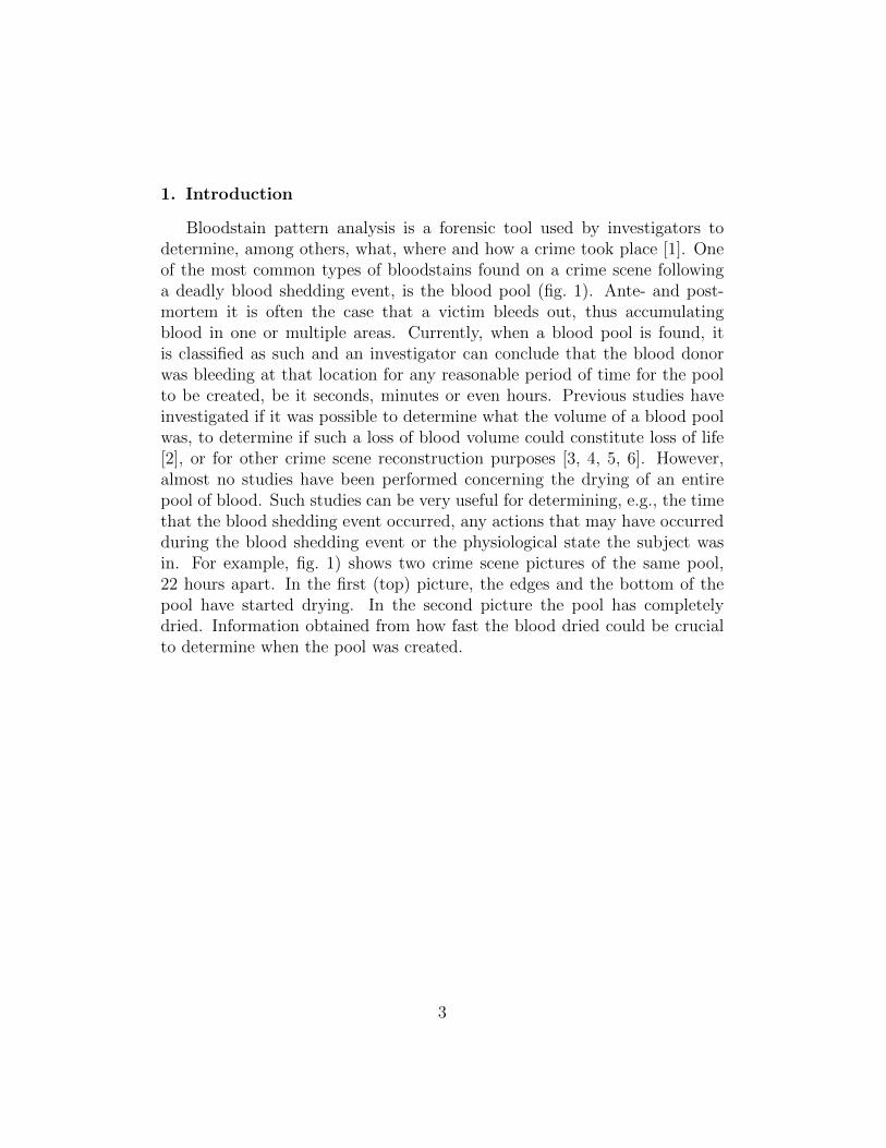
The second stage starts when the gelation rim is created around the pool.The gelation front propagates inwards, towards the centre of the stain, as thepool continues to dry. (III) The third stage starts as soon as the rim turnsblack and starts to crack, indicating that the rim is desiccating. The transi-tion from the red to black colour is referred to as the drying front. Duringthis stage both the gelation and drying front propagate towards the centre ofthe stain. (IV) Once the gelation front reaches the centre of the stain, the en-tire stain has gelified and evaporation of fluid is mainly driven by the porousmedia drying dynamics [7]. The drying front and cracks propagate towardsthe middle of the stain. (V) Finally, the drying front reaches the centre ofthe pool. The pool has almost completely desiccated. During this last stage,the entire pool is black in colour. As the last liquid evaporates, the remainscontract and the cracks reach the middle of the stain. Accordingly, flakesare separated and partially or completely detach from the surface. We haveobserved the five stages described above for every pool we created. However,it should be clear that a pool does not dry in a uniform manner. Instead,one part of the blood pool may be fully desiccating (left side fig. 4) whileanother part is still in a gel-like state (right side fig. 4). Consequently, thecentre of the pool, i.e., the location where all cracks come together, does notnecessarily has to be the geometrical centre of the pool. Furthermore, theduration of any one stage and complete desiccation can differ between bloodpools, depending on the humidity, temperature, shape and size of the pooland the kind of surface.
10
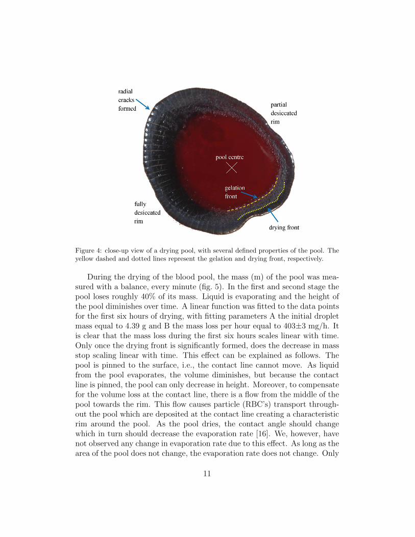
Figure 4: close-up view of a drying pool, with several defined properties of the pool. Theyellow dashed and dotted lines represent the gelation and drying front, respectively.
During the drying of the blood pool, the mass (m) of the pool was mea-sured with a balance, every minute (fig. 5). In the first and second stage thepool loses roughly 40% of its mass. Liquid is evaporating and the height ofthe pool diminishes over time. A linear function was fitted to the data pointsfor the first six hours of drying, with fitting parameters A the initial dropletmass equal to 4.39 g and B the mass loss per hour equal to 403±3 mg/h. Itis clear that the mass loss during the first six hours scales linear with time.Only once the drying front is significantly formed, does the decrease in massstop scaling linear with time. This effect can be explained as follows. Thepool is pinned to the surface, i.e., the contact line cannot move. As liquidfrom the pool evaporates, the volume diminishes, but because the contactline is pinned, the pool can only decrease in height. Moreover, to compensatefor the volume loss at the contact line, there is a flow from the middle of thepool towards the rim. This flow causes particle (RBC’s) transport through-out the pool which are deposited at the contact line creating a characteristicrim around the pool. As the pool dries, the contact angle should changewhich in turn should decrease the evaporation rate [16]. We, however, havenot observed any change in evaporation rate due to this effect. As long as thearea of the pool does not change, the evaporation rate does not change. Only
11

once a critical amount of liquid has been depleted from the pool, in this casemore than 55%, does the evaporation rate change. At this point the liquidand gel areas of the pool are diminishing, accordingly the evaporation ratediminishes as well, explaining the deviation from the linear trend after sixhours in fig. 5. The largest visual change happens during stage III wherethe entire stain transforms from a liquid/gel to a solid. Finally, 21% of theinitial mass remains in this case.
Figure 5: the mass of a blood pool as a function of time with some pictures of the poolat several moments in time. The vertical dashed lines represent the time at which thepictures in fig. 3 where taken. The red line is the mass of the pool, the dashed line a linearfit to the data, from t=0 till t=8.2 h, the time the pool transitioned into stage III. Thepercentages are the amount of mass left at that specific point in time.
Multiple blood pools were created and recorded with varying haematocritvalues and under different humidities. The mass of those pools are shown infig. 6b(inset). Not surprisingly, it is clear that the larger the mass of a pool,the longer it takes for the pool to dry. The mass decreases linearly with timeuntil roughly 50% to 70% of the mass has been depleted, at which point theslope diminishes and the mass becomes constant as the last liquid evaporatesfrom the pool. Moreover, the left-over mass is dependent on both the initialvolume of the pool and the haematocrit value of the blood, which is in accor-
12

dance with the findings of [5, 6]. As the pools were deposited under variousenvironmental humidities; these findings indicate that the drying speed, i.e.,the slope of the linear part of the drying curves, becomes steeper with de-creasing humidity. In other words, the higher the humidity, the longer theblood pool takes to dry. However, a more in-depth investigation is requiredto quantify how the drying speed depends on the humidity and other fac-tors, as temperature, contact angle and size of the pool might influence thedrying speed a lot. To have a better insight into the drying of the pools, wenormalized the mass according to:
mnorm =m−mf
mi −mf
(2)
Here mi is the initial mass, mf is the final mass and tf is the final drying timedefined as the point in time where the change in mass is less than 0.01 g/h.In fig. 6a, the normalized mass was plotted as a function of the normalizedtime to show that independent of humidity, mass, haematocrit value, or totaldrying time the mass diminishes similarly for every pool. We show that thisdimensionless rescaling is valid for a range of masses (3.5 g < mi < 7 g) anda range of humidities (22% < H < 57%). This rescaling is the first steptowards a unified theory concerning the drying of blood pools and maybeeven pools in general.
Although the literature concerning the drying and evaporation of bloodpools is poor, there has been extensive studies concerning the drying of gels[17, 18, 19, 20], which show considerable similarities. The drying process ofgels has three distinct drying stages. During the first stage the evaporationrate is constant and the volume decrease of the gel is equal to the volume ofliquid lost by evaporation [18, 19]. Once a critical point is reached, the volumeof the gel stops to decrease and cracking may occur. According to a studyof Dwivedi [20], the evaporation rate of water from alumina gels during thisfirst stage is comparable to the evaporation rate of pure water. Subsequentto the first stage, gels undergo two so-called ’falling rate’ stages where theevaporation rate decreases towards zero. During the first falling rate stagethe liquid flows through partially empty pores, followed by a second fallingrate stage, where the liquid diffuses its vapour to the surface. Comparably,drying blood exhibits a similar behaviour to water and a drying gel. Afterblood pool creation, blood dries with a constant rate of evaporation (fig.5). Subsequently, the drying rate decreases and cracking occurs, similar toa gel. Consequently, drying blood has analogous characteristics to that of
13

both water and a gel.
0.0 0.2 0.4 0.6 0.8 1.00.0
0.2
0.4
0.6
0.8
1.0b
Humidity Hct 22% 41% 29% 44% 32% 43% 34% 44% 35% 50% 35% 45% 42% 44% 46% 44% 51% 43% 54% 44% 57% 47%
(m-m
f)/(m
i-mf)
t/tf
a
0 2 4 6 8 10 12 14 16 180
1
2
3
4
5
6
7
m (g
)
t(h)
Figure 6: a) the normalized mass as a function of normalized time of the blood poolsand b) the mass of the pools as a function of time (inset), for different humidities andhaematocrit values.
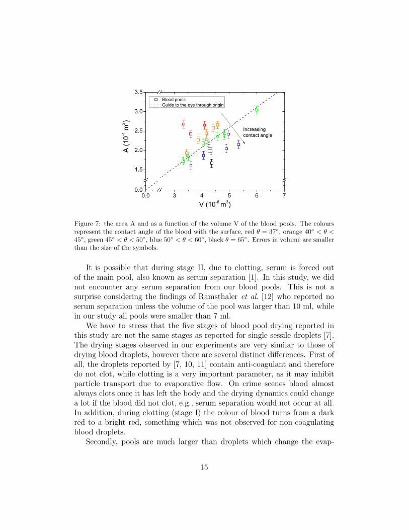
The size of the pool is dependent on two important parameters. The firstone is the volume of the blood, namely, the larger the volume of the blood, thelarger the area will be the blood spreads over (fig. 7). The second parameter isthe contact angle θ, i.e., the wettability of the surface the blood lies on, whichwas calculated using the approach described in the supplementary materials(Eq. S6). If a surface is wetting (θ < 90◦) then the blood spreads over amuch larger area then if the surface was non-wetting (θ > 90◦). Accordingly,the larger the contact angle between blood and surface, the smaller the areacovered by the blood. This is reflected well by our results (fig. 7) which showthat blood pools with a larger contact angle deviate from the average (dashedline), i.e., they have a smaller area. These factors are very important for thedrying dynamics. Namely, a higher volume blood pool takes a longer timeto dry, whereas an increase in area and a lower contact angle can both speedup the drying process.
14
0.0 3 4 5 6 70.0
1.5
2.0
2.5
3.0
3.5
Blood pools Guide to the eye through origin
A (1
0-4 m
2 )
V (10-6 m3)
Increasingcontact angle
Figure 7: the area A and as a function of the volume V of the blood pools. The coloursrepresent the contact angle of the blood with the surface, red θ = 37◦, orange 40◦ < θ <45◦, green 45◦ < θ < 50◦, blue 50◦ < θ < 60◦, black θ = 65◦. Errors in volume are smallerthan the size of the symbols.
It is possible that during stage II, due to clotting, serum is forced outof the main pool, also known as serum separation [1]. In this study, we didnot encounter any serum separation from our blood pools. This is not asurprise considering the findings of Ramsthaler et al. [12] who reported noserum separation unless the volume of the pool was larger than 10 ml, whilein our study all pools were smaller than 7 ml.
We have to stress that the five stages of blood pool drying reported inthis study are not the same stages as reported for single sessile droplets [7].The drying stages observed in our experiments are very similar to those ofdrying blood droplets, however there are several distinct differences. First ofall, the droplets reported by [7, 10, 11] contain anti-coagulant and thereforedo not clot, while clotting is a very important parameter, as it may inhibitparticle transport due to evaporative flow. On crime scenes blood almostalways clots once it has left the body and the drying dynamics could changea lot if the blood did not clot, e.g., serum separation would not occur at all.In addition, during clotting (stage I) the colour of blood turns from a darkred to a bright red, something which was not observed for non-coagulatingblood droplets.
Secondly, pools are much larger than droplets which change the evap-
15

oration dynamics and the final appearance of the remains. Drying blooddroplets show mobile fragmented cracking patterns at the corona and finecracking patterns at the middle and periphery of the droplet [7]. The crack-ing patterns of blood droplets have been extensive investigated and resemblethe cracking patterns of colloidal gels [10, 11, 21], i.e., the peripheral cracksof a blood droplet divide the remains in polygonal shaped cells. However,the cracking patterns of an entire (coagulating) blood pool are quite dissimi-lar from a single blood droplet. With blood pools we observe long elongatedcracking patterns which propagate towards the middle of the stain, that turnblack when completely desiccating. Once more, the size and thickness of thepool and coagulation of the blood can be the main reasons why blood poolsshow very different cracking patterns from those of single blood droplets.The cracks of a blood pool are similar to those observed by Pauchard et al.for a colloidal suspension (with a low ionic strength I = 0.4 mol/l) [22] andfor a drying suspension of latex particles (0.1 µm) resulting from directionalgrowth of fractures [23]. Cracks closely follow the drying front and the widthbetween cracks is directly related to the thickness of the sample, with a pref-actor depending on gel thickness, physicochemical properties, adhesion ontosubstrate and desiccation conditions [23, 24].
Finally, a single blood droplet only shows a gelation front which partlypropagates from the rim towards the middle. In contrast, a drying bloodpool shows both a gelation front and a drying front that is created at the rimand completely propagates towards the middle of the stain. It is specificallythe evolution of the drying fronts that define our different stages.
This study showed clearly that the drying dynamics of a pool of bloodcould be identified. However many more parameters would need to be in-vestigated in further studies. Such parameters would be the influence ofhumidity and temperature, but as well the influence of the substrate whichwould mainly change the contact angle, and thus drying rate since it wouldchange the spreading of the pool. Moreover it would be interesting to con-sider more the influence of the shape of the pool. Finally, size of the pool isvery important. In these experiments, each pool was in the order of 4 ml,while in practice they might be much larger or even much smaller. For thelatter, it is necessary to distinguish when a volume of liquid is considered apool or a droplet. We suggest that a pool should be defined as having a flatsurface. Accordingly, in our experiments given a contact angle of roughly45◦, the minimum volume of a pool should be much more than 170 µl (seesupplementary materials, Eq. S11), which is the case in our experiments.
16

5. Conclusion
In this study, for the first time the drying dynamics of pools of whole bloodwere investigated, for a range of 3.5 to 4.5 ml. We were able to distinguishfive different drying stages, each with their own characteristics. The massof a blood pool diminishes in a very reproducible manner, first linearly intime and then approaches a constant value. Additionally, we were able tocollapse all mass curves onto a single curve by normalizing the mass andtime of drying. The general knowledge concerning blood pools within thefield of bloodstain pattern analysis is very limited at most. This work is astep forward in the classification and characterization of blood pools withinthis field. Prospectively, the results of this study may be used for crimescene reconstruction or for future investigations into determining the timethe blood shedding event occurred. Finally, we verified that the size of theblood pools is directly related to its volume and the wettability of the surface.This result could be used to estimate the original volume of a dried bloodpool, to answer the question if the amount of blood could constitute loss oflife. We anticipate this study to be of considerable importance for forensicsand bloodstain pattern analysis as a whole.
6. Acknowledgments
Of the Institut de Recherche Criminelle de la Gendarmerie Nationale wewould like to thank the bloodstain pattern group for helping in this investi-gation, the nurses from the infirmary for helping us with the blood collectionand all volunteers for giving their blood. This work received a financial grantfrom the French Agence Nationale de la Recherche in the frame of the projectANR-13-BS09-0026. Also, this work has been carried out in the framework ofthe Labex MEC (ANR-10-LABX-0092) and of the A*MIDEX project (ANR-11-IDEX-0001-02), funded by the ”Investissements dAvenir” French Govern-ment program managed by the French National Research Agency (ANR).
[1] S. H. James, P. E. Kish, Sutton, Principles of Bloodstain Pattern Anal-ysis, Theory and Practice, CRC, 2005.
[2] H. F. Bartz, Estimating original bloodstain volume: the development ofa new technique relating volume and surface area, Ph.D. thesis, Depart-ment of Biology, Laurentian University, Sudbury, Ontario (2003).
17

[3] H. Lee, Estimation of original volume of bloodstains, Identification News9 (1986) 4.
[4] S. P. Sant, S. I. Fairgrieve, Exsanguinated blood volume estimation usingfractal analysis of digital images*, Journal of forensic sciences 57 (3)(2012) 610–617.
[5] N. Laan, R. H. Bremmer, M. C. G. Aalders, K. G. de Bruin, Volume de-termination of fresh and dried bloodstains by means of optical coherencetomography, J. Forensic Sci. 59 (1) (2014) 34–41.
[6] N. Laan, K. G. de Bruin, D. Slenter, J. Wilhelm, M. Jermy, D. Bonn,Bloodstain pattern analysis: implementation of a fluid dynamic modelfor position determination of victims, Scientific reports 5.
[7] D. Brutin, B. Sobac, B. Loquet, J. Sampol, Pattern formation in dryingdrops of blood, Journal of fluid mechanics 667 (2011) 85–95.
[8] B. Sobac, D. Brutin, Structural and evaporative evolutions in desiccatingsessile drops of blood, Physical Review E 84 (1) (2011) 011603.
[9] W. B. Zeid, D. Brutin, Influence of relative humidity on spreading, pat-tern formation and adhesion of a drying drop of whole blood, Colloidsand Surfaces A: Physicochemical and Engineering Aspects 430 (2013)1–7.
[10] W. B. Zeid, J. Vicente, D. Brutin, Influence of evaporation rate oncracks formation of a drying drop of whole blood, Colloids and SurfacesA: Physicochemical and Engineering Aspects 432 (2013) 139–146.
[11] B. Sobac, D. Brutin, Desiccation of a sessile drop of blood: cracks, foldsformation and delamination, Colloids and Surfaces A: Physicochemicaland Engineering Aspects 448 (2014) 34–44.
[12] F. Ramsthaler, J. Schlote, C. Wagner, J. Fiscina, M. Kettner, The ringphenomenon of diluted blood droplets, International journal of legalmedicine 130 (3) (2016) 731–736.
[13] P. De Gennes, F. Brochard-Wyart, D. Quere, Capillarity and wettingphenomena: drops, bubbles, pearls, waves, Springer Science & BusinessMedia, 2004.
18

[14] R. D. Deegan, O. Bakajin, T. F. Dupont, G. Huber, S. R. Nagel, T. A.Witten, Capillary flow as the cause of ring stains from dried liquid drops,Nature 389 (6653) (1997) 827–829.
[15] J. P. Riddel, B. E. Aouizerat, C. Miaskowski, D. P. Lillicrap, Theories ofblood coagulation, Journal of Pediatric Oncology Nursing 24 (3) (2007)123–131.
[16] H. Hu, R. G. Larson, Evaporation of a sessile droplet on a substrate,The Journal of Physical Chemistry B 106 (6) (2002) 1334–1344.
[17] T. Sherwood, The drying of solidsi, Industrial & Engineering Chemistry21 (1) (1929) 12–16.
[18] F. Moore, The mechanism of moisture movement in clays with particularreference to drying-a concise review, Transactions of the British CeramicSociety 60 (1961) 517–539.
[19] H. Macey, Clay-water relationships and the internal mechanisms of dry-ing, Transactions of the British Ceramic Society 41 (2009) 73–121.
[20] R. Dwivedi, Drying behaviour of alumina gels, Journal of materials sci-ence letters 5 (4) (1986) 373–376.
[21] L. Pauchard, Patterns caused by buckle-driven delamination in desic-cated colloidal gels, EPL (Europhysics Letters) 74 (1) (2006) 188.
[22] L. Pauchard, F. Parisse, C. Allain, Influence of salt content on crack pat-terns formed through colloidal suspension desiccation, Physical ReviewE 59 (3) (1999) 3737.
[23] L. Pauchard, M. Adda-Bedia, C. Allain, Y. Couder, Morphologies re-sulting from the directional propagation of fractures, Physical Review E67 (2) (2003) 027103.
[24] V. Lazarus, L. Pauchard, From craquelures to spiral crack patterns:influence of layer thickness on the crack patterns induced by desiccation,Soft Matter 7 (6) (2011) 2552–2559.
19