motion detection and prediction through spike-timing
TRANSCRIPT

Motion Detection and Prediction through Spike-TimingDependent Plasticity
A. P. Shon†, R. P. N. Rao†and T. J. Sejnowski‡§†Department of Computer Science and EngineeringUniversity of WashingtonSeattle, WA 98195‡Computational Neurobiology LaboratoryThe Salk Institute10010 N. Torrey Pines RoadLa Jolla, CA 92037§Department of BiologyUniversity of California at San DiegoLa Jolla, CA 92037
E-mail:[email protected],[email protected],[email protected]
Submitted to: NET
PACS numbers: 87.18b,87.18Hf,87.18Sn

Motion Detection and Prediction through Spike-Timing Dependent Plasticity 2
Abstract. We describe a possible mechanism for the formation of direction- and velocity-selective cells in visual cortex through spike-timing dependent learning. We contrast the casewhere only feedforward excitation and inhibition signals are provided to visual neurons withthe case where both feedforward and feedback signals are provided. In the feedforward-only case, neurons become selective for a broad range of velocities centered around thetraining velocity. However, we show that direction selectivity in this case is stronglydependent on delayed feedforward inhibition and in contrast to experimental results, becomesdramatically weaker when inhibition is reduced. When feedback connections are introduced,direction selectivity becomes much more robust due to predictive delays encoded in recurrentactivity. Direction selectivity persists in the face of decreasing inhibition in a manner similarto experimental findings. The model predicts that direction selective cells should exhibitanticipatory activity due to recurrent excitation and suggests a pivotal role for spike-timingdependent plasticity in shaping cortical circuits for visual motion detection and prediction.
1. Introduction
In both the neocortex and in sub-cortical structures such as the hippocampus, researchershave observed a striking dependence of synaptic plasticity on the relative order of pre- andpost-synaptic spikes: typically, a synapse is strengthened if an input spike arrives a fewmilliseconds before an output spike; a reversal in the order of spiking causes a decreasein synaptic strength [17, 4]. This phenomenon has been labeled “spike-timing dependentplasticity” (STDP). Modeling studies have demonstrated the importance of STDP in temporalsequence learning [19, 1, 24, 18, 25, 26] and coincidence detection [10, 32].
In this paper, we investigate how the interaction between STDP and recurrentconnections affects the development of motion detection circuits in the the visual cortex.Using networks of integrate-and-fire neurons that adapt their connections using STDP, weshow how specific types of neural circuits might develop varying degrees of directionselectivity. Our results show how STDP could enable cortical circuits to develop predictiveresponses to moving inputs through learned patteens of lateral connections. Specifically, weshow the following:
(i) Feedforward-only excitation and inhibition can cause emergence of direction selectivity.Modifying excitatory connections using an STDP learning rule leads to an asymmetryin the weights which, coupled with slow inhibition, can cause neurons to spikepreferentially in one direction of motion. However, only a few spikes are emitted by eachneuron, meaning that appreciable amounts of noise may disrupt direction selectivity inrealistic neural circuits.
(ii) Addition of fixed-strength recurrent inhibitory connections to the above model allowscompetition between neurons, enabling multiple directions to be encoded.
(iii) Addition of recurrent excitatory connections modified through STDP results in muchstronger and more robust direction selective responses, compared to cases where onlyfeedforward excitation is present.

Motion Detection and Prediction through Spike-Timing Dependent Plasticity 3
(iv) Our model predicts that recurrent connections modified using STDP can lead to“predictive coding” in the neocortex, where cells fire slightly before receivingfeedforward inputs from the lateral geniculate nucleus (LGN). As a consequence, corticalcells should continue to propagate a wave of predictive activity for a short time even whenthe original stimulus is taken away.
1.1. Prior Work
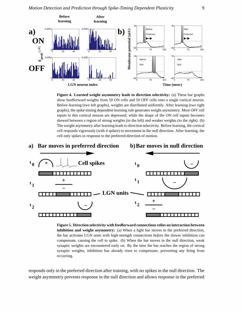
A classic model for direction-selectivity is the Barlow-Levick model [3] which postulatesa spatial discrepancy between excitation and delayed inhibition in the receptive field of adirection selective neurons. The basic idea is that motion in one direction (the “preferreddirection”) causes the neuron to fire because excitation arrives before the delayed inhibition.Motion in the opposite direction (the “null direction”) recruits the delayed inhibition first,which arrives just in time to counteract any excitation from the other side of the receptivefield (the (cf. Fig. 5 in this paper).
More recent computational models have postulated a variety of mechanisms for directionselectivity in the visual cortex, including probabilistic feedforward synapses [5], short-term synaptic depression [7, 29], a combination of feedforward spike-timing dependentplasticity and synaptic depression [6, 27], rate-based Hebbian learning [34, 9], and specializedconnectivity schemes [2, 14, 28, 31, 16, 15, 21, 20].
1.2. Contributions of this Paper
An important aspect of direction selectivity that has thus far gone uninvestigated is theinteraction between STDP, inhibition, and recurrent connectivity in the visual cortex. Weinvestigated this question using a series of simulations based on networks of integrate andfire neurons with plastic synapses. We found that modification of peak conductances ofexcitatory synapses alone allows single cortical neurons to become direction selective as inthe Barlow-Levick model. Starting from simple feedforward-only connectivition schemes, weinvestigated the development of direction selectivity in networks with increasingly complex(and increasingly realistic) connection schemes as summarized below:
• Experiment 1: A single model neuron received a mix of feedforward excitation andinhibition from ON/OFF cells in the LGN. The results demonstrate that a difference intime constants for excitatory and inhibitory currents is sufficient to allow to formation ofweak direction selectivity, where cell responses are comparable to background activityrates.
• Experiment 2: In addition to receiving feedforward ON/OFF inputs, model neuronsinhibited each other recurrently. We found that this leads to competition between theneurons, allowing them to partition the input space and code for different velocities.
• Experiment 3: We tested the effects of adding feedback excitation and inhibition, inaddition to feedforward inputs. The results indicate that such a scheme leads to robust

Motion Detection and Prediction through Spike-Timing Dependent Plasticity 4
Frame number
LGN
uni
t
a)
1 50 100
1
50
100−100 0 100
−0.32
0.5
1.23b)
0 15 30−1
0
1c)
0 15 30−1
0
1d)
Figure 1. Raw input and LGN filters: (a) Example of raw inputs for a light bar moving ata velocity of 1 LGN unit per time step. (b) Spatial filter used in the LGN module. The filteris formed as the difference of Gaussian curves. (c) Temporal filter for ON cells in the LGN,formed as the difference of Gaussians. (d) Temporal filter for LGN OFF cells, formed as thedifference of Gaussians.
direction selectivity. This selectivity is resilient to changes in inhibition strength, a resultconsistent with experimental observations.
• Experiment 4: We investigated the role of recurrent excitatory connections in mediatingprediction and delay compensation in the visual pathway. We found that STDP allowsrecurrently connected model cortical neurons to establish “predictive waves” of activity,allowing them to fire in anticipation of inputs from the LGN.
2. Methods
Our model consists of two subsystems: a lower-level model that represents the retina and LGNand a cortical model. The retina-LGN model takes as input moving 1-dimensional bars andgenerates spike trains. The cortical model receives these spike trains as input and producesvoltage traces using integrate-and-fire dynamics [13]. All simulations used an integration rateof 1 msec per simulation step. To quantify the direction selectivity of a neuron, we denoted

Motion Detection and Prediction through Spike-Timing Dependent Plasticity 5
the direction of motion that elicited the maximum respose as “preferred” and the oppositedirection as “null,” and used the following direction selectivity index(DSI):
DSI = 1 −#null spikes
#preferred spikeswhere the numerator and denominator in the fraction refer to the number of spikes fired
by the neuron for a bar moving in the null and preferred direction respectively.
2.1. Retina-LGN model
The preprocessing step of our system models the ON/OFF center-surround filteringmechanisms in the retina and the lateral geniculate nucleus (LGN) [33]. We modeled thesemechanisms using a set of spatiotemporal filters intended to model the combined effects ofretino-geniculate processing. Inputs for all experiments in this paper used a 1-dimensionallight bar 10 retinal/LGN units in width. The bar moved on every time step with a constantvelocity set to an integer number of retinal/LGN units, with the constant velocity varying fromexperiment to experiment. Fig. 1 (a) shows an example of one light bar moving at a velocityof 1 unit per time step. Images of these moving bars were preprocessed by convolving them(using a Fourier transform) with a spatial filter FSPATIAL given by a difference of Gaussianfunctions (Fig. 1 (b)). The output of the spatial filter was passed through two temporal filtersFON and FOFF , each formed as a difference of Gaussian functions (Fig. 1 (c) and (d)).
The spatiotemporal outputs of the filters described above model the relative firing ratesover time for LGN “ON” and “OFF” units. These time-varying firing rates were fed as inputto a Poisson-rate spike generator, which generated two spike trains (one for the “ON” units,another for the “OFF” units). In addition to these input spikes, uniformly-distributed spikeswere added to each LGN spike train to model noisy spontaneous activity in the LGN andretina. Fig. 2 (a) and (b) show the spatiotemporal outputs of the “ON” and “OFF” filters to themoving bar stimulus depicted in Fig. 1 (a). Fig. 2 (c) and (d) show the corresponding “ON”and “OFF” spike trains that the cortical model receives as input.
2.2. Synapse model
We model synaptic impulse responses using the alpha function:
g (t) =t exp (−t/τpeak)
τpeak exp(−1)(1)
where τpeak defines the peak time of the alpha function, and both t and τpeak are relative to aspike input to the synapse at t = 0.

Motion Detection and Prediction through Spike-Timing Dependent Plasticity 6
a)
1 50 100
1
50
100−40
−20
0
20
40b)
1 50 100
1
50
100−40
−20
0
20
40
1 50 100
1
50
100
Frame number
LGN
uni
t
c)
1 50 100
1
50
100
Frame number
LGN
uni
t
d)
Figure 2. FFT traces and spike rasters: (a) Traces following LGN FFT processing for ONunits. (b) Traces following LGN FFT processing for OFF units. (c) Spike raster correspondingto activity of LGN ON cells, produced using a Poisson process. (d) Spike raster correspondingto activity of LGN OFF cells.
Parameter Value UnitsExcitatory reversal potential (Esyn) 0 mVExcitatory peak time (τ exc
peak) 10 msecExcitatory maximum peak conductance (gmax
syn ) 0.02 µSInhibitory reversal potential (E inh
syn) -80 mVFeedforward inhibitory peak time (τFFinh
peak ) 40 msecFeedforward inhibitory peak conductance (ginhFF
syn ) 0.0018 µSFeedback inhibitory peak time (τFBinh
peak ) 5 msecFeedback inhibitory peak conductance (ginhFB
syn ) 0.025(Exp.3)0.0055(Exp.4)
µS

Motion Detection and Prediction through Spike-Timing Dependent Plasticity 7
a)I
E
ON
OFF
V1
−80 −60 −40 −20 0 20 40 60 80−1.25
0
1
Time (msec)
∆ w
b)
Figure 3. Experiment 1: Feedforward architecture and STDP learning window: (a) Inthe first group of experiments, a single cortical cell receives inputs from ON and OFF-selectivecells from the LGN. Each connection consists of a plastic excitatory connection and a fixed-strength inhibitory connection. (b) STDP learning window used in our experiments. Thenegative lobe of the window is larger than the positive lobe to facilitate competition betweensynaptic weights of individual neurons.
Table 2.2: Synapse parameters
2.3. Cortical neuron model
We modeled cortical neurons as leaky integrate-and-fire neurons [13]. A second-order Runge-Kutta solver was used to perform integration of the neural membrane voltage. In addition tothe input spike trains from the retina-LGN model, each cortical neuron also received Poisson-distributed current to model noisy background inputs to the neurons. Each model corticalneuron received a separate set of feedforward excitatory connections from the LGN “ON”cells and “OFF” cells. Each excitatory connection was paired with a fixed-strength feedfor-ward inhibitory connection (see Fig. 3 (a)). In experiments 2, 3, and 4, fixed-strength feedbackinhibitory connections were present between all cortical neurons (with no self-connections).In experiments 3 and 4, excitatory feedback connections were also present. Table 2.3 summa-rizes neural parameters used in our experiments.
Parameter Value UnitsCapacitance (C) 0.5 nFResistance (R) 40 MΩ
Resting potential (Eleak) -60 mVThreshold voltage (Vth) -40 mVRefractory period (τref ) 5 msecPoisson-distributed noise magnitude (η) 0.35 nA
Table 2.3: Cortical neuron parameters

Motion Detection and Prediction through Spike-Timing Dependent Plasticity 8
2.4. Learning rule
Learning rules for STDP are typically based on a temporally asymmetric window thatdetermines the sign and amount of synaptic modification as a function of the time-differencebetween pre- and postsynaptic spiking (e.g. [4]). The learning window we used captures theshape and temporal extent of the window observed in physiological experiments and is shownin Fig. 3 (b). Note that in keeping with the observations for firing rate stability noted in [12](see also [30]), which state that the negative lobe of the synaptic learning kernel should belarger than the positive lobe, our learning rule has a negative lobe 1.25 times the size of thepositive lobe. The learning window is multiplied by a learning rate parameter ∆g to determinethe magnitude of synaptic modification for a given time step. In all simulations shown here,∆g for all synapses was set to the constant value 10−4.
3. Results
3.1. Experiment 1: Feedforward connections only
A mismatch in the time constants for inhibitory and excitatory synapses, in conjunction witha spike-timing dependent rule, can lead to development of direction selectivity. Because ofthe asymmetry in the synaptic learning kernel, repeated exposure to bars moving in the samedirection will cause an asymmetry in the excitatory LGN synaptic weights. When a bar movesacross the retina in the learned preferred direction, it will first encounter a group of high-valued synaptic weights, causing the cortical neuron to fire. In contrast, when the bar movesin the opposite (null) direction, it will first encounter a group of synapses with low peakconductance. By the time the bar reaches the high-valued weights, feedforward inhibitionwill have risen sufficiently to prevent the cortical neuron from firing. Fig. 5 demonstratesthis idea, similar to the direction-selective neural detector first proposed by Barlow andLevick [3]. As an initial proof of concept, experiment 1 shows how a single neuron receivingonly feedforward excitation and inhibition and Poisson-distributed noise can learn directionselectivity.
3.1.1. Training paradigm We trained a single cortical neuron using a light bar that moved ata velocity of 5 LGN units per simulation step. We performed 10 training iterations, where eachiteration consisted of moving the bar from left to right across the simulated retina and applyingspike-timing dependent learning to modify the excitatory connections from the retinal/ LGNsystem. Each iteration used a different Poisson-generated raster of LGN input spikes, ensuringthat the feedforward excitatory weights are biased in general toward forward motion withoutovertraining for one particular sequence of input spikes. Each iteration ran for 350 msec.
3.1.2. Weak direction selectivity Fig. 4 shows how, despite a uniform distribution beforetraining, a marked asymmetry forms in the excitatory feedforward connections as a resultof applying STDP over multiple iterations. Fig. 6 demonstrates how the neuron respondswith 2 spikes in the preferred direction and 3 spikes in the null direction before training, and
Motion Detection and Prediction through Spike-Timing Dependent Plasticity 9
Afterlearning
Beforelearning
gpe
ak
0 20 40 0
0.0051
0 20 40 0
0.0051
0 20 40 0
0.0051
0 20 40 0
0.0051
0 75 150−80
50
Before
Preferred
0 75 150−80
50
Before
Null
0 75 150−80
50
After
Preferred
0 75 150−80
50
After
Null
Mem
bran
e po
tent
ial (
mV
)
LGN neuron index Time (msec)
b)a)ON
OFF
(µS)
Figure 4. Learned weight asymmetry leads to direction selectivity: (a) These bar graphsshow feedforward weights from 50 ON cells and 50 OFF cells onto a single cortical neuron.Before learning (two left graphs), weights are distributed uniformly. After learning (two rightgraphs), the spike-timing dependent learning rule generates weight asymmetry. Most OFF cellinputs to this cortical neuron are depressed, while the shape of the ON cell inputs becomesskewed between a region of strong weights (to the left) and weaker weights (to the right). (b)The weight asymmetry after learning leads to direction selectivity. Before learning, the corticalcell responds vigorously (with 4 spikes) to movement in the null direction. After learning, thecell only spikes in response to the preferred direction of motion.
+−
t 1
+t 0 Cell spikes
t 1 −
t 0 −
t 2 −+−
t 2
LGN units
Bar moves in preferred direction Bar moves in null directiona) b)
Figure 5. Direction selectivity with feedforward connections relies on interaction betweeninhibition and weight asymmetry: (a) When a light bar moves in the preferred direction,the bar activates LGN units with high-strength connections before the slower inhibition cancompensate, causing the cell to spike. (b) When the bar moves in the null direction, weaksynaptic weights are encountered early on. By the time the bar reaches the region of strongsynaptic weights, inhibition has already risen to compensate, preventing any firing fromoccurring.
responds only in the preferred direction after training, with no spikes in the null direction. Theweight asymmetry prevents response in the null direction and allows response in the preferred

Motion Detection and Prediction through Spike-Timing Dependent Plasticity 10
0 1 2 3 4 5 6 7 8 9 10−3
−2
−1
0
1
2
3
Bar velocity (LGN units/time step)
# sp
ikes
(+
pre
ferr
ed /
− o
ppos
ite) Training
velocity
Figure 6. Learning causes direction selectivity across a range of velocities: A singlecortical neuron trained using a velocity of 5 LGN units per msec demonstrates directionselectivity across a range of velocities, with a notable decline in selectivity for highervelocities.
direction.Unfortunately, as Fig. 6 shows, the response in the preferred direction remains weak;
only 2 spikes are generated after training. Although the asymmetry between feedforwardexcitation and inhibition can generate direction selectivity, it is clearly inadequate for creatingenough spikes to overcome large-scale noise fluctuations. We defer a discussion of robustdirection selectivity using recurrent connections to experiment 3 in section 3.3 below.
3.2. Experiment 2: Competitive feedback inhibition
Our previous experiment demonstrated the ability of a single neuron to learn directionselectivity when the neuron is exposed to light bars moving in a single direction. In thenext set of simulations, we investigated whether groups of neurons recurrently connected byinhibitory synapses can learn to code for multiple directions of motion simultaneously.

Motion Detection and Prediction through Spike-Timing Dependent Plasticity 11
0 1 2 3 4 5 6 7 8 9 10−3
−2
−1
0
1
2
3
Bar velocity (LGN units/time step)
# sp
ikes
(+ p
refe
rred
/ −
oppo
site
) Training velocity
0 1 2 3 4 5 6 7 8 9 10−3
−2
−1
0
1
2
3
Bar velocity (LGN units/time step)
# sp
ikes
(+ p
refe
rred
/ −
oppo
site
)
Training velocity
0 1 2 3 4 5 6 7 8 9 10−3
−2
−1
0
1
2
3
Bar velocity (LGN units/time step)
# sp
ikes
(+ p
refe
rred
/ −
oppo
site
)
Training velocity
1 2 3
Neuron 2 Neuron 3
Neuron 1
b)
c) d)
LGN
a) V1I
I
Figure 7. Experiment 2: Inhibitory feedback connections permit learning of multiplevelocities: (a) In the second group of experiments, a collection of 3 cortical cells areinterconnected using fixed-strength inhibitory synapses. The 3 neurons are exposed to 2different velocities (in this case, +1 and -1, representing two different directions of motion).(b,c,d) Neurons 1 and 2 code for the opposite direction of motion as neuron 3.
3.2.1. Training paradigm We trained a group of 3 cortical neurons on two differentdirections of motion. The first direction involved a bar moving left to right at a velocity of 1retinal/LGN unit per time step; the second direction involved a bar moving right to left at avelocity of 1 unit per time step. Each pass of the light bar over the retinal/LGN unit constitutedone iteration of the simulation. We applied the spike-timing dependent learning rule over 20total iterations, 10 for the left-to-right bar and 10 for the right-to-left bar. Again, each iterationran for 350 msec. The neurons were all-to-all connected (with no self-connections) usinginhibitory synapses with a peak synaptic conductance ginhFB
peak = 0.025µS.
3.2.2. Partitioning the set of input sequences Given slight initial biases in the feedforwardexcitatory weights, some neurons may be expected to respond more vigorously than others tobars moving in a particular direction. These vigorously-responding neurons will inhibit theless-responsive neurons. In turn, only the vigorously-responding neurons will modify theirfeedforward excitatory synapses sufficiently to create an asymmetry that codes for motion inone particular direction. Fig. 7 (a) shows a schematic diagram of this arrangement. Fig. 7 (b),(c), and (d) demonstrate responses of the 3 cortical neurons to motion in each direction. Twoof the neurons code for motion in the left-to-right direction; the other codes for motion in theright-to-left direction.
Fig. 8 shows the resulting asymmetry in feedforward excitatory weights. Note that

Motion Detection and Prediction through Spike-Timing Dependent Plasticity 12
125
5012
3
0
6
x 10−3
125
5012
3
0
6
x 10−3
125
5012
3
0
6
x 10−3
125
5012
3
0
6
x 10−3
Corticalneuron
LGNneuron
a) b)
d)c)
gpeak
(µS)
Figure 8. Feedforward weights reflect competition between cortical cells: (a,c) Beforelearning, weights are uniformly distributed. (b,d) After learning, competition between corticalneurons has caused an asymmetry in the weights of neurons 1 and 2 that causes spiking in onedirection of motion, while neuron 3’s weights respond to motion in the opposite direction.
neurons 1 and 2 display weights that code for the opposite direction of motion as neuron3.
Fig. 9 demonstrates how the weak direction selectivity developed as a result of STDPin feedforward-only connections drops off as feedforward and feedback inhibitory strengthsare reduced from 100% of training inhibition down to 0% in 20% decrements. This resultcontrasts with biological findings that complex cells maintain direction selectivity even wheninhibition is greatly reduced. This leads us to conclude that although competitive inhibitioncan allow cortical neurons to code for direction selectivity, additional, excitatory recurrentsynapses (cf. Section 3.3) are necessary to cause robust learning of direction selectivity.
3.3. Experiment 3: Feedback excitatory and inhibitory connections
Our previous experiments showed that mismatch between feedforward excitation andinhibition time constants is sufficient to create an asymmetry in feedforward excitatoryweights, causing direction selectivity, and that mutually inhibiting groups of cortical neuronscan compete to code for stimuli moving in different directions. However, since experiments

Motion Detection and Prediction through Spike-Timing Dependent Plasticity 13
Fra
ctio
nal i
nhib
ition
Velocity (LGN units/timestep) 0 1 2 3 4 5 6 7 8 9 10
1
0.8
0.6
0.4
0.2
0
−0.5
0
0.5
1
Figure 9. Feedforward-only learning causes dropoff in direction selectivity as inhibitionis decreased: A single neuron is trained without feedback inhibition or feedback excitationon sequences with velocities of 5 LGN units per time step. After training, direction selectivityis tested over a range of velocities on the range 1..10. For each velocity, direction selectivityis measured when feedforward inhibition is set at 100% of training inhibition, 80%, ... 0%.Selectivity drops off markedly for higher velocities than the training velocity and for reducedinhibition.
1 and 2 only employed feedforward connections, the direction selectivity developed by thecortical neurons was relatively weak. Intuitively, having recurrent excitatory connectionsbiased in the learned direction of motion should help the cortical neurons code much morestrongly for stimuli moving in that direction. In experiment 3, we investigated the effects ofadding recurrent excitatory synapses that are modified by STDP, along with weak non-plasticrecurrent inhibitory synapses.
3.3.1. Training paradigm Our simulated cortical network for this experiment consistsof a chain of 11 integrate-and-fire neurons connected all-to-all (no self-connections) withexcitatory and inhibitory synapses (Fig. 10). The recurrent inhibitory synapses are initializedto constant fixed values of ginhFB
peak = 0.0055µS, and the recurrent excitatory synapses areinitialized to random, uniformly-distributed values from 0 to 0.005µS. We assume that eachneuron receives input from a patch of the retinal/LGN system that does not overlap with the

Motion Detection and Prediction through Spike-Timing Dependent Plasticity 14
V1
LGN
EI
Figure 10. Experiment 3: Network with recurrent excitation and inhibition: The thirdset of experiments covers the case where feedback excitation and inhibition and feedforwardexcitation and inhibition are present in the network.
0 1 2 3 4 5 6 7 8 9 10−8
0
8
Bar velocity (LGN units/timestep
# sp
ikes
(+
pre
ferr
ed /
− o
ppos
ite)
Training
velocity
0 1 2 3 4 5 6 7 8 9 10−21
0
21
Bar velocity (LGN units/timestep
# sp
ikes
(+
pre
ferr
ed /
− o
ppos
ite) Training
velocityb)a)
Figure 11. Recurrent connections allow robust direction selectivity: In contrast to thesingle neuron shown in Fig. 6, responses in the preferred direction are represented by numerousspikes. Responses in the null direction remain weak. (a) shows responses with inhibitionat 100% of initial; (b) shows responses when inhibition is set at 60% of initial. Note thedifference in scale of the y axes for the two subgraphs.
receptive field of any other cortical cell in the chain. Further, the feedforward excitatoryweights were set to the values learned in the previous experiments, so an asymmetry biasingthe network toward weak responses in the preferred direction already exists. The recurrentexcitatory weights were modified according to the STDP rule as the network was exposed toa moving bar 10 retinal/LGN units wide moving at a velocity of 5 retinal/LGN units per timestep. Again, each pass of the light bar over the retinal/LGN unit constituted one iteration ofthe simulation, and each iteration ran for 725 msec. 10 iterations comprised the total trainingset for the network.

Motion Detection and Prediction through Spike-Timing Dependent Plasticity 15
24
68
10
24
68
100
0.01
0.02
0.03
0.04
0.05
0
0.005
0.01
0.015
0.02
0.025
0.03
0.035
0.04
0.045
0.05
24
68
10
24
68
10 0
0.001
0.002
0.003
0.004
0.005
0
0.001
0.002
0.003
0.004
0.005a) b)
Postsynapticneuron Presynaptic
neuronneuron
PresynapticPostsynaptic
neuron
gpeak
(µS)
gpeak
(µS)
Figure 12. Recurrent weights show asymmetry after training: (a) Shows recurrent synapticweights before training. (b) Shows weights from the same network after training. A clearasymmetry results from being trained on bars moving in the preferred direction. Note thedifference in scale from (a).
3.3.2. Strong direction selectivity Fig. 11 shows that STDP causes an asymmetry in theexcitatory recurrent connections that leads to robust direction selectivity. Fig. 11(a) showsthe number of spikes fired by the neuron in the middle of the chain when presented with barsmoving at velocities from 0 . . . 10. Compared to the results for feedforward-only excitationin Fig. 6, the neuron displays much more vigorous activity in the preferred direction, whilefiring either 0 or 1 spikes in the null direction for all velocities except 0. Strong directionselectivity persists until inhibition is lowered to 20% to 0% of normal (see Fig. 13). Notethe higher velocities in particular display much more robust direction selectivity as comparedto the feedforward-only case. Our findings are consistent with other modeling studies, forexample, the work of Suarez, Koch, and Douglas [31], who found that asymmetric, excitatoryrecurrent connections are necessary to replicate biological data and ensure robust directionselectivity.
4. Model Predictions
Our model of STDP-driven selectivity for direction and motion makes several experimentallytestable predictions. In particular, it is known that STDP allows a network of neurons topredictively encode sequences [1, 24, 25, 26]. We investigated the implications of thesefindings within the context of motion detection and direction selectivity.
4.1. Experimental paradigm
We used the 11-neuron cortical network described in section 3.3 (after training the recurrentexcitatory weights) to determine whether moving bars could generate predictive activity. Inthis experiment, we assume that synaptic weights have stabilized to represent a preferred

Motion Detection and Prediction through Spike-Timing Dependent Plasticity 16
Velocity (LGN units/timestep)
Fra
ctio
nal i
nhib
ition
0 1 2 3 4 5 6 7 8 9 10
1
0.8
0.6
0.4
0.2
0
−0.5
0
0.5
1
Figure 13. Learning with recurrent excitatory feedback causes robust directionselectivity even when inhibition is dropped: A mix of inhibitory and excitatory connectionson both the feedforward and the feedback synapses is trained on a moving bar. The resultingnetwork displays robust direction selectivity across a range of velocities, even as feedforwardinhibition is decreased.
direction of motion, and therefore turn off synaptic plasticity while running our simulations.We present two experimental setups:
• Predictive firing: In this setup, we move a bar across the retina-LGN system, thenexamine the activity of the 5 middle neurons to determine whether the onset of activityprecedes the appearance of the bar in the receptive field of the neurons. The bar isconsidered to impinge on the receptive field of the neurons as soon as the rightmost edgeof the bar encounters the leftmost retina-LGN unit corresponding to the leftmost corticalneuron.
• Continuing predictive activity: In this setup, we examined the dynamics of modelneuron responses in the network when a moving bar was abruptly stopped after an initialperiod of motion.

Motion Detection and Prediction through Spike-Timing Dependent Plasticity 17
−50 0 500
0.2
0.4
0.6
0.8
1
Time (msec)
Res
pons
e st
reng
thMovingFlashed
Figure 14. Recurrent connections cause predictive waves of activity: A moving stimulus(green line) causes recurrent connections to fire predictively, allowing cells to spike before thestimulus reaches their receptive fields. In contrast, when a flashed stimulus is provided (redline), the model predicts a peak in activity only after the flash has occurred.
4.2. Predictive firing
Fig. 14 shows the results from the first experimental setup. We contrast the results when theinput is a moving bar with the results when the input is a single flashed bar. To generatethe curves shown here, we convolve the mean activity of the middle 3 neurons in the case ofmoving stimuli with a Gaussian with mean 0 and standard deviation 0.1, and in the case offlashed stimuli with mean 0 and standard deviation 0.05.
Our model predicts a sharp onset of activity for the flashed stimulus, and that activityfor the moving stimulus should not be as sharply-peaked and begin a few milliseconds(approximately 20 msec in the model) before the arrival of the moving stimulus on thereceptive field of the leftmost neuron. Further, our model predicts that no such predictivefiring will occur in direction-selective cells when exposed to a bar moving in the null direction.

Motion Detection and Prediction through Spike-Timing Dependent Plasticity 18
1 2 3 4 5 6 7 8 9 10 110
0.2
0.4
0.6
0.8
1
Neuron number
Res
pons
e st
reng
th20 msec40 msec60 msec80 msec100 msec
Figure 15. Predictive activity continues to propagate when moving bar is stopped: After20 msec of exposing the network to a moving bar, we turn off all inputs from the LGN.Recurrent connections continue to propagate the activity even in the absence of external input.In a larger cortical model, this activity would gradually be reduced due to recurrent inhibition.Here, the small number of simulated recurrent neurons and asymmetry in the weights acts toreduce activity as time passes.
4.3. Continuing predictive activity
Fig. 15 shows the results from the second experimental setup. We begin with a bar movingat a velocity of 5 LGN units per simulation step. After 20 milliseconds, input from the LGNstops completely. Our model predicts that, even in the absence of continued LGN input, apropagating wave of activity should continue for some time as a result of recurrent excitation.Fig. 15 shows this effect in the model network; the figure plots the mean location of activitywithin the network of 11 cortical neurons over time. Cortical activities were measuredevery 20 msec, and plotted as Gaussians whose means are located at the mean locus ofcortical activity and whose standard deviations are given by the standard deviation of corticalactivity. In long chains of recurrently connected neurons, this continued predictive firing willeventually cease as recurrent inhibition overcomes excitation; in our small simulated network,activity stops as the wave of firing reaches the end of the chain.

Motion Detection and Prediction through Spike-Timing Dependent Plasticity 19
Fixed
circuitsbetweenconnectionsinhibitory
Plastic excitatory connectionswithin circuits
Common input for all circuitsFigure 16. Proposal for cortical architecture: We propose that cortical columns actto partition the set of input sequences, with nearby columns competitively inhibiting oneanother and excitatory intracolumn connections coding for temporal correlations. Thissetup is reminiscent of the expectation maximization (EM) approach to finding a model forprobabilistic input data.
5. Conclusions
We have shown how STDP causes different configurations of model cortical neurons to learnto detect motion in a particular direction. We demonstrated 4 main results:
(i) STDP allows single neurons receiving feedforward excitatory and inhibitory connectionsto develop weak direction selectivity.
(ii) A network of mutually competitive, inhibitory neurons can learn to code for multipledifferent directions.
(iii) A network with both feedforward as well as STDP-driven recurrent excitatoryconnections develops robust direction selectivity.
(iv) Recurrently connected networks of direction selective neurons can predictively encodethe direction of stimulus motion and fire in anticipation of feedforward inputs.
Our model predicts that recordings from visual cortical columns should reveal movingwaves of activity when the retina is exposed to moving bars, and that the activity shouldbegin slightly before the bar reaches the receptive field of the cortical neuron (i.e. it shouldbe “predictive”). Waves of activity should continue for a short while in cortex even after thestimulus is removed. These trends may represent a general coding strategy used throughoutthe neocortex: chains of excitatory neurons provide a top-down prediction of how a stimuluswill evolve with time, biasing the activity of lower-level sensory neurons, an idea consistentwith recent predictive coding models [22, 23].

Motion Detection and Prediction through Spike-Timing Dependent Plasticity 20
Our results suggest the following model for the development of cortical directionselectivity and sequence learning in general: chains of recurrently connected neurons learningto code for a particular direction of motion interact competitively with other chains throughrecurrent inhibition (see Fig. 16). For any given sequence of inputs, one recurrent chainmay “win out” over its neighbors to code for a particular temporal sequence, spikingsufficiently to prevent neurons in other chains from spiking significantly. The “winning”chain of neurons would modify their recurrent excitatory synapses according to the STDPlearning rule, so that the winning chain is more likely to respond vigorously to the temporalinput sequence in the future. The mutually competitive interaction of chains of recurrentlyconnected neurons, interspersed with STDP-driven learning, is strongly reminiscent of thewell-known expectation maximization (EM) algorithm [8, 11] from statistics and machinelearning, raising the intriguing possibility that the neocortex utilizes statistically-motivatedprinciples for learning temporal sequences.
Acknowledgments This work is being supported by the Sloan Foundation, an NSF awardfrom the BITS program, and a National Defense Science and Engineering GraduateFellowship to APS.
[1] L. F. Abbott and K. I. Blum. Functional significance of long-term potentiation for sequence learning andprediction. Cereb. Cortex, 6:406–416, 1996.
[2] E. H. Adelson and J. Bergen. Spatiotemporal energy models for the perception of motion. J. Opt. Soc.Am. A, 2(2):284–299, 1985.
[3] H. Barlow and W. Levick. The mechanism of directionally selective units in rabbit’s retina. J. Phsyiol.(Lond.), 178:477–504, 1965.
[4] G. Bi and M. Poo. Synaptic modifications in cultured hippocampal neurons: Dependence on spike timing,synaptic strength, and postsynaptic cell type. J. Neurosci., 18(24):10464–10472, 1998.
[5] N. Buchs and W. Senn. Learning direction selectivity through spike-timing dependent modification ofneurotransmitter release probability. Neurocomputing, 38–40:121–127, 2001.
[6] N. Buchs and W. Senn. Spike-based synaptic plasticity and the emergence of direction selective simplecells: simulation results. J. Computational Neurosci., 13:167–186, 2002.
[7] F. Chance, S. Nelson, and L. Abbott. Temporal characteristics of v1 cells arising from synaptic depression.In J. Bower, editor, Computational Neuroscience, Trends in Research 1998. Amsterdam: Elsevier Press,1998.
[8] A. Dempster, N. Laird, and D. Rubin. Maximum likelihood from incomplete data via the EM algorithm.J. Royal Statistical Soc., Series B, 39(1):1–38, 1977.
[9] J. C. Feidler, A. B. Saul, A. Murthy, and A. L. Humphrey. Hebbian learning and the development ofdirection selectivity: the role of geniculate response timings. Network: Computation in Neural Systems,8:195–214, 1997.
[10] W. Gerstner, R. Kempter, J. L. van Hemmen, and H. Wagner. A neuronal learning rule for sub-millisecondtemporal coding. Nature, 383:76–81, 1996.
[11] H. Hartley. Maximum likelihood estimation from incomplete data. Biometrics, 14:174–194, 1958.[12] R. Kempter, W. Gerstner, and J. L. van Hemmen. Intrinsic stabilization of output rates by spike-time
dependent Hebbian learning. Neural Computation, 13:2709–2742, 2001.[13] C. Koch. Biophysics of Computation: Information Processing in Single Neurons. New York: Oxford
University Press, 1999.[14] C. Koch and T. Poggio. The synaptic veto mechanism: does it underlie direction and orientation selectivity
in the visual cortex? In D. Rose and V. Dobson, editors, Models of the Visual Cortex, pages 408–419.New York, NY: Wiley, 1985.
[15] M. Livingstone. Mechanisms of direction selectivity in macaque V1. Neuron, 20:509–526, 1998.

Motion Detection and Prediction through Spike-Timing Dependent Plasticity 21
[16] R. Maex and G. A. Orban. Model circuit of spiking neurons generating directional selectivity in simplecells. J. Neurophysiol., 75(4):1515–1545, 1996.
[17] H. Markram, J. Lubke, M. Frotscher, and B. Sakmann. Regulation of synaptic efficacy by coindence ofpostsynaptic aps and epsps. Science, 275:213–215, 1997.
[18] M. R. Mehta and M. Wilson. From hippocampus to V1: Effect of LTP on spatiotemporal dynamics ofreceptive fields. In J. Bower, editor, Computational Neuroscience, Trends in Research 1999. Amsterdam:Elsevier Press, 2000.
[19] A. A. Minai and W. Levy. Sequence learning in a single trial. In Proceedings of the 1993 INNS WorldCongress on Neural Networks II, pages 505–508. New Jersey: Erlbaum, 1993.
[20] P. Mineiro and D. Zipser. Analysis of direction selectivity arising from recurrent cortical interactions.Neural Computation, 10(2):353–371, 1998.
[21] M. Miyashita, D.-S. Kim, and S. Tanaka. Cortical direction selectivity without directional experience.NeuroReport, 8(5):1187–1191, 1997.
[22] R. P. N. Rao and D. H. Ballard. Dynamic model of visual recognition predicts neural response propertiesin the visual cortex. Neural Computation, 9(4):721–763, 1997.
[23] R. P. N. Rao and D. H. Ballard. Predictive coding in the visual cortex: A functional interpretation of someextra-classical receptive field effects. Nature Neuroscience, 2(1):79–87, 1999.
[24] R. P. N. Rao and T. J. Sejnowski. Predictive sequence learning in recurrent neocortical circuits. InAdvances in Neural Information Processing Systems 12, pages 164–170. Cambridge, MA: MIT Press,2000.
[25] R. P. N. Rao and T. J. Sejnowski. Spike-timing dependent Hebbian plasticity as temporal differencelearning. Neural Computation, in press, 2001.
[26] R. P. N. Rao and T. J. Sejnowski. Self-organizing neural systems based on predictive learning. Phil. Trans.R. Soc. Lond. A, 361, 2003.
[27] W. Senn and N. Buchs. Spike-based synaptic plasticity and the emergence of direction selective simplecells: mathematical analysis. J. Computational Neurosci., 14:119–138, 2003.
[28] R. M. Shapley, R. C. Reid, and R. Soodak. Spatiotemporal receptive fields and direction selectivity. InM. S. Landy and J. A. Movshon, editors, Computational Models of Visual Processing, pages 109–118.Cambridge, MA: MIT Press, 1992.
[29] A. P. Shon and R. P. N. Rao. Learning temporal patterns by redistribution of synaptic efficacy. InJ. Bower, editor, Computational Neuroscience, Trends in Research 2003. Amsterdam: Elsevier Press,(to be published).
[30] S. Song, K. D. Miller, and L. F. Abbott. Competitive Hebbian learning through spike-timing dependentsynaptic plasticity. Nature Neuroscience, 3:919–926, 2000.
[31] H. Suarez, C. Koch, and R. Douglas. Modeling direction selectivity of simple cells in striate visual cortexwithin the framework of the canonical microcircuit. J. Neurosci., 15(10):6700–6719, 1995.
[32] R. E. Suri and T. J. Sejnowski. Spike propagation synchronized by temporally asymmetric hebbianlearning. Biological Cybernetics, 87(5–6):440–445, 2002.
[33] B. Wandell. Foundations of Vision. Sunderland, MA: Sinauer, 1995.[34] S. Wimbauer, O. Wenisch, K. Miller, and J. van Hemmen. Development of spatiotemporal receptive fields
of simple cells: I. Model Formulation. Biological Cybernetics, 77:453–461, 1997.