moving and handling patients with actual or suspected ... · moving and handling patients with...
TRANSCRIPT

MOVING AND HANDLINGPATIENTS WITH ACTUAL ORSUSPECTED SPINAL CORD
INJURIES (SCI)
Produced by the Spinal Cord Injury Centres
of the United Kingdom and Ireland
Initiated by
Multidisciplinary Association of
Spinal Cord Injury Professionals
Supported by an education grant from Huntleigh Healthcare
Endorsed by

4
CONTENTSPage
Foreword 5
Key point for trainers 6
Definition – level of injury and extent of paralysis 7
Dermatome and myetome maps 8
American Spinal Injury Association
– Standard Neurological Classification of Spinal Injury 9
Pictorial Guidelines
– Adapted ATLS head hold for actual or potential cervical spinal injury 11
– Application of a one-piece collar 12
– Application of a two-piece collar 13
– Acute tetraplegic spinal logroll – method 1 14
– Acute paraplegic spinal logroll 15
– Airway protection 16
– Removal from vacuum mattress 17
– Lateral transfer using a spinal board 18
– Lateral transfer using scoop stretcher 19
– Acute tetraplegic spinal logroll – method 2 20
– Mechanised turn for postural change 21
– Postural alignment 22
– Adjusting skin loading 23
– Assisted cough 24
References 25
Acknowledgements 26

5
FOREWORDThe integrated care pathway for acute spinal cord injuries (SCI) patients involves numerous
transfers between surfaces, wards and departments or even between different hospitals
before eventual admission to a specialist care facility. Wherever there is a reasonable
suspicion of acute SCI, the aim is to maintain full spinal alignment during any moving and
handling activity. Careful handling, positioning and turning, on every occasion, can prevent
or significantly reduce patient pain and discomfort. It will also reduce the potential for skin
damage and secondary spinal cord trauma (Harrison 2007).
These pictorial guidelines are provided as a resource for moving and handling trainers to
support the promotion of best practice. There are numerous scenarios associated with the
initial management of acute patients presenting within hospital with ‘actual’, ‘potential’ or
‘uncleared’ spinal cord injuries
The management of this project was coordinated between the Multidisciplinary Association
of SCI Professionals (MASCIP) and the Spinal Injuries Association (SIA) with sponsorship
provided by Huntleigh Healthcare. The clinicians within the project team that developed this
resource combined the knowledge, skills and experiences of healthcare professionals
employed within the 12 UK and Irish SCI Centres. They were supported by clinicians
representing Emergency Departments, Critical Care, Orthopaedic and Neurosciences
Departments within District General and University Teaching Hospitals. Additional
assistance was also provided by medical device manufacturers to showcase the generic
range of equipment available to support the moving and handling of SCI patients.
The initial review of these guidelines was principally undertaken by members of the UK and
Irish Forum for SCI Multi-professional Education (SCIMPE), the SIA Academy and Moving
and Handling Specialists with appropriate post graduate qualifications. The final review was
undertaken by a broad spectrum of practicing healthcare professionals and members of
university nursing schools with a role responsibility for teaching moving and handling of
patients.
Please note that these pictorial guidelines focus on specific key elements
associated with the moving and handling of ‘actual’, ‘potential’ or ‘uncleared’ SCI
patients. These guidelines make no reference to the fundamental practical,
professional, and legislative principles of safe moving and handling practices, which
should already be implicit. These guidelines must therefore be used with reference
to the organizations moving and handling safe of systems of work, operational
guidelines / policies, current equipment provision and national legislation.

6
KEY POINTS FOR TRAINERSUp to six members of staff may be required to work together in order to undertake routine
turning and transfer procedures and they must have supreme confidence in their ability to
work as a team. This can provide challenging within teams consisting of members of
different disciplines. All moving and handling must be coordinated by a nominated team
leader and undertaken with a quiet confidence in the team’s ability. Gaining the attention,
confidence and co-operation of the conscious patient before attempting any manoeuvre
will enhance the team’s efforts to maintain spinal alignment throughout the procedure.
The team leader for any manoeuvre will always be identified as the person in the position
closest to the patient’s head from where the patient’s alignment throughout the manoeuvre
can be monitored. The team leader is also responsible for checking and recording the
patient’s sensory and motor function in all four limbs at the beginning and end of a
manoeuvre.
A properly justified, implemented and sustained programme of two-hourly turning can
deliver multi-system benefits to patients with SCI during the acute bedrest stage. These
benefits go far beyond the simple prevention of pressure ulcers (Hawkins et al, 1999).
During spinal shock, paralysed limbs are completely flaccid and care should be taken to
prevent patients’ limbs falling from the surfaces of beds and trolleys or becoming trapped
in side rails. A patient whose flaccid arm falls from a bed, trolley or table may suffer
disruption of the rotator cuff and shoulder joint, resulting in a second disabling condition. A
leg allowed to fall under the same circumstances could pull a paralysed patient onto the
floor.
A wide range of equipment is available to facilitate the movement and transfer of a patient
with an acute SCI, increasing both staff and patient safety. Before investing in any
equipment of this nature, staff in general areas should consult with their specialist peers for
advice. Where applicable, manual support of the head and neck should be maintained
during any flat surface transfers as an additional safeguard – even if a cervical collar is in
situ. If cervical traction is in place, the traction cord should be shortened to maintain the
pull of the traction weights during transportation. Alternatively, the traction cord may be tied
off to the end of the scoop stretcher or spinal board.
After every manoeuvre, the patient’s position and alignment should be checked, and the
skin loading adjusted as required, in particular to ensure that the patient’s buttocks are not
allowed to compress against each other when supine. Manual separation of each buttock
from its neighbour at the end of each turn usually suffices.

DEFINITIONSLEVEL OF INJURY AND EXTENT OF PARALYSIS
Spinal Cord Injured Person(SCI)
An individual with a definitive neurological impairment due to trauma or disease of thespinal cord
Complete Lesion This term is used to define injuries where no sensation or motor activity is preserved in thelowest part of the spinal cord (S4 / 5) and there is not motor function at least three levelsbelow the cord injury
Incomplete Lesion This term is used to define injuries where there is some preservation of sensation and / orvoluntary movements below the level of the spinal cord injury
Level of injury and extent of paralysis
C4InjuryTETRAPLEGIAResults in complete paralysis below the neck
C6InjuryTETRAPLEGIAResults in partial paralysis of hands and armsas well as lower body
T4InjuryPARAPLEGIAResults in paralysis below the chest
L1InjuryPARAPLEGIAResults in paralysis below the waist
Cervical vertebrae(neck)
Thoracic vertebrae(attached to ribs)
Lumbar vertebrae(lower back)
Sacral vertebrae
Coccygeal vertebrae (tail bone)
SPINAL CORD
7

C2-C3 NeckC4 Upper shoulder
Upper anterior chestC5 Lateral shoulderC6 Radial forearm
ThumbIndex finger
C7 Middle fingerMedian strip of palmBack of hand
C8 Ring and little fingerUlnar forearm
T1-T2 Proximal medial armAxilla
T2-T12 –T4 Nipple lineT7 Lower coastal marginT10 UmbilicusT12 GroinL1-L2 Proximal anterior thighL3 Anterior kneeL4 Anterior lower legL5 Great Toe
Medial dorsum of footS1 Lateral border of foot
SoleAlong Achilles tendon
S3, S4, S5 Genitals & saddle area
Neck musclesDiaphragm (Phrenic Nerve)TrapeziusDeltoid, BicepsExtensor carpi radialis
TricepsExtensor digitorum
Flexor digitorum
Hand Intrinsics (T2)
Intercostals–Abdominals (T7-L2)__Ileo-psoasAdductors (L2)
Quadriceps
Medial hamstringsAnterior tibialis
Lateral hamstringsPosterior tabialisPeroneals
Extensor digitorumExtensor halluxisGastroenemiusSoleus
Anal/Bulbocavernosus reflexes(S2,S3,S4)
Bladder, Lower Bowel
MOTOR
Affects of Lesion at Level
SENSORY
DERMATOME & MYETOME MAPS
8

9
AMERICAN SPINAL INJURY ASSOCIATION STANDARD NEUROLOGICAL CLASSIFICATION OF SPINAL INJURY
(distal phalanx of middle finger)
(little finger)
Patient Name ________________________________________
Examiner Name ______________________________________
Date/Time of Exam ____________________________________
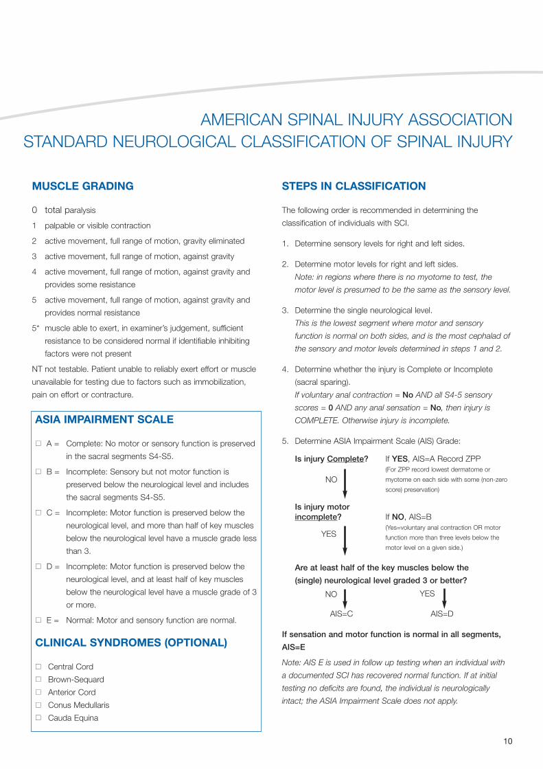
MUSCLE GRADING
0 total paralysis
1 palpable or visible contraction
2 active movement, full range of motion, gravity eliminated
3 active movement, full range of motion, against gravity
4 active movement, full range of motion, against gravity and
provides some resistance
5 active movement, full range of motion, against gravity and
provides normal resistance
5* muscle able to exert, in examiner’s judgement, sufficient
resistance to be considered normal if identifiable inhibiting
factors were not present
NT not testable. Patient unable to reliably exert effort or muscle
unavailable for testing due to factors such as immobilization,
pain on effort or contracture.
AMERICAN SPINAL INJURY ASSOCIATION STANDARD NEUROLOGICAL CLASSIFICATION OF SPINAL INJURY
STEPS IN CLASSIFICATION
The following order is recommended in determining the
classification of individuals with SCI.
1. Determine sensory levels for right and left sides.
2. Determine motor levels for right and left sides.
Note: in regions where there is no myotome to test, the
motor level is presumed to be the same as the sensory level.
3. Determine the single neurological level.
This is the lowest segment where motor and sensory
function is normal on both sides, and is the most cephalad of
the sensory and motor levels determined in steps 1 and 2.
4. Determine whether the injury is Complete or Incomplete
(sacral sparing).
If voluntary anal contraction = No AND all S4-5 sensory
scores = 0 AND any anal sensation = No, then injury is
COMPLETE. Otherwise injury is incomplete.
5. Determine ASIA Impairment Scale (AIS) Grade:
Is injury Complete? If YES, AIS=A Record ZPP(For ZPP record lowest dermatome or
myotome on each side with some (non-zero
score) preservation)
Is injury motor incomplete? If NO, AIS=B
(Yes=voluntary anal contraction OR motor
function more than three levels below the
motor level on a given side.)
Are at least half of the key muscles below the
(single) neurological level graded 3 or better?
AIS=C AIS=D
If sensation and motor function is normal in all segments,
AIS=E
Note: AIS E is used in follow up testing when an individual with
a documented SCI has recovered normal function. If at initial
testing no deficits are found, the individual is neurologically
intact; the ASIA Impairment Scale does not apply.
NO
NO
YES
YES
ASIA IMPAIRMENT SCALE
nn A = Complete: No motor or sensory function is preserved
in the sacral segments S4-S5.
nn B = Incomplete: Sensory but not motor function is
preserved below the neurological level and includes
the sacral segments S4-S5.
nn C = Incomplete: Motor function is preserved below the
neurological level, and more than half of key muscles
below the neurological level have a muscle grade less
than 3.
nn D = Incomplete: Motor function is preserved below the
neurological level, and at least half of key muscles
below the neurological level have a muscle grade of 3
or more.
nn E = Normal: Motor and sensory function are normal.
CLINICAL SYNDROMES (OPTIONAL)
nn Central Cord
nn Brown-Sequard
nn Anterior Cord
nn Conus Medullaris
nn Cauda Equina
10

The SIA Academy is the training arm of the Spinal Injuries Association which promotes training, education,social research, Spinal Cord Injury awareness and best practice in living with spinal cord injury
11All rights, including copyright © of the guideline's content is owned by the SIA, MASCIP and Huntleigh. This publication is for information and illustration purposes only. Opinions expressed should not be construedas medical advice. The teaching of these procedures and the management of the spinal cord injured person should only be undertaken by a suitably qualified and authorised health care professional.
Explain to the patient what is happening and why. A suitably qualified and experienced health careworker will be designated as Team Leader. TheTeam Leader positions self at top of trolley / bed,placing hands either side of patient’s head. Withfingers spread wide, slides both hands downwardsso the thumb rests either below the jaw or abovethe clavicle and the fingers are spread behind theneck encompassing C7. If sandbags / headblocksare present an assistant removes them, one at atime and the Team Leader brings each hand intoposition individually. Forearms are then broughttogether either side at the back of the head.
Prior to rolling the patient, care must be taken toposition the bed height at optimum level to reduceexcessive forward trunk flexion of the Team Leader.Patient is then rolled on the command of the TeamLeader. To accommodate this roll, Team Leader maybe required to adopt a side flexed position. Notefingers crossed behind cervical spine as describedabove.
In order to maintain a comfortable head hold duringthe logroll, the Team Leader releases top hand andmaintaining contact with the skin throughout, moveshands slowly to the top of the patient’s head withfingers spread wide. They should then adjust theirbase of support (feet and legs) to a morecomfortable and sustainable position whilemaintaining the head in the aligned position.
Shows adapted ATLS head hold from the oppositeside showing alignment nose – chin – sternum. A chair can be made available for the Team Leaderto sit down during prolonged holding to enable theelbows to be rested on a pillow. The Team Leadermust be aware that they are allowed to return thepatient to the supine position if they feel the strain ofmaintaining the turn becomes excessive and beyondtheir limitations. In patients with broad shoulders, apillow or pad can be used to support the TeamLeader’s underlying arm but it must be of the correctdepth to maintain spinal alignment.
ADAPTED ATLS HEAD HOLD FOR ACTUAL OR POTENTIAL CERVICAL SPINAL INJURY
Advanced trauma life support manual and training stipulate a standardized approach to head holding in the event of actual or suspected spinal injury. The healthcare worker responsible for headholding is designated as the Team Leader and directs all patient movement. However, the degree of lateral flexion experienced by the Team Leader during logrolling is excessive and this represents an
adaptation of the current technique as recommended by American College of Surgeons’ Committee on Trauma (ACS). (2008) Advanced Trauma Life Support Manual for Physicians (8th edition).American College of Surgeons Press, Chicago.
1 2 3 4

The SIA Academy is the training arm of the Spinal Injuries Association which promotes training, education,social research, Spinal Cord Injury awareness and best practice in living with spinal cord injury
12All rights, including copyright © of the guideline's content is owned by the SIA, MASCIP and Huntleigh. This publication is for information and illustration purposes only. Opinions expressed should not be construedas medical advice. The teaching of these procedures and the management of the spinal cord injured person should only be undertaken by a suitably qualified and authorised health care professional.
In order to maintain a secure positionafter fitting, collars must be fitted againstbare skin. Clothing may need to bemoved aside or cut away in order tofacilitate this. Jewellery and earrings mustbe removed before fitting of collar. Check sensory and motor function andpositional awareness in all four limbsbefore application.
Explain to the patient what is happeningand why. A suitably qualified andexperienced health care worker firstmeasures the patient’s neck againstanatomical landmarks (picture 1) andthen adjusts the collar to the appropriatesize against a visual scale in accordancewith the manufacturer’s instructions andthe collar design.
Prior to the fitting of any collar, manualhead holding must be in place. Fingersmust be positioned to encompass asmuch of the patient’s head as possiblebut without obscuring the ears so thatthe patient can hear explanations andinstructions throughout the procedure.
With the patient’s head secured, the 1stassistant – team leader gently feeds theback piece of the collar into position,pressing the collar into the mattresssurface to prevent friction with thepatient’s skin.
The 2nd assistant assists as necessary tomanoeuvre the collar back into its correctposition.
The front of the collar is now broughtround into position and the velcro strapfastening is secured. Most collarsincorporate cutaway panels to ensurevisualisation of underlying skin,anatomical structures, dressings etc.
Check sensory and motor function andpositional awareness in all four limbsagain after application in comparison withpre-application assessment.
Check sizing and fitting of collar forsecurity and patient comfort and makeany adjustments prior to any furtherpatient movement.
Carer holding head now moves handsdown to patient’s shoulders maintainingtactile contact with collar surfacethroughout.
Fingers move inwards behind back ofcollar to encompass collar back andforearms move inwards to secure patient’shead against lateral movements. Manualhead hold can now be released unlessfurther movement of patient is required.
APPLICATION OF A ONE-PIECE COLLARThe need to apply a properly sized and fitted hard cervical collar as an aid to spinal protection should always be considered whenever there is evidence or suspicion of actual or potential cervical
spinal trauma or spinal cord injury. Cervical collars should only be fitted by suitably qualified and authorised healthcare professional or rescue first aider in accordance with manufacturer’s guidelinesand locally established practice guidelines. The person who measures the neck should be the person who sizes the collar. Due to the potential for hidden spinal metabolic disease or deformity the
fitting of a hard cervical collar in a patient aged more than 55 years should be approached with some caution lest it cause or compound a cervical injury.
1
6
2
7
3
8
4
9
5
10

The SIA Academy is the training arm of the Spinal Injuries Association which promotes training, education,social research, Spinal Cord Injury awareness and best practice in living with spinal cord injury
13All rights, including copyright © of the guideline's content is owned by the SIA, MASCIP and Huntleigh. This publication is for information and illustration purposes only. Opinions expressed should not be construedas medical advice. The teaching of these procedures and the management of the spinal cord injured person should only be undertaken by a suitably qualified and authorised health care professional.
Explain to the patient what is happening and why. A suitably qualified and authorised healthcareprofessional first measures the patient’s neck againstanatomical landmarks using a sizing guide providedby the collar manufacturer (see picture 1) and thenselects the most appropriate collar in accordancewith the manufacturer’s instructions and the collardesign. This collar is also available with a sizeadjustable front piece.
Prior to the fitting of any collar manual head holdingmust be in place. Fingers must be positioned toencompass as much of the patient’s head aspossible but without obscuring the ears so thatpatient can hear explanations and instructionsthroughout the procedure.
With the patient’s head secured, the 1st assistant –team leader gently feeds the back piece of the collar into position, pressing it into the mattressto prevent friction with the patient’s skin. The 2ndassistant assists as necessary to manoeuvre thecollar back into its correct position.
The front-piece of the collar is then brought intoposition by an assistant who brings the collar pieceup and under the chin in a straight line. Curling andflexing of the collar piece before fitting ensures amore comfortable fit.
The velcro fastenings are then secured. Checksensory and motor function and positionalawareness in all four limbs again after application incomparison with pre-application assessment.
Picture depicts position of hands during a logroll tofacilitate removal of collar back for visualisation orexamination of underlying skin or posterior cervicalspine. This model of collar has a small window toallow for ventilation but also useful for examiningstatus of any underlying surgical wound or dressing.
Head holding as at end position of adapted ATLSlogrolling (see ATLS and Tetra logroll). Handpositioning is same whether patient presents with orwithout collar in situ. This picture also shows analternative and smaller size of back piece providedfor this model of collar and designed to reduceincidence of collar-derived occipital pressure ulcers,particularly within critical care environments.
APPLICATION OF A TWO-PIECE COLLARThe application of a properly sized and fitted two-piece cervical collar is usually undertaken within hospital environments as an aid to continued spinal protection in patients with actual or suspectedcervical spinal or spinal cord injury. Two-piece collars usually replace extrication collars within the first 48 hours of admission. All models of two-piece cervical collars should only be fitted by suitablyqualified and authorised healthcare professional in accordance with manufacturer’s guidelines and locally established practice guidelines. The person who measures the neck should be the personwho sizes the collar. Due to the potential for hidden spinal metabolic disease or deformity the fitting of a two-piece cervical collar in a patient aged more than 55 years should be approached with
some caution lest it cause or compound a cervical injury.
1
5
2
6
3
7
4
8
Cervical Collar Sizing GuideReproduced with kind permission of Aspen Medical Products.

The SIA Academy is the training arm of the Spinal Injuries Association which promotes training, education,social research, Spinal Cord Injury awareness and best practice in living with spinal cord injury
14All rights, including copyright © of the guideline's content is owned by the SIA, MASCIP and Huntleigh. This publication is for information and illustration purposes only. Opinions expressed should not be construedas medical advice. The teaching of these procedures and the management of the spinal cord injured person should only be undertaken by a suitably qualified and authorised health care professional.
ACUTE TETRAPLEGIC SPINAL LOGROLL – Method 1During an acute tetraplegic logroll the patient’s head and vertebral column must be kept in alignment when rolling from supine to side-lying and vice versa. During this manoeuvre the alignment of the vertebral
column and the body as a whole is maintained through the manual support provided by the turning team. (1st assistant – Team leader & acute head hold in accordance with adapted ATLS procedure; 2nd assistant – shoulder level; 3rd assistant – hip level; 4th assistant – lower leg level; 5th assistant – operating the bed controls, supporting arms, checking patient’s skin, placing pillows in situ etc)
Logrolling on a trolley in the Emergency Department or within a wardsetting on a normal hospital bed or tilt and turn bed is essential toenable examination of the back and necessary for relieving pressure onthe skin, hygiene, bowel care and postural chest drainage. The followingtechnique is applicable in all clinical settings.
Team leader undertakes acute initial head hold in accordance withadapted ATLS procedure. 5th assistant passively positions patient’sarms across chest but above diaphragm. This is important as the armsare paralysed and may fall down causing injury to the shoulder joint.
2nd assistant reaches over patient. First hand on shoulder and secondhand on top of hip. 5th assistant supports patient’s arm during thisaction.
3rd assistant positions hands. First hand at hip level alongside the 2ndassistant, and second hand underneath furthest thigh.
4th assistant positions hands. First hand under the knee of the furthestleg, and second hand under the ankle of the same leg.
Close up of hand positions – ensure all parties are in contact with thepatients natural skeletal landmarks and not just adipose tissue.
1
4
2
5
3
6
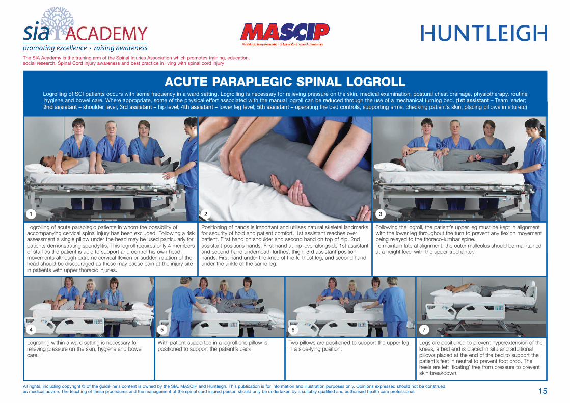
Logrolling within a ward setting is necessary forrelieving pressure on the skin, hygiene and bowelcare.
With patient supported in a logroll one pillow ispositioned to support the patient’s back.
Two pillows are positioned to support the upper legin a side-lying position.
Legs are positioned to prevent hyperextension of theknees, a bed end is placed in situ and additionalpillows placed at the end of the bed to support thepatient’s feet in neutral to prevent foot drop. Theheels are left ‘floating’ free from pressure to preventskin breakdown.
Logrolling of acute paraplegic patients in whom the possibility ofaccompanying cervical spinal injury has been excluded. Following a riskassessment a single pillow under the head may be used particularly forpatients demonstrating spondylitis. This logroll requires only 4 membersof staff as the patient is able to support and control his own headmovements although extreme cervical flexion or sudden rotation of thehead should be discouraged as these may cause pain at the injury sitein patients with upper thoracic injuries.
Positioning of hands is important and utilises natural skeletal landmarksfor security of hold and patient comfort. 1st assistant reaches overpatient. First hand on shoulder and second hand on top of hip. 2ndassistant positions hands. First hand at hip level alongside 1st assistantand second hand underneath furthest thigh. 3rd assistant positionhands. First hand under the knee of the furthest leg, and second handunder the ankle of the same leg.
Following the logroll, the patient’s upper leg must be kept in alignmentwith the lower leg throughout the turn to prevent any flexion movementbeing relayed to the thoraco-lumbar spine.To maintain lateral alignment, the outer malleolus should be maintainedat a height level with the upper trochanter.
The SIA Academy is the training arm of the Spinal Injuries Association which promotes training, education,social research, Spinal Cord Injury awareness and best practice in living with spinal cord injury
15All rights, including copyright © of the guideline's content is owned by the SIA, MASCIP and Huntleigh. This publication is for information and illustration purposes only. Opinions expressed should not be construedas medical advice. The teaching of these procedures and the management of the spinal cord injured person should only be undertaken by a suitably qualified and authorised health care professional.
1 2 3
4 5 6 7
ACUTE PARAPLEGIC SPINAL LOGROLLLogrolling of SCI patients occurs with some frequency in a ward setting. Logrolling is necessary for relieving pressure on the skin, medical examination, postural chest drainage, physiotherapy, routinehygiene and bowel care. Where appropriate, some of the physical effort associated with the manual logroll can be reduced through the use of a mechanical turning bed. (1st assistant – Team leader; 2nd assistant – shoulder level; 3rd assistant – hip level; 4th assistant – lower leg level; 5th assistant – operating the bed controls, supporting arms, checking patient’s skin, placing pillows in situ etc)

The SIA Academy is the training arm of the Spinal Injuries Association which promotes training, education,social research, Spinal Cord Injury awareness and best practice in living with spinal cord injury
16All rights, including copyright © of the guideline's content is owned by the SIA, MASCIP and Huntleigh. This publication is for information and illustration purposes only. Opinions expressed should not be construedas medical advice. The teaching of these procedures and the management of the spinal cord injured person should only be undertaken by a suitably qualified and authorised health care professional.
AIRWAY PROTECTION
Traumatic SCI occurs without warning and casualtiesoften present having recently eaten or, more often,having drunk a significant amount of alcohol prior tothe accident. Vomiting is common following SCI andusually occurs at the scene or during transportationto hospital. Vomiting is a particular hazard in childrenand those experiencing near-drowning followingaquatic SCI incidents. The risk of aspiration ishighest whilst the SCI patient is positioned supine asthey are unable to adequately protect their ownairway. Fully operational suction equipment shouldbe available at all times.
As this picture illustrates, paramedical andemergency care staff are trained in turning patientssecured appropriately on spinal boards usingminimum numbers of staff initially. This turningtechnique is enabled by first bearing down on thenear side of the spinal board with one hand toinduce a rolling movement before reaching across tothe opposite side with the other hand to facilitateturning.
A patient can only be removed from the spinal boardor placed in side lying if initial screening does notidentify spinal cord trauma. To maintain airwayprotection once the patient has been removed fromthe spinal board, rapid logrolling by 4 health workerswith an additional person needed to performsuctioning is the usual response to a vomiting SCIpatient in A&E but the availability of staff can delayan immediate response or compromise spinalalignment.
Whenever a significant risk of vomiting exists it ispreferable to retain the patient on a spinal board untilprimary screening and examinations have beencompleted and the patient can then be positioned ona trolley or in a bed in a lateral side-lying position toimprove airway clearance. Management of thevomiting SCI patient will also include administrationof an appropriate anti-emetic and gastricdecompression via nasogastric tube.

The SIA Academy is the training arm of the Spinal Injuries Association which promotes training, education,social research, Spinal Cord Injury awareness and best practice in living with spinal cord injury
17All rights, including copyright © of the guideline's content is owned by the SIA, MASCIP and Huntleigh. This publication is for information and illustration purposes only. Opinions expressed should not be construedas medical advice. The teaching of these procedures and the management of the spinal cord injured person should only be undertaken by a suitably qualified and authorised health care professional.
Emergency Department staff prefer whereverpossible not to have trauma patients arriving onvacuum mattresses direct from the scene of anaccident as removal requires additional logrolling of apatient in pain and with unknown injuries. Inaddition, the vacuum mattress is not suitable to useas a splint for patients with acute pelvic fracturesunless they have other means of pelvic splinting insitu. If the fracture is unstable the patient maycontinue to “bleed out” on releasing the mattressand collapse.
Positioning a patient with actual or suspectedspinal injury in a vacuum mattress (not illustrated).The patient is scoop transferred onto the mattress atthe scene. And the mattress folded around theirbody and secured with straps. The mattress is filledwith tiny silicone beads which vacuum mould to thepatient when all of the air is pumped out. Themattress is then loaded onto a spinal board orscoop stretcher because it lacks a suitably rigidbase. Special care is required to ensure that thehead and cervical spine are properly supportedthroughout these operations. Once in hospital, thevacuum mattress can be used to continue to protectthe patient during flat surface-to-surface transfersand the patient can even be x-rayed or scannedwith the vacuum mattress in place if required.
Removing a patient with actual or suspectedspinal injury from a vacuum mattress.First apply manual cervical spinal protection whetheror not a cervical collar is in situ before undoing all ofthe mattress straps. Do not cut any straps.
Now open the air valve on the outer mattresssurface to let the air reinflate the mattress. Themattress sides will now become flexible again sothat they can be unwrapped from around thepatient. The air is then pumped out of the mattressagain providing a flat surface from which the patientcan then be retrieved using a flat lifting scoopstretcher hoist.
A flat lifting scoop stretcher hoist is the preferredtransfer method. An alternative option is to use alateral transfer board and sliding sheet to transfer apatient on a scoop stretcher between surfaces.
REMOVAL FROM VACUUM MATTRESSThe vacuum mattress is the preferred device to provide spinal protection during inter-hospital transfers of critically-ill patients or those with serious trauma such as acute spinal or spinal cord injuries, in
accordance with manufacturer’s instructions and locally established practice guidelines.
1 2 3 4

The SIA Academy is the training arm of the Spinal Injuries Association which promotes training, education,social research, Spinal Cord Injury awareness and best practice in living with spinal cord injury
18All rights, including copyright © of the guideline's content is owned by the SIA, MASCIP and Huntleigh. This publication is for information and illustration purposes only. Opinions expressed should not be construedas medical advice. The teaching of these procedures and the management of the spinal cord injured person should only be undertaken by a suitably qualified and authorised health care professional.
Team assemble and team leader explains procedureand confirms all team members understandindividual role in procedure. Team leader appliesadapted ATLS head hold if appropriate and rest ofturning team take position for logrolling patient.Before logrolling commences, 5th assistant placesspinal board in position. Note board held in angleposition for insertion and not flat.
Team leader gives command to roll and team turnpatient in unison onto chosen side. Whilst it is mostcommon to turn patient onto left side to facilitatemedical examination (as picture above) but inpatients with cervical injury it is often preferable toturn patient on to their right side to avoid inducingvaso-vagal cardiac syncope. 5th member placesspinal board in situ angled against the patient’s back.
Team leader gives command to return patient tosupine position and checks alignment. Anynecessary adjustments in position to maintainalignment are made before head blocks and strapsare applied. If the patient might require to be tiltedon the spinal board for pressure relief or there is arisk of vomiting, additional padding is inserted toprevent inappropriate lateral movement.
With the patient now secured, team membersshould reassemble in different positions beforeundertaking the transfer to reduce the potential forpostural strain. This may not always be possible dueto staffing numbers and the experience of the staffavailable at the time.
The new team leader continues to provide manualsupport for the cervical spine as rest of teamprepare to insert a patient transfer board beneaththe spinal board. By bearing down on the edge ofthe spinal board, the two team members on the leftof the picture can induce a slight raising of the spinalboard on the opposite side allow for an easierinsertion of a sliding aid. If appropriate, a slidingsheet or patient roller can also be introducedbeneath the spinal board at this time.
The team leader gives the command to slide thepatient across between the two flat surfaces ingradual stages. Never attempt to transfer the patientacross in one movement.
The team reassembles to logroll the patient again toremove the spinal board.Again, wherever staffing numbers and experienceallow, team members should reassemble in differentpositions before undertaking the final logroll toreduce the potential for repetitive strain.
As the spinal board is removed, the 5th member ofthe team takes the opportunity to inspect thepatient’s underlying skin for signs of pressuredamage after transfer.
LATERAL TRANSFER USING A SPINAL BOARDAlthough spinal boards were primarily designed to be used during the extrication and evacuation of casualties to hospital, they still serve a useful purpose after admission, along with scoop stretchers to
protect the patient’s spine from lateral forces experienced during sliding transfers between flat surfaces, in accordance with manufacturer’s guidelines and locally established practice guidelines.
1
5
2
6
3
7
4
8
The SIA Academy is the training arm of the Spinal Injuries Association which promotes training, education,social research, Spinal Cord Injury awareness and best practice in living with spinal cord injury
19All rights, including copyright © of the guideline's content is owned by the SIA, MASCIP and Huntleigh. This publication is for information and illustration purposes only. Opinions expressed should not be construedas medical advice. The teaching of these procedures and the management of the spinal cord injured person should only be undertaken by a suitably qualified and authorised health care professional.
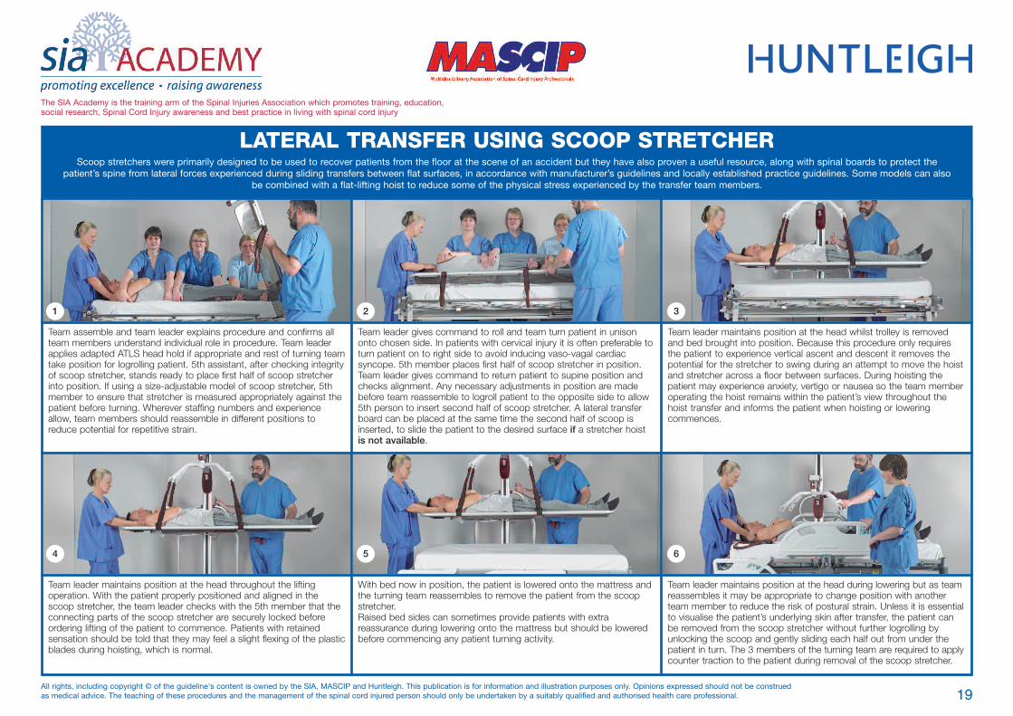
LATERAL TRANSFER USING SCOOP STRETCHERScoop stretchers were primarily designed to be used to recover patients from the floor at the scene of an accident but they have also proven a useful resource, along with spinal boards to protect the
patient’s spine from lateral forces experienced during sliding transfers between flat surfaces, in accordance with manufacturer’s guidelines and locally established practice guidelines. Some models can alsobe combined with a flat-lifting hoist to reduce some of the physical stress experienced by the transfer team members.
Team assemble and team leader explains procedure and confirms allteam members understand individual role in procedure. Team leaderapplies adapted ATLS head hold if appropriate and rest of turning teamtake position for logrolling patient. 5th assistant, after checking integrityof scoop stretcher, stands ready to place first half of scoop stretcherinto position. If using a size-adjustable model of scoop stretcher, 5thmember to ensure that stretcher is measured appropriately against thepatient before turning. Wherever staffing numbers and experienceallow, team members should reassemble in different positions toreduce potential for repetitive strain.
Team leader gives command to roll and team turn patient in unisononto chosen side. In patients with cervical injury it is often preferable toturn patient on to right side to avoid inducing vaso-vagal cardiacsyncope. 5th member places first half of scoop stretcher in position.Team leader gives command to return patient to supine position andchecks alignment. Any necessary adjustments in position are madebefore team reassemble to logroll patient to the opposite side to allow5th person to insert second half of scoop stretcher. A lateral transferboard can be placed at the same time the second half of scoop isinserted, to slide the patient to the desired surface if a stretcher hoistis not available.
Team leader maintains position at the head whilst trolley is removedand bed brought into position. Because this procedure only requiresthe patient to experience vertical ascent and descent it removes thepotential for the stretcher to swing during an attempt to move the hoistand stretcher across a floor between surfaces. During hoisting thepatient may experience anxiety, vertigo or nausea so the team memberoperating the hoist remains within the patient’s view throughout thehoist transfer and informs the patient when hoisting or loweringcommences.
Team leader maintains position at the head throughout the liftingoperation. With the patient properly positioned and aligned in thescoop stretcher, the team leader checks with the 5th member that theconnecting parts of the scoop stretcher are securely locked beforeordering lifting of the patient to commence. Patients with retainedsensation should be told that they may feel a slight flexing of the plasticblades during hoisting, which is normal.
With bed now in position, the patient is lowered onto the mattress andthe turning team reassembles to remove the patient from the scoopstretcher.Raised bed sides can sometimes provide patients with extrareassurance during lowering onto the mattress but should be loweredbefore commencing any patient turning activity.
Team leader maintains position at the head during lowering but as teamreassembles it may be appropriate to change position with anotherteam member to reduce the risk of postural strain. Unless it is essentialto visualise the patient’s underlying skin after transfer, the patient canbe removed from the scoop stretcher without further logrolling byunlocking the scoop and gently sliding each half out from under thepatient in turn. The 3 members of the turning team are required to applycounter traction to the patient during removal of the scoop stretcher.
1 2 3
4 5 6

The SIA Academy is the training arm of the Spinal Injuries Association which promotes training, education,social research, Spinal Cord Injury awareness and best practice in living with spinal cord injury
20All rights, including copyright © of the guideline's content is owned by the SIA, MASCIP and Huntleigh. This publication is for information and illustration purposes only. Opinions expressed should not be construedas medical advice. The teaching of these procedures and the management of the spinal cord injured person should only be undertaken by a suitably qualified and authorised health care professional.
ACUTE TETRAPLEGIC SPINAL LOGROLL – Method 2Logrolling of SCI patients occurs with some frequency in a ward setting. Logrolling is necessary for relieving pressure on the skin, medical examination, postural chest drainage, physiotherapy, routinehygiene and bowel care. Where appropriate, some of the physical effort associated with the manual logroll can be reduced through the use of a mechanical turning bed. (1st assistant – Team leader; 2nd assistant – shoulder level; 3rd assistant – hip level; 4th assistant – lower leg level; 5th assistant – operating the bed controls, supporting arms, checking patient’s skin, placing pillows in situ etc)
A team of five health care workers are required to logroll a patient withacute tetraplegia. All commands come from the team leader who alsotakes responsibility for protecting the patient’s cervical spine. Teamleader takes position first applying an adapted ATLS head hold. 2ndassistant in line holds shoulders and hip, 3rd assistant holds hip andunder upper thigh, 4th assistant holds under knee and under ankle. 5thassistant is responsible for operating bed controls, checking patient’sskin and placing pillows in situ.
On the command of the team leader, the 5th assistant presses thecontrol button to start the bed turning towards the turning team. As theangle of turn increases, the turning team members provide support forthe patient’s body as required, to maintain spinal alignment. The teamleader adjusts their position in accordance with the increasing turningangle in order to prevent excessive lateral leaning.
Once the patient has achieved a suitable side-lying position the teamleader orders the 5th assistant to stop turning the bed any further andthen orders the turning team to hold the patient in position while the5th assistant returns the bed to flat position again. During this part ofthe turn, the 4th assistant must ensure that the patient’s upper leg iskept in alignment with the lower leg throughout the turn. To maintainlateral alignment, the outer malleolus should be maintained at a heightlevel with the upper trochanter.
With the bed now flat again the 5th assistant inspects the patient’s skinfor any signs of pressure damage. Additional cares such as bowelcare, hygiene, and sheet changing can also be performed while thepatient is in this position.
With the patient supported in a logroll one pillow is positioned tosupport the patient’s back and two pillows are positioned to supportthe upper leg in a side-lying position.
On the command of the team-leader the patient is lowered down ontothe pillows by the turning team and their position adjusted to ensureproper alignment. Legs are positioned to prevent hyperextension of theknees, a bed end is placed in situ and additional pillows placed at theend of the bed to support the patient’s feet in neutral to prevent footdrop. The heels are left ‘floating’ free from pressure to prevent skinbreakdown (not illustrated).
1 2 3
4 5 6

Team insert pillows under both arms and legs for patient comfort andalignment.
Legs are positioned to prevent hyperextension of the knees, a bed endis placed in situ and additional pillows placed at the end of the bed tosupport the patient’s feet in neutral to prevent foot drop. The heels areleft ‘floating’ free from pressure to prevent skin breakdown (notillustrated)
Team leader undertakes acute initial head hold in accordance withadapted ATLS procedure. 2nd assistant provides contact guard againstinappropriate patient movement, 3rd assistant positions pillow betweenthe legs to maintain hip abduction.
2nd and 3rd assistant provide contact guard against inappropriatemovement of the patient during mechanical turning of the bed. Team leader gives the command when all the team are in position tocommence the turning of the bed.
4th assistant checks inclinometer fitted to the bed and stops the bedat the required degree of tilt.
The SIA Academy is the training arm of the Spinal Injuries Association which promotes training, education,social research, Spinal Cord Injury awareness and best practice in living with spinal cord injury
21All rights, including copyright © of the guideline's content is owned by the SIA, MASCIP and Huntleigh. This publication is for information and illustration purposes only. Opinions expressed should not be construedas medical advice. The teaching of these procedures and the management of the spinal cord injured person should only be undertaken by a suitably qualified and authorised health care professional.
MECHANISED TURN FOR POSTURAL CHANGEThe availability of a mechanical turning bed can enhance the experience of turning in alignment for patients with actual or suspected spinal injury. This is particularly beneficial for tetraplegic patients,
patients with multiple trauma and acute chest complications, as well as for patients whose size causes a significant risk for staff during routine manual turning. (1st assistant – Team leader & acute headhold in accordance with adapted ATLS procedure; 2nd assistant – shoulder level; 3rd assistant – hip level; 4th assistant – operating the bed controls, supporting arms, checking patient’s skin, placing
pillows in situ etc)
1 2 3
4 5

The SIA Academy is the training arm of the Spinal Injuries Association which promotes training, education,social research, Spinal Cord Injury awareness and best practice in living with spinal cord injury
22All rights, including copyright © of the guideline's content is owned by the SIA, MASCIP and Huntleigh. This publication is for information and illustration purposes only. Opinions expressed should not be construedas medical advice. The teaching of these procedures and the management of the spinal cord injured person should only be undertaken by a suitably qualified and authorised health care professional.
POSTURAL ALIGNMENTPhysical landmarks are visualised to demonstrate postural alignment of the spine during turning and positioning of SCI patients.
During all patient movements all commands comefrom the team leader who also takes responsibility formonitoring the physical alignment of the patient’sspine during and after turning and transferprocedures by monitoring the alignment of bodylandmarks.
From their sight position at the patient’s head theycan monitor the alignment of the nose, sternum andpubic symphysis. They can also observe lateralalignment of shoulders, ribcage, hips and legs forsigns of spinal rotation. When at rest, the headshould be supported to maintain mid-line positionusing pads or blocks.
The accompanying pictures illustrate correct posturalalignment of SCI patients following turning andtransfer procedures. Upper limbs should besupported in a position that guards againstcontractures of elbow, wrist and fingers until thepatient is assessed for splints.
Legs are positioned to prevent hyperextension ofthe knees, a bed end is placed in situ andadditional pillows placed at the end of the bed tosupport the patient’s feet in neutral to prevent footdrop. The heels are left ‘floating’ free from pressureto prevent skin breakdown.
1
2
3
4
5
6
7

The SIA Academy is the training arm of the Spinal Injuries Association which promotes training, education,social research, Spinal Cord Injury awareness and best practice in living with spinal cord injury
23All rights, including copyright © of the guideline's content is owned by the SIA, MASCIP and Huntleigh. This publication is for information and illustration purposes only. Opinions expressed should not be construedas medical advice. The teaching of these procedures and the management of the spinal cord injured person should only be undertaken by a suitably qualified and authorised health care professional.
ADJUSTING SKIN LOADINGAt the end of a turning and positioning episode, the SCI patient has a tendency to place undue pressure upon the underlying bony surfaces and weight-bearing areas as they are unable to adjust the
loading pressure upon their skin without assistance.
Adjusting skin loading should form part of the routine at the end of a turnor transfer. The adjusted skin loading needs to initially focus on thebuttocks, to ensure the natal cleft is separated, this is a two-personprocedure and in the acute stage a third person may be used to stabilisethe site of injury also. Once the buttock skin loading has been adjustedand the patient has been aligned to the satisfaction of the team leader,the team will disperse to their other duties. However one nurse remainsto perform the procedure. Adjusting limb skin loading is better learnt as apractical technique so this poster serves only as an illustration.
There is no lifting involved in this technique. The carer places both oftheir hands under the patient’s shoulder blade with palms uppermostand gently draws them out towards them, allowing the naturalresistance of the patient’s bodyweight to create a slight traction thatredistributes the surface area of the skin as the hands are withdrawn.
Keeping their hands in the same position the carer now moves theirhands under the patients arm and hand until they move out fromunder the patient’s body completely.
The carer now moves to the patient’s lower bodyand places both hands palms uppermost under thepatient’s buttock. Carer must ensure that they avoidany twisting or prolonged stooping of their trunkduring this procedure.
Again moving slowly and without attempting to liftupwards, the carer begins to draw their hands alongunder the patient’s buttock and down the leg untilthey again move out from under the patient’s bodycompletely.
The carer then moves to the opposite side of thebed and repeats the procedure for the other side ofthe patient’s body. After completion, any pillowsnecessary to maintain patient alignment are placedin situ, the patient made comfortable and the bedspace restored.
The benefit of this technique is firstly that it ensuresthe broadest distribution of underlying skin pressurein patients who are unable adjust their positionindependently. Secondly, with an increasingincidence of SCI patients with incomplete sensoryloss, it reduces patient discomfort during periods ofenforced bed rest, reducing the number of requestsby the patient to care staff for additional andunplanned turning and repositioning during the day.
1 2 3
4 5 6 7

The SIA Academy is the training arm of the Spinal Injuries Association which promotes training, education,social research, Spinal Cord Injury awareness and best practice in living with spinal cord injury
24All rights, including copyright © of the guideline's content is owned by the SIA, MASCIP and Huntleigh. This publication is for information and illustration purposes only. Opinions expressed should not be construedas medical advice. The teaching of these procedures and the management of the spinal cord injured person should only be undertaken by a suitably qualified and authorised health care professional.
ASSISTED COUGHParalysis of the abdominal muscles causes severe impairment of forced expiration. The cough mechanism will be altered in SCI patients with a neurological level of T11 and above. The higher the level oflesion the more likely the patient will require assistance with coughing. Patients with complete cervical spinal cord lesions are at greatest risk of respiratory complications. Medical advice should always be
sought first before attempting assisted coughing in new SCI patients, those with chest injuries, cardiovascular disease, abdominal trauma or disease or who are pregnant.
Two-person technique: Clear verbal direction and co-ordinationbetween the person(s) helping and the patient is essential for thesetechniques to be successful.Stand on either side of the bed. Each person places their hands on theupper and lower ribs of the same side with their fingers spread andpointing upwards and centrally. As the patient attempts to cough, pushinwards and upwards simultaneously. This method may not be suitablefor a patient who has an unstable spine because if the actions are notperformed simultaneously it introduces rotation of the thorax.
This two person method is preferred if spinal stability is a considerationas both people are pushing bilaterally which will minimise rotation.Stand on either side of the bed. Each person places one forearmacross the upper abdomen of the patient with their other hand on theupper or lower ribs of both sides of the chest. As the patient attemptsto cough, push inwards simultaneously.
Single person technique: spread your hands anteriorly around thelower rib cage and upper abdomen. With your elbows extended pushinwards and upwards with both arms as the patient attempts to cough.Arms must be kept extended for this technique to work effectively, itmay therefore not be appropriate to use if the patient’s bed does notlower to a suitable height.
1 2 3

25
REFERENCES
American College of Surgeons’ Committee on Trauma (ACS).(2008) Advanced Trauma Life Support Manual forPhysicians (8th edition). American College of Surgeons Press,Chicago.
Harrison P (ed) (2007) Managing Spinal Cord Injury: The First48 Hours. Spinal Injuries Association. Milton Keynes.
Hawkins S, Stone K, Plummer L. (1999) An holistic approach toturning patients. Nursing Standard.; 14: (3) 52 – 56

ACKNOWLEDGMENTSName Contact Details
Lead AuthorPaul Harrison Clinical Development Officer
Princess Royal Spinal Injuries CentreNorthern General HospitalSheffield
Guideline Contributors(In Alphabetical order)
Heather Abraham SisterYorkshire Spinal Injuries CentrePinderfields General Hospital Wakefield
Ann Bell Clinical EducatorThe Spinal Cord Injuries UnitMusgrave Park HospitalBelfast
Sharon Budd Trauma PractitionerRoyal Derby HospitalDerby
Mary Collins Trauma Ward ManagerNorfolk and Norwich University Foundation HospitalNorwich
Sandra Cosgrove Clinical Educator The Regional Spinal Injuries CentreSouthport and Formby District HospitalSouthport
Laurie Duffy Respiratory Nurse SpecialistQueen Elizabeth National Spinal Injuries Centre for ScotlandSouthern General HospitalGlasgow
Kathryn Evans Nurse ManagerThe Rockwood Spinal Injuries UnitRockwood HospitalCardiff
Name Contact Details
Caroline Fallon Clinical Development SpecialistHuntleigh UK310 – 312 Dallow RoadLuton
Amanda Fletcher Senior Staff NurseAccident & Emergency DepartmentQueen Elizabeth Hospital King’s Lynn
Theresa Flynn Moving and Handling Manager St Vincent’s University Hospital, Dublin Link to Dublin Hospitals Group Risk Management Forum
Angela Gall MASCIP Chair & BASICS LinkConsultantLondon Spinal Injuries CentreRoyal National Orthopaedic HospitalStanmore
Sarla Gandhi Neuro Pathway LeaderUniversity of Central LancashirePreston
Graham Gentry ITU Manager and Coordinator South Thames, ITU NetworkAshford and St Peter's HospitalChertsey
Steve Hancock Paediatric Critical Care Consultant Link to UK Paediatric Retrieval TeamsSheffield Children's Hospital Sheffield
Kyla Honing Neuro ITU ManagerDerriford HospitalPlymouthRoyal Derby HospitalDerby
26

Name Contact Details
Di Llewellyn Spinal Specialist NurseTrauma-Orthopaedic DepartmentLeicester Royal InfirmaryLeicester
Andrea Macarthur Clinical Educator, Paediatric ICU Royal Manchester Link to PICU NetworkRoyal Manchester Children's HospitalManchester
Karen Mackay SisterAccident & Emergency DepartmentNorfolk & Norwich University HospitalNorwich
Fanche McCourt Education CoordinatorThe National Rehabilitation HospitalDublin
Mary Muir Clinical Development ManagerHuntleigh UK310 – 312 Dallow RoadLuton
Sheila Nevin Ward ManagerThe Spinal Cord Injuries UnitMusgrave Park HospitalBelfast
Corinne Newman Moving & Handling AdvisorQueen Elizabeth HospitalKing's Lynn
Fiona O’Hara Nurse ManagerThe Northern Regional Spinal Injuries CentreSouth Clevedon HospitalMiddlesborough
Michele Paterson Clinical Educator Queen Elizabeth National Spinal Injuries Centre for ScotlandSouthern General HospitalGlasgow
Name Contact Details
Nicola Pennington Trauma PractitionerStepping Hill HospitalStockport
Donna Poole Education Lead / Lecturer PractitionerLondon Spinal Injuries CentreRoyal National Orthopaedic HospitalStanmore
Pauline Robertson Clinical EducatorLondon Spinal Injuries CentreRoyal National Orthopaedic HospitalStanmore
Karen Scott-Scarth SisterTrauma-Orthopaedic DepartmentRoyal Liverpool University Hospital Liverpool
Anne Seaman Lead NurseDuke of Cornwall Spinal Treatment CentreSalisbury District General Hospital Salisbury
Nicky Sharpe Moving and Handling CoordinatorSheffield Teaching Hospitals NHS TrustNorthern General HospitalSheffield
Kathryn Sherrington Clinical EducatorThe National Spinal Injuries CentreStoke Mandeville HospitalAylesbury
Karen Whitehurst SisterCritical Care DepartmentUniversity Hospital of North StaffordshireStoke-on-Trent
Cath Waterhouse Clinical Educator & Nurse Lecturer, Neurosciences, BANN LinkRoyal Hallamshire Hospital Sheffield
27

28
Supported and produced from an educational grant from