mri diagnostic red flags
TRANSCRIPT
MRI Diagnostic Red Flags
Tony Traboulsee, MD Associate Professor
(Medicine/Neurology) Head, UBC MS and NMO
Programs
UBC MS and NMO Research Programs
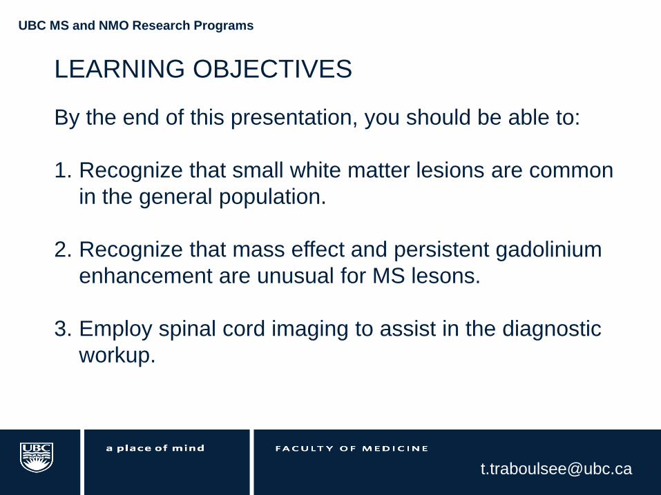
LEARNING OBJECTIVES
By the end of this presentation, you should be able to:
1. Recognize that small white matter lesions are common
in the general population.
2. Recognize that mass effect and persistent gadolinium
enhancement are unusual for MS lesons.
3. Employ spinal cord imaging to assist in the diagnostic
workup.
UBC MS and NMO Research Programs
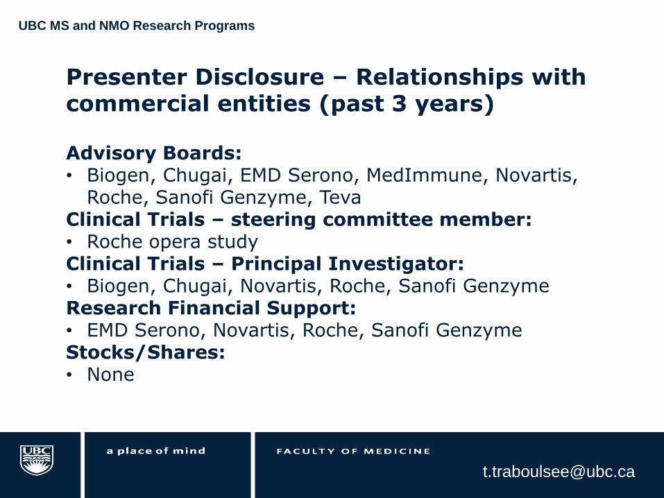
Presenter Disclosure – Relationships with commercial entities (past 3 years)
Advisory Boards: • Biogen, Chugai, EMD Serono, MedImmune, Novartis,
Roche, Sanofi Genzyme, Teva Clinical Trials – steering committee member: • Roche opera study Clinical Trials – Principal Investigator: • Biogen, Chugai, Novartis, Roche, Sanofi Genzyme Research Financial Support: • EMD Serono, Novartis, Roche, Sanofi Genzyme Stocks/Shares: • None
UBC MS and NMO Research Programs
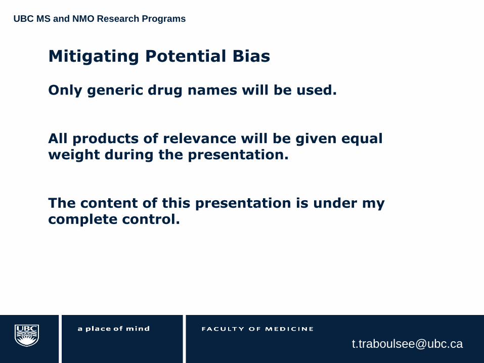
Mitigating Potential Bias
Only generic drug names will be used. All products of relevance will be given equal weight during the presentation. The content of this presentation is under my complete control.
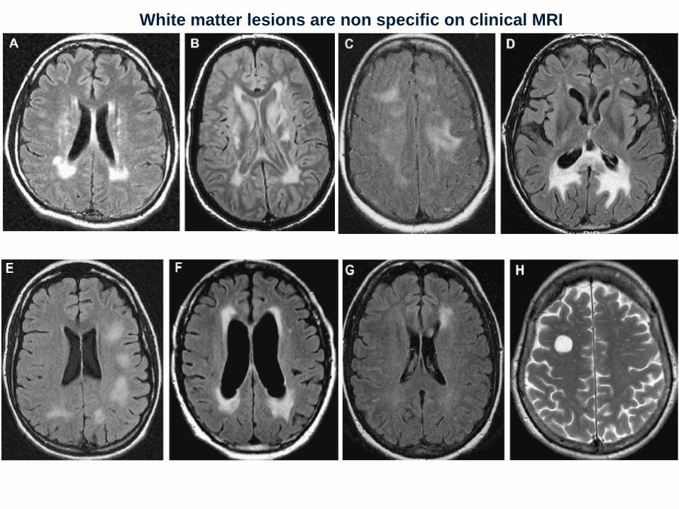
White matter lesions are non specific on clinical MRI
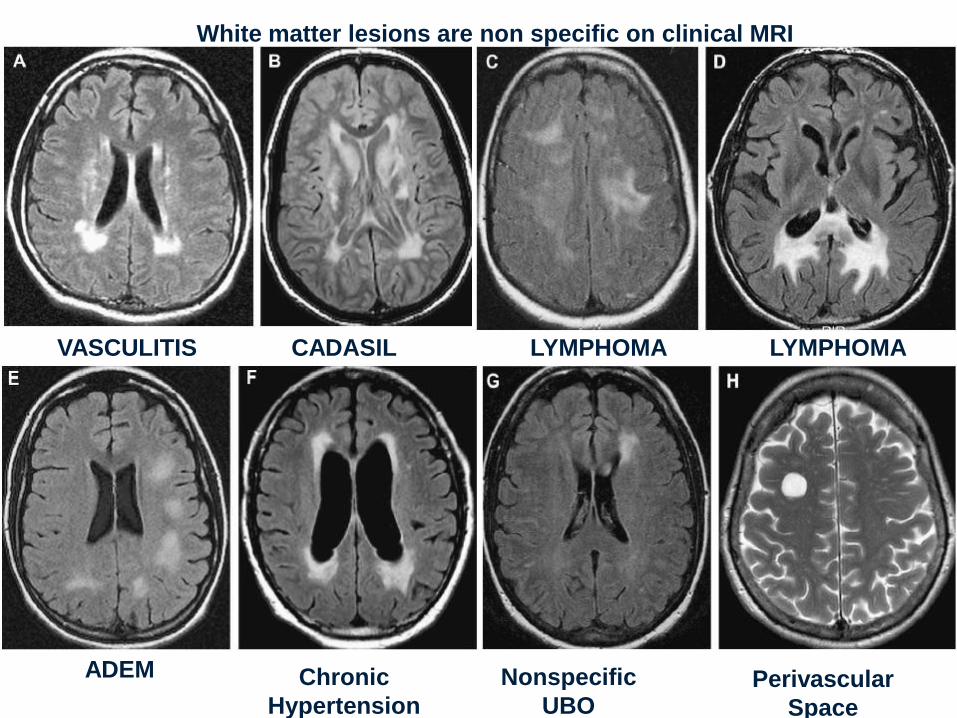
CADASIL LYMPHOMA VASCULITIS
ADEM Chronic
Hypertension
LYMPHOMA
Nonspecific
UBO Perivascular
Space
White matter lesions are non specific on clinical MRI
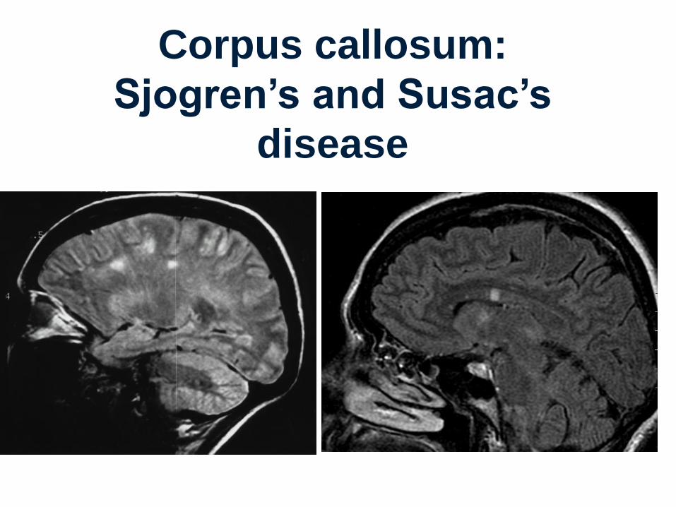
Corpus callosum:
Sjogren’s and Susac’s
disease
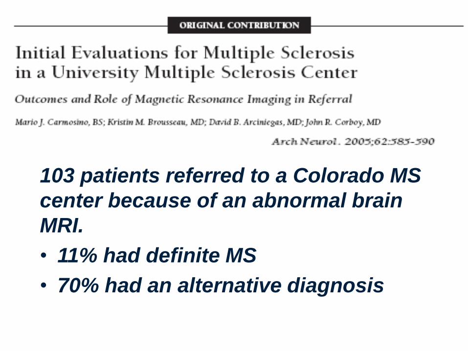
103 patients referred to a Colorado MS
center because of an abnormal brain
MRI.
• 11% had definite MS
• 70% had an alternative diagnosis
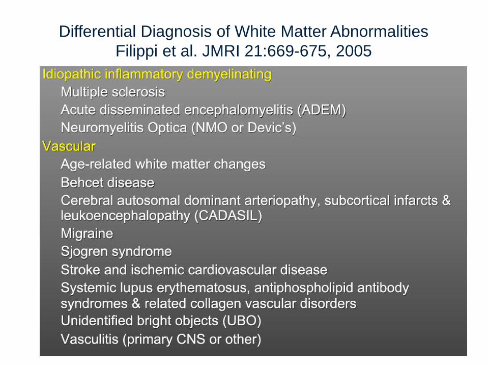
Differential Diagnosis of White Matter Abnormalities
Filippi et al. JMRI 21:669-675, 2005
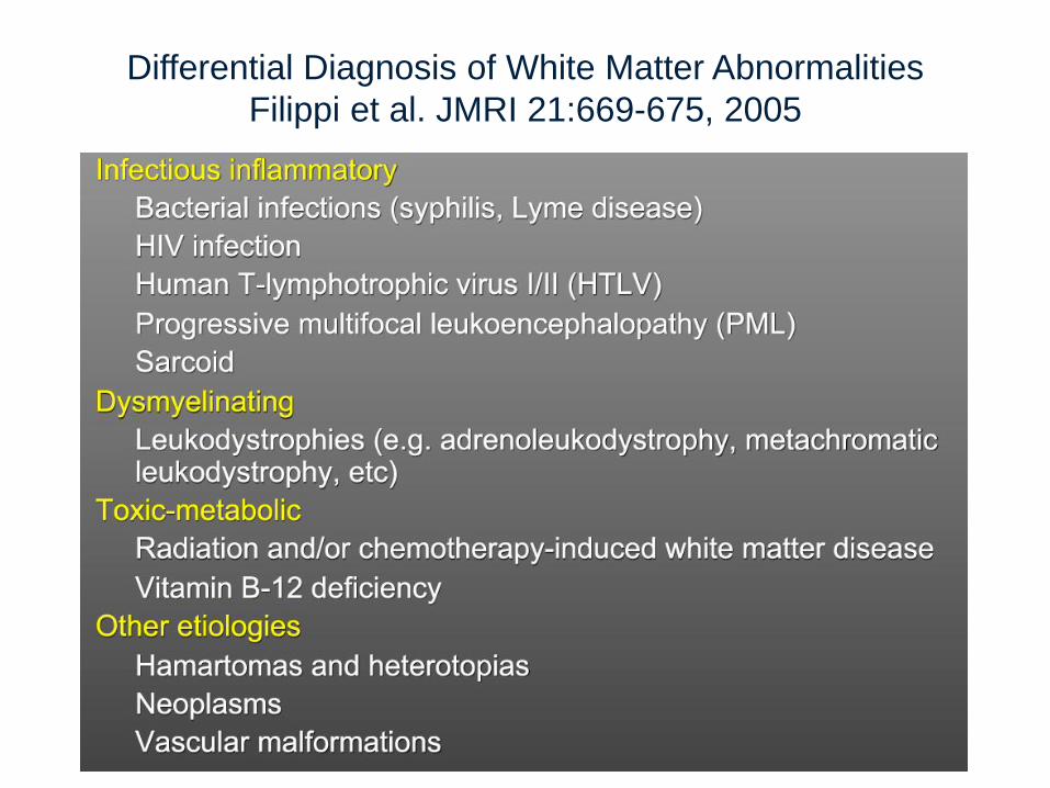
Differential Diagnosis of White Matter Abnormalities
Filippi et al. JMRI 21:669-675, 2005
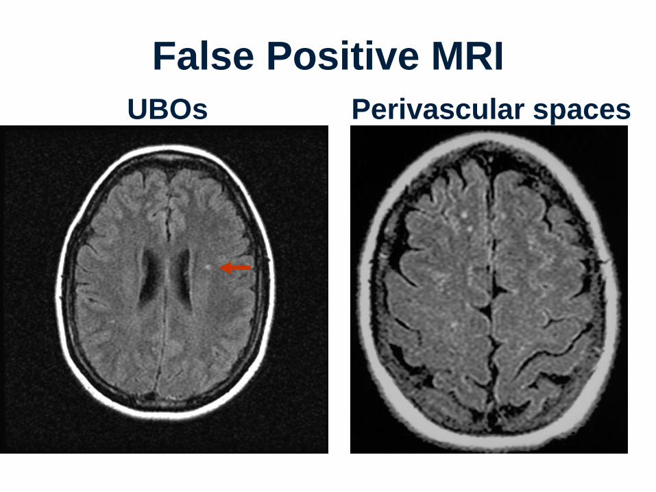
False Positive MRI
UBOs Perivascular spaces
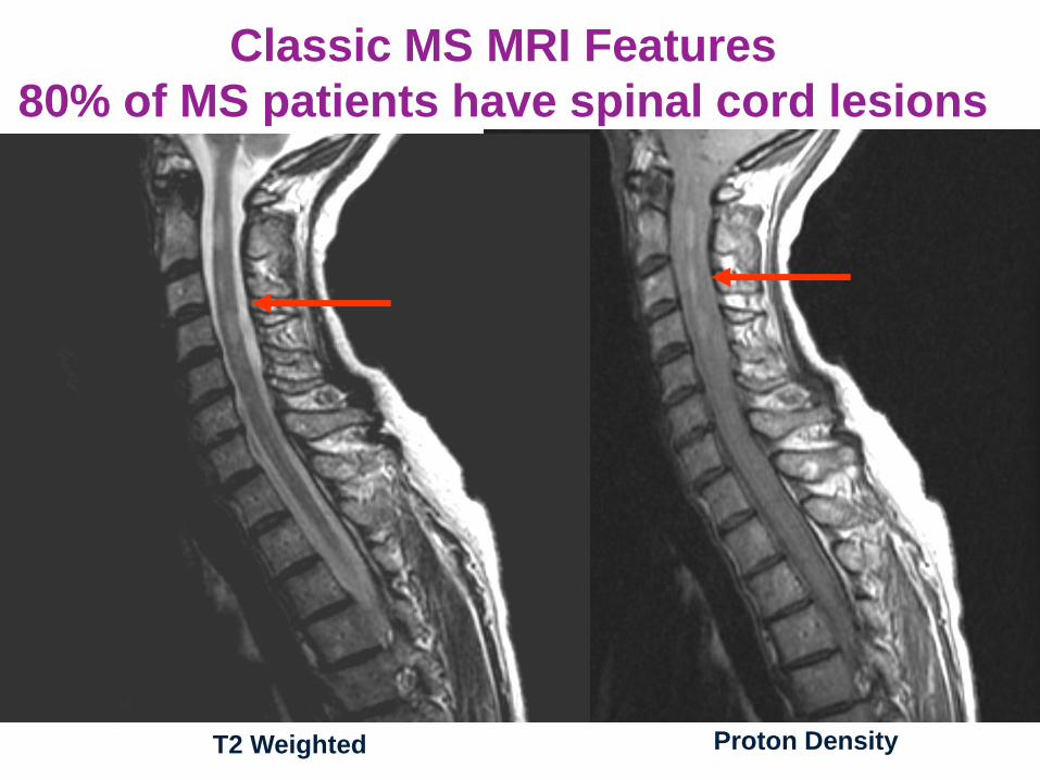
Classic MS MRI Features
80% of MS patients have spinal cord lesions
T2 Weighted Proton Density
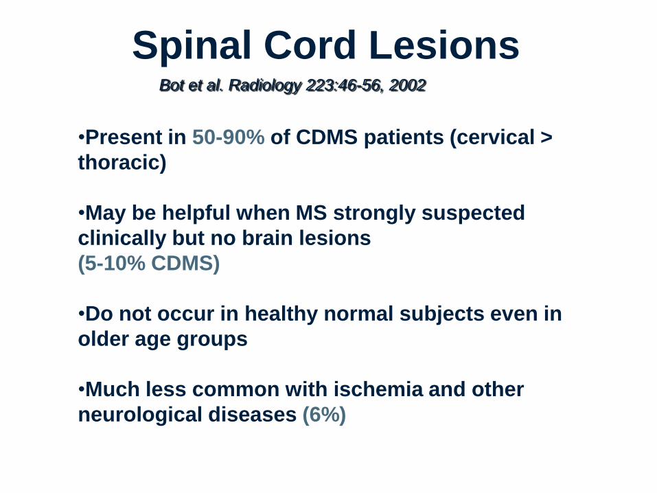
Spinal Cord Lesions
•Present in 50-90% of CDMS patients (cervical >
thoracic)
•May be helpful when MS strongly suspected
clinically but no brain lesions
(5-10% CDMS)
•Do not occur in healthy normal subjects even in
older age groups
•Much less common with ischemia and other
neurological diseases (6%)
Bot et al. Radiology 223:46-56, 2002
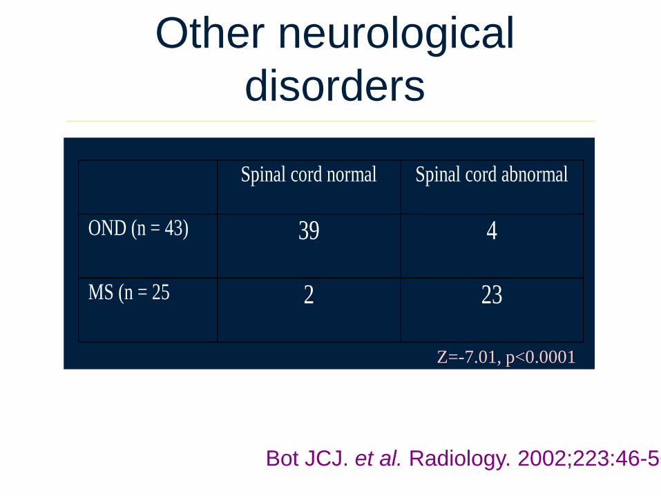
Spinal cord normal Spinal cord abnormal
OND (n = 43) 39 4
MS (n = 25 2 23
Other neurological
disorders
Z=-7.01, p<0.0001
Bot JCJ. et al. Radiology. 2002;223:46-56
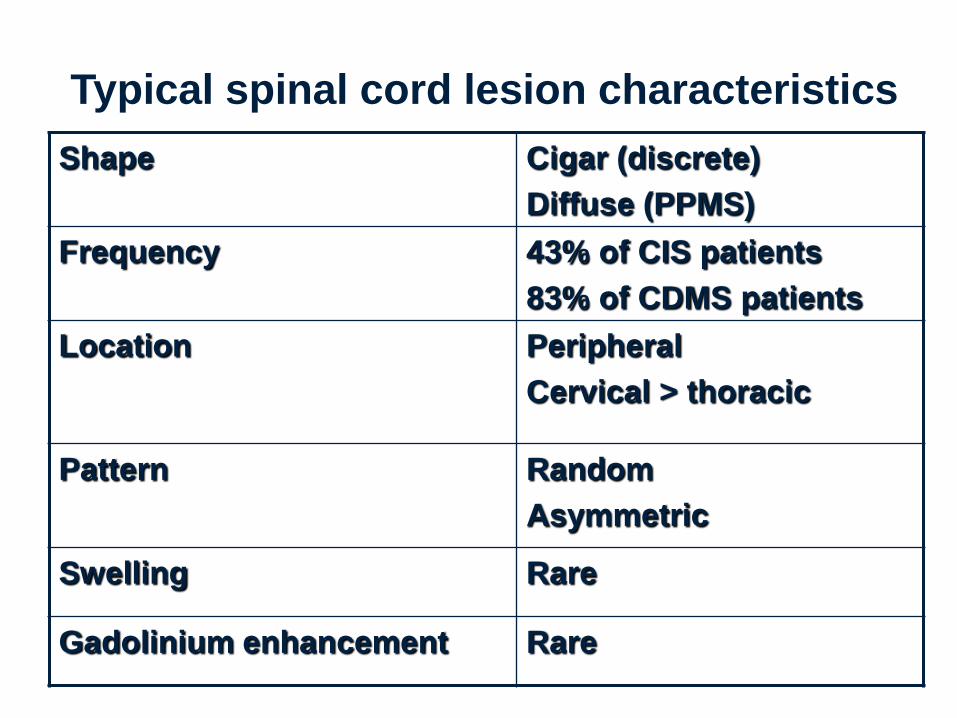
Typical spinal cord lesion characteristics
Shape Cigar (discrete)
Diffuse (PPMS)
Frequency 43% of CIS patients
83% of CDMS patients
Location Peripheral
Cervical > thoracic
Pattern Random
Asymmetric
Swelling Rare
Gadolinium enhancement Rare
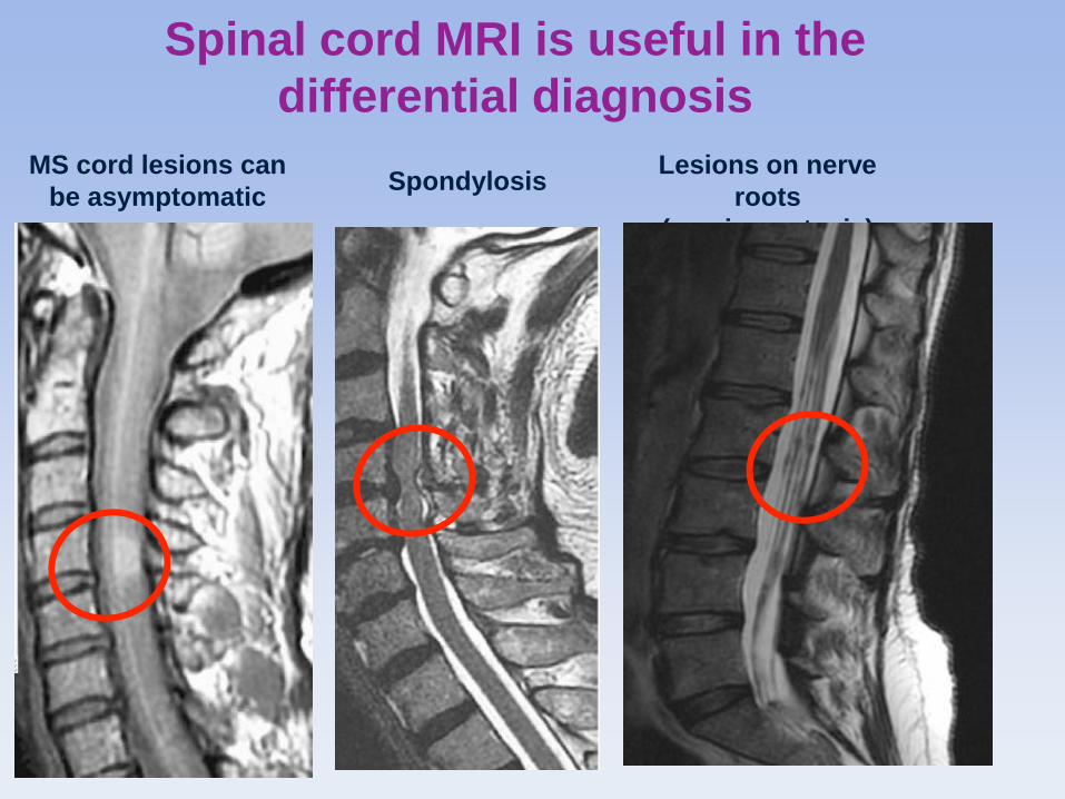
Spinal cord MRI is useful in the
differential diagnosis
MS cord lesions can
be asymptomatic
Lesions on nerve
roots
(carcinomatosis)
Spondylosis
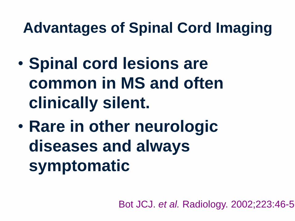
Advantages of Spinal Cord Imaging
• Spinal cord lesions are
common in MS and often
clinically silent.
• Rare in other neurologic
diseases and always
symptomatic
Bot JCJ. et al. Radiology. 2002;223:46-56

Large and unusual appearing
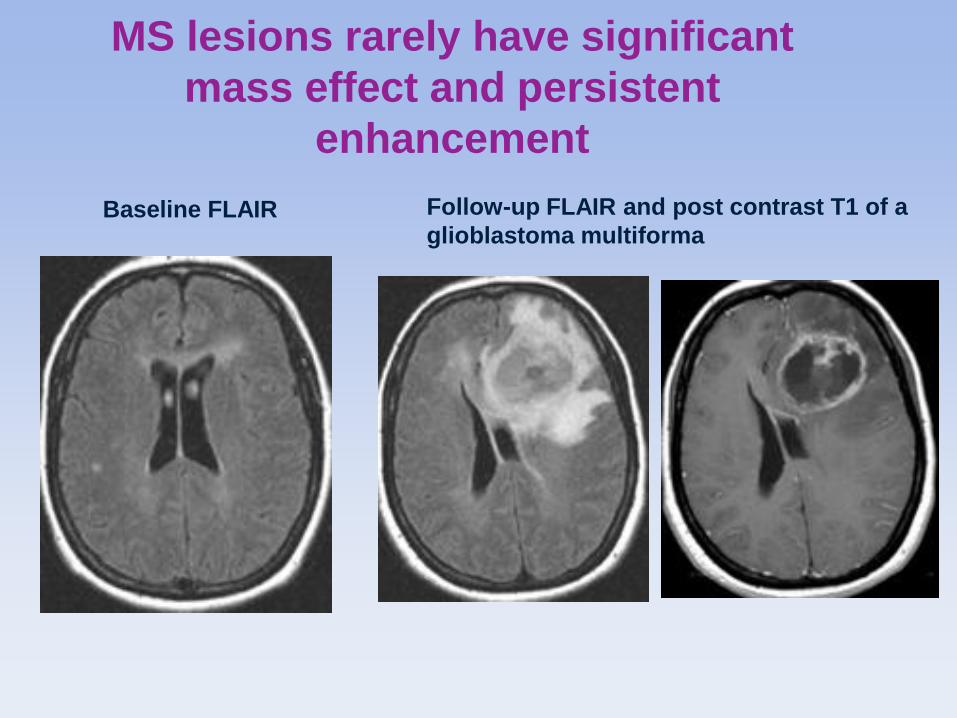
MS lesions rarely have significant
mass effect and persistent
enhancement
Baseline FLAIR Follow-up FLAIR and post contrast T1 of a
glioblastoma multiforma
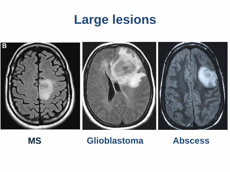
Large lesions
MS Glioblastoma Abscess
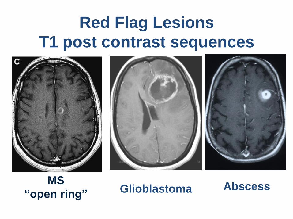
Red Flag Lesions
T1 post contrast sequences
Glioblastoma Abscess MS
“open ring”
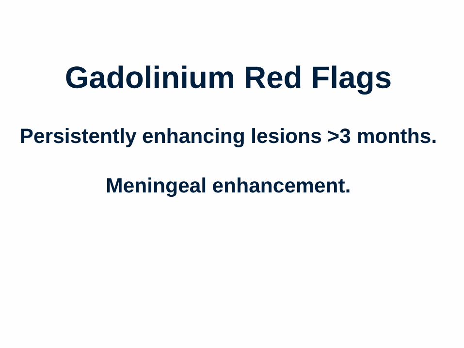
Gadolinium Red Flags
Persistently enhancing lesions >3 months.
Meningeal enhancement.
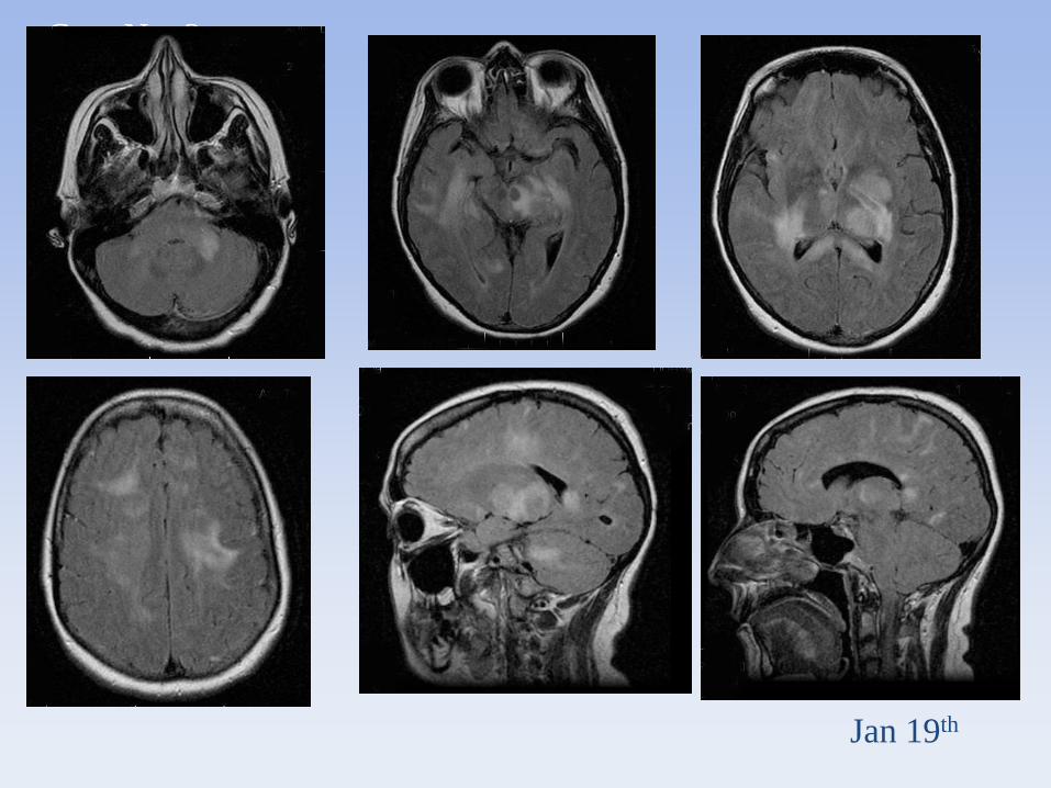
Case No. 3
Jan 19th
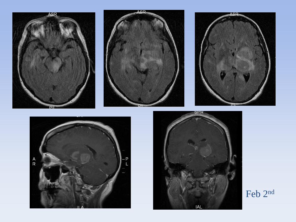
Feb 2nd
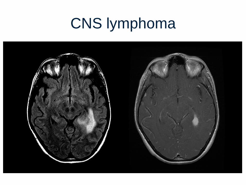
CNS lymphoma
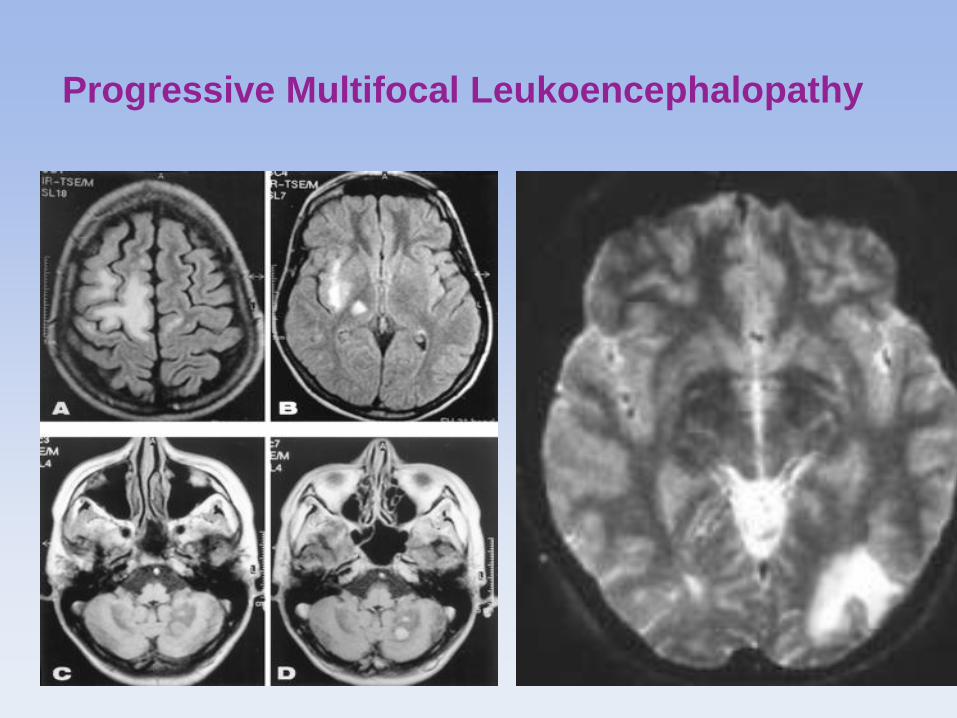
Progressive Multifocal Leukoencephalopathy
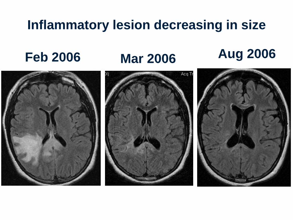
Inflammatory lesion decreasing in size
Feb 2006 Aug 2006 Mar 2006
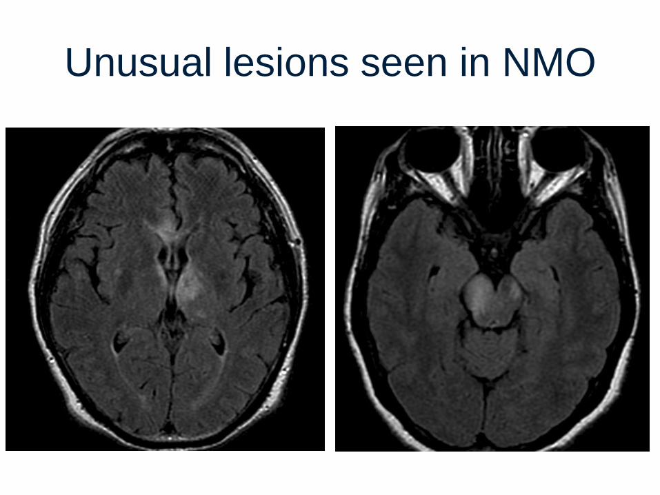
Unusual lesions seen in NMO
LESCL on Sagittal T2 MS Lesion on Sagittal T2
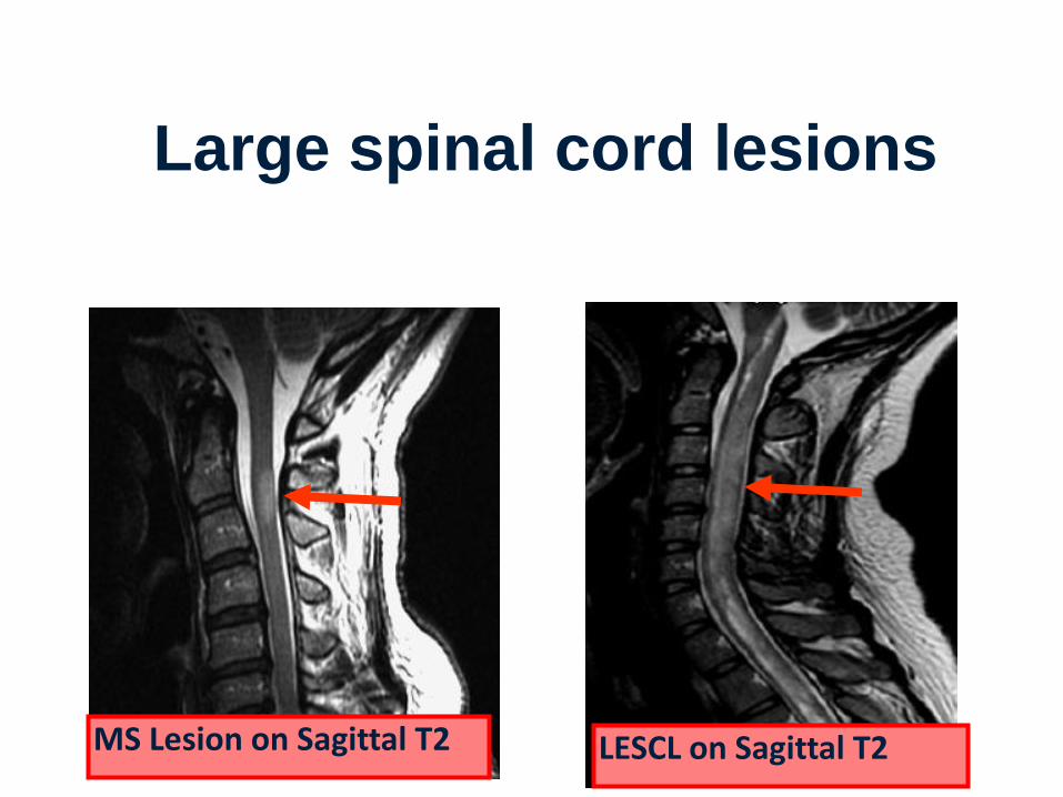
Large spinal cord lesions
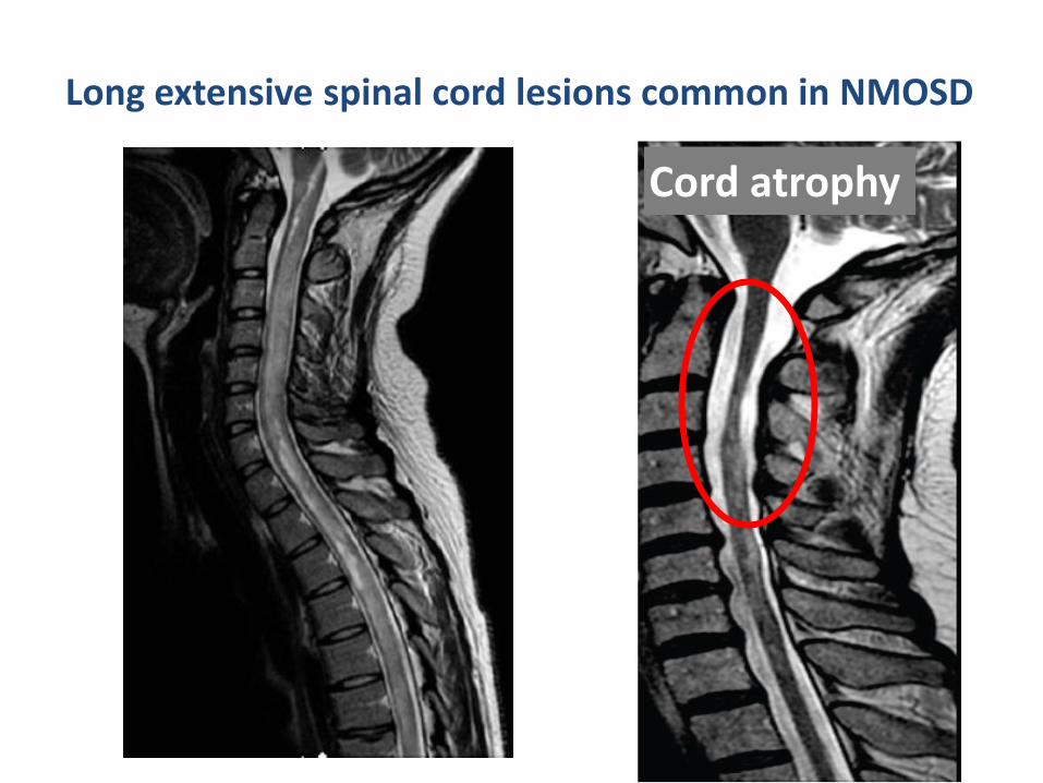
Long extensive spinal cord lesions common in NMOSD
Cord atrophy
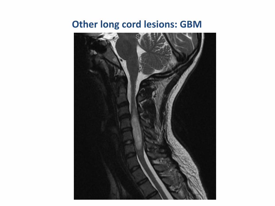
Other long cord lesions: GBM
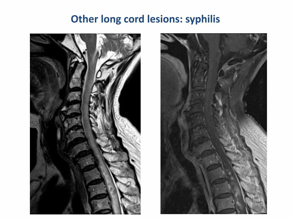
Other long cord lesions: syphilis
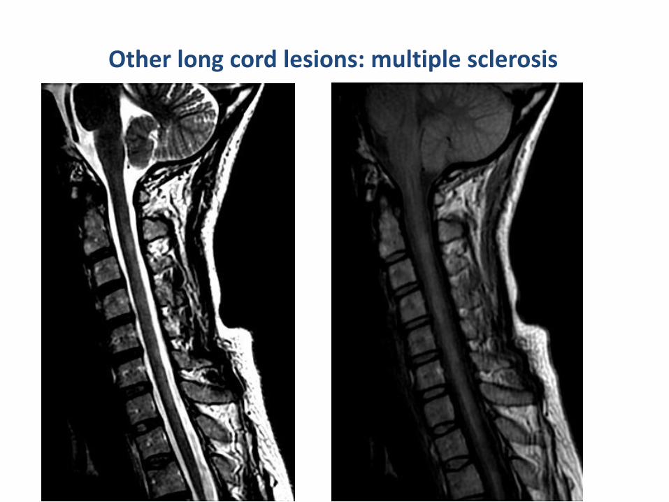
Other long cord lesions: multiple sclerosis
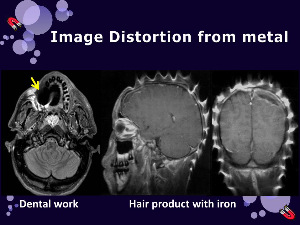
Dental work Hair product with iron
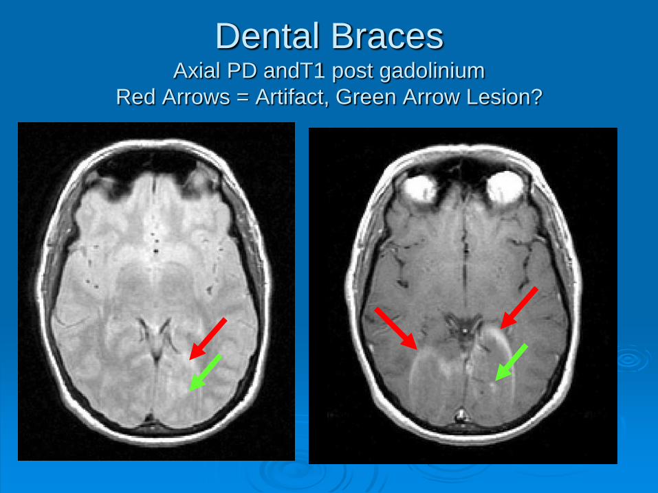
Dental Braces Axial PD andT1 post gadolinium
Red Arrows = Artifact, Green Arrow Lesion?
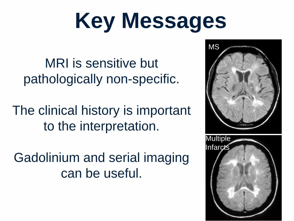
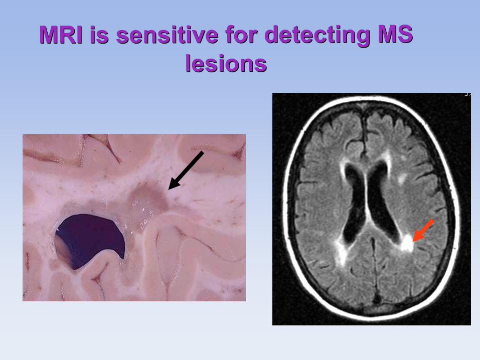
Key Messages
MRI is sensitive but
pathologically non-specific.
The clinical history is important
to the interpretation.
Gadolinium and serial imaging
can be useful.
MS
Multiple
Infarcts