mri of the knee - amazon s3 · meniscus flounce meniscal ossicle plicae discoid meniscus...
TRANSCRIPT

MRI of the Knee:
Part 4 - normal variants that may simulate disease
Mark Anderson, M.D. University of Virginia

Learning Objectives
• identify a cortical desmoid and describe its typical appearance and location on MR images
• discuss the most common normal variants in the pediatric knee that may simulate pathology on MR imaging.
• list the four types of synovial plicae in the knee as well as their clinical significance.
• At the end of the presentation, each participant should be able to:

The Knee: normal variants
Bipartite patella Dorsal defect of the patella Cortical desmoid Distal femoral epiphyseal irregularity Posterior “stripe” Juvenile cartilage signal intensity Terminal sulcus cartilage “thinning” Semimembranosus insertions Lateral inferior geniculate vessels Meniscus flounce Meniscal ossicle Plicae Discoid meniscus Fabello-fibular ligament Meniscofibular ligament Popliteofibular lgament
Tibial attachment of the biceps femoris
Transverse meniscal ligament Meniscofemoral ligaments Oblique meniso-meniscal ligament Double barreled PCL Meniscal root attachments Patello-meniscal ligament Fabella Cyamella Accessory popliteus tendon Bifurcated popliteus 3rd head of the gastrocnemius muscle Bifurcating sartorius tendon

The Knee: normal variants
Bone Bipartite patella Dorsal defect of the patella Cortical desmoid Irregular ossification vs. “juvenile OCD” Posterior stripe
Cartilage
Juvenile cartilage signal intensity Terminal sulcus cartilage “thinning” Upper trochlear “defect”
Menisci
Meniscal roots Transverse ligament Meniscofemoral ligaments Semimembranosus insertion Lateral inferior geniculate vessels Meniscal ossicle
Plicae
Medial patellar Suprapatellar Infrapatellar
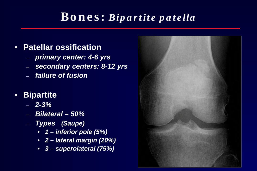
Bones: Bipartite patella
• Patellar ossification – primary center: 4-6 yrs – secondary centers: 8-12 yrs – failure of fusion
• Bipartite
– 2-3% – Bilateral – 50% – Types (Saupe)
• 1 – inferior pole (5%) • 2 – lateral margin (20%) • 3 – superolateral (75%)
4 yr old male

Bones: Bipartite patella
• Patellar ossification – primary center: 4-6 yrs – secondary centers: 8-12 yrs – failure of fusion
• Bipartite
– 2-3% – Bilateral – 50% – Types (Saupe)
• 1 – inferior pole (5%) • 2 – lateral margin (20%) • 3 – superolateral (75%)
4 yr old male

Bones: bipartite patella
• Symptomatic – acute / chronic trauma
• fracture / avulsion • may be overlooked as etiology
• MRI – edema along margins
Kavanagh, Skeletal Radiol 2007
53 pts – knee pain – only MRI finding:
edema along bipartite patella

Bones: dorsal defect of the patella
• Unknown etiology
• Incidence – 0.3 – 1% / bilat - up to 30% – may be seen with bipartite
• Appearance – well circumscribed – round, lytic lesion – superolateral patella
• MRI – lack of edema – evaluate overlying cartilage
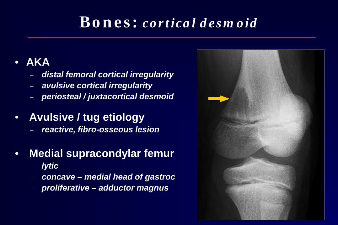
Bones: cortical desmoid
• AKA – distal femoral cortical irregularity – avulsive cortical irregularity – periosteal / juxtacortical desmoid
• Avulsive / tug etiology – reactive, fibro-osseous lesion
• Medial supracondylar femur
– lytic – concave – medial head of gastroc – proliferative – adductor magnus
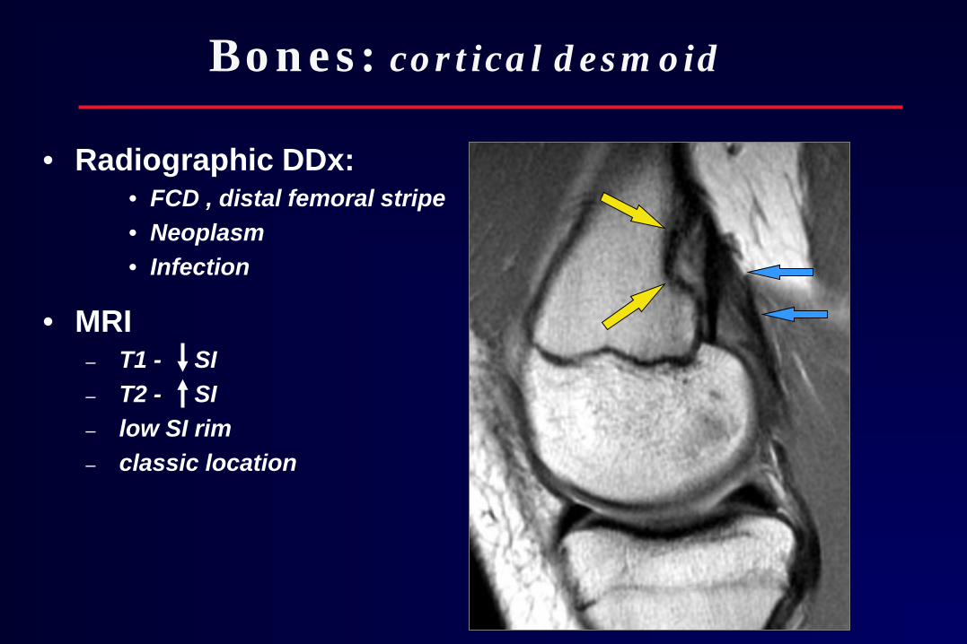
Bones: cortical desmoid
• Radiographic DDx: • FCD , distal femoral stripe • Neoplasm • Infection
• MRI – T1 - SI – T2 - SI – low SI rim – classic location
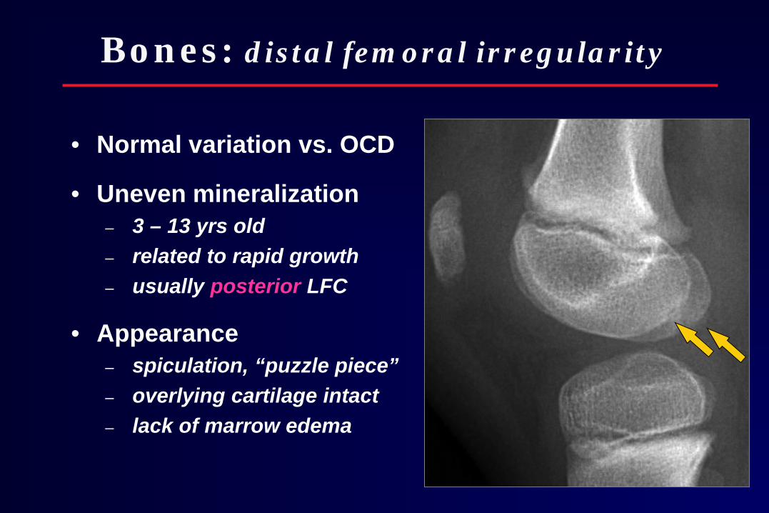
Bones: distal femoral irregularity
• Normal variation vs. OCD
• Uneven mineralization – 3 – 13 yrs old – related to rapid growth – usually posterior LFC
• Appearance – spiculation, “puzzle piece” – overlying cartilage intact – lack of marrow edema

11 yr old male

Bones: “Juvenile OCD”
• “Juvenile” OCD – open physes – mean age: 12-13 yrs – central 1/3 + intercondylar – adjacent edema common
• Vs. “Adult” – better prognosis (80% resolve) – more commonly bilateral + LFC – MRI signs of fragment instability less predictive than in adult
Gebarski, Pediatr Radiol 2005 Kijowski, Radiology 2008

Med Lat
10 yr old male
Med Lat

Med Med
Lat
4 years later (14 yo)
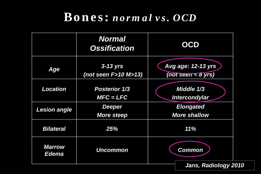
Bones: normal vs. OCD
Normal Ossification
OCD
Age
3-13 yrs (not seen F>10 M>13)
Avg age: 12-13 yrs (not seen < 8 yrs)
Location
Posterior 1/3 MFC = LFC
Middle 1/3 Intercondylar
Lesion angle Deeper More steep
Elongated More shallow
Bilateral
25%
11%
Marrow Edema
Uncommon
Common
Jans, Radiology 2010
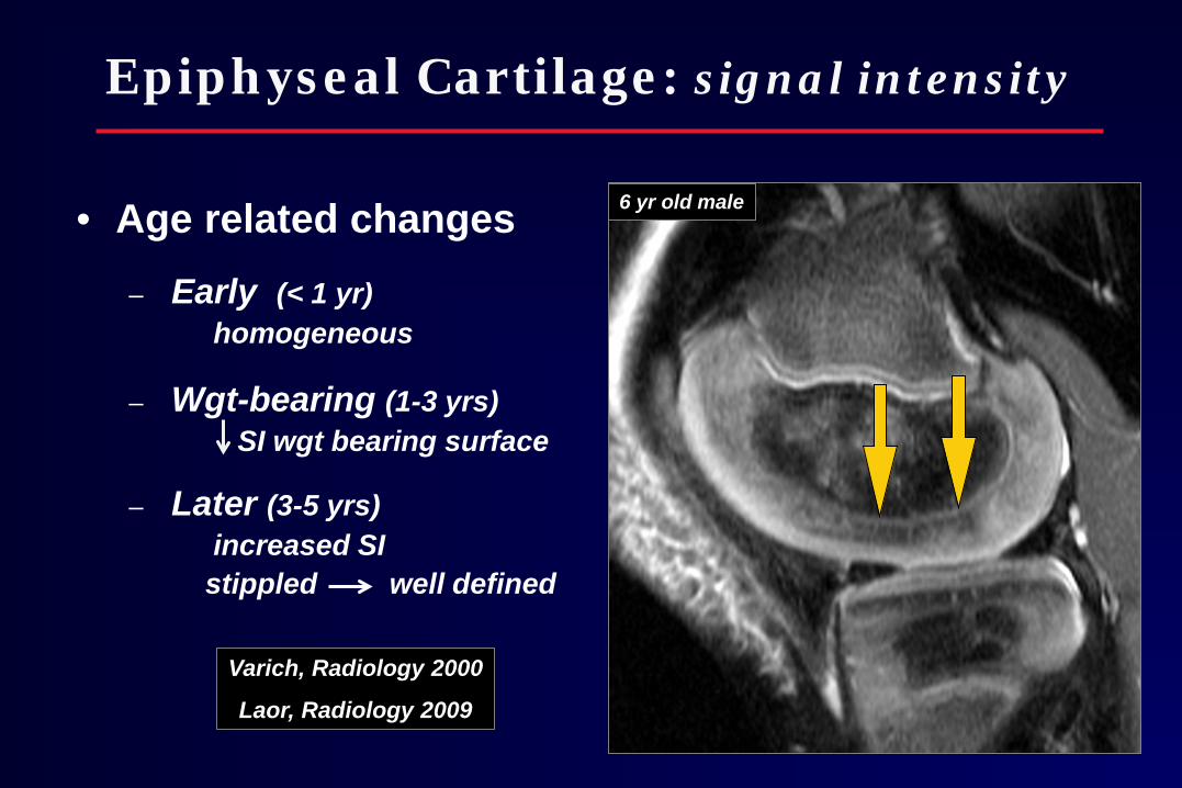
Epiphyseal Cartilage: signal intensity
• Age related changes
– Early (< 1 yr) homogeneous
– Wgt-bearing (1-3 yrs) SI wgt bearing surface
– Later (3-5 yrs) increased SI stippled well defined
2 yr old female 4 yr old male 6 yr old male
Varich, Radiology 2000
Laor, Radiology 2009
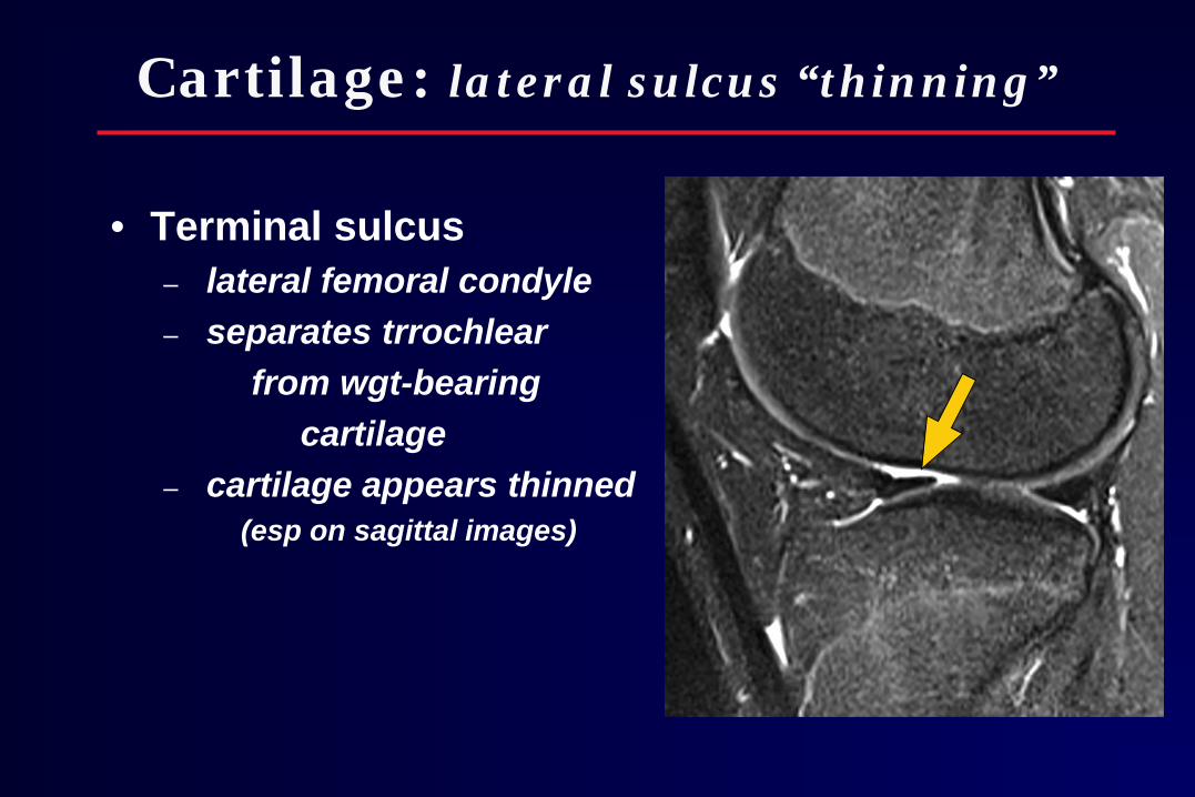
Cartilage: lateral sulcus “thinning”
• Terminal sulcus – lateral femoral condyle – separates trrochlear from wgt-bearing cartilage – cartilage appears thinned
(esp on sagittal images)
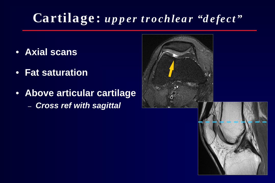
Cartilage: upper trochlear “defect”
• Axial scans
• Fat saturation
• Above articular cartilage – Cross ref with sagittal

Cartilage: upper trochlear “defect”
• Axial scans
• Fat saturation
• Cross-reference sagittal – above articular cartilage
• Asymmetric cartilage – lateral extends more proximally
LAT MED
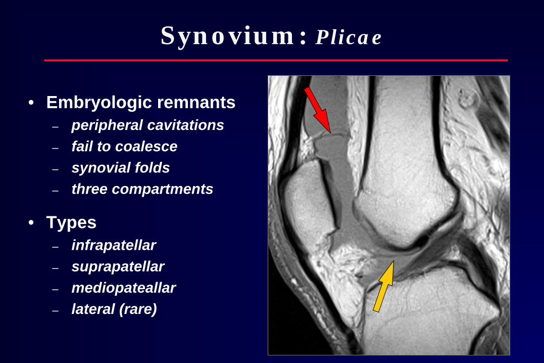
Synovium: Plicae
• Embryologic remnants – peripheral cavitations – fail to coalesce – synovial folds – three compartments
• Types – infrapatellar – suprapatellar – mediopateallar – lateral (rare)
INFRAPAT
MEDIAL
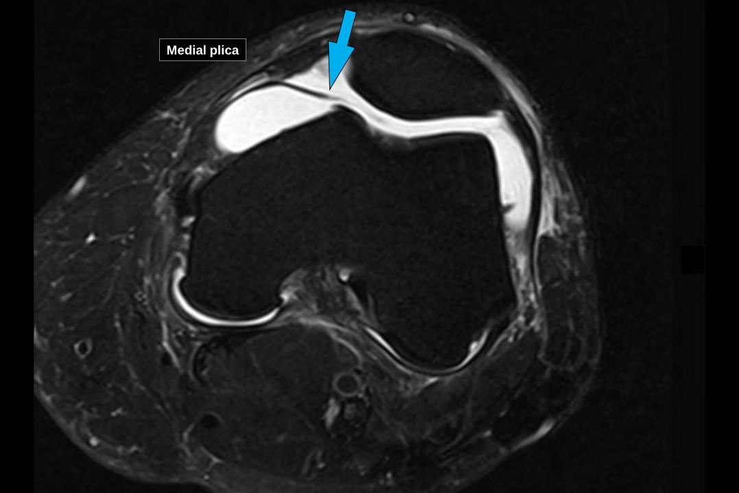
Medial plica
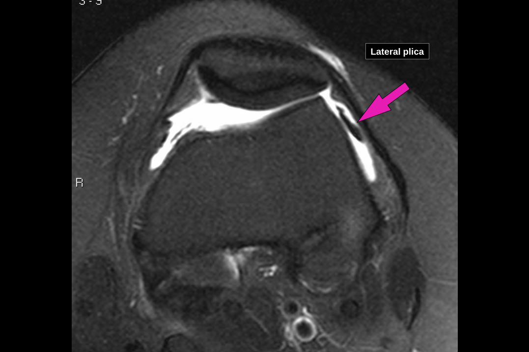
Lateral plica

Synovium: Plicae
• Plica Syndrome? – mediopatellar – thickens – impinges on femur/patella – cartilage “impingement” lesion
• MR Findings – appearance does not correlate with symptoms
Boles, JCAT 2004 Weckstrom, The Knee 2010
Demirag, Knee Surg Sports Traumatol Arthrosc 2006

The Knee: normal variants
Bone Bipartite patella Dorsal defect of the patella Cortical desmoid Irregular ossification vs. “juvenile OCD” Posterior stripe
Cartilage
Juvenile cartilage signal intensity Terminal sulcus cartilage “thinning” Upper trochlear “defect”
Menisci
Meniscal roots Transverse ligament Meniscofemoral ligaments Semimembranosus insertion Lateral inferior geniculate vessels Meniscal ossicle
Plicae
Medial patellar Suprapatellar Infrapatellar