mrs. penley. the fluid that leaves capillaries and goes into tissues it flows towards the heart only
TRANSCRIPT
Lymphatic SystemMrs. Penley

Lymph• The fluid that leaves capillaries and goes into
tissues• It flows TOWARDS the heart only
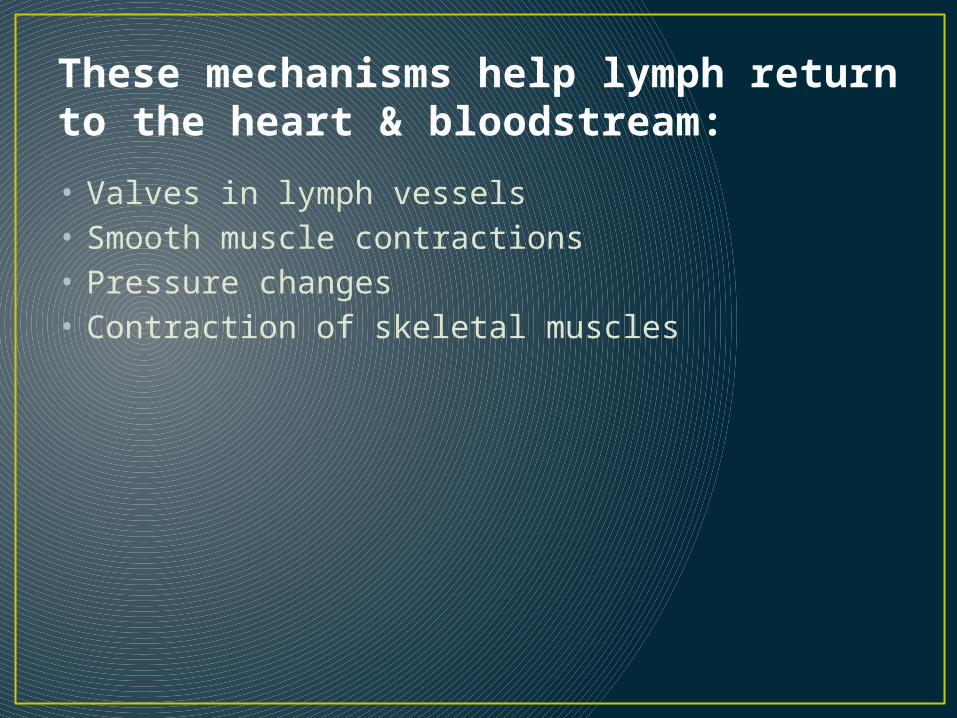
These mechanisms help lymph return to the heart & bloodstream:
• Valves in lymph vessels• Smooth muscle contractions• Pressure changes• Contraction of skeletal muscles
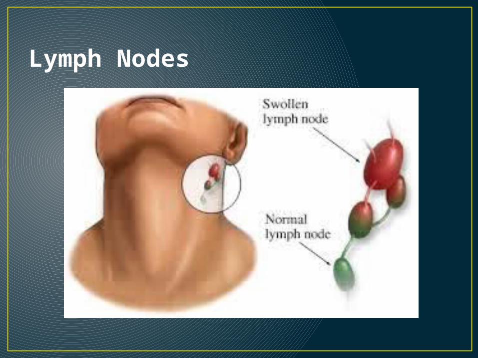
Lymph Nodes
Lymph nodes act as…• Filters• Remove foreign materials• Contain lymphocytes (T cells) http://
www.youtube.com/watch?v=RO6qmpApyDM • Contain macrophages http://
www.youtube.com/watch?v=7WGyq82oMkM
Characteristics of macrophages • Act as antigen presenters• Engulf foreign particles• Secrete monokines• Considered “big eaters” of immune system
Immunology• The study of immunity
What is an antigen? • An antigen is any substance that causes your
immune system to produce antibodies against it.• An antigen may be a foreign substance from the
environment such as chemicals, bacteria, fungi, viruses, or pollen grains.
• An antigen may also be formed within the body, as with bacterial toxins or tissue cells.
What is an antibody? • Antibodies are large Y-shaped proteins.• They are recruited by the immune system to identify and
neutralize foreign objects like bacteria and viruses. • Each antibody has a unique target known as the antigen
present on the invading organism.• This antigen is like a key that helps the antibody in identifying
the organism. This is because both the antibody and the antigen have similar structure at the tips of their “Y” structures.
• Just like every lock has a single key, an antibody has a single antigen key. When the key is inserted into the lock, the antibody activates, tagging or neutralizing its target.
• The production of antibodies is the main function of the humoral immune system.
• http://www.news-medical.net/health/Antibody-What-is-an-Antibody.aspx
Classes of antibodies• Antibodies are composed of a light chain protein
and a heavy chain protein that come together and form a Y-shaped structure.
• The base of the Y is a conserved region that all antibodies have in common, while the tips of the forks of the Y are unique to each antibody.
• The tips react with the antigen, while the conserved base interacts with the immune system.
• Five types of antibodies are formed in the body: IgG, IgM, IgA, IgD and IgE (notice they spell MADGE)
IgG• Immunoglobulin G (IgG) is the most abundant circulating antibody,
making up 80% of the total antibodies and 75% of that found in serum. • It contains a single antibody protein complex, with two heavy chains and
two light chains. • IgG is the second type of antibody synthesized in response to an
infection and is the only antibody that can pass through the wall of small blood vessels to access antigens present in the extracellular spaces.
• It is also the only antibody capable of crossing the placenta in humans, where it confers the mother's immunity onto the fetus and newborn.
• This immunity protects a baby for the first 6-12 months of its life and allows it time for its own immune system to mature.
• IgG is particularly effective at attacking extracellular viruses and protein toxins and is also capable of activating the classic pathway of the complement cascade. It helps to prevent the systemic spread of infection and facilitates recovery from many infections.
• Finally, IgG is the antibody that serves as an efficient handle for phagocytes
IgM• IgM is the largest antibody, with five Y structures being joined by
their Fc regions in a circular configuration.• A J chain (another polypeptide) links the five antibodies together.
IgM is present in serum, making up about 10 % of antibodies in the blood.
• The presence of its ten antigen reactive sites helps agglutinate or clump antigens (see the explanation of this term in the next section), making it easier for the immune system to eliminate them.
• IgM is more efficient than IgG at activating the complement pathway.
• IgM is synthesized by plasma cells early in a primary infection and is very important in slowing or stopping the spread of a pathogen during the initial stages of an illness.
• IgM is also found on mature B cells in a monovalent form, where it serves as a receptor.
IgA• IgA is present in serum, mucus, saliva, tears, sweat and milk. • Two subclasses with different heavy chains are made, IgA1 and
IgA2. IgA1 is synthesized in the bone marrow and makes up most of the serum IgA.
• IgA2 is synthesized by B cells present in MALT (mucosa-associated lymphoid tissue). NOTE: MALT includes your tonsils & Peyer’s patches
• IgA in breast milk interferes with the colonization of the GI tract by harmful microorganisms in the first few months of life. The mother's IgA in the GI tract of newborns keeps these pathogens at low populations, preventing them from causing serious disease, but still allowing the stimulation of the infant's own immune system. The newborn thus develops its own immunity while being partially protected by the mother. IgA molecules do not activate the classical complement pathway, but may activate the alternative complement pathway.
IgE• IgE is a monomeric antibody that accounts for only
0.002 % of the total serum antibodies. • Almost all IgE is bound to tissue cells, especially mast
cells and eosinophils in various parts of the body. • Contact of IgE with antigen leads to release of a set of
signal molecules from the mast cells, which effectively recruits various agents of the immune response to fight the infection.
• IgE and MALT serve to detect penetrating pathogens and amplify the immune response in an area leading to the repulsion of the invader.
• Antigen reactions with IgE are also responsible for atopic allergic reactions (e.g., hives, asthma, hay fever etc.)
IgD• IgD is found on the surface of B-lymphocytes and
together with monomeric IgM, serves as antigen receptor for the activation of B cell as described previously.
• IgD is monovalent (one site of attachment).
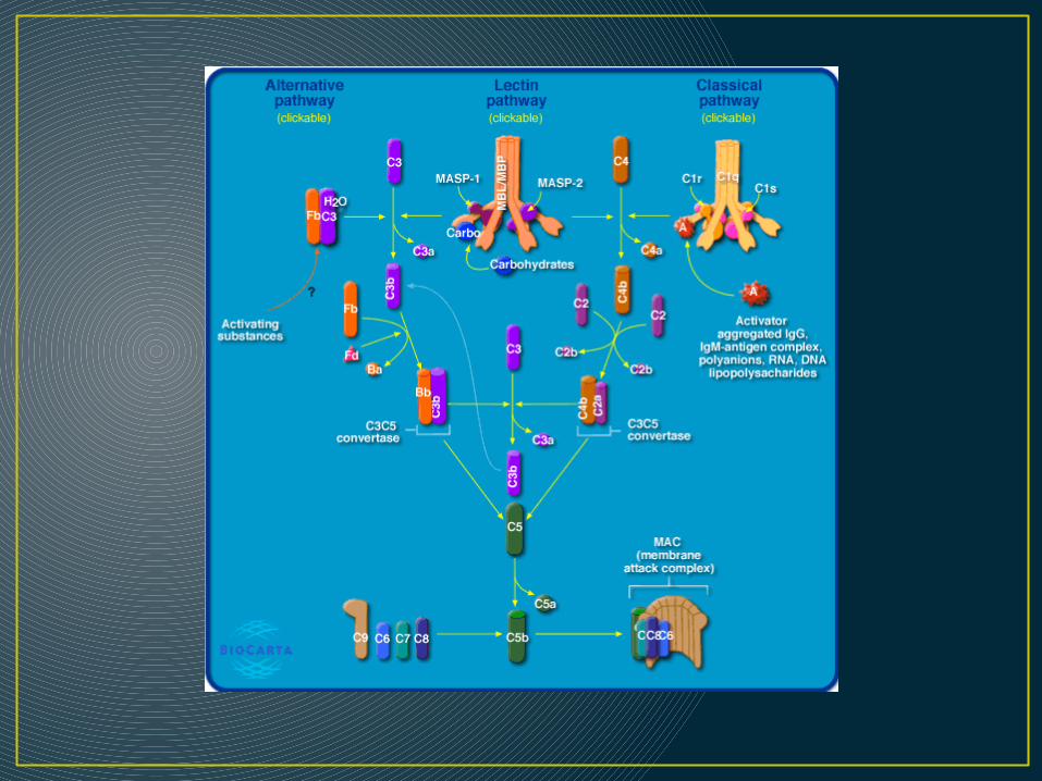
What exactly is this “complement pathway” • The complement pathway consists of a series of
over thirty proteins in plasma that are part of the immune response.
• Activation of the complement system lyses bacterial cells, forms chemotactic peptides by attracting immune cells, and increases phagocytotic clearance of infecting cells.
• Picture on next slide
Whoa…What’s the difference between those T-cells and B-cells, anyway?
• Let’s take a breather and do a little thoughtful Ed activity here.
• Compare and Contrast T-cells and B-cells
Some things you should have gleaned from your activity….• B cell and T cell are lymphocytes responsible for the third line of
defense in your body. Basically defending your body from invading antigens (i.e. germs).
The main difference between the two are:
- B cell are responsible for the antibody-mediated response while T cells are responsible for cell-mediated response.- B cell produced in bone marrow. T cell produced in bone marrow & lymph nodes. - When detected antigen, B cell undergoes clonol expansion and create different B cell, some are memory B cell which remembers the germ. Most become plasma cells which produces antibody (germ killer) to fight the antigen. - T cell's main role is to destroy infected cells and cancer cells and to coordinate acquired immune response.
• Note: Antigen presentation is necessary for the activation and cloning of T-cells.

Some ways they are alike…
• Each B cell and T cell is specific for a particular antigen. What this means is that each is able to bind to a particular molecular structure.
• They are integral membrane proteins. • They are present in thousands of identical copies
exposed at the cell surface. • They are made before the cell ever encounters an
antigen. • They are encoded by genes assembled by the
recombination of segments of DNA.
Remember….• B cells get immunocompetence (the ability to
develop an immune response following exposure to an antigen) in the BONE MARROW
• AND• Lymphocytes that are able to mount an immune
response are called immunocompetent
How do antibodies inactivate antigens?• 1. They complement fixation• 2. Precipitation• 3. Agglutination• 4. Neutralization
Complement fixation• Complement fixation. Antibodies IgM and IgG
bind to enemy cells and change shape, exposing their complement-binding sites
• This initiates the binding of complement to the enemy cell surface and leads to inflammation, phagocytosis, immune clearance, and cytolysis
• Complement fixation is the primary mechanism of defense against foreign cells such as bacteria and mismatched erythrocytes.
• It makes it easier for phagocytes to ingest and destroy bacteria.
Precipitation• a similar process in which antibodies link antigen
molecules (not whole cells) together. • This creates large Ag–Ab complexes that are too
large to remain dissolved in solution. • These complexes can be removed by immune
clearance or phagocytized by eosinophils in the connective tissues.
Agglutination• It is effective not only in mismatched blood
transfusions, but more importantly as a defense against bacteria.
• An antibody molecule has 2 to 10 binding sites; thus, it can bind to antigen molecules on two or more enemy cells at once and stick them together
• This immobilizes microbes and antigen molecules and prevents them from spreading through the tissues.
Neutralization• Only certain regions of an antigen are pathogenic
—for example, the parts of a toxin molecule or virus that enable these agents to bind to human cells.
• Antibodies can neutralize an antigen by masking these active regions (i.e. The antibodies bind to specific sites on bacteria toxins and block their “bad” effects) .
Chemotaxis• The migration of phagocytes and WBCs (white
blood cells) to an inflamed area along a chemical gradient
• Q: When would this happen?
The body’s 1st line of defense • Skin & mucous membranes
The body’s non-specific defenses include:• Fever• Inflammatory response• Intact skin• Natural killer cells (these are not to be confused
with T or B cells) http://www.youtube.com/watch?v=HNP1EAYLhOs
The inflammatory Response• The inflammatory response is the body's natural
response that occurs immediately following tissue damage.
• It's main functions are to defend the body against harmful substances, dispose of dead or dying tissue and to promote the renewal of normal tissue.
Characteristics of inflammatory response include…
• Pain (due to chemicals released by damaged cells).
• Swelling or Edema (due to an influx of fluid into the damaged region).
• Redness (due to vasodilatation- the widening of blood vessels and bleeding in the joint or structure).
• Heat (due to an increase in blood flow to the area).
• Loss of function (due to increased swelling and pain).
http://www.youtube.com/watch?v=_bNN95sA6-8
Chemicals released during the inflammatory response do the following…
• Dilate the blood vessels• Capillaries become leaky• Attract phagocytes• Activate natural pain receptors
The immunity you receive from a vaccine• Artificially acquired active immunity
Lymphatic system organs• Spleen• Tonsils• Thymus• Peyer’s Patches
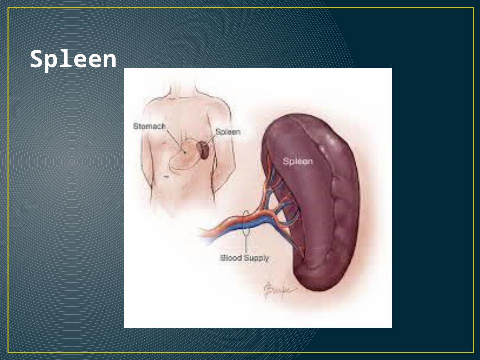
Spleen
• The spleen destroys worn out red blood cells.• It is located in the upper far left part of the
abdomen, to the left of the stomach. • The spleen varies in size and shape between
people, but it’s commonly fist-shaped, purple, and about 4 inches long.
• The spleen also helps fight certain kinds of bacteria that cause pneumonia and meningitis
Spleen conditions• Enlarged Spleen (Splenomegaly): An enlarged spleen, usually caused
by viral mononucleosis (“mono”), liver disease, blood cancers (lymphoma and leukemia), or other conditions.
• Ruptured spleen: The spleen is vulnerable to injury, and a ruptured spleen can cause serious life-threatening internal bleeding and is a life-threatening emergency. An injured spleen may rupture immediately after an injury, or in some cases, days or weeks after an injury.
• Sickle cell disease: In this inherited form of anemia, abnormal red blood cells block the flow of blood through vessels and can lead to organ damage, including damage to the spleen. People with sickle cell disease need immunizations to prevent illnesses their spleen helped fight.
• Thrombocytopenia (low platelet count): An enlarged spleen sometimes stores excessive numbers of the body’s platelets. Splenomegaly can result in abnormally few platelets circulating in the bloodstream where they belong.
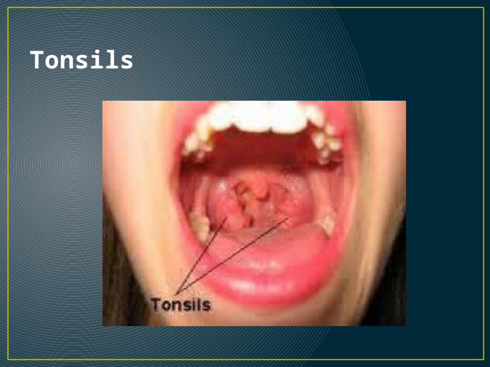
Tonsils
• The tonsils (palatine tonsils) are a pair of soft tissue masses located at the rear of the throat (pharynx).
• Each tonsil is composed of tissue similar to lymph nodes, covered by pink mucosa (like on the adjacent mouth lining). Running through the mucosa of each tonsil are pits, called crypts.
Tonsil Conditions• Acute tonsillitis: A bacteria or virus infects the tonsils, causing
swelling and a sore throat. The tonsil may develop a gray or white coating (exudate).
• Chronic tonsillitis: Persistent infection of the tonsils, sometimes as a result of repeated episodes of acute tonsillitis.
• Peritonsillar abscess: An infection creates a pocket of pus next to the tonsil, pushing it toward the opposite side. Peritonsillar abscesses must be drained urgently.
• Acute mononucleosis: Usually caused by the Epstein-Barr virus, “mono” causes severe swelling in the tonsils, fever, sore throat, rash, and fatigue.
• Strep throat: Streptococcus, a bacterium, infects the tonsils and throat. Fever and neck pain often accompany the sore throat.
• Enlarged (hypertrophic) tonsils: Large tonsils reduce the size of the airway, making snoring or sleep apnea more likely.
• Tonsilloliths (tonsil stones): Tonsil stones, or tonsilloliths, are formed when this trapped debris hardens, or calcifies.
Tonsillectomy & Adenoidectomy• http://
www.entusa.com/tonsils_adenoid_surgery.htm • Note: not for the queasy
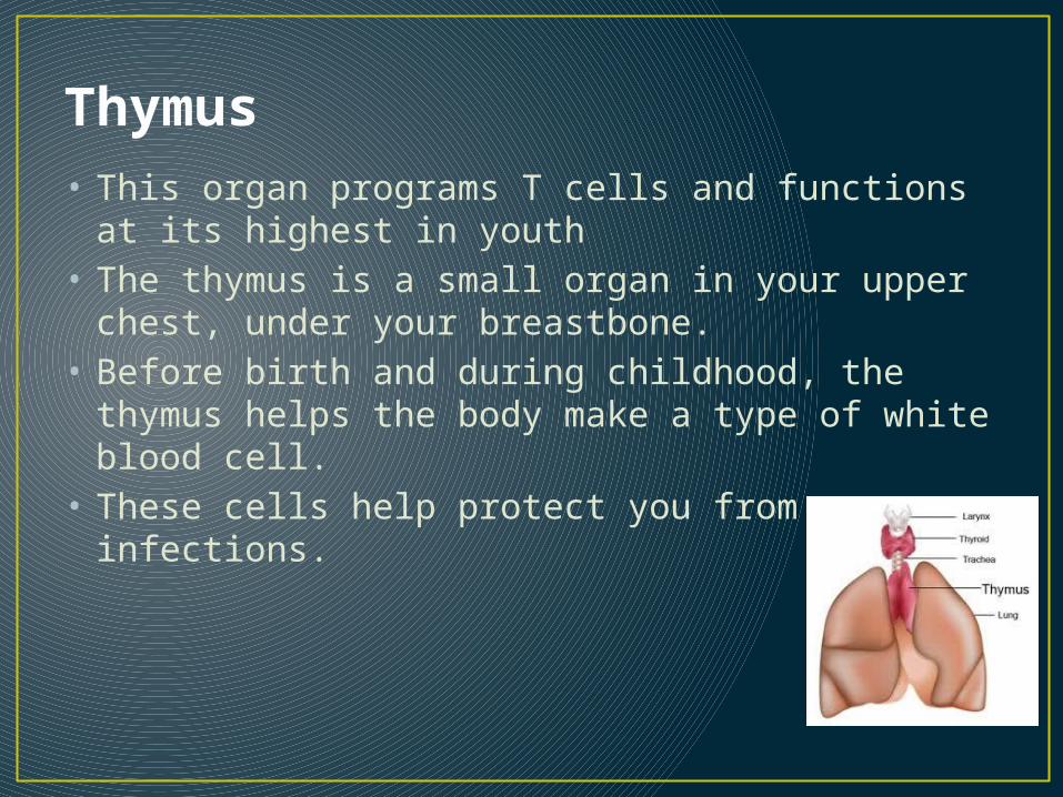
Thymus• This organ programs T cells and functions at its
highest in youth• The thymus is a small organ in your upper chest,
under your breastbone. • Before birth and during childhood, the thymus
helps the body make a type of white blood cell. • These cells help protect you from infections.
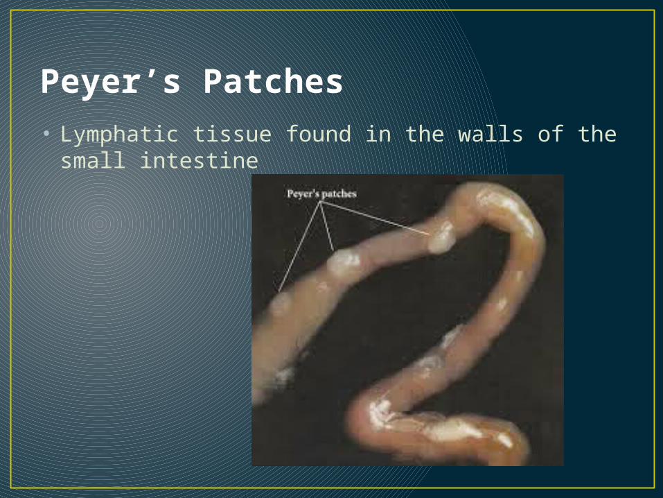
Peyer’s Patches• Lymphatic tissue found in the walls of the small
intestine
• Peyer's patches are areas of specialized tissue in the lower area of the small intestine that work to distinguish friend from foe as food passes through the gastrointestinal tract.
• Sensitized cells inside these areas identify antigens and decide whether they are harmless, associated with foods that the person is consuming for nutrition, or harmful, and linked with organisms like bacteria that could try to colonize the body.
• As food passes by, the antigens in the food are presented to the tissue and it determines whether it recognizes them and how they are classified.
• In addition to recognizing antigens and triggering the appropriate response, the Peyer's patches can also learn to identify new antigens, storing this information for future reference to make the immune system more effective.
Vaccines Only work for diseases like…• Pneumonia• Tetanus• Polio• Meaasles
Types of autoimmune diseases• Grave’s Disease http://
www.webmd.com/a-to-z-guides/understanding-graves-disease-basics
• Rheumatoid arthritis http://www.webmd.com/rheumatoid-arthritis/guide/rheumatoid-arthritis-basics
• Multiple sclerosis http://www.webmd.com/multiple-sclerosis/default.htm?names-dropdown=RI
• Type I Diabetes Mellitus http://www.webmd.com/diabetes/types-of-diabetes-mellitus
What’s “UP” with Fevers?• Also known as “PYREXIA”• Fevers are caused by chemicals called pyrogens
flowing in the bloodstream. • Pyrogens make their way to the hypothalamus in the
brain, which is in charge of regulating body temperature.
• When pyrogens bind to certain receptors in the hypothalamus, body temperature rises.
• One common pyrogen is called Interleukin-1 (IL-1). IL-1 is produced by white blood cells called macrophages when they come into contact with certain bacteria and viruses.
• IL-1 has multiple purposes, one of which is to signal other white blood cells, called helper T cells, into action.
Fevers, cn’t….• Average body temperature is about 98.6°F or
37°C, and temperatures above 100.4°F or 38°C are generally considered to be febrile.
• Fevers…….• Speed up the repair process• Increases metabolic rate (this is why your heart rate gets
faster)• Denature proteins
Stimulate liver and spleen to gather zinc and iron
Last but not least….• The process that neutrophils use to get across the
capillary walls is called diapedesis• Cells infected by viruses secrete the protein
interferon to protect nearby cells• Lymph from the left arm goes back into the heart
through the thoracic duct• Your genes determine the ability of your being
able to recognize foreign substances.
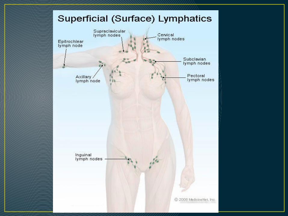
Be able to recognize….• The lymphatic organs on a diagram• Lymph capillary, lymph node, valve, blood
capillary, and vein on a diagram