mrsi detects abnormalities in normal-appearing frontal ... · lower frontal lobe gray matter naa...
TRANSCRIPT
MRSI Detects Abnormalities in Normal-Appearing Frontal Lobe of Sturge-Weber Syndrome Patients
Z. Kou1, M. Li1, Q. Jiang2, N. Seraji3, Y. Xuan1, E. Haacke1, H. T. Chugani3, C. Juhasz3, and J. Hu1 1Radiology, Wayne State University School of Medicine, Detroit, MI, United States, 2Henry Ford Hospital, 3Wayne State University
Introduction: Sturge-Weber syndrome (SWS) is characterized by vascular malformations present in the skin (port wine stain), eye, and meninges (leptomeningeal angioma) [1]. Seizures are the most common neurologic complications [2]. The intracranial vascular anomaly is most often unilateral and involves the occipital and parietal lobes. Neuroimaging techniques have been routinely employed in the assessment of children with SWS; structural MRI abnormalities are most common in the posterior brain regions. However, it is still unknown whether the frontal lobe, which is often spared by the vascular malformation, presents abnormalities on MRS. This is a clinically important issue, since motor impairment, which is one of the most feared complications beside seizures, could become more severe as the frontal lobe is progressively involved in the disease process. The goal of this study was to determine whether Magnetic Resonance Spectroscopic Imaging (MRSI) can improve detection of frontal lobe involvement in children with SWS.
Materials and Methods: Sixteen children (7 boys, mean age + SD = 5.2 + 3 years, range: 0.9-10.4 years) with the clinical diagnosis of Sturge-Weber syndrome and unilateral hemispheric involvement, based on post-contrast MRI and FDG PET were prospectively included. All MRI/MRS studies were performed in a Siemens Sonata 1.5 Tesla whole-body clinical imager (CTI/Siemens, Erlangen, Germany). A standard head coil was used both for MRI and 1H MRS. MRSI spectra were acquired in an axial plane passing through the parietal and frontal lobes above the level of the thalamus, including region(s) showing leptomeningeal angioma on previous and current MRI scans. Typical measurement parameters were: FOV = 200x200 mm2; phasing-encoding steps = 24x24, TR = 1280 ms, TE = 280 ms, slice thickness = 15 mm, signal averages = 2, and acquisition time = 10:12 minutes. Motor and intellectual functions were assessed via neuropsychological evaluation by neuropsychologists who were blind to all neuroimaging results.
Results: Eight children presented normal-appearing frontal lobes on conventional MRI, but 7 of them showed abnormal NAA and/or choline content in the frontal lobe of the affected hemisphere. Lower frontal lobe gray matter NAA was associated with earlier onset of seizures (r = 0.76; p = 0.04) and was an excellent predictor of motor function (r=-0.89, p<0.001).
Frontal lobe metabolite abnormalities in patients with vs. without frontal lobe involvement on structural MRI. Eight children showed frontal lobe abnormalities on structural MRI. Children with frontal lobe involvement on structural MRI had significantly lower level of NAA (reflected by lower NAA asymmetry ratios) in both frontal lobe gray and white matter when compared to patients without frontal MRI involvement (gray matter: 0.79 + 0.08 vs. 0.97 + 0.08, respectively, p < 0.001; white matter: 0.87 + 0.1 and 1.01 + 0.13, respectively, p = 0.03).
In patients with no frontal lobe involvement on structural MRI, a ≥10% NAA decrease was seen in 3 patients (1 in both in the gray and white matter, 2 in the white matter only); however, ≥10% increase of frontal NAA values was found in 3 other children (all in white matter).
Individual frontal lobe choline values showed abnormal increase in 4 patients in the subgroup with abnormal frontal lobe on MRI, and in 3 patients with normal frontal lobe on structural MRI (1 in both gray and white matter, 1 in gray matter only and 1 in white matter only). Two patients had a ≥10% decrease of frontal lobe choline in the same subgroup. Altogether, all but one child with normal frontal lobe on conventional MRI showed abnormal NAA and/or choline values in the frontal lobe.
MRSI and clinical variables. NAA asymmetry in the frontal lobe gray and white matter correlated with motor function (r = - 0.89; p < 0.001 and r = - 0.80; p = 0.03, respectively, after Bonferroni correction), i.e., lower NAA in the frontal lobe was associated with more severe motor impairment. Among the clinical seizure variables, NAA asymmetry in the frontal lobe gray matter showed a positive correlation with the age of seizure onset (r = 0.76; p = 0.04 after correction), indicating lower NAA values in children with early onset of seizures. Regression analysis showed that only frontal lobe NAA asymmetry (p = 0.01) but not age at seizure onset (p = 0.6) was an independent predictor of motor functions.
Discussion and Conclusions: The current study represents the largest series to date where MRSI was employed to analyze cerebral biochemical changes in children with SWS. The results show that in addition to an almost universal abnormality of NAA and choline compounds in the posterior regions which show angiomatous involvement, these metabolites are also often abnormal in the frontal lobe even in patients with no apparent structural abnormality detected by conventional MRI in this region. Furthermore, the severity of frontal NAA abnormalities appears to be a strong, independent predictor of motor functions. In conclusion, MRS imaging may be a useful complementary imaging tool for assessment of patients with Sturge-Weber syndrome, particularly to address frontal lobe involvement in children who show no apparent structural abnormalities in the frontal lobe on conventional MRI. Early detection of frontal lobe damage in such children may facilitate early interventions (such as intense physiotherapy or timely presurgical evaluation if seizures remain uncontrolled) to minimize neuro-cognitive consequences of the disease.
References: 1. Bebin EM, Gomez MR, Prognosis in Sturge-Weber disease: comparison of unihemispheric and bihemispheric involvement. J Child Neurol, 1988. 3: p. 181-4. 2. Roach ES, Neurocutaneous Syndromes. Pediatr Clin North Am, 1992. 39: p. 591-620.
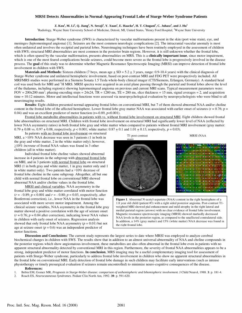
T1 post-contrast MRSI (NAA map)
Figure 1. Abnormal N-acetyl-aspartate (NAA) content in the right hemisphere of a 1.8 year old child (patient #3) with a right sided posterior angioma. Post-contrast T1-weighted MRI showed pial enhancement and mild atrophy in the right lateral and medial parietal region (arrows) with no clear evidence of frontal lobe involvement. Magnetic resonance spectroscopic imaging (MRSI) showed markedly decreased NAA levels in the posterior region, as compared to the unaffected contralateral side. In addition, a 14% (gray matter) and 15% (white matter) NAA decrease was found in the right frontal lobe.
Proc. Intl. Soc. Mag. Reson. Med. 16 (2008) 2081