multifactorial t-cell hypofunction that is reversible...
TRANSCRIPT
Cancer Therapy: Preclinical
Multifactorial T-cell Hypofunction That Is Reversible CanLimit the Efficacy of Chimeric Antigen Receptor–TransducedHuman T cells in Solid Tumors
Edmund K. Moon1, Liang-Chuan Wang1, Douglas V. Dolfi2, Caleph B. Wilson3, Raghuveer Ranganathan1,Jing Sun1, Veena Kapoor1, John Scholler3, Ellen Pur�e4, Michael C. Milone3, Carl H. June3, James L. Riley2,E. John Wherry2, and Steven M. Albelda1
AbstractPurpose: Immunotherapy using vaccines or adoptively transferred tumor-infiltrating lymphocytes (TIL)
is limited by T-cell functional inactivation within the solid tumor microenvironment. The purpose of this
study was to determine whether a similar tumor-induced inhibition occurred with genetically modified
cytotoxic T cells expressing chimeric antigen receptors (CAR) targeting tumor-associated antigens.
ExperimentalDesign:HumanTcells expressingCAR targetingmesothelinorfibroblast activationprotein
and containing CD3z and 4–1BB cytoplasmic domains were intravenously injected into immunodeficient
mice bearing large, established human mesothelin-expressing flank tumors. CAR TILs were isolated from
tumors at various time points and evaluated for effector functions and status of inhibitory pathways.
Results:CART cells were able to traffic into tumorswith varying efficiency and proliferate. Theywere able
to slow tumor growth, but did not cause regressions or cures. The CAR TILs underwent rapid loss of
functional activity that limited their therapeutic efficacy. This hypofunction was reversible when the T cells
were isolated away from the tumor. The cause of the hypofunction seemed to be multifactorial and was
associated with upregulation of intrinsic T-cell inhibitory enzymes (diacylglycerol kinase and SHP-1) and
the expression of surface inhibitory receptors (PD1, LAG3, TIM3, and 2B4).
Conclusions: Advanced-generation human CAR T cells are reversibly inactivated within the solid tumor
microenvironment of some tumors by multiple mechanisms. The model described here will be an
important tool for testing T cell–based strategies or systemic approaches to overcome this tumor-induced
inhibition. Our results suggest that PD1 pathway antagonism may augment human CAR T-cell function.
Clin Cancer Res; 20(16); 4262–73. �2014 AACR.
IntroductionAdoptive T-cell transfer (ACT) is a form of immunother-
apy that has demonstrated increasing promise as a thera-peutic option for cancer (1–3). ACT using cytotoxic T cellsthat have been genetically modified to express a chimericantigen receptor (CAR) specifically targeting a tumor-asso-ciated antigen (TAA) or a cancer stromal antigen offers theadvantages of specific, high-affinity binding of target cells in
a major histocompatibility class (MHC)–independent fash-ion, optimization of T-cell activation via incorporation ofdifferent internal costimulatory domains (so-called"advanced generation"CARs), and relatively straightforwardand efficient ex vivo preparation (4).
Recently, some dramatic tumor regressions in patientswith hematologic malignancies using CARs targeting theB-cell antigen CD19 have been reported (3). This hasspurred a growing interest inusing this approach for a varietyof solid tumors (5, 6). However, if CAR T cells behavesimilarly to endogenous T cells [or to ex vivo expandedtumor-infiltrating lymphocytes (TIL); refs. 7–10], it is likelythat the efficacy of the infused T cells will be limited by anumber of factors, including (i) inhibitory effects of tumor-derived cytokines, (ii) metabolic challenges (i.e., lack ofarginine or tryptophan), (iii) a microenvironment charac-terized by hypoxia and low pH, (iv) negative effects ofintratumoral immune-suppressor cells (5, 6, 11–13), (v)intrinsic inhibitory pathways mediated by upregulatedinhibitory receptors reacting with their cognate ligandswithin the tumor (14, 15), and (vi) intracellular inhibitorypathways that are engaged after T-cell activation, which
Authors' Affiliations: 1Division of Pulmonary, Allergy, and Critical Care,Department of Medicine, 2Department of Microbiology and Institute forImmunology, 3Department of Pathology and Laboratory Medicine, Abram-son Family Cancer Research Institute, Perelman School of Medicine; and4Department of Animal Biology, School of Veterinary Medicine, Universityof Pennsylvania, Philadelphia, Pennsylvania
Note: Supplementary data for this article are available at Clinical CancerResearch Online (http://clincancerres.aacrjournals.org/).
Corresponding Author: Edmund K. Moon, University of Pennsylvania,1015F Abramson Research Center, 3615 Civic Center Boulevard, Phila-delphia, PA 19104. Phone: 215-573-9918; Fax: 2155734469; E-mail:[email protected]
doi: 10.1158/1078-0432.CCR-13-2627
�2014 American Association for Cancer Research.
ClinicalCancer
Research
Clin Cancer Res; 20(16) August 15, 20144262
on October 14, 2018. © 2014 American Association for Cancer Research. clincancerres.aacrjournals.org Downloaded from
Published OnlineFirst June 11, 2014; DOI: 10.1158/1078-0432.CCR-13-2627

function to inhibit T-cell receptor (TCR) pathways andeffector functions (16). Examples of surface-inhibitoryreceptors on TILs include CTLA4, PD1, LAG3, 2B4, andTIM3 (17, 18). Examples of upregulated intracellular inhi-bitors in TILs are phosphatases (i.e., SHP1 that dephosphor-ylates TCR kinases such as Lck and ZAP70; ref. 19), ubiqui-tin-ligases (i.e., cbl-b; ref. 20), and kinases [i.e., diacylgly-cerol kinase (DGK), which inactivates diacylglycerol; ref. 21]Because advanced-generation CAR T cells have intrinsic
costimulatory activity [i.e., cytoplasmic domains fromCD28 and/or 4–1BB (CD137)], it is possible that they aremore resistant to these inhibitory forces. For example, thereare data supporting the ability of 4–1BB costimulation toblunt the anergy response (22–24). However, there are nodata studying the same protective ability of 4–1BB in CAR-modified T cells. Furthermore, a significant portion of thesedata was from research in murine T cells (23, 25). Thepurpose of this study was to develop a model in whichsuppression of T-cell function using advanced-generationhuman CAR T cells could be studied.
Materials and MethodsGenerationofmesoCARconstruct and lentivirus vectorpreparationThe single-chain Fv domain of the anti-mesothelin anti-
body (scFv SS1), originally provided by Dr. Ira Pastan (26)(National Cancer Institute/NIH, Bethesda, MD) was previ-ously subcloned into the lentiviral vector pELNSbearing theEF1a promoter and incorporated the CD3z and 4–1BBintracellular TCR signaling domains (27). A variant of themesoCAR construct incorporating a myc-tag between thescFv SS1 and the CD8 hinge was generated to allow forclearer detection of surface mesoCAR expression on TILsharvested from mouse flank tumors. Construction of asimilar CAR, but targeting murine fibroblast activationprotein (FAP), has been described previously (28).
Cell linesFor mesoCAR studies, a human mesothelioma cell line
derived from a patient’s tumor was used––EMP (parental).Because EMP did not have baseline expression of the TAA
mesothelin, it was transduced with a lentivirus to stablyexpress human mesothelin (the transduced cell line wasnamed EMMESO). Mesothelin expression level is shown inSupplementary Fig. S1.
Mouse 3T3Balb/C cells (3T3p) were purchased from theAmerican Type Culture Collection. Mouse FAP-expressing3T3Balb/C cells (3T3mFAP)were created by lentiviral trans-duction of the parental line with murine FAP (28).
All lines were also transduced to stably express fireflyluciferase (called EMPffluc, EMMESOffluc, 3T3p-ffluc, and3T3mFAP-ffluc). The culture conditions are described inSupplementary Methods.
Isolation, bead activation, transduction, expansion ofprimary human T lymphocytes, and T-cell effectorassays
These protocols are described in the SupplementaryMethods.
AnimalsAll animal experiment protocols were approved and
conducted in accordancewith the Institutional Animal Careand Use Committee. NOD/scid/IL2rg�/� (NSG) mice werebred in the Animal Services Unit of the Wistar Institute andChildren’s Hospital of Philadelphia. Femalemicewere usedfor experiments at 10 to 16 weeks of age.
In vivo xenograft experimentsA total of 5 � 106 EMMESO tumor cells were injected in
the flanks of NSG mice in a solution of X-Vivo media(Lonza) and Matrigel (BD Biosciences). After tumors wereestablished (100–200 mm3), the mice were randomlyassigned to one of three intravenous (tail-vein) treatmentgroups: (i) 20 � 106 nontransduced (NT) T cells (Dyna-beads activated T cells), (ii) 20 � 106 mesoCAR T cells(Dynabeads activated T cells transduced with mesoCAR),and (iii) saline. Tumors were measured using calipers andtumor volumes were calculated using the formula (p/6) �(length) � (width)2. An additional experiment was per-formed in which mice bearing EMMESO flank tumors wereinjected with 20 � 106 FAPCAR T cells. FAP is highlyexpressed on the tumor fibroblasts, which make up about6.3% of the digested EMMESO tumors.
At different time points, tumors were harvested, micro-dissected, and digested in a solution of 1:2 DNAse:collage-nase with rotation at 37�C for 1 hour. Digested tumors werethen filtered through 70-mm nylon mesh cell strainers, andred blood cells were lysed if needed (BD Pharm Lyse; BDBiosciences). After single-cell suspensions were achieved, 1� 106 cells were placed in standard FACS tubes and werestained with fluorochrome-conjugated anti-human CD45or CD3 antibodies.
Groups contained 10 mice each. The in vivo experimentswere repeated three times in independent fashion.
Ex vivo TIL analysisAfter digestion, TILs were isolated by using an anti-
human CD45 PE antibody (BD Biosciences) with the
Translational RelevanceAdoptive T cell transfer (ACT) using T cells genetically
modified to express chimeric antigen receptors (CAR)against tumor-associated antigens (TAA) has showngreat promise in the treatment of blood-borne malig-nant disease, but may be limited by the strong immu-nosuppressive environment within solid tumors. Wepresent a novel model demonstrating that reversibletumor-induced hypofunction of CAR T cells does occurin solid tumors. This model will be important in under-standing the mechanisms of this effect and developingstrategies to reduce or eliminate this hypofunction,paving the way for future clinical trials.
Solid Tumor Induction of CAR T-cell Hypofunction
www.aacrjournals.org Clin Cancer Res; 20(16) August 15, 2014 4263
on October 14, 2018. © 2014 American Association for Cancer Research. clincancerres.aacrjournals.org Downloaded from
Published OnlineFirst June 11, 2014; DOI: 10.1158/1078-0432.CCR-13-2627

EasySEP PE Selection Kit (STEMCELL Technologies;#18551). Once isolated, TILs were analyzed in three differ-ent ways: (i) luciferase-based killing assays, (ii) intracellularcytokine expression, and/or (iii) measurement of antigen-induced T-cell IFNg secretion (refer to SupplementaryMethods for detailed protocols).
AntibodiesRefer to Supplementary Methods for details.
InhibitorsThe SHP1 inhibitor sodium stibogluconate (SSG) was
purchased from EMD Millipore (567565). Two differentinhibitors of DGK were purchased from Sigma [DGKinh1,(nonspecific inhibitor), D5919; DGKinh2 (a isoformspecific), D5794]. Functional antibody against PDL1 wasa generous gift from Dr. Gordan Freeman (29). HumanIL2 (Proleukin; Prometheus Laboratories Inc. Dana FarberCancer Institute, Boston, MA) was acquired through theHospital of the University of Pennsylvania pharmacy.Dose–response curves were performed for both DGKinhibitors and for SSG, and the highest doses that didnot induce direct tumor cell killing were used. Both DGKinhibitors (1 mmol/L) and SSG (25 mg/mL) were alsodemonstrated to be appropriate in other published inves-tigations (30, 31). All inhibitory studies were done twicein independent fashion with comparable results.
ImmunoblottingLysates of T cells (40 mg) before and/or after activation
with beads were run on SDS-PAGE gels, transferred, andimmunoblotted using standard approaches. Primary andsecondary antibodies used are described in SupplementaryMethods.
Statistical analysisAll results were expressed as means � SEM as indicated.
For studies comparing two groups, the student t test wasused. For comparisons of more than two groups, we usedone-way ANOVA with appropriate post hoc testing. Differ-ences were considered significant when P < 0.05.
ResultsIntravenous injection of humanmesoCAR T cells slowsbut does not eradicate human tumors
The human mesothelin–expressing mesotheliomatumor cell line EMMESO was injected into the flanks ofNSG mice and allowed to grow to a size between 100 and200 mm3. At that time, tumor-bearing mice were givenone intravenous injection of 20 million T cells [mesoCARexpression was approximately 50% (data not shown)].Significant slowing of tumor growth was seen after a delayof 14 days (Fig. 1A); however, unlike our experiencewith another mesothelioma cell line (27, 32), no tumorregression or cures were noted. Injection of NT T cellshad minimal antitumor effects when compared withthe saline-treated control (Fig. 1A), indicating that thereduction in tumor growth observed in animals treated
with T cells expressing mesoCAR was specifically a resultof the mesoCAR.
Human mesoCAR T cells traffic into tumors andproliferate
To understand why we did not see tumor regression, wefirst evaluated whether our tumor antigen had been lost.Interestingly, immunohistochemistry of EMMESO tumorsat 40 days after T-cell injection showed robust and uniformexpression of mesothelin (Supplementary Methods andSupplementary Fig. S2).
We next evaluated the number and phenotype of thehuman T cells approximately 40 days after CAR T-cellinjections by removing and digesting the tumors andspleens and identifying human CD3þ cells by flow cyto-metry. In EMMESO tumors from mice injected with meso-CAR T cells, we noted that approximately 10% of thedigested cells (8.2 � 106) were human T cells. In contrast,in EMMESO tumors injectedwithNT T cells, only 0.1% (2.5� 105) of the digest was human T cells (Fig. 2A and B). Incontrast with our findings in the tumors, CAR T-cell per-sistence was much lower than NT T cells in spleens (Fig. 2CandD).Wealso examined tumors at earlier timepoints (Fig.2E and F). The percentage and number of intratumoral CART cells increased steadily over time, starting from very smallnumbers detectable at day 5.
0
200
400
600
800
1,000
1,200
1,400
1,600
0 4 7 11 14 18 21 26 32 39
Fla
nk
tum
or
size
(m
m3 )
No. of days post i.v. mesoCAR T cell
EMMESO tumor s/p mesoCAR T cell i.v.
Control
NT T cell
mesoCAR
A
B
0
200
400
600
800
0 4 7 9 11 16 18 21
Fla
nk
tum
or
size
(m
m3 )
No. of days post i.v. FAPCAR T cell
EMMESO tumor s/p FAPCAR T cell i.v.
Control
FAPCAR
*
*
Figure1. CARTcells slow tumorgrowthbutdonotcause regression.A,5�106 EMMESO tumor cells were injected into the flanks of NSGmice. Afterthey grew to approximately 200 mm3 in size, 20 � 106 mesoCAR T cellswere injected via tail vein and tumors were measured for 39 days afterinjection. T cells were able to slow growth by day 18 but were unable toeradicate flank tumors. B, 5� 106 EMMESO tumor cells were injected intothe flanks of NSGmice. After they grew to approximately 150mm3 in size,20 � 106 FAPCAR T cells were injected via tail vein and tumors weremeasured for 41 days after injection. T cells were able to slow growth byday 11 but were unable to eradicate flank tumors; �, P < 0.05.
Moon et al.
Clin Cancer Res; 20(16) August 15, 2014 Clinical Cancer Research4264
on October 14, 2018. © 2014 American Association for Cancer Research. clincancerres.aacrjournals.org Downloaded from
Published OnlineFirst June 11, 2014; DOI: 10.1158/1078-0432.CCR-13-2627

Tumor-infiltrating humanmesoCAR T cells continue toexpress CAR receptors on their surfaceTo evaluate loss or downregulation of the surface expres-
sion of the CARs, we injected mice bearing EMMESOtumors with T cells expressing a CAR that had been engi-neered to express amyc-tag in the extracellular domain.CARexpression (using an anti-myc antibody) on human T cellsfrom tumors 40 days after injection was compared with theexpression on CAR T cells that had been originally injected(Fig. 2G). In this experiment, the percentage of TILs expres-sing CAR on their surface increased to more than 42%compared with 13% at the time of injection.
Tumor-infiltrating human CAR T cells becomehypofunctionalThe data above suggested that although the human T cells
were present in large numbers, they had become hypofunc-
tional. Given that the level of CAR expression on the CARTILs was equal to or greater than that of the cells beforeinjection (Fig. 2G), we compared their functional activity.We isolated andanalyzed themesoCARTILs fromEMMESOtumors 40 days after injection (all studies were startedimmediately after isolation) and compared them with thesame batch of mesoCAR T cells that had been used for theoriginal injection and frozen away ("cryo mesoCAR").These cells were studied after thawing and incubating at37�C in 5% CO2 for 18 hours to mirror handling beforeinjection.
When we added cryo mesoCAR T cells and flank meso-CAR TILs to cultured EMMESO cells expressing firefly lucif-erase (EMMESOffluc) at a 20:1 ratio for 18 hours, the cryomesoCAR T cells were highly efficient in killing tumorcells (>95%), whereas the mesoCAR TILs killed only about10% (Fig. 3A; P < 0.001). Similarly, whereas cryo mesoCAR
0
2
4
6
8
10
12
NT T CAR T
hCD
3+ c
ells
(% o
f tot
al c
ells
)in
tum
or
0.0E+00
2.0E+06
4.0E+06
6.0E+06
8.0E+06
1.0E+07
1.2E+07
NT T CAR T
abs
# hC
D3
cells
in tu
mor
B
0
0.1
0.2
0.3
0.4
NT T CAR T
hCD
3+ c
ells
(% o
f tot
al c
ells
)in
spl
een
0.0E+00
1.0E+04
2.0E+04
3.0E+04
4.0E+04
NT T CAR T
abs
# hC
D3
cells
in s
plee
n
C
D
A
0
2
4
6
8
10
12
Day 5 Day 17 Day 39
hCD
3+ c
ells
(% o
f tot
al c
ells
)
0.0E+00
2.0E+06
4.0E+06
6.0E+06
8.0E+06
1.0E+07
Day 5 Day 17 Day 39
abs
# of
hC
D3
cells
in tu
mor
E F
P < 0.05 P < 0.05 P < 0.05
P < 0.05
CD45
myc
13.6% 42.7%
Myc-tagged mesoCAR(at time of injection)
Myc-tagged mesoCAR(at time of harvest)
G104
10486.4
0
0
0
0 57.3
103
103
102
102
101
101100
104
103
102
101
100
100 104103102101100
Figure 2. CAR T cells infiltrate, survive, proliferate, and retain transgene expression in tumors. 39 days after tail vein T-cell injection, flank tumors wereharvested and digested and the quantity of human CD3þ cells was assessed. The percentage (A) and absolute number (B) of intratumoral CAR T cellswere much higher than NT T cells [9.7% (8.2 � 106) CAR T cells vs. 0.16% (2.5 � 105) NT T cells (P < 0.05)]. The percentage (C) and absolute number(D) of intrasplenic CAR T cells weremuch lower than NT T cells [<0.05% (3.89� 102) CAR T cells vs. 0.25% (2.82� 104) NT T cells (P < 0.05)]. The percentage(E) and absolute number (F) of intratumoral CAR T cells were also assessed at earlier time points and demonstrated a marked increase over time [0.07%(2.16 � 104) in the first week after T-cell injection to 9.7% (7.98 � 106) in the sixth week after T-cell injection]. G, when myc-tagged mesoCAR TILs wereisolated from flank tumors 40 days after injection, they retained surface expression of myc-tagged mesoCAR after infiltration into tumors. Percentage ofMyc-tag–expressing T cells after infiltration was actually higher than at the time of injection (42.7% vs. 13.6%).
Solid Tumor Induction of CAR T-cell Hypofunction
www.aacrjournals.org Clin Cancer Res; 20(16) August 15, 2014 4265
on October 14, 2018. © 2014 American Association for Cancer Research. clincancerres.aacrjournals.org Downloaded from
Published OnlineFirst June 11, 2014; DOI: 10.1158/1078-0432.CCR-13-2627
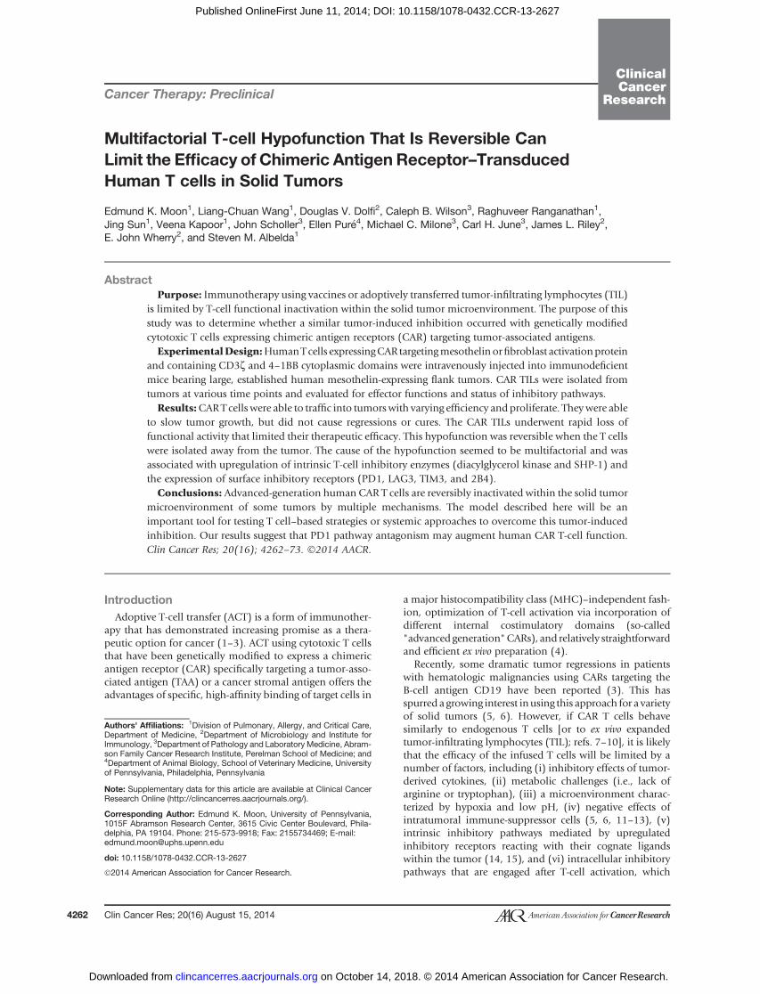
T cells released large amounts of IFNg into the supernatant,the mesoCAR TILs secreted very little (Fig. 3B; P < 0.001).Neither mesoCAR TILs nor cryo mesoCAR T cells hadsignificant antitumor activity against EMPffluc, demonstrat-ing that the response was specific to mesothelin TAA. Theex vivo killing ability of mesoCAR T cells isolated fromthe spleens of the same tumor-bearing mice demonstratedmuch less loss of function than the T cells isolated fromthe tumor, suggesting that these effects were specific to thetumor microenvironment (Fig. 3C and D).
We next examined the ability of the CAR T cells and CARTILs to produce cytokines (using intracellular cytokinestaining by flow cytometry; Fig. 4A). When exposed toalbumin-coated beads (control), neither the cryomesoCART cells nor fresh mesoCAR TILs (CD4 and CD8 cells)produced IFNg . After exposure tomesothelin-coated beads,a clear subpopulation of CD4 (0.59%) and CD8 (2.07%)cryo mesoCAR T cells made IFNg (Fig. 4A, left). In contrast,
the mesoCAR TILs did not produce IFNg after exposure totheir surrogate antigen (Fig. 4A, right) despite having rel-atively high expression of mesoCAR on the surface (Fig.2G). We saw very similar responses with IL2 production(Supplementary Fig. S3). We next exposed the same cells toPMA/ionomycin (Fig. 4B). Approximately 20%of the fresh-ly thawed mesoCAR T cells made IFNg (Fig. 4B, bottomleft). In contrast with the results with the beads, a similarpercentage of themesoCARTILs (22%) produced IFNg afterPMA/ionomycin (Fig. 4B, bottom right).
We also assessed the ability of the T cells to signal byassessing phospho-ERK expression using immunoblotting20minutes after exposure to beads (Fig. 4C). Phospho-ERKexpression was minimal in both types of CAR T cells afterexposure to control beads. Cryo mesoCAR T cells exhibitedERK activation via phosphorylation after exposure to bothmesothelin and anti-CD3/CD28–coated beads (Fig. 4C,left). In contrast, no phospho-ERK was detected in the
A B
0
20
40
60
80
100
Cryo mesoCAR Splenic mesoCAR
% K
illin
g
C
0
1,000
2,000
3,000
4,000
Cryo mesoCAR Splenic mesoCAR
D
0
20
40
60
80
100
Cryo mesoCAR Flank mesoCAR
% K
illin
g EMPffluc
EMMESOffluc
0
5,000
10,000
15,000
20,000
Cryo mesoCAR Flank mesoCAR
IFN
γ (p
g/m
L)
IFN
γ (p
g/m
L)
IFN
γ (p
g/m
L)
EMPfflucEMMESOffluc
0
5
10
15
20
25
30
Cryo FAPCAR Flank FAPCAR
% K
illin
g 3t3p-ffluc
3t3mFAP-ffluc
E
P < 0.001 P < 0.001
P < 0.01
0
1,000
2,000
3,000
4,000
Cryo FAPCAR Flank FAPCAR
3T3p-ffluc
3T3mFAP-ffluc
F P < 0.01
Figure 3. CAR T cells undergo tumor-induced hypofunction of cytolytic and cytokine secretion ability. A, cytotoxicity: flank mesoCAR TILs from day 39demonstratedhypofunctional killing of EMMESOffluccomparedwith cryomesoCART-cell controls (98.7%vs. 12.3%,P<0.001). TILs/Tcellswere coculturedwith firefly luciferase expressing EMMESO tumors at 20:1 E:T ratio for 18 hours. Measurement of luciferase activity of remaining tumor cells was used tocalculate percentage killing. B, IFNg secretion: flank mesoCAR TILs demonstrate hypofunctional secretion of IFNg in response to EMMESOffluc tumorcomparedwith cryomesoCART-cell controls (17,387 vs. 1,689 pg/mL,P < 0.001). Coculturewas performed at a ratio of 20:1 (effector to target) for 18 hours at37�C, in 5%CO2. EMPffluc targets were also tested to confirm antigen-specific tumor killing ability of both the cryomesoCAR T cells and themesoCAR TILs.C, isolated spleen-infiltrated T cells demonstratedmuch less loss of tumor killing function as compared with TILs (A) and (D) much less loss of tumor-inducedIFNg secretion as compared with TILs (B). E, flank FAPCAR TILs from day 28 demonstrated hypofunctional killing of 3T3mFAP-ffluc compared withcryo FAPCART-cell controls (24%vs. 0%,P <0.01). TILs/T cellswere coculturedwith firefly luciferase expressing 3T3p and 3T3mFAP tumors at 10:1 E:T ratiofor 18 hours. Measurement of luciferase activity of remaining tumor cells was used to calculate percentage killing. F, IFNg secretion: flank FAPCAR TILsdemonstrate hypofunction secretion of IFNg in response to 3T3mFAP-ffluc tumor compared with cryo FAPCAR T-cell controls (2,653.7 vs. 60.85 pg/mL,P < 0.01).
Moon et al.
Clin Cancer Res; 20(16) August 15, 2014 Clinical Cancer Research4266
on October 14, 2018. © 2014 American Association for Cancer Research. clincancerres.aacrjournals.org Downloaded from
Published OnlineFirst June 11, 2014; DOI: 10.1158/1078-0432.CCR-13-2627

mesoCAR TILs after exposure to mesothelin or anti-CD3/CD28–coated beads (Fig. 4C, right).It was also of interest to determine the kinetics of induc-
ing T-cell hypofunction. MesoCAR TILs were thus isolatedfrom tumors at 5, 17, and 39 days after injection, and theirability to kill tumor cells ex vivo was determined (Fig. 4D).MesoCAR TILs isolated at day 5 were still highly active,killing 85% of EMMESO tumor cells as assessed by our invitro killing assay. In contrast, mesoCAR TILs isolated ondays 17 and 39 had progressive hypofunction.
The hypofunction seen in human mesoCAR TILs isreversibleTo determine whether the hypofunction was reversible,
mesoCAR TILs isolated from day 39 tumors were "rested"for 24 hours away from the tumor in either media aloneor media plus low-dose IL2 and then had their effectorfunctions assessed. As shown in Fig. 5A and B, substantialrecovery of killing ability and IFNg release was seen24 hours at 37�C, 5% CO2 (away from tumor). The pres-ence of IL2 in the resting media accelerated recovery, but
was not required (Supplementary Fig. S4). We saw a similarrecovery in the ability to produce cytokines (at the single-cell level using flow cytometry) for IFNg and TNFa inresponse to EMMESO tumor (Supplementary Fig. S5).
The ability to phosphorylate ERK after surrogate antigenstimulation or ligation of the endogenous TCR was alsorestored after 24 hours of "rest" (Fig. 5C). Thus, a profoundbut reversible functional impairment exists in human CARTILs in mice with progressive tumor growth.
Human mesoCAR TILs express increased levels ofinhibitory receptors
We next evaluated the expression of four inhibitoryreceptors, that have been previously described in hypofunc-tional TILs isolated from humans, using flow cytometryon (i) the cryomesoCAR T cells that were used for injection,(ii) freshly isolated mesoCAR TILs from EMMESO tumor atday 39, and (iii) "recovered" mesoCAR TILs that had beenremoved from EMMESO tumor and "rested" 24 hours(Supplementary Table S1). CAR TILs expressed high levelsof inhibitory receptors. These levels were generally much
A
Unt
reat
edP
MA
/iono
myc
in
CryomesoCAR T cells
Fresh mesoCAR TILs
IFNγ
IFNγ CD45
CD4
CD8
FSC
0.33
0.09 0.59
2.07
0.18 0.07
0.05 0.010.539
0.32
21.7 22.8
B
Alb
umin
Mes
othe
lin
CD
3/C
D28
Alb
umin
Mes
othe
lin
CD
3/C
D28
Cryo mesoCAR T cells
Cryo mesoCAR T-cells
Fresh mesoCAR TILs
Fresh mesoCAR TILs
p-ERK
TotalERK
C
0
20
40
60
80
100
Day 5 Day 17 Day 39
% K
illin
g
D
Albumin beads Albumin beadsMesothelin beads Mesothelin beads
Figure 4. CAR TIL hypofunction progresses rapidly after injection andmay be due to defects in proximal signaling. A, upon exposure to either albumin-labeledbeads (negative control) or mesothelin-labeled beads, the mesoCAR TILs were found to be hypofunctional in their ability to produce IFNg in response to theantigen-coated beads. B, upon exposure to 0.1 mg/mL PMA and 2 mg/mL ionomycin, the mesoCAR TILs demonstrated preserved ability to produce IFNg asmeasured by intracellular cytokine staining by flow cytometry. C, upon exposure to either albumin-labeled beads (negative control), mesothelin-labeledbeads, or CD3/CD28 beads (positive control) for 15 minutes, mesoCAR TILs were found to be hypofunctional in their ability to produce signal viaphosphorylation of ERK in response to antigen or CD3/28 stimuli. D, when flank tumors were harvested at early (day 5), mid (day 17), and late (day 39) timepoints, isolated EMMESO TILs were cocultured with firefly luciferase expressing EMMESO tumor at 20:1 E:T ratio. After 18 hours, percentage killingwas calculated after measuring luciferase activity of the remaining tumor cells. We demonstrated that EMMESO TILs undergo rapidly increasingtumor-induced hypofunction after i.v. injection.
Solid Tumor Induction of CAR T-cell Hypofunction
www.aacrjournals.org Clin Cancer Res; 20(16) August 15, 2014 4267
on October 14, 2018. © 2014 American Association for Cancer Research. clincancerres.aacrjournals.org Downloaded from
Published OnlineFirst June 11, 2014; DOI: 10.1158/1078-0432.CCR-13-2627

lower after 24 hours of recovery away from the tumormicroenvironment. For the CD4 CAR TILs, PD1 went from73% to 53%, LAG3 went from 63% to 3%, and TIM3 wentfrom 24% to 1%. 2B4 expression was high and remainedelevated after rest (67% to 88%). For the CD8 CAR TILs,PD1went from 26% to 21%, LAG3went from 48% to 13%,and TIM3 went from 56% to 1%. 2B4 expression was highand remained elevated after rest (96% to 98%).
We also evaluated three of these inhibitory receptors onthe human T cells that could be isolated from the spleens ofthe EMMESO mice. Interestingly, the expression levels ofPD1, TIM3, and LAG3 were all lower on the splenic T cellscomparedwith the TILs (Supplementary Table S2), support-ing the hypothesis that the tumor microenvironmentinduces the upregulation of inhibitory receptors.
Human mesoCAR TILs express increased levels ofintracellular inhibitory enzymes
We also explored the expression levels of two intrinsicinhibitors of T-cell function that have been implicated inTIL dysfunction, SHP1 and DGK, using immunoblotting(Fig. 5D). The levels of both isoforms of DGK (a and z), aswell as the phosphorylated form of SHP1 (pSHP1), were
significantly elevated in mesoCAR TILs that were freshlyisolated from EMMESO flank tumor compared with restedTILs. This was also confirmed for DGKa using flow cyto-metry in which 23% of fresh EMMESO TILs expressedDGKa. Expression was undetectable after overnight rest(data not shown).
Blockade of inhibitors in human mesoCAR T cellsenhances their ex vivo killing function
Given these expression data, we studied the potentialfunctional importance of specific inhibitory pathways inmesoCAR TILs by introducing available blocking agentsduring the ex vivokillingandcytokine release assays.Additionof an anti-PDL1 antibody significantly restored the killingactivity and ability to secrete IFNg by themesoCAR TILs (Fig.6A and B). The relatively high dose of 10 mg/mL anti-PDL1antibody was based on previously published investigationsin cancer immunotherapy (33). Addition of either a type I ortype II DGK inhibitor also significantly increased the killingability (Fig. 6C), but without significantly increasing tumor-induced IFNg secretion (Fig. 6D). Addition of the SHP1inhibitor, SSG, slightly inhibited the killing ability of cryomesoCAR T cells, but significantly increased that of the
0
10
20
30
40
50
60
70
Pre rest Post rest
% K
illin
g
0
5,000
10,000
15,000
20,000
25,000
30,000
35,000
Pre rest Post rest
IFN
g (p
g/m
L)
P < 0.001P < 0.05
Alb
umin
Mes
othe
lin
CD
3/C
D28
Fresh TILs
p-ERK
TotalERK
Alb
umin
Mes
othe
lin
CD
3/C
D28
Recovered TILs
DGKa
pSHP1
Total SHP1
β-Actin
DGKz
A B
C D
Figure 5. CAR TIL cytolytic and cytokine secreting functions recover after rest away from tumor. DGK and pSHP1 expression is increased in hypofunctionalCAR TILs. Human TILs isolated from flank EMMESO tumors at day 39 and "rested" away from tumor for 24 hours demonstrated significant recovery intheir EMMESOffluc tumor killing ability (A) and their ability to secrete IFNg in response to EMMESOffluc tumor (B). C, compared with freshly harvestedEMMESO TILs, resting away from tumor led to recovery of antigen-specific signaling function of EMMESO TILs asmeasured by detection of phosphorylatedERK. D, DGKa and DGKz and pSHP1 significantly elevated in TILs freshly isolated from EMMESO. Expression of both isoforms of DGK as well as pSHP1decreased dramatically after overnight rest of TILs.
Moon et al.
Clin Cancer Res; 20(16) August 15, 2014 Clinical Cancer Research4268
on October 14, 2018. © 2014 American Association for Cancer Research. clincancerres.aacrjournals.org Downloaded from
Published OnlineFirst June 11, 2014; DOI: 10.1158/1078-0432.CCR-13-2627

mesoCAR TILs (Fig. 6E), as well as significantly increasingtumor-induced IFNg secretion (Fig. 6F).
Tumor-infiltrating human T cells against other tumorsand other TAAs also become hypofunctionalTo investigate the generalizability of our findings, we
studied mesoCAR T cells in an additional human mesothe-lioma model (M30 cells) and saw virtually identical induc-tion of hypofunction as we did with EMMESO (data notshown). Todemonstrate that these effectswere independentof the specific TAA,we evaluatedT cellsmodifiedwith aCARdirected against murine FAP, which is expressed on themouse fibroblasts in the stroma formed in the EMMESOflank tumors. Similar to our findings using murine T cellsand mouse tumors (28), injection of 107 FAPCAR T cellssignificantly slowed the growthof EMMESOtumors, but didnot eradicate them by day 41 after injection (Fig. 1B).FAPCAR TILs isolated from flank tumors at day 28 postinjection (by methods described above) also demonstratedprofound hypofunction in cytolytic activity and IFNg secre-tion similar to that seen in themesoCARTILs (Fig. 3E andF).
DiscussionAdoptive T cell transfer (ACT) using chimeric antigen
receptor–transduced T cells has demonstrated increasingpromise as a therapeutic option for cancer, especially inhematopoietic tumors (3, 34).Given that T-cell inactivationhas been reported in solid tumors (5, 6, 11), the purpose ofthis study was to develop a model in which this processcould be assessed using advanced-generation humanCAR Tcells.We found that a single intravenous injectionof humanmesoCAR T cells or FAPCAR T cells into immunodeficientmice induced a significant decrease in the tumor growth ratecompared with that of controls, but did not result in tumorregressions or cures. By harvesting and analyzing the tumorsat multiple time points, we were able to show that thisfailure to cure the tumors was not due to loss of tumorantigen, loss of the CAR on the surface of the T cells, nor afailure of the CAR T cells to accumulate within the tumor.Instead, our data show that loss of antitumor efficacy wasdue to a progressive loss of CAR T-cell effector functionprimarily caused by the tumor microenvironment. This T-cell hypofunction is very similar to that describedpreviously
0
20
40
60
80
Cryo mesoCAR mesoCAR TIL
% K
illin
g
P < 0.01
1
10
100
1,000
10,000
100,000
Cryo mesoCAR mesoCAR TILs
IFN
g/m
L P < 0.05
1
10
100
1,000
10,000
100,000
Cryo mesoCAR mesoCAR TILsIF
Ng
(pg
/mL
)
No inhibitor
DGK I inhibitor
DGK II inhibitor
0
20
40
60
80
Cryo mesoCAR mesoCAR TIL
% K
illin
g
SSG 25 μg/mL
P < 0.01
1
10
100
1,000
10,000
100,000
Cryo mesoCAR mesoCAR TIL
IFN
g (p
g/m
L)
No inhibitor
SSG 25 μg/mL
A B
C D
E F
No inhibitor
anti-PDL1 10 μg/mL
P < 0.05
No inhibitor
anti-PDL1 10 μg/mL
No inhibitor
0
20
40
60
80
Cryo mesoCAR mesoCAR TIL
% K
illin
g
No inhibitor
DGK I inhibitor
DGK II inhibitor
P < 0.01
P < 0.01
Figure 6. PDL1 blockade and DGK inhibition are able to restore CAR TIL function. 10 mg/mL of anti-PDL1 antibody was added to the coculture killing assay andwas able to restore mesoCAR TIL killing of EMMESOffluc (A) and tumor-induced mesoCAR TIL secretion of IFNg after fresh isolation from flank tumor (B).C, 1 mmol/L of type I and II DGK inhibitor was added to the coculture killing assay and was able to restore mesoCAR TIL killing of EMMESOffluc with minimalincrease in tumor-inducedmesoCARTILsecretionof IFNg after fresh isolation from flank tumor (D). 25mg/mLofSSGwasadded to thecoculturekillingassayandwas able to restore mesoCAR TIL killing of EMMESOffluc (E) and tumor-induced mesoCAR TIL secretion of IFNg after fresh isolation from flank tumor (F).
Solid Tumor Induction of CAR T-cell Hypofunction
www.aacrjournals.org Clin Cancer Res; 20(16) August 15, 2014 4269
on October 14, 2018. © 2014 American Association for Cancer Research. clincancerres.aacrjournals.org Downloaded from
Published OnlineFirst June 11, 2014; DOI: 10.1158/1078-0432.CCR-13-2627
in mouse and human TILs (7–9, 18, 35) and in nongene-tically modified adoptively transferred T cells in mousemodels (10, 36, 37).
Interestingly, although the CAR TILs isolated fromtumors had a profound but reversible functional defect,they accumulated to high numbers in the tumor microen-vironment. This suggests that the dysfunctional CAR TILssurvive and retain proliferative capacity, and is consistentwith the hypothesis that the 4–1BB cytoplasmic domain inthe CAR supports T-cell survival, but does not protect T cellsfrom hypofunction. Our animal data, thus, predict thateven though human CAR T cells may accumulate withinsolid tumors in patients, their efficacy may be limited by aprogressive inactivation within certain solid tumors. Therelative kinetics of CAR T-cell accumulation versus theirrate of inactivationwithin tumorswill ultimately determinethe overall antitumor efficacy, and this balancewill likely betumor specific.
Given these observations, we evaluated the nature ofthis tumor-induced CAR T-cell hypofunction. One featurewas that the hypofunction was rapidly and almost fullyreversible when theCAR TILs were removed from the tumormicroenvironment. Although supplemental IL2 acceleratedthe recovery, it was not necessary. We believe that thisreversibility was due to a general recovery of T-cell functiondue to the removal of inhibitory factors that reside inthe tumor microenvironment. However, it is formally pos-sible that our observations were due to a selection andexpansion of the most functional TILs, with a concomitantloss of the hypofunctional TILs during the rest period. Thispossibility is difficult to exclude and may require advancedmolecular strategies like "bar-coding" the T cells to trackthem during the rest period. However, we find the expla-nation of recovery of antitumor functionmore likely, basedon published work from other groups’ investigations (38–40). If one assumes that the CAR TILs are not irreversiblyinactivated, it may be best to describe their phenotype as"hypofunctional" rather than "exhausted" or "anergic,"which infer a permanent state of dysfunction (41, 42).
A second feature of our hypofunctional CARTILswas thatalthough TIL hypofunction was evident by 17 days postin-jection, tumor volume did not progress dramatically at thatpoint but rather "plateaued." This was likely due to abalance between progressively decreasing TIL function (Fig.4D) and increasing number of TILs in the tumor (Fig. 2E) asa result of the proliferative 4–1BB signaling incorporatedinto the CAR construct. Potentially, combining strategiesthat increase the rate or duration of TIL proliferation anddecrease TIL hypofunction would widen the "therapeuticsweet spot," leading to tumor regression.
A third feature of our hypofunctional CAR TILs was thatthe block seemed to be "proximal" in the T-cell signalingpathway. After stimulation by antigen or by CD3/CD28crosslinking, CAR TILs failed to secrete cytokines or tophosphorylate ERK. CAR TILs also had defects in the phos-phorylation of Lck, ZAP70, and SLP76 (data not shown).However, when the CAR TILs were exposed to PMA/iono-mycin, bypassing the early TCR activation steps, the cells
were fully capable of making cytokines. Although thesefeatures of reversibility and defects in proximal signalinghave been well documented previously in mouse (17, 43)and to some extent, human TILs (44, 45), the exactmechan-isms responsible for this phenotype remain elusive. Bothcytoplasmic and cell-surface candidates have been impli-cated. For instance, Rappl and colleagues elucidated onepossible mechanism of terminally differentiated, late-stageT-cell hypofunction as being due to impaired TCR synapseformation from immobility of TCR membrane surfacecomponents, which can be bypassed by introducing afirst-generation CAR construct that confers tumor reactivityby signaling through normal T-cell signaling components(46). The mechanism of the hypofunction described hereseems to be different as demonstrated by dysfunctionalproximal T-cell signaling. The described in vivo model isinadequate to assess TCR function as it lacks human anti-gen-presenting cells.
With regard to the mechanisms of hypofunction, weevaluated two intrinsic inhibitory enzymes previously iden-tified in hypofunctional TILs. The first candidate was thephosphatase SHP1, which can inactivate a number of thekinases in the early TCR signaling cascade (19, 47, 48).Investigators have shown in mice that TILs have elevatedSHP1activity and that inhibitionof SHP1 innonlytic TILs invitro restored their tumor-cytolytic ability (43). Conversely,mice deficient in SHP1 showed increased effector T-cellactivity (48). The second candidate was DGK, a key enzymethat inactivates diacylglycerol, a downstream messengernecessary for translating the TCR signal into T-cell stimu-lation (21, 44, 49). Loss of DGK leads to resistance to T-cellanergy and increased CD8 T-cell function (21, 44, 49). Ourgroup has recently shown that loss of the a or z isoform ofDGK augments murine and human CAR T-cell effectorfunction (31, 50). Both SHP1 and DGK have been shownto be upregulated in hypofunctional TILs (21, 44, 49, 51).
Our data support the importance of both SHP1 andDGKin the induction of hypofunction of the human CAR TILs.First, CAR TILs had high expression of both SHP1 andDGK,which rapidly declined 24 hours after the TILs wereremoved from the tumors. Second, when we blocked DGKor SHP1 activity using chemical inhibitors in our ex vivokilling assay, we were able to reduce the defects in tumorkilling,with lesser effects on IFNg secretion.Consistentwiththese data, Prinz and colleagues demonstrated increasedCD8 activity in human TILs from renal cell cancers afterblocking DGK (44). The factors within the tumor micro-environment that upregulate these inhibitory enzymes, aswell as approaches to inhibit these enzymes in vivo, areactive areas of research in our laboratory. In addition tocytoplasmic inhibitory pathways, it has become increasing-ly recognized that expression of some cell-surface inhibitoryreceptors on T cells can induce dysfunction (18). Overall,we observed a similar phenotype as that described byprevious studies in human TILs, with increased expressionof PD1, TIM3, Lag3, and 2B4 (52–55). Interestingly, ourdata suggest that the tumor microenvironment seemed toplay an important role in "shaping" the expression pattern
Moon et al.
Clin Cancer Res; 20(16) August 15, 2014 Clinical Cancer Research4270
on October 14, 2018. © 2014 American Association for Cancer Research. clincancerres.aacrjournals.org Downloaded from
Published OnlineFirst June 11, 2014; DOI: 10.1158/1078-0432.CCR-13-2627
of specific inhibitory receptors on the T cells. First, theexpression of inhibitory receptors wasmuch higher on TILs,rather than on human T cells isolated from spleens of thesame animals. Second, after overnight "rest" away from thetumor microenvironment, expression levels of most ofthese receptors decreased dramatically. Finally, we wereable to test the functional significance of the PD1/PDL1interaction in the EMMESO model. Addition of a blockingPDL1 antibody to our ex vivo CAR TIL killing assay was ableto restore the defect in tumor cell killing.We believe the model that we have described here has
many potential advantages and possible uses. First, it allowsstudy of human (not mouse) CAR T cells. This is importantas we have found the behavior of human CAR T cells to bevery different that of murine CAR T cells with regard to keyparameters such as persistence, sensitivity to activation-induced cell death, and the activation state at the time ofinjection. Furthermore, it allows the study of CAR T cellsthat are prepared identically to those being injected intopatients. This should enhance the generalizability of thefindings to clinical trials. Second, the use of a uniformpreparation of CAR T cells prepared from one donor allowsone to focus onmicroenvironmental differences rather thanintrinsic T-cell variability. Third, the use of CAR T cellstargeted to specific targets allows the study of antigen-specific T-cell interactions rather than just general TCRactivation induced by CD3/CD28 beads or PMA/ionomy-cin stimulation, as has been used in most studies of humanTILs. Fourth, the model uses well-characterized humansolid tumor cells that can be geneticallymodified, if needed.This allows the systematic study of different tumor micro-environments. Although we have used flank tumors forconvenience, orthotopic tumor cell placement is possible(and probably desirable).Most importantly, the CAR T cellsstudied in themodel seem to very closely resemble naturallyoccurring human TILs in all of our assays. Finally, the CART-cell hypofunction in thismodel seems to be generalizableacross different tumors and CARs recognizing differenttargets. The in vivo experiment detailed above was repeatedusing other MPM cell lines in the NSG xenograft model.After a single intravenous dose of mesoCAR T cells in NSGmice bearing flank tumors from another human MPM cellline (M30), we observed slowing of tumor growth (with nocures), and the mesoCAR TILs also demonstrated tumor-induced hypofunction that improved with overnight restaway from tumor. Thehypofunctionwas alsodemonstratedin T cells redirected against a nonmesothelin antigen(mFAP) after infiltration in our NSG xenograft model.Whether the immunosuppression occurred because ofEMMESO tumor or themurine fibroblasts within the tumorstroma will be investigated. Other groups have demonstrat-ed that tumor-associated fibroblasts also play a significantimmunosuppressive role (56).Limitations of the preclinical model should also be
acknowledged. The NSG mice have a hybrid immune sys-tem, thus limiting the ability to study the effects of impor-tant tumor immune cells such at T-regulatory cells, naturalkiller (NK) cells, dendritic cells, B cells, and myeloid cells.
The innate immune cells present in NSG mice are notisologous with the human T cells and tumor cells. The useofmicewithmore fully "humanized" immune systemsmaybe advantageous. Finally, theremay be some species incom-patibility issues (i.e., some ligands in murine microenvi-ronment might not react with human TCRs). Despite thesepotential problems, the CAR TILs isolated from the tumorsdo seem to strongly resemble human TILs, suggesting thatthe key interactions that induce T-cell hypofunction arepresent.
In addition to studying mechanisms of tumor-inducedT-cell hypofunction, models such as ours will allow testingof a wide variety of therapeutic approaches in an in vivosetting where anti-human reagents can be used. One strat-egy would be systemic administration of agents that mightaffect the tumor microenvironment. These could includeagents like (i) inhibitors of soluble inhibitory mediatorssuch as TGFb or PGE2, (ii) activating cytokines such as IL2,IL12, IL15, or type I interferons, (iii) small molecularinhibitors of intrinsic inhibitors like DGK and SHP1, (iv)agents thatmight alter tumor pHor oxidative status, and (v)anti–T-cell inhibitory receptor antibodies like anti-PD1,anti-PDL1, anti-TIM3, etc. Another promising strategy willbe to introduce genetic changes into thehumanT cells alongwith the CAR to prevent or modulate hypofunction. Thisapproach would allow highly specific alterations and avoidany systemic toxicity. There are awide variety of possibilitiesthat include introduction of new gene genes such as cyto-kines (i.e., IL12; refs. 57) or stimulatory proteins, such asconstitutively active AKT (58). It should also be possibleto express shRNA, dominant-negative constructs, or intra-cellular antibodies to reduce or block the expression oractivities of inhibitory surface receptors (such as the TGFbreceptor; ref. 59), PD1 and Tim3 or intrinsic inhibitors(such as DGK and SHP1). On the basis of our presentfindings, we plan to test anti-PD1 antagonists with CART cells in future clinical trials.
Disclosure of Potential Conflicts of InterestM.C. Milone reports receiving commercial research support from and is a
consultant/advisory board member for Novartis. C.H. June reports receivinga commercial research grant and speakers’ bureau honoraria from Novartis.E.J. Wherry reports receiving commercial research grants from Bristol-MyersSquibb, Janssen Pharmaceuticals, and Ono Pharmaceutical, and has owner-ship interest (including patents) in Genentech. No potential conflicts ofinterest were disclosed by the other authors.
Authors' ContributionsConception and design: E.K. Moon, L.-C. Wang, D.V. Dolfi, C.B. Wilson,E. Pur�e, J.L. Riley, S.M. AlbeldaDevelopment of methodology: E.K. Moon, L.-C. Wang, D.V. Dolfi,C.B. Wilson, J. Sun, J. Scholler, E. Pur�e, M.C. Milone, C.H. June, E.J. Wherry,S.M. AlbeldaAcquisitionofdata (provided animals, acquired andmanagedpatients,provided facilities, etc.): E.K. Moon, L.-C. Wang, D.V. Dolfi, R. Ranga-nathan, V. Kapoor, E.J. Wherry, S.M. AlbeldaAnalysis and interpretation of data (e.g., statistical analysis, biosta-tistics, computational analysis): E.K. Moon, L.-C. Wang, D.V. Dolfi,V. Kapoor, E. Pur�e, M.C. Milone, J.L. Riley, E.J. Wherry, S.M. AlbeldaWriting, review, and/or revision of the manuscript: E.K. Moon,L.-C. Wang, C.B. Wilson, C.H. June, S.M. AlbeldaAdministrative, technical, or material support (i.e., reporting or orga-nizing data, constructing databases): E.K. Moon, V. KapoorStudy supervision: E.K. Moon, J.L. Riley, E.J. Wherry, S.M. Albelda
Solid Tumor Induction of CAR T-cell Hypofunction
www.aacrjournals.org Clin Cancer Res; 20(16) August 15, 2014 4271
on October 14, 2018. © 2014 American Association for Cancer Research. clincancerres.aacrjournals.org Downloaded from
Published OnlineFirst June 11, 2014; DOI: 10.1158/1078-0432.CCR-13-2627
AcknowledgmentsThe authors thank the Human Immunology Core at the University of
Pennsylvania for supplying the healthy donor T cells used for the studies inthis article and Albert Lo, Kazim Panjwani, Michael Allegreza, and SanjanaKirloskar for their assistance with some of the experiments.
Grant SupportThis work was supported by the National Cancer Institute (NCI) grants
(P01 CA 66726–07/R01 CA 1477–95/RO1 CA172/RO1 CA141144/K08 CA
163941–01), a grant from the Cancer Research Institute, Novartis Pharma-ceuticals Corporation, and the Lung Cancer Translational Center of Excel-lence at University of Pennsylvania.
The costs of publication of this articlewere defrayed in part by the paymentof page charges. This article must therefore be hereby marked advertisementin accordance with 18 U.S.C. Section 1734 solely to indicate this fact.
Received September 29, 2013; revised April 7, 2014; accepted April 24,2014; published OnlineFirst June 11, 2014.
References1. Escudier B. Emerging immunotherapies for renal cell carcinoma. Ann
Oncol 2012;23 Suppl 8:viii35–40.2. Rosenberg SA, Yang JC, Sherry RM, Kammula US, Hughes MS, Phan
GQ, et al. Durable complete responses in heavily pretreated patientswith metastatic melanoma using T-cell transfer immunotherapy. ClinCancer Res 2011;17:4550–7.
3. Porter DL, Levine BL, Kalos M, Bagg A, June CH. Chimeric antigenreceptor-modified T cells in chronic lymphoid leukemia. N Engl J Med2011;365:725–33.
4. Sadelain M, Brentjens R, Riviere I. The basic principles of chimericantigen receptor design. Cancer Discov 2013;3:388–98.
5. Gilham DE, Debets R, Pule M, Hawkins RE, Abken H. CAR-T cells andsolid tumors: tuning T cells to challenge an inveterate foe. Trends MolMed 2012;18:377–84.
6. Lipowska-Bhalla G, Gilham DE, Hawkins RE, Rothwell DG. Targetedimmunotherapy of cancer with CAR T cells: achievements and chal-lenges. Cancer Immunol Immunother 2012;61:953–62.
7. Gajewski TF. Failure at the effector phase: immune barriers at the levelof the melanoma tumor microenvironment. Clin Cancer Res 2007;13(18 Pt 1):5256–61.
8. MescherMF,PopescuFE,GernerM,HammerbeckCD,Curtsinger JM.Activation-induced non-responsiveness (anergy) limits CD8 T cellresponses to tumors. Semin Cancer Biol 2007;17:299–308.
9. Powell JD. The induction and maintenance of T cell anergy. ClinImmunol 2006;120:239–46.
10. RussAJ,Wentworth L, XuK,RakhmilevichA,SeroogyCM,SondelPM,et al. Suppression of T-cell expansion by melanoma is exerted onresting cells. Ann Surg Oncol 2011;18:3848–57.
11. Baitsch L, Fuertes-Marraco SA, Legat A, Meyer C, Speiser DE. Thethree main stumbling blocks for anticancer T cells. Trends Immunol2012;33:364–72.
12. Gajewski TF, Woo SR, Zha Y, Spaapen R, Zheng Y, Corrales L, et al.Cancer immunotherapy strategies based on overcoming barriers withinthe tumor microenvironment. Curr Opin Immunol 2013;25:268–76.
13. Mayer A, Vaupel P. Hypoxia, lactate accumulation, and acidosis:siblings or accomplices driving tumor progression and resistance totherapy? Adv Exp Med Biol 2013;789:203–9.
14. Abate-Daga D, Hanada KI, Davis JL, Yang JC, Rosenberg SA, MorganRA. Expression profiling of TCR-engineered T cells demonstratesover-expression of multiple inhibitory receptors in persisting lympho-cytes. Blood 2013;122:1399–410.
15. Park HJ, Kusnadi A, Lee EJ, Kim WW, Cho BC, Lee IJ, et al. Tumor-infiltrating regulatory T cells delineated by upregulation of PD-1 andinhibitory receptors. Cell Immunol 2012;278:76–83.
16. Zheng Y, Zha Y, Gajewski TF. Molecular regulation of T-cell anergy.EMBO Rep 2008;9:50–55.
17. Vazquez-Cintron EJ, Monu NR, Frey AB. Tumor-induced disruption ofproximal TCR-mediated signal transduction in tumor-infiltrating CD8þ
lymphocytes inactivates antitumor effector phase. J Immunol 2010;185:7133–40.
18. Crespo J, Sun H, Welling TH, Tian Z, ZouW. T cell anergy, exhaustion,senescence, and stemness in the tumor microenvironment. Curr OpinImmunol 2013;25:214–21.
19. PalingNR,WelhamMJ.Role of the protein tyrosine phosphataseSHP-1 (Src homology phosphatase-1) in the regulation of interleukin-3-induced survival, proliferation and signalling. Biochem J 2002;368(Pt3):885–94.
20. Hopewell EL, Zhao W, Fulp WJ, Bronk CC, Lopez AS, Massengill M,et al. Lung tumor NF-kappaB signaling promotes T cell-mediatedimmune surveillance. J Clin Invest 2013;123:2509–22.
21. Zhong XP, Olenchock BA, Koretzky GA. The role of diacylglycerolkinases in T cell anergy. Ernst Schering Found Symp Proc 2007;3:139–49.
22. Habib-Agahi M, Phan TT, Searle PF. Co-stimulation with 4–1BB ligandallows extended T-cell proliferation, synergizes with CD80/CD86 andcan reactivate anergic T cells. Int Immunol 2007;19:1383–94.
23. Wilcox RA, Tamada K, Flies DB, Zhu G, Chapoval AI, Blazar BR, et al.Ligation of CD137 receptor prevents and reverses established anergyof CD8þ cytolytic T lymphocytes in vivo. Blood 2004;103:177–84.
24. Zhang H, Merchant MS, Chua KS, Khanna C, Helman LJ, Telford B,et al. Tumor expression of 4–1BB ligand sustains tumor lytic T cells.Cancer Biol Ther 2003;2:579–86.
25. Wilcox RA, Flies DB, ZhuG, Johnson AJ, TamadaK, Chapoval AI, et al.Provision of antigen and CD137 signaling breaks immunological igno-rance, promoting regression of poorly immunogenic tumors. J ClinInvest 2002;109:651–9.
26. Ho M, Feng M, Fisher RJ, Rader C, Pastan I. A novel high-affinityhuman monoclonal antibody to mesothelin. Int J Cancer 2011;128:2020–30.
27. Carpenito C, Milone MC, Hassan R, Simonet JC, Lakhal M, SuhoskiMM, et al. Control of large, established tumor xenografts with genet-ically retargeted human T cells containing CD28 and CD137 domains.Proc Natl Acad Sci U S A 2009;106:3360–5.
28. Wang LS, Lo A, Scholler J, Sun J, Majumdar RS, Kapoor V, et al.Targeting fibroblast activation protein in tumor stroma with chimericantigen receptor T cells can inhibit tumor growth and augment hostimmunitywithout severe toxicity.Cancer ImmunolRes2013;2:154–66.
29. Brown JA, Dorfman DM,Ma FR, Sullivan EL,MunozO,WoodCR, et al.Blockade of programmed death-1 ligands on dendritic cells enhancesT cell activation and cytokine production. J Immunol 2003;170:1257–66.
30. Yi T, Pathak MK, Lindner DJ, Ketterer ME, Farver C, Borden EC.Anticancer activity of sodium stibogluconate in synergy with IFNs.J Immunol 2002;169:5978–85.
31. Riese MJ, Wang LC, Moon EK, Joshi RP, Ranganathan A, June CH,et al. Enhanced effector responses in activated CD8þ T cells deficientin diacylglycerol kinases. Cancer Res 2013;73:3566–77.
32. Zhao Y, Moon E, Carpenito C, Paulos CM, Liu X, Brennan AL, et al.Multiple injections of electroporated autologous T cells expressing achimeric antigen receptor mediate regression of human disseminatedtumor. Cancer Res 2010;70:9053–61.
33. Pilon-Thomas S, Mackay A, Vohra N, Mule JJ. Blockade of pro-grammed death ligand 1 enhances the therapeutic efficacy of com-bination immunotherapy against melanoma. J Immunol 2010;184:3442–9.
34. Brentjens RJ, Davila ML, Riviere I, Park J, Wang X, Cowell LG, et al.CD19-targeted T cells rapidly induce molecular remissions in adultswith chemotherapy-refractory acute lymphoblastic leukemia. SciTransl Med 2013;5:177ra138.
35. Radoja S, Frey AB. Cancer-induced defective cytotoxic T lymphocyteeffector function: another mechanism how antigenic tumors escapeimmune-mediated killing. Mol Med 2000;6:465–79.
36. Zippelius A, Batard P, Rubio-Godoy V, Bioley G, Lienard D, Lejeune F,et al. Effector function of human tumor-specific CD8 T cells in
Moon et al.
Clin Cancer Res; 20(16) August 15, 2014 Clinical Cancer Research4272
on October 14, 2018. © 2014 American Association for Cancer Research. clincancerres.aacrjournals.org Downloaded from
Published OnlineFirst June 11, 2014; DOI: 10.1158/1078-0432.CCR-13-2627

melanoma lesions: a state of local functional tolerance. Cancer Res2004;64:2865–73.
37. Gajewski TF,MengY, BlankC, Brown I, Kacha A, Kline J, et al. Immuneresistance orchestrated by the tumormicroenvironment. Immunol Rev2006;213:131–45.
38. Mantovani A, RomeroP, PaluckaAK,Marincola FM. Tumour immunity:effector response to tumour and role of the microenvironment. Lancet2008;371:771–83.
39. Sakuishi K, Apetoh L, Sullivan JM, Blazar BR, Kuchroo VK, AndersonAC. Targeting Tim-3 and PD-1 pathways to reverse T cell exhaustionand restore anti-tumor immunity. J Exp Med 2010;207:2187–94.
40. Radoja S, Saio M, Schaer D, Koneru M, Vukmanovic S, Frey AB. CD8(þ) tumor-infiltrating T cells are deficient in perforin-mediated cytolyticactivity due to defective microtubule-organizing center mobilizationand lytic granule exocytosis. J Immunol 2001;167:5042–51.
41. Chappert P, Schwartz RH. Induction of T cell anergy: integration ofenvironmental cues and infectious tolerance. Curr Opin Immunol2010;22:552–9.
42. Wherry EJ. T cell exhaustion. Nat Immunol 2011;12:492–9.43. MonuN,FreyAB.Suppressionof proximal T cell receptor signalingand
lytic function in CD8þ tumor-infiltrating T cells. Cancer Res 2007;67:11447–54.
44. Prinz PU,Mendler AN,Masouris I, Durner L, Oberneder R, Noessner E.High DGK-alpha and disabled MAPK pathways cause dysfunction ofhuman tumor-infiltrating CD8þ T cells that is reversible by pharmaco-logic intervention. J Immunol 2012;188:5990–6000.
45. Whiteside TL. Signaling defects in T lymphocytes of patients withmalignancy. Cancer Immunol Immunother 1999;48:346–52.
46. Rappl G, Riet T, Awerkiew S, Schmidt A, Hombach AA, Pfister H, et al.The CD3-zeta chimeric antigen receptor overcomes TCR hypo-responsiveness of human terminal late-stage T cells. PLoS ONE2012;7:e30713.
47. ChemnitzJM,ParryRV,NicholsKE, JuneCH,RileyJL.SHP-1andSHP-2 associate with immunoreceptor tyrosine-based switch motif of pro-grammed death 1 upon primary human T cell stimulation, but onlyreceptor ligationpreventsTcell activation. J Immunol 2004;173:945–54.
48. Fowler CC, Pao LI, Blattman JN, Greenberg PD. SHP-1 in T cellslimits the production of CD8 effector cells without impacting the
formation of long-lived central memory cells. J Immunol 2010;185:3256–67.
49. Sanjuan MA, Jones DR, Izquierdo M, Merida I. Role of diacylglycerolkinase alpha in the attenuation of receptor signaling. J Cell Biol2001;153:207–20.
50. Riese MJ, Grewal J, Das J, Zou T, Patil V, Chakraborty AK, et al.Decreased diacylglycerol metabolism enhances ERK activation andaugments CD8þ T cell functional responses. J Biol Chem 2010;286:5254–65.
51. Koneru M, Schaer D, Monu N, Ayala A, Frey AB. Defective proximalTCR signaling inhibits CD8þ tumor-infiltrating lymphocyte lytic func-tion. J Immunol 2005;174:1830–40.
52. Ahmadzadeh M, Johnson LA, Heemskerk B, Wunderlich JR, DudleyME, White DE, et al. Tumor antigen-specific CD8 T cells infiltrating thetumor express high levels of PD-1 and are functionally impaired. Blood2009;114:1537–44.
53. Ngiow SF, von Scheidt B, Akiba H, Yagita H, Teng MW, Smyth MJ.Anti-TIM3 antibody promotes T cell IFN-gamma-mediated antitumorimmunity and suppresses established tumors. Cancer Res 2011;71:3540–51.
54. Goldberg MV, Drake CG. LAG-3 in cancer immunotherapy. Curr TopMicrobiol Immunol 2011;344:269–78.
55. Chlewicki LK, Velikovsky CA, Balakrishnan V, Mariuzza RA, Kumar V.Molecular basis of the dual functions of 2B4 (CD244). J Immunol2008;180:8159–67.
56. Nazareth MR, Broderick L, Simpson-Abelson MR, Kelleher RJ Jr,Yokota SJ, Bankert RB. Characterization of human lung tumor-asso-ciated fibroblasts and their ability to modulate the activation of tumor-associated T cells. J Immunol 2007;178:5552–62.
57. Chmielewski M, Abken H. CAR T cells transform to trucks: chimericantigen receptor-redirected T cells engineered to deliver inducible IL-12 modulate the tumour stroma to combat cancer. Cancer ImmunolImmunother 2012;61:1269–77.
58. Sun J, Dotti G, Huye LE, Foster AE, Savoldo B, GramatgesMM, et al. Tcells expressing constitutively active Akt resist multiple tumor-asso-ciated inhibitory mechanisms. Mol Ther 2010;18:2006–17.
59. Iyer S, Wang ZG, Akhtari M, Zhao W, Seth P. Targeting TGFbetasignaling for cancer therapy. Cancer Biol Ther 2005;4:261–6.
www.aacrjournals.org Clin Cancer Res; 20(16) August 15, 2014 4273
Solid Tumor Induction of CAR T-cell Hypofunction
on October 14, 2018. © 2014 American Association for Cancer Research. clincancerres.aacrjournals.org Downloaded from
Published OnlineFirst June 11, 2014; DOI: 10.1158/1078-0432.CCR-13-2627

2014;20:4262-4273. Published OnlineFirst June 11, 2014.Clin Cancer Res Edmund K. Moon, Liang-Chuan Wang, Douglas V. Dolfi, et al. in Solid Tumors
Transduced Human T cells−Efficacy of Chimeric Antigen Receptor Multifactorial T-cell Hypofunction That Is Reversible Can Limit the
Updated version
10.1158/1078-0432.CCR-13-2627doi:
Access the most recent version of this article at:
Material
Supplementary
http://clincancerres.aacrjournals.org/content/suppl/2014/06/06/1078-0432.CCR-13-2627.DC1
Access the most recent supplemental material at:
Cited articles
http://clincancerres.aacrjournals.org/content/20/16/4262.full#ref-list-1
This article cites 59 articles, 26 of which you can access for free at:
Citing articles
http://clincancerres.aacrjournals.org/content/20/16/4262.full#related-urls
This article has been cited by 23 HighWire-hosted articles. Access the articles at:
E-mail alerts related to this article or journal.Sign up to receive free email-alerts
Subscriptions
Reprints and
To order reprints of this article or to subscribe to the journal, contact the AACR Publications Department at
Permissions
Rightslink site. Click on "Request Permissions" which will take you to the Copyright Clearance Center's (CCC)
.http://clincancerres.aacrjournals.org/content/20/16/4262To request permission to re-use all or part of this article, use this link
on October 14, 2018. © 2014 American Association for Cancer Research. clincancerres.aacrjournals.org Downloaded from
Published OnlineFirst June 11, 2014; DOI: 10.1158/1078-0432.CCR-13-2627