multifunctional nanocarriers for enhanced tumor delivery1737/... · multifunctional nanocarriers...
TRANSCRIPT

MULTIFUNCTIONAL NANOCARRIERS FOR ENHANCED
TUMOR DELIVERY
Doctoral Thesis Presented
By
ANJALI VASANT APTE
Advisor: Dr. Vladimir P. Torchilin
To
The Graduate School of Bouvé College of Health Sciences
In Partial Fulfillment of the Requirements for the Degree of
Doctor of Philosophy in Pharmaceutical Sciences with Specialization in
Pharmaceutics and Drug Delivery Systems
NORTHEASTERN UNIVERSITY
BOSTON, MASSACHUSETTS
April, 2012

~ i ~
ABSTRACT
A tumor is an abnormally growing mass of cells. Ordinary chemotherapeutic agents used
to prevent tumor growth produce more side effects and less than desired therapeutic
effect. Targeted drug-delivery systems are necessary to provide and maintain an optimum
drug concentration in target tissues and cells. Long-circulating nanoparticles have the
ability to passively and actively target tumors and deliver the anticancer drugs. Passive
targeting can be achieved by prolonged circulation and retention of the drug-loaded
nanoparticles in the tumor, via enhanced permeability and retention (EPR) effect. Active
targeting is possible by attaching various ligands to the nanoparticles to specifically target
the tumors. These multifunctional particles help in improving the pharmacokinetic profile
of the drug, improve the drug bioavailability and reduce undesired side-effects in the non-
target organs. The main long-term goal of this project is the development of
multifunctional micelles and liposomes that increase the delivery of anticancer drugs to
the tumor.
Passive targeting of nanoparticles can be achieved by using polyethylene glycol-
phosphatidylethanolamine (PEG-PE) coating to prevent uptake of the nanoparticles by
macrophages and make the particle long-circulating. For active targeting, several
moieties can be attached to the particle surface. Anti-nucleosome monoclonal antibody
2C5 recognizes a broad variety of tumor cells via the tumor cell surface-bound
nucleosomes. It can recognize murine and human tumor cells, but not normal cells. Cell-
penetrating peptides (CPPs) have shown enhanced transport of cargoes through the
plasma membrane into the cells. Stimuli-sensitive PEG-hydrazone (Hz)-PE conjugate

~ ii ~
(with a low pH-sensitive hydrazone bond between PEG and PE) which is stable at normal
pH and can shield the CPP function.
In this study, micelles and liposomes were modified with a CPP-PEG-PE conjugate to
improve the transport of these nanocarriers into the cells. Since CPPs are susceptible to
enzymatic degradation and non-specific cellular interactions, they were shielded by the
PEG-Hz-PE conjugate. At normal pH (as found in circulation), the PEG-Hz-PE shielded
the CPP and at low pH (as found in tumors), when this polymer underwent hydrolysis,
PEG detached to expose the CPP and increased the internalization of the micelles and
liposomes. CPP modified pH-sensitive micelles loaded with anticancer drug paclitaxel,
improved tumor cell association and cytotoxicity in vitro. Intratumor injections of these
micelles demonstrated apoptosis in tumors developed subcutaneously in mice and
established this proof of concept.
Liposomes were further modified with mAb 2C5-PEG-PE. Recognition of the tumor cells
during the first phase of delivery was imparted by the attached mAb 2C5. During the
second phase of delivery, the exposure of the shielded TATp upon hydrolysis of pH-
sensitive polymer at the slightly acidic pH of the tumor cell environment, improved the
uptake of drug-loaded liposomes by tumor cells. Thus, tumor cell recognition and uptake
of the liposomes was obtained in a controlled fashion. Moreover, the growth of
subcutaneously developed drug-resistant and drug-sensitive tumors in nude mice was
inhibited and substantial decrease in the mean tumor weights was obtained by
intravenous injection of these drug-loaded multifunctional liposomes. In this way,
nanoparticles can be modified with multiple functionalities to improve tumor targeting
and enhance the effect of an anticancer agent.

~ iii ~
ACKNOWLEDGEMENTS
I would like to take this opportunity to express my gratitude towards everyone who made
my doctoral study possible.
First and foremost, I would like to thank Dr. Vladimir P. Torchilin for giving me a
chance to work in his laboratory and providing me financial support throughout my PhD
years. His mentoring and advice helped me immensely in developing my scientific
thought. He gave me the independence of developing my own experiments and at the
same time provided guidance that instilled confidence in me as a scientist.
I would like to express my sincere gratitude to the members of my thesis committee: Dr.
S. John Gatley, Dr. Akio Ohta, Dr. Roger Giese and Dr. Rebecca Carrier for their
valuable time and guidance. The feedback they gave me was extremely helpful.
I can‟t thank Dr. Jacob Grunwald enough with whom I started my thesis project. His
supervision and mentoring in the initial stages of my thesis developed the foundation of
my scientific research. Special thanks to Dr. Erez Koren who guided me through all the
hurdles in the project and with whom I have two publications.
My heartfelt thanks to Dr. William Hartner for all the support during animal studies and
editing my manuscripts and abstracts throughout my graduate study. I would also like to
thank Dr. Tatyana Levchenko and Dr. Dmitriy Mongayt for their assistance in my work.
They have provided very important inputs in my work and helped me whenever I needed
their help.

~ iv ~
I would also like to thank the Department of Pharmaceutical Sciences, Bouve College of
Health Sciences, Northeastern University who accepted me first in the Master of Science
and later in the PhD program. The staff at the Department of Pharmaceutical Sciences,
Roger Avelino, Rosalee Robinson and Sarom Lay has been extremely helpful. My
friends, colleagues and post-docs in the lab have also immensely contributed to the
success of my thesis project. This wouldn‟t have been a successful project without the
discussions I had with them and the inputs they gave.
And last but not the least the continuous support, help and love throughout all these years
that my family has provided me. It has been the strongest backbone of my academic life.
I can‟t express in words the gratitude I feel towards my grandmother who always stressed
the importance of education and wished that I achieve the highest possible degree. She
has been my source of inspiration throughout. I can never imagine how I would have
completed this dissertation without the support of my parents. The financial support they
provided was the most beneficial thing that I needed when I came to this University. I am
thankful to my sister who has always encouraged me through difficult times. And finally
I would like to thank my dear husband who gave me the emotional strength and stability
that I needed to achieve my dreams.

~ v ~
TABLE OF CONTENTS
ABSTRACT ....................................................................................................................... i
ACKNOWLEDGEMENTS .................................................................................................... iii
LIST OF TABLES ...................................................................................................................... x
LIST OF FIGURES ........................................................................................................ xi
1. INTRODUCTION ................................................................................................................... 1
1.1. STATEMENT OF THE PROBLEM............................................................................... 1
1.1.1. Rationale for target-specific delivery ........................................................................1
1.1.2. Passive targeting of nanoparticles .............................................................................2
1.1.3. Active targeting of nanoparticles ..............................................................................4
1.2. REVIEW OF THE LITERATURE ................................................................................ 5
1.2.1. Tumor physiology and lowered extracellular pH … ..................................................... 5
1.2.2. Micelles and liposomes for drug delivery to tumors … ............................................5
1.2.3. In vivo longevity of nanoparticles .................................................................................... 9
1.2.3.1. Importance of PEG coating ..........................................................................9
1.2.3.2. PEGylation of liposomes ...........................................................................10
1.2.3.3. Use of detachable PEG ..............................................................................12
1.2.4. Ligands for active targeting ........................................................................................... 14
1.2.4.1. Anti-nucleosome monoclonal antibody, 2C5 .............................................14
1.2.4.2. Tumor-specific ligands ..............................................................................16
1.2.4.3. Cell-penetrating peptides ...........................................................................18
1.2.5. Multifunctional nanocarriers ......................................................................................... 20

~ vi ~
1.2.5.1. Concept of multifunctionality ....................................................................20
1.2.5.2. Development of multifunctional nanocarriers .............................................. 20
1.2.6. Tumors and the drug resistance phenomenon .............................................................. 25
2. OBJECTIVES AND SPECIFIC AIMS ....................................................................27
3. EXPERIMENTAL DESIGN AND METHODS ......................................................28
3.1. Materials ................................................................................................................28
3.2. TATp cleavage properties and shielding methodologies ...................................29
3.2.1. Synthesis of TATp-PEG1000-PE ...............................................................................29
3.2.2. Preparation of TATp-modified micelles (TM) and
TATp- modified liposomes (TL) ......................................................................................30
3.2.3. Characterization and determination of cleavage of TATp in TM and TL ..............30
3.2.4. Cell interaction studies ............................................................................................31
3.2.5. Cytotoxicity studies with TATp liposomes ............................................................33
3.2.6. Preparation and characterization of shielded, non pH-sensitive micelles ............... 34
3.3. TATp-modified pH-sensitive micelles ..................................................................35
3.3.1. Synthesis of hydrolysable PEG conjugate (PEG2000-Hz-PE)...................................35
3.3.2. Preparation and characterization of TATp-modified pH-sensitive micelles ........... 36
3.3.3. Determination of kinetics of hydrolysis of hydrazone bond and
TATp in pH-sensitive-micelles ................................................................................................ 36
3.3.4. Cell interaction studies ............................................................................................37

~ vii ~
3.3.5. In vitro cell viability studies ...................................................................................38
3.3.6. In vivo studies- Determination of apoptosis by TUNEL assay ...............................38
3.4 Multifunctional TATp-containing, pH-sensitive, mAb 2C5-modified liposomes
........................................................................................................................................................ 40
3.4.1. Synthesis of pNP-PEG3400-PE and 2C5 mAb modification ....................................40
3.4.2. Preparation of multifunctional TATp-modified, pH-sensitive, immuno-liposomes
............................................................................................................................................41
3.4.3. Determination of optimum shielding of TATp with long chain PEG2000-Hz-PE ... 42
3.4.4. Characterization of liposomes .................................................................................42
3.4.4.1. Measurement of particle size and zeta potential .........................................42
3.4.4.2. Specific activity of mAb 2C5 on liposomal surface by ELISA ..................43
3.4.5. Cell interaction studies ............................................................................................43
3.4.6. In vitro cell viability studies ...................................................................................44
3.4.7. Determination of P-gp levels in SKOV3 drug-resistant and sensitive cells ...........45
3.4.8. In vivo studies of multifunctional liposomes in drug-resistant
and drug-sensitive tumors ......................................................................................................... 45
4. RESULTS AND DISCUSSION .................................................................................47
4.1. TATp cleavage properties and shielding methodologies ......................................47
4.1.1. Preparation and characterization of TATp-micelles(TM) and
TATp-Liposomes (TL) .....................................................................................................47
4.1.2. Determination of cleavage properties of TATp in TM and TL ..............................48
4.1.3. Cell interaction studies ............................................................................................49

~ viii ~
4.1.4. In vitro cytotoxicity of doxorubicin-loaded TATp liposomes ................................53
4.1.5. Characterization of non-pH-sensitive steric shielding of TATp-micelles. ............... 54
4.2. TATp-modified pH-sensitive micelles ..................................................................57
4.2.1. Preparation and characterization of TATp-modified pH-sensitive micelles ..........57
4.2.2. Determination of hydrolysis of hydrazone bond and cleavage of TATp in TATp-
modified pH-sensitive micelles ................................................................................................ 58
4.2.3. In vitro studies .........................................................................................................61
4.2.3.1. Cell interaction studies .................................................................................61
4.2.3.2. Cell viability studies ....................................................................................64
4.2.4. In vivo studies- Determination of apoptosis by TUNEL assay ...............................65
4.3. Multifunctional TATp-containing, pH-sensitive, mAb 2C5 containing liposomes
....................................................................................................................................................... 68
4.3.1. Concept of multifunctional TATp-modified, pH-sensitive, immuno-liposomes ....68
4.3.2. Preparation and characterization of multifunctional liposomes ..............................69
4.3.2.1. Determination of optimum PEG2000-Hz-PE shielding ................................70
4.3.2.2. Determination of particle size and zeta potential ........................................72
4.3.2.3. Specific activity of modified mAb 2C5 by indirect ELISA .......................75
4.3.3. Cell interaction studies ............................................................................................77
4.3.4. In vitro cytotoxicity studies ....................................................................................80
4.3.5. Development of drug-resistance in sensitive cell lines ...........................................83
4.3.6. P-gp expression studies to determine/confirm drug-resistant and sensitive cells ...84
4.3.7. Cell interaction studies with SKOV3 drug resistant and sensitive cells .................85

~ ix ~
4.3.8. In vitro cytotoxicity studies ....................................................................................88
4.3.9. In vivo studies of multifunctional liposomes in drug-resistant
and drug-sensitive tumors ......................................................................................................... 90
4.3.9.1. Tumor growth inhibition studies .................................................................90
4.3.9.2. Analysis of changes in body weight of mice ..............................................93
4.3.9.3. Determination of tumor weight ...................................................................94
4.3.9.4. Determination of apoptosis by TUNEL assay ............................................95
5. CONCLUSIONS .........................................................................................................98
6. REFERENCES ..........................................................................................................100
7. APPENDIX ................................................................................................................119
7.1. List of publications .................................................................................................119

~ x ~
LIST OF TABLES
Table 1 Nanoscaled systems for systemic cancer therapy .................................................2
Table 2 Zeta Potential of TATp-modified micelles and liposomes .................................48
Table 3 Size range and zeta potential of TATp-modified pH-sensitive micelles at pH 5.0
and pH 7.4 ..........................................................................................................................57
Table 4 Zeta potentials of liposomal formulations ...........................................................74
Table 5 IC50 values of unmodified and surface modified Doxil multifunctional immuno-
liposomes with HeLa cells .................................................................................................82
Table 6 IC50 values of unmodified and surface modified Lipodox multifunctional
immuno-liposomes with SKOV3 drug-sensitive and drug-resistant cells ........................89

~ xi ~
LIST OF FIGURES
Figure 1. Schematic of enhanced permeability and retention effect for passive targeting
of nanoparticles ...................................................................................................................3
Figure 2. Self assembling PEG-PE micelles ......................................................................7
Figure 3. Structure of a liposome ........................................................................................8
Figure 4. Structure of a TATp-modified nanoparticle with TATp groups exposed on the
particle surface ..................................................................................................................21
Figure 5. Schematic diagram depicting the shielding of TATp by the long PEG chains in
the pH-sensitive-TATp-micelle and the exposure of TATp as a result of the hydrolysis of
hydrazone bond of PEG-Hz-PE at acidic pH .....................................................................22
Figure 6. Schematic of the effect of low pH on TATp-modified, pH-sensitive, immuno-
liposomes composed of a pH-degradable PEG2000-Hz-PE with a long PEG block, TATp-
PEG1000-PE with a short PEG block, and mAb2C5-PEG3400-PE .......................................24
Figure 7. Schematic of P-gp efflux of drug and prevention of P-gp drug efflux by
encapsulation in a nanoparticle ..........................................................................................26
Figure 8. RR fragment obtained on treatment of TATp with trypsin, as analyzed by
HPLC ................................................................................................................................49
Figure 9. Fluorescence microscopy and flow cytometry analysis showing interaction of
TATp-liposomes upon trypsin/trypsin inhibitor pre-treatment, with B16F10 and HeLa
cells ...................................................................................................................................51

~ xii ~
Figure 10. Fluorescence microscopy and flow cytometry analysis showing interaction of
TATp- micelles upon trypsin/trypsin inhibitor pre-treatment, with B16F10 cells ...........52
Figure 11. In vitro cytotoxicity of TATp-Doxil formulations towards B16-F10 and HeLa
cells ...................................................................................................................................53
Figure 12. PEG shielding of TATp-micelles ..................................................................55
Figure 13. Representative TEM image of TATp-modified pH-sensitive micelles ..........58
Figure 14. HPLC analysis of the cleavage of TATp and hydrolysis of hydrazone bond in
TATp-modified pH-sensitive-micelles .......................................................................59, 60
Figure 15. Kinetics of the cleavage of TATp and hydrolysis of pH-sensitive hydrazone
bond in TATp-modified pH-sensitive-micelles pre-incubated at pH 5 ............................61
Figure 16. Fluorescence microscopy and flow cytometry analysis showing the
internalization of TATp-modified pH-sensitive micelles pre-incubated at pH 5 or pH 7.4,
with B16-F10 or HeLa cells ..............................................................................................63
Figure 17. In vitro cytotoxicity of paclitaxel loaded TATp-modified pH-sensitive-
micelles preincubated at pH 5 or pH 7.4 on B16 F10 cells ..............................................65
Figure 18. Detection of apoptotic activity by fluorescence microscopy in B16F10 tumor
sections as shown by TUNEL staining .............................................................................66
Figure 19. Schematic of the effect of low pH on TATp-modified pH-sensitive immuno-
liposomes composed of a pH-degradable PEG2000-Hz-PE with a long PE block, TATp-
PEG1000-PE with a short PEG block, and mAb2C5-PEG3400-PE .......................................69
Figure 20. Zeta potentials of TATp-modified liposomes shielded with increasing
quantity of PEG2000-Hz-PE ...............................................................................................70

~ xiii ~
Figure 21. Particle size of liposomes with increasing mol % of PEG coating ................71
Figure 22. Particle sizes of modified Doxil and Lipodox preparations and a TEM image
of Doxil and multifunctional immuno-Doxil ...............................................................72, 73
Figure 23. Specific activity of mAb 2C5 on the surface of Doxil (a) and Lipodox (b) by
indirect ELISA. ............................................................................................................75, 76
Figure 24. Fluorescence microscopy of multifunctional liposomes and other liposomal
controls with NIH-3T3, MCF-7 and HeLa cells ...............................................................77
Figure 25. Flow cytometry analysis of multifunctional immuno-Doxil® and other
liposomal controls with NIH-3T3, MCF-7 and HeLa cells ...............................................79
Figure 26. In vitro cytotoxicity of multifunctional TATp-modified, pH-sensitive,
immuno-Doxil and other controls with HeLa cells .....................................................80, 81
Figure 27. Pgp expression in SKOV3 drug-sensitive and drug-resistant cells ................84
Figure 28. Fluorescence microscopy of multifunctional immuno-liposomes and other
liposomal controls with SKOV3 drug-sensitive and resistant cells ..................................85
Figure 29. Flow cytometry analysis of multifunctional immuno-liposomes and other
liposomal controls with SKOV3 drug-sensitive and resistant cells ..................................87
Figure 30. In vitro cytotoxicity of multifunctional TATp-modified, pH-sensitive,
immuno- Lipodox and other controls with SKOV3 drug-resistant and drug-sensitive cells
............................................................................................................................................88
Figure 31. Effect of intravenous administration of liposomal preparations on tumor
growth in nude mice bearing SKOV-3 drug-sensitive and drug-resistant tumors .......90,91

~ xiv ~
Figure 32. Effect of intravenous administration of liposomal preparations on the body
weight in nude mice bearing SKOV-3 drug-resistant and drug-sensitive tumors ............93
Figure 33. Final tumor weights in nude mice bearing SKOV-3 drug-sensitive and drug-
resistant tumors following intravenous administration of liposomal preparations .... 94, 95
Figure 34. Detection of apoptotic activity by fluorescence microscopy in SKOV-3 drug-
sensitive tumor sections as shown by TUNEL staining ....................................................96
Figure 35. Detection of apoptotic activity by fluorescence microscopy in SKOV-3 drug-
resistant tumor sections as shown by TUNEL staining ....................................................97
~ 1 ~
1. INTRODUCTION
1.1. STATEMENT OF THE PROBLEM
1.1.1. Rationale for target-specific delivery.
A major problem associated with the use of a drug is its tendency to distribute uniformly
in the body. This usually leads to undesirable side-effects on otherwise healthy organs
and tissues, possible inactivation of the drug and low bioavailability in the organ of
interest [1]. As a result, a larger amount of drug is required to achieve the high local
concentration needed for a therapeutic effect but will exacerbate the side effects.
Many targeted drug delivery systems have been developed to overcome these problems.
Use of such delivery systems can reduce the quantity of the drug required, deliver the
drug specifically to the targeted pathological site, improve therapeutic outcome and
reduce the side effects.
Over the past few decades, nanoparticles have come into wide-spread practice as drug
delivery systems. Some examples include polymeric and metallic nanoparticles,
dendrimers, microcapsules, micelles and liposomes. They can be used for diagnosis,
treatment and monitoring of cancer. Some nanoparticles that are commercially available/
in clinical trials are summarized in the following table:

~ 2 ~
Table 1. Nanoscaled systems for systemic cancer therapy [2]
Micelles and liposomes have been much studied and have repeatedly been shown to
provide significant in vitro and in vivo anti-tumor effects of the loaded drug.
1.1.2. Passive targeting of nanoparticles.
An important characteristic of the nanoparticles is their tailorability to suit the needs
peculiar to a tumor‟s physiology or other particular features that make the tumor
targetable. However, while manipulating their physical characteristics, the effect of a
nanoparticle on in vivo behavior needs to be taken into consideration [3]. Some of the
important features of the tumor microenvironment include: its non-uniform arrangement
of vasculature, hypoxia and high metabolic activity [4], [5]. The frequency of cell
~ 3 ~
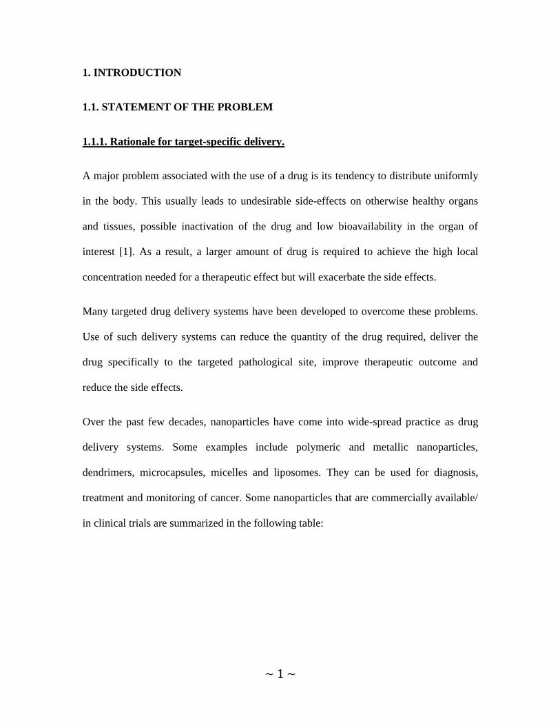
division is very high compared to the adjoining tissues and delivery of oxygen and other
nutrients is not adequate. To support its growth, tumor tissue recruits development of new
blood vessels from the already existing ones, a process called angiogenesis [6]. The
arrangement of these vessels is non-uniform and unorganized, with loops and
trifurcations in the vessels. Endothelial cell junction gaps of 100-700 nm diameter
increase the vascular leakiness [7], [8]. This „leaky vasculature‟ enables nanoparticles
such as micelles and liposomes with molecular weights of ≤ 40 KDa and sizes between
10-400 nm to extravasate into the tumor interstitium. This, in addition to the poor
lymphatic drainage found in the tumors, leads to retention of the nanoparticles in the
tumor, a phenomenon known as the „enhanced permeability and retention effect‟[9], [10].
Passive targeting of the drugs is thus possible with these nanoparticles (Figure 1).
Figure 1. Schematic of enhanced permeability and retention effect for passive targeting of
nanoparticles.
To accumulate optimally in the target area, micelles and liposomes should also circulate
for an extended period of time in comparison to the free drug [11]. To make these
nanoparticles long-circulating, coating them with PEG or “PEGylation” is necessary
Normal vasculature(Tight)
Tumor vasculature(Leaky)
Nanoparticles
Vascular cutoff size (100-700nm)

~ 4 ~
which can be achieved with PEG-PE, a conjugate of polyethylene glycol and
phosphatidylethanolamine [12],[13]. The hydrophilic PEG organizes a tight network of
water around the nanoparticle and prevents its recognition, uptake by macrophages and
clearance by the RES (reticulo-endothelial system) organs such as liver, spleen and
kidneys [14]. This “stealth” property imparted by the PEG makes the nanoparticle long-
circulating [15], [16] and results in a 10-100 fold increased tumor accumulation
compared to the non-PEGylated nanoparticle [11, 17-18]. Repeated passages through the
tumor vasculature increase the half-life of the drug in the circulation.
1.1.3. Active targeting of nanoparticles.
To “actively” target nanoparticles towards the tumor tissue and enhance the effects of
drugs delivered by these systems, several modalities can be attached to their outer
surface. Cell penetrating peptides (CPPs) increase the intracellular penetration of these
carriers [19]. However, these peptides are susceptible to enzymatic degradation and non-
specific cellular interactions [20]. In order to confine their function to the tumor cell
surface, CPPs can be shielded with a pH-sensitive polymer that degrades at the slightly
more acidic extracellular pH of tumor cells. To impart a tumor recognition capability,
various ligands and antibodies can be added to the surface of the nanoparticle [21-22].
In conclusion, an optimized multifunctional carrier should be long-circulating with
attached moieties that can actively target the tumor cells to improve the delivery of the
drug-containing carrier.

~ 5 ~
1.2. REVIEW OF THE LITERATURE.
1.2.1. Tumor physiology and lowered extracellular pH.
Tumor cells typically undergo rapid cell division which reduces the levels of oxygen and
other nutrients to inadequate levels. The presence of hypoxia and low nutrient levels
stimulates the cells to increase glycolysis for the production of ATP which leads to the
generation of elevated amounts of lactic acid [23], [4]. Thus, intracellular proton
concentration increases, and the affected cells respond by pumping out these excess
protons. The ion pumps/ ion exchangers/ proton pumps present on the membrane are
known to be particularly efficient in cancer cells and help in the removal of protons into
the extracellular space [24], [4]. Tumor vasculature is abnormal and the blood flow
distribution is therefore heterogeneous and relatively slow in some areas. This leads to
the variable accumulation of protons and metabolic acids in the extracellular space [5].
All these factors contribute to the lowered pH of the tumor extracellular space (pHe). The
range of pHe observed in tumors was 5.66-6.78 and depended upon the type of tumor and
its location [25].
1.2.2. Micelles and liposomes for drug delivery to tumors
Ideally, pharmaceutical nanocarriers should be cheap, easy to prepare, biodegradable,
biocompatible, non-toxic, non-immunogenic, small in size, high in drug loading capacity,
show prolonged circulation and accumulate in the desired site [26]. Micelles and

~ 6 ~
liposomes can fulfill these criteria and provide a surface that can be modified for
particular requirements of a pathological site.
Micelles are colloidal particles with a size in the range of 5-50 nm. Amphiphilic
molecules that make up micelles self-assemble spontaneously in an aqueous environment
where the hydrophobic fragment forms the core and hydrophilic fragment forms the
corona of the micelle [27]. Since the core of the micelle is hydrophobic, poorly water
soluble drugs can be encapsulated in the core and be protected from exposure to the
aqueous phase, thus enabling enhanced drug delivery of water-insoluble drugs [28].
Micelles made from diacyllipids conjugated to PEG are more stable than the conventional
amphiphilic polymeric micelles because of an increase in the hydrophobic interactions
between the lipid chains of the micelle core (Figure 2). This contributes to the
development of critical micelle concentration (CMC) values of 10-5
to 10-6
M for micelles
prepared from diacyllipid-PEG conjugates [29] which are significantly lower (at least
100-fold) than conventional plain micelles. A low CMC denotes that micelle integrity
will be maintained upon dilution in blood and hence will show good drug retention [30].
Modification of micelles with PEG makes them long-circulating and a high concentration
of drug can be maintained in the blood circulation for a longer period of time. This also
enhances drug targeting to areas with low blood supply or with low amount of surface
antigen where a longer circulation time is essential for the drug activity [30-31].
An important characteristic of micelles is their delivery of drugs to tumors with a smaller
vascular pore cut-off size when the larger liposomes are not able to deliver the drugs [32].

~ 7 ~
Figure 2. Self assembling PEG-PE micelles with (1) hydrophobic core, (2) hydrophilic corona,
(3) nonpolar drugs solubilized within the micelle core, (4) polar drugs adsorbed on micelle
surface, (5) substances with intermediate polarity distributed between the core and the corona
[30].
Liposomes are phospholipid nanoparticles with a size range of 100- 1000 nm and consist
of a lipid bilayer with an aqueous core (Figure 3). Hydrophilic compounds can be
entrapped in the core and hydrophobic compounds within the lipid bilayer, thus limiting
the pharmaceutical‟s side-effects and premature inactivation in the blood circulation [33].
They can also be made long-circulating by the incorporation of PEG chains on their
surface [34].
Liposomes and micelles can be modified with a number of targeting moieties, including
but not limited to antibodies and their Fab‟ fragments, proteins/ peptides, polysaccharides
and low-molecular weight ligands for specific recognition and binding to a targeted site.

~ 8 ~
Figure 3. Structure of liposome with (1) hydrophilic drug encapsulated in the aqueous core and
(2) hydrophobic drug entrapped in the lipid bilayer [30].
An example of successful clinical application of a PEG-coated liposome is Doxil ®, a
doxorubicin-loaded long-circulating PEGylated liposome. The half life of doxorubicin in
this formulation is approximately 45 hrs in humans as compared to 10 hrs for free
doxorubicin. Both, the clearance rate and the volume of distribution are low, with more
than 90% of the drug remaining encapsulated while in the circulation [35]. The side-
effects associated with free doxorubicin are strongly diminished which makes this
formulation clinically useful.
Liposomes modified with TAT-peptide on the surface enhance delivery of drugs and
genes in vivo [36] . Micelles can also be modified with cell-penetrating peptides. For
example, drug-loaded micelles with TATp attached to the surface improved delivery to
tumors and enhanced apoptosis of the tumor cells [37]. Monoclonal antibody 2C5/ 2G4
have been attached to liposomes and micelles to specifically target them to tumors and to
ischemic myocardium [11, 38-44]. Folate receptor-targeted liposomes encapsulating
anticancer drugs to target tumor cells have been prepared. pH-responsive micelles
encapsulating anticancer drugs have been formulated as well [45-47]. In response to the
low pH found in tumors, the pH-sensitive polymer degrades and the drug is released.

~ 9 ~
Various combinations of these targeting moieties can generate a multifunctional
nanoparticle.
In this study, in order to obtain an improved drug delivery to tumors, micelles and
liposomes have been developed with a cell-penetrating peptide, a pH-sensitive polymer
and monoclonal antibody 2C5 on their surface.
1.2.3. In vivo longevity of nanoparticles.
1.2.3.1. Importance of PEG coating.
As stated before, PEGylation of nanoparticles increases their circulation time by
preventing opsonization by macrophages and accumulation in RES organs. The other
important characteristics of such PEGs include low toxicity and immunogenicity, high
polymer chain flexibility and minimum interference with the biological properties of the
drug. Since it is possible to modify only one group of the PEG chain to either add ligands
or make any other conjugate, the resulting compound does not form cross-linked
aggregates [30]. PEG is available in various molecular weights and the length of the head
group can be selected according to specific needs.
Nanoparticles can be made long-circulating by addition of PEG since it remains stable in
the circulation [34]. It is removed unchanged by filtration via the kidneys.
Studies have shown that plain, non-PEGylated liposomes follow saturable and non-linear
kinetics whereas PEGylated liposomes follow dose-independent, non-saturable, log-
linear kinetics which makes them better candidates for drug delivery [48-49].

~ 10 ~
1.2.3.2. PEGylation of liposomes.
Theoretically, the amount of PEG-lipid able to incorporate into vesicles is directly
correlated with the membrane elasticity, in this case, the cholesterol content of the
bilayers and the PEG chain length [50]. Leroux and co-workers showed that increasing
the mol% of PEG (up to15 mol %) did not increase the carrier‟s size and did not
influence their removal from the bloodstream [51].
It has been observed that PEG attached to the liposome surface reduces van der Waals
forces of attraction and increases repulsive forces (steric and electrostatic) [52-53]. The
conformation of PEG chains on the liposomal surface depends on the PEG chain length
and on the density of PEG chains covering the nanoparticle surface [54] and [55]. A
study of the interactions between proteins and PEGylated surfaces shows that a high
surface PEGylation density is more important than the chain length in the prevention of
protein interactions with the surface. Grafted PEG can assume two conformations
(mushroom and brush) depending on the concentration of the surface PEG. At a low
density of PEG (up to 4 mol %), the PEG chains of the same length will acquire the
“mushroom” conformation. The same PEG molecules will have a “brush” conformation
if the density of PEG chains on the nanoparticle is above 4 mol%. The “mushroom”
conformation of PEGs results in contraction of PEG chains and increases the likelihood
of creating “gaps” in the PEG shell, while the movements of PEG in “brush”
configuration are more restricted. Previous experimental data indicates that a brush
conformation provides the most effective opsonin repulsion [56]. With more overlap of
the PEG chains and increasing PEG chain surface density, repulsion of proteins becomes

~ 11 ~
increasingly efficient [57]. Also, longer chains fill the gaps between less densely grafted
PEG chains [58].
Increasing the molar ratios of methoxy PEG: phospholipid to 15 mol %, increases the
circulation time of liposomes. However there is an upper limit to the amount of methoxy
PEG-lipids that can be incorporated into lipid bilayers. Above this, mixed micelles are
formed and the bilayer collapses [59].
Garbuzenko, et al have used scaling mathematical models to describe the effect of up to
25 mol% of PEG2k-DSPE in the presence or absence of cholesterol and the effect of
phosphatidylcholine saturation on the size and the lipid bilayer packing of large
unilamelar vesicles [60]. They concluded that when a matrix lipid (PC), cholesterol and
PEG2k-DSPE were combined, the optimal biological stability of the vesicles was
achieved at 7±2 mol % PEG-DSPE. Their results also clearly show that, with increased
PEG-DSPE mol% a decrease in the packing parameter of the vesicles is obtained.
However, the combination of phosphatidylcholine, cholesterol and grafted PEG
maintains the vesicle as a lipid bilayer for higher mol % of PEG (as seen for PC: PEG-
DSPC liposomes). These results correlate with the findings of Hristova and Needham,
who incorporated 15 mol% PEG5k-lipid in phospholipid/cholesterol vesicles [50].
Furthermore, Allen and co-workers [61] have described and characterized liposomal
formulations, containing DOPE/CHEMS with up to 20 mol% of mPEG. In order to
increase the molar ratio of mPEG-lipid on the liposomal surface, they added, on top of
the pre-formed vesicles already containing 10 mol% of mPEG-DSPE, an additional 10
mol% mPEG-DSPE in the form of micelles using the post-insertion technique.
Approximately 20 mol% mPEG-DSPE was present in the outer leaflet of the liposomes,

~ 12 ~
confirming that a high mol% of PEG is achievable [61]. While some investigators find
destabilization of lipid lamellar structures only after the incorporation of more than 30-40
mol% of mPEG-lipid, others have mentioned much lower amounts [59, 62-65]. An
explanation for this wide disparity of PEG mol% ranges is probably the different
carriers/techniques used for PEG surface incorporation.
1.2.3.3. Use of detachable PEG.
Despite the attractive properties of PEG, there are several drawbacks. Firstly, the
PEGylated nanoparticles can accumulate in the tumors but if the drug is not released from
this stable formulation at the required rate/amount; tumor cell killing is not possible.
Secondly, for a nanoparticle taken up by endocytosis, the PEG coat may prevent
endosomal escape and release of drug in the cytoplasm [30].
Therefore, detachable PEGs have been used as an alternative to avoid this problem. Here,
the chemistry has been developed in such a way that the PEG is stable in the circulation,
increases the half-life of the encapsulated drug and detaches from the nanoparticle just
before it enters the tumor cell to obtain controlled drug release [66].
pH- cleavable bonds are used in polymers to make them detachable when pH changes.
Typically, there is a drop in the pH from the normal value of 7.4 in systemic arterial
blood to 6 or lower in extracellular fluid of a tumor cell and in endosomes, infarcts and
inflammatory areas. Such a polymer is stable at normal pH but degrades at this low pH.

~ 13 ~
PEG-PE conjugates can be made pH-sensitive by introduction of cleavable bonds such as
vinyl-esters, double esters and hydrazones which are stable at pH 7.4 but degrade at
lower pH [67-68]. This destabilization may be due to swelling of the polymer, increase/
decrease in the permeability of the polymer, degradation of the pH-sensitive bond or
changes in the network of pH-sensitive bonds [69], [70].
Thus, different types of systems can be developed for drug delivery using pH-sensitive
polymers containing PEG:
i. The pH-sensitive conjugate of a long chain PEG is incorporated in a drug-loaded
nanoparticle. In this way, certain functions present on the surface of the nanoparticle are
kept „hidden‟ in the PEG coat and exposed only near the tumor cells where necessary.
ii. The drug is attached to the polymer by a pH-sensitive bond and, as the bond breaks,
the attached drug is released.
iii. The drug is encapsulated in the polymer and is released when the polymer degrades at
lower pH.
iv. The pH-sensitive polymers/ lipids undergo destabilization at the low pH of
endosomes, fuse with the endosomal membrane and cause endosomal leakage that
releases the encapsulated drug/DNA in the cytoplasm and prevents the lysosomal
degradation of the carrier‟s contents.

~ 14 ~
1.2.4. Ligands for active targeting
1.2.4.1. Anti-nucleosome monoclonal antibody, 2C5.
Nucleosomes are a complex of a DNA subunit and four pairs of histone proteins. They
are the repeating subunits of chromatin. Apoptically dying tumor cells release
nucleosomes which bind to the neighboring live tumor cells via their nucleosome-specific
receptors.
These nucleosomes are the antigens for the anti-nucleosome autoantibody, 2C5 [71-72].
It is a monoclonal antibody belonging to the class of antibodies of the IgG2a isotype. It
recognizes murine and human tumor cells through their surface-bound nucleosomes, with
a very low affinity for the normal cells [71]. It inhibits tumor growth of both murine and
human origin. Studies using mAb 2C5 alone demonstrated accumulation in various
unrelated human tumors in vivo with a tumor-cell to normal-cell ratio ranging from 2.7 to
13.4 indicating higher tumor accumulation compared to normal cells [73]. Prophylactic
and established tumor therapy studies have demonstrated significant tumor growth
suppression in nude mice.
This mAb 2C5 can be conjugated to the surface of micelles or liposomes via
incorporation of pre-formed 2C5-PEG-PE micelles [74]. In this method, pNP-PEG-PE
(PEG-PE with the activated para nitrophenyl carbonyl group) bind to the amino group of
mAb 2C5 to form 2C5-PEG-PE conjugate [74]. This conjugate forms micelles which are
then incorporated via a post-insertion technique into preformed micelles and liposomes.
This post-insertion technique has previously shown to yield a stable association of
~ 15 ~
proteins, peptides and other polymers with liposomes [75-76]. Approximately 70-80 mAb
2C5 molecules can be attached to a single liposome [22].
When used at a sub-therapeutic concentration, mAb 2C5 can be a targeting moiety for
recognizing tumor cells. When attached to the surface of micelles or liposomes, it
specifically targets the tumor cells and delivers drug locally, further minimizing non-
specific interactions. The target of mAb 2C5 can be found on various types of tumors and
is not restricted to a specific tumor antigen, which makes it a “universal” tumor-targeting
antibody [71-72].
Previous studies have reported preparation of 2C5-conjugated Doxil using a sub-
therapeutic amount of mAb 2C5 without any significant loss of doxorubicin [77]. This
preparation had the ability to recognize nucleosomes and bound 3 to 8-folds greater to
tumor cells in vitro compared to non-modified Doxil and IgG-Doxil (IgG was used as a
non-specific antibody, which as expected showed no preferential attachment with the
tumor cells). With both human and murine tumor cells, 5 to 8-folds higher IC50 values
were obtained compared to Doxil alone [38, 78].
Although 2C5-Doxil demonstrated a 3-fold higher tumor accumulation compared to
Doxil and IgG modified Doxil, an increase in clearance and reduction in half-life was
observed compared to plain Doxil [79]. This was because of the Fc-mediated uptake and
clearance by RES which occurs when the whole antibody e.g. 2C5 is attached to the
liposome as compared to the use of only Fab‟ portion of the antibody, where Fc-mediated
uptake is not possible. The circulation time of 2C5-Doxil was 16 hours while that for
Doxil was 21 hrs. However, 2C5-Doxil was still long-circulating, demonstrated
preferential accumulation in tumor and had a rapid therapeutic effect [79]. The
~ 16 ~
accumulation of 2C5-Doxil was less EPR-dependent and took fewer passages within 4-6
hr window and so resulted in a higher tumor accumulation. Marked reduction in tumor
volume was obtained within a week after the onset of therapy. Additionally, an enhanced
anti-cancer effect was obtained compared to the original, un-modified Doxil. 2C5-Doxil
also demonstrated significant therapeutic efficacy against intracranial human brain tumor
xenografts in nude mice [80].
Similarly, specific binding and a preferential accumulation of mAb 2C5-micelles was
observed in tumors developed in mice. A greater inhibition of tumor growth in mice was
observed with paclitaxel-loaded 2C5-micelles than with paclitaxel-loaded plain micelles
[81].
These studies provided background support for the development of multifunctional
liposomes with mAb 2C5 attached to the surface, the development of which will be
discussed in the following sections.
1.2.4.2. Tumor-specific ligands.
Tumor cells over express certain proteins and receptors, e.g. epidermal growth factor
receptor (EGFR), transferrin receptor (TfR), folate receptor (FR), vasoactive intestinal
peptide (VIP) receptor, integrins, matrix metalloproteinases (MMP), HER2 (Herceptin) to
name a few.
Fab‟ fragment of monoclonal antibody, C225 (Cetuximab®) or EMD72000
(Matuzumab®), which binds to EGFR was covalently linked to drug-loaded liposomes
and this preparation showed enhanced cell association and cytotoxicity compared to non-
targeted liposomes in EGFR over-expressing cancer cell line HCT116 [82]. Enhanced

~ 17 ~
therapeutic effect of these targeted, drug-loaded immuno-liposomes was demonstrated in
vivo in MDA-MB 468 tumor models developed in nude mice [83].
Transferrin and anti-transferrin antibody can be conjugated to drug-loaded nanoparticle
surface to impart tumor-specific targeting and enhanced cytotoxicity in transferrin over-
expressing cancer cells. Anti-Bcr-Abl siRNA/ asDNA loaded nanoparticles have shown
increased cell transfection and lowered cell viability in chronic myeloid leukemia cell
lines which express higher amounts of transferrin [21]. Tumor targeting via folate-
modified liposomes is an approach used to target tumor cells over-expressing folate
receptor. Doxorubicin and 5-fluorouracil loaded-liposomes targeting folate receptor have
shown enhanced in vitro and in vivo cytotoxicity [84]. Anti-HER2 antibody labeled Doxil
has shown enhanced anti-cancer effect compared to Doxil alone [85].
Similarly, reduction in tumor growth was observed in mice treated with MMP2-labeled
nanoparticles and nanoparticles modified with Fab‟ fragment of anti-β1-integrin antibody
[86-87] .
Many other tumor-specific ligands have been made use of for active targeting of the
nanoparticles towards tumor cells and obtain a better effect than the non-targeted ones.
The type of tumor to be treated plays an important role in the choice of the targeting
ligand.

~ 18 ~
1.2.4.3. Cell-penetrating peptides.
Cell penetrating peptides (CPPs) are short peptides which, when attached to cargoes can
transport them through the plasma membrane into the cells. These proteins contain
domains called protein transduction domains (PTD), which are mainly involved in the
transport. TAT peptide [88] [89], penetratins [90], transportan [91], amphiphilic model
peptide [92], signal sequence-based peptides [93] and synthetic polyarginines (e.g. Arg9)
are examples of CPPs. TAT peptide (TATp) is derived from the transactivator of
transcription (TAT) protein encoded by human immunodeficiency virus type I (HIV-I).
Residues 47-57 (YGRKKRRQRRR) constitute the PTD of this peptide [89].
The exact mechanism of entry of TATp is somewhat controversial, since there are many
studies that support various methods of internalization. One group showed that lipid-raft-
dependent endocytocis is the mode of entry of TATp [94]. According to another group,
the mode of entry of full length TAT protein is mediated by heparan sulfate
proteoglycans (HSPG), whereas short TAT peptide (TAT basic peptide) does not involve
binding to HSPG [95]. It has also been demonstrated that full length-TAT and
heterologous proteins fused to the transduction domain of TAT enter by a caveolae-
mediated pathway [96]. The mechanism of entry also seems to depend on the size of
cargo/ molecule attached to TATp [97]. When TATp is attached to small molecules,
electrostatic interactions promote cell penetration [98] [99], and when attached to larger
cargoes, the mode of entry is via macropinocytosis [100].
TATp conjugation to antisense oligonucleotides increased the delivery of these
oligonucleotides when compared to the unconjugated oligonucleotides [101]. PEGylated
nanoparticles (NLPs) encapsulating a plasmid DNA and with TATp attached to the

~ 19 ~
surface, had a significantly higher transfection than NLPs without the TATp moiety
[102]. Micelles [37], [103] and liposomes [104], [36] with attached TATp enhanced the
uptake as compared to their non-TATp conjugated counterparts. Levchenko, et al
demonstrated that TATp-liposome uptake was independent of temperature and the
localization of these liposomes occurred initially in the cytoplasm and moved later to the
perinuclear region [105]. TATp-liposomes when injected intratumorally, improved gene
delivery to intracranial human brain tumor xenografts in nude mice [106].
However, this peptide is unstable because of its composition which consists mainly of
positively charged amino acids (six arginines and two lysines). This composition makes it
highly susceptible to degradation by proteolytic enzymes. The sequence of TATp can be
cleaved at six sites by trypsin. Analysis of stability of TATp in plasma showed that TATp
is cleaved in human plasma due to plasma enzymes [107]. As a result, its potential as
mediator of cellular delivery is restricted [107],[108].
In the present studies, micelles were prepared with a short length PEG-PE, TAT peptide
conjugated to PEG1000 -PE and PEG2000-Hz-PE. Under these conditions, the TATp
conjugate remains shielded under normal pH conditions by the long pH-sensitive
polymer and it is exposed under acidic pH conditions, such as at a tumor, where the pH
sensitive bond is degraded. Thus, TAT peptide is protected from the proteolytic enzymes
in the circulatory system and, after being exposed at the target tissue, can exert its effect.
These studies represent examples where simple modifications with tumor targeting
ligands and cell-penetrating peptides provide a successful drug delivery system that can
improve the original formulation.

~ 20 ~
1.2.5. Multifunctional nanocarriers.
1.2.5.1. Concept of multifunctionality.
In response to the continued push to bring about a more effective therapeutic outcome,
proposals are being advanced to develop better drug delivery systems (DDS). An in vivo
benefit of such a nanocarrier is the ultimate goal of a DDS. Several DDS have shown
good effects clinically, but the challenge of overcoming unwanted side-effects remains.
To make a formulation better targeted to the appropriate pathological site, various
functional moieties can be attached to a nanocarrier for simultaneous or sequential but
rapid accumulation, and a therapeutic or diagnostic action.
In this context, the goal for such a micelle or liposome is a greater circulation half-life,
better tumor targeting, increased tumor accumulation, responsiveness to stimuli such as
low pH or high temperature, increased tumor cell penetration and marked suppression of
tumor growth compared to the non-modified formulation. In other words, a micelle or
liposome should be made multifunctional by addition of multiple functionalities to their
surface [109]. The active targeting moieties that have been mentioned before, namely
TATp and mAb 2C5 can be attached to micelles and/or liposomes to make them
multifunctional.
1.2.5.2. Development of multifunctional nanocarriers.
The development of multifunctional nanocarriers is a challenge because the individual
moieties having different functions must be attached in such a way that their individual

~ 21 ~
action is maintained and can be made useful in a controlled fashion. Engineering of the
moieties to make them act in a coordinated way can be accomplished by attaching them
to the micelle/ liposome surface by attachment of the targeting ligands via PEG spacer
arms of differing lengths. In this way, the ligand can be outside the PEG coat and
available for binding its target [30].
The distal terminal of PEG can be functionalized so that an activated functional group of
the ligand can be attached. The ligand-PEG-PE conjugate can be added during the
preparation of micelles or liposomes or via co-incubation of micelles formed from the
conjugate with the liposomes via post-insertion method as described before [74].
In the present studies, in a first step towards the development of multifunctional
nanocarriers, micelles and liposomes were developed with TATp attached to a short
length PEG-PE (Figure 4). The TATp exposed on the surface of the micelles and
liposomes promoted enhanced cell penetration. But since this peptide is non-specific and
prone to proteolytic degradation as studied in vitro by analytical and biological
techniques, in the second step, we have introduced another moiety, a shielding pH-
sensitive PEG.
Figure 4. Structure of a TATp-modified nanoparticle with TATp groups exposed on the particle
surface.

~ 22 ~
In this scheme, micelles containing PEG1000-PE, TAT peptide conjugated to PEG1000 -PE
and PEG2000-Hz-PE were prepared. While the TATp conjugate remained shielded by the
longer PEG2000-Hz-PE at physiological pH, it is „exposed‟ under acidic pH conditions,
characteristic of a tumor, where the pH sensitive bond is degraded. The pH-sensitive PEG
increases the circulation time and prevents proteolysis of the TATp but degrades at the
target site to expose the cell penetrating TAT peptide. When exposed at the target tissue,
TATp increases micelle penetration and delivers more drug to the tumor, thus inducing
increased cell killing compared to the plain micelles (Figure 5).
Figure 5. Schematic diagram depicting the shielding of TATp by the long PEG chains in the pH-
sensitive-TATp-micelle and the exposure of TATp as a result of the hydrolysis of hydrazone bond
of PEG-Hz-PE at acidic pH.

~ 23 ~
Finally, in order to develop a multifunctional liposome that shows better therapeutic
effect than the commercially available preparation, multifunctional Doxil® containing
TAT-peptide moieties, sterically shielded with a degradable pH-sensitive hydrazone bond
between long shielding PEG chains and PE (PEG2000-Hz-PE conjugate) was prepared
(Figure 6). The nucleosome-specific antibody (mAb 2C5), was attached to a longer PEG
chain (2C5-PEG3400-PE). During the first phase of delivery, PEGylated liposomes can
accumulate in the tumor via the EPR effect (a “passive” targeting effect) and additionally
in the tumor cells via the mAb 2C5 (“active” targeting). In the second phase of delivery,
within the acidified milieu (typical of solid tumors or ischemic tissues) these carriers can
lose their PEG coating by hydrolysis of a hydrazone-containing pH-sensitive polymer,
and penetrate inside the cells via the action of exposed TATp moieties. Thus, specific
tumor targeting can be achieved along with an improved drug delivery which helps to
achieve a better therapeutic effect than the commercial doxorubicin liposomes.

~ 24 ~
Figure 6. Schematic of the effect of low pH on TATp-modified pH-sensitive immunoliposomes
composed of a pH-degradable PEG2000-Hz-PE with a long PEG block, TATp-PEG1000-PE with a
short PEG block, and mAb2C5-PEG3400-PE.

~ 25 ~
1.2.6. Tumors and the drug resistance phenomenon.
Multidrug resistance (MDR) is a phenomenon whereby the tumor becomes resistant to
the anticancer drug and higher doses of drug are necessary for the same cytotoxic/
therapeutic effect. Various mechanisms implicated in this phenomenon include altered
metabolism of drug, changes in the amount/structure of drug targets present on the tumor
cells, increased DNA repair activity and reduction in drug accumulation due to reduced
uptake or increased drug efflux [110-111]. One of the major pathways for MDR is the
enhanced drug efflux caused by over expression of P-gp (P-glycoprotein).
P-gp belongs to ATP-binding cassette transporter superfamily and is encoded by a MDR1
gene [112]. Anticancer drugs such as doxorubicin and several other compounds are
removed from the cells by this efflux pump which leads to decreased cytotoxic activity of
the drug [111].
Approaches to overcome MDR have included use of:
1. PgP blockers/modulators which can inhibit the drug efflux [113-114].
2. Inhibitors of MDR gene expression [115-117].
3. Chemical modification of the drug [118-121].
4. Novel drug delivery systems such as nanoparticles [122-128].
In this case, encapsulation in nanoparticles has been used/ adapted to overcome drug
resistance (Figure 7).
Free drug enters a cell by diffusion. Being closer to the membrane where P-gp pumps are
located, it is spatially available for drug efflux. When encapsulated in a nanoparticle, the

~ 26 ~
drug undergoes endocytosis and the contents of the nanoparticle are released near the
nuclear region and hence the drug is less prone to efflux by P-gp [127, 129].
Figure 7. Schematic of P-gp efflux of drug and prevention of P-gp drug efflux by encapsulation in
a nanoparticle.
Other possible mechanisms by which these multifunctional nanocarriers can be expected
to overcome P-gp efflux are:
i. Prolonged circulation in the tumor can be provided by the PEG.
ii. Higher accumulation at the target cells can be obtained by the combined effect of mAb
2C5 and the cell penetrating peptide.
iii. mAb2C5-Doxil specifically internalized by tumor cells and can help in localization of
drug load inside the cells near the nucleus [38].
Based on these previous findings and proposed mechanisms, multifunctional liposomes
were developed for the treatment of multidrug resistant tumors.
P-gpDrug
Nucleus
P-gpMultifunctional Lipodox
Nucleus

~ 27 ~
2. OBJECTIVES AND SPECIFIC AIMS
OBJECTIVE:
To develop multifunctional nanocarriers for efficient tumor targeting and drug delivery
using
TATp
pH-sensitive polymer
mAb 2C5
SPECIFIC AIMS:
1. To study cleavage properties of TATp using trypsin as a model enzyme, its effect on
TATp-mediated cell association of micelles and liposomes and the effect produced upon
shielding of TATp.
2. To prepare and characterize TATp-modified pH-sensitive micelles containing
paclitaxel and study their cell targeting and cytotoxic effect on tumor cells in vitro and in
vivo.
3a. To develop and characterize doxorubicin loaded multifunctional liposomes modified
with TATp, pH-sensitive polymer and mAb 2C5 and to evaluate their cell targeting and
cytotoxic effect on tumor cells in vitro.
3b. To evaluate the cell targeting and cytotoxic effects of the multifunctional liposomes
on drug-sensitive and drug-resistant tumor cells in vitro and in vivo.

~ 28 ~
3. EXPERIMENTAL DESIGN AND METHODS
3.1. Materials.
Diacyllipid-polyethylene glycols (PEG750-PE, PEG1000-PE, PEG2000-PE, PEG5000-PE),
1,2-dipalmitoyl-sn-glycero-3-phosphothioethanol (DPPE-SH), 1,2-dioleoyl-sn-glycero-3-
phosphoethanolamine) (DOPE), NHS-PEG1000-Maleimide, fully hydrogenated soy
phosphatidylcholine (HSPC) and L-α-phosphatidylcholine (egg), were purchased from
Avanti polar lipids (Alabaster, AL). mPEG2000-SH was from Laysan Bio Inc, 4-(4-N-
Maleimidophenyl) butyric acid hydrazide hydrochloride (MPBH) was obtained from
Pierce Biotechnology Inc, and 4-acetyl phenyl maleimide was from Acros organics.
TATp (12 mer: CysTyrGlyArgLysLysArgArgGlnArgArgArg; mol. mass 1663 Da) was
synthesized by Tufts University Core Facility, Boston, MA. Trypsin from porcine
pancreas (Type IX-S, 13000-20000 units/mg) and trypsin-chymotrypsin inhibitor from
soybean were purchased from Sigma (St. Louis, MO). Triethylamine (TEA), 9-
Fluorenylmethyl chloroformate (FMOC-Cl) and cholesterol were purchased from Sigma-
Aldrich. Poly-oxyethylene3400-bis (p-nitrophenyl carbonate) [PEG (pNP)2] was
purchased from Laysan Bio Inc (Alab, Alabama). Fluoromount-G was from Southern
Biotechnology Associates Inc (Birmingham, AL), and Cell Titer Blue cell viability assay
kit was from Promega (Madison, WI). The mAb 2C5 was produced in ascites via I.P.
injection of 1.5×10⁶
hybridoma cells/ml into pristine primed 4 week old Balb/C male
mice. The production and the purification of the mAb 2C5 were carried out by Harlan
Bioproducts (Indiannapolis, IL) using the cell line from our laboratory. Control bovine
antibody IgG was obtained from MP Biomedicals LLC (Ohio, USA). Doxil®, a
commercially available preparation of doxorubicin in PEGylated liposomes (ALZA

~ 29 ~
Corp.), was purchased from Pharmaceutics Inc. (West Roxbury, MA). Lipodox ®, a
generic of Doxil was purchased from SunPharma, India. Trypsin from porcine pancreas
(type IX-S), chymotrypsin-trypsin inhibitor, heparinase-I from Flavobacterium
heparinum, and lyophilized human plasma (reconstituted with deionized water) were
purchased from Sigma-Aldrich (St. Louis, MO). Cell lines (B16-F10, HeLa, NIH 3T3,
MCF-7 and SKOV-3 drug-resistant and drug-sensitive) were purchased from the
American Type Culture Collection (Manassas, VA). All cell culture media, heat-
inactivated fetal bovine serum (FBS), and concentrated solutions of
penicillin/streptomycin stock solutions were from Cellgro® (Herndon, VA). All other
chemicals and solvents were of analytical grade, purchased from Fischer Scientific and
used without further purification. TATp-PEG1000-PE (TATp-conjugate), PEG2000-Hz-PE
(pH sensitive polymer) and pNP-PEG3400-PE were prepared in-house.
Methods:
3.2. TATp CLEAVAGE PROPERTIES AND SHIELDING METHODOLOGIES.
3.2.1. Synthesis of TATp-PEG1000-PE.
TATp-PEG1000-PE conjugate was synthesized as described previously [47] with some
modifications. Briefly, DOPE was reacted with ~ 1.5-fold molar excess of NHS-PEG
1000-maleimide by stirring for 2 h in chloroform at room temperature with a 3-fold molar
excess of triethylamine. A 2-fold molar excess of TATp-Cys was then added, and the
reaction was continued with stirring overnight. The solvent was evaporated, and the
product was freeze-dried overnight. The excess of TATp-Cys was separated from the
product by gel filtration chromatography. The solvent was evaporated, product was

~ 30 ~
freeze-dried overnight, and the dried product was dissolved in 0.5 mL of water and
loaded onto a Sephadex G25 column (length, 10–15 cm; diameter, 0.5–1 cm). Fractions
were collected and monitored by TLC using silica plates (mobile phase of
chloroform/methanol 80:20% v/v), and TATp-PEG-PE was visualized with
phosphomolybdic acid and Dragendorff spray reagents.
3.2.2. Preparation of TATp-modified micelles (TM) and TATp-modified liposomes
(TL).
A lipid film was made by mixing chloroform solutions of PEG1000-PE and TATp-
PEG1000-PE in the mole ratio 95:5. When necessary, 1 mol % of the fluorescent probe,
dipalmitoyl phosphatidylethanolamine (lissamine-rhodamine B) (Rh-PE) was added. The
final concentration of the lipids was 3.3 mg/ml. This dried film was suspended in
phosphate buffered saline, PBS, pH 7.4. The hydrated mixture was vortexed for 5 min.
TM form spontaneously. TATp liposomes were prepared by hydrating a dried lipid film
of egg phosphatidylcholine, cholesterol and TATp-PEG1000-PE in the mole ratio 65:30:5.
This film was lyophilized overnight and hydrated with PBS, pH 7.4. The solution was
extruded 21 times through polycarbonate filters of size 200 nm, using a hand-held
extruder (Avanti, Alabaster, AL). The final concentration of the lipids was 2.5 mg/ml.
3.2.3. Characterization and determination of cleavage of TATp in TM and TL.
The micelles and liposomes were divided into three groups, the first group was treated
with trypsin (0.1mg/ml), the second group was treated with a mixture of trypsin+ trypsin

~ 31 ~
inhibitor and the third group was kept as is. All the solutions were incubated at 370C for
10 min. These formulations were characterized by dynamic light scattering (DLS) using a
N4 submicron particle system (Coulter Corporation, Fl) and Zeta Phase Analysis Light
Scattering (PALS) using a Zeta potential analyzer (Brookhaven, Holtsville, NY) .
The cleavage of TATp was followed by HPLC using the proteolytic enzyme, trypsin. An
aliquot of 10 µl was removed from micelles or liposomes. One µl of trypsin solution in
PBS, pH 7.4 (1mg/ml) was added to this aliquot to get a final trypsin concentration of 0.1
mg/ml. This solution was incubated at 37 0
C for 10 min, following which, 1 µl of borate
buffer pH 10.0 and FMOC-chloride solution (0.4 mg/ml) were added. This mixture was
kept at room temperature for a further 10 min. It was diluted with 400 µl water and
analyzed by following the RR fragment resulting from trypsinolysis of TATp using the
HPLC method already established in the lab [107].
3.2.4. Cell interaction studies.
Fluorescence microscopy studies:
Murine melanoma (B16F10) and human cervical cancer (HeLa) cells were grown at
370C, 5% CO2 using Dulbecco‟s modified eagle‟s medium (DMEM) supplemented with
10 % v/v Fetal Bovine Serum and 1% v/v antibiotics. These cells were grown in 6 well
plates containing a sterile coverslip in each well to about 70% confluency. The
formulations prepared were Rh-PE labeled plain micelles or liposomes and Rh-PE
labeled TATp-micelles or TATp-liposomes. Two solutions of trypsin (2mg/ml each)
were prepared in PBS, pH 7.4. One solution was incubated with trypsin- chymotrypsin
inhibitor (20 mg/ml) in 1:1 v/v ratio and another solution was incubated with PBS 1:1 v/v

~ 32 ~
ratio, at 37 0C for one hour, so as to have both the trypsin solutions in the same
concentration and in the same experimental conditions. Trypsin solution without the
inhibitor was added to one of the micelle/liposomal formulations. The final concentration
of trypsin on the micelles/liposomes was 0.1 mg/ml. Trypsin solution incubated with
inhibitor was added to another micelle/liposome solution (with the final concentration of
inhibitor 1 mg/ml and trypsin concentration 0.1mg/ml). These formulations were
incubated for one hour at 37 0
C, for maximum action of trypsin on TATp containing
formulations. A third sample of TAT micelles/ liposomes was kept as is. The plain
micelle/liposome sample was also left untreated. After one hour, the samples were added
to the respective wells of the 6 well plates, in serum free media, so that the final
concentration of lipids was 0.3 mg/ml. HOECHST 33342 was used to stain the cells at a
concentration of 5 µg/ml for 15 min. The media was then removed, cells were washed
and the coverslips were mounted cell side down on glass slides using the Fluoromount-G
(Southern Biotechnology Associates, Birmingham, AL) mounting media and observed
using a Nikon Eclipse E400 microscope under epifluorescence with a Rhodamine/TRITC
filter [47].
FACS analysis:
For FACS analysis, the micelles/liposomes were treated in a similar way with the trypsin/
inhibitor solutions. They were then added to the respective wells in 6-well plate and
incubated for one hour at 370
C. The media was removed; the cells were washed with
PBS. They were harvested using a 0.25% trypsin, 0.1% EDTA solution, centrifuged at
1000 rpm and the supernatant was removed. The pellet was resuspended in cold PBS, pH

~ 33 ~
7.4. The cellular uptake/attachment efficiency was determined by FACScan™ (Beckton
Dickinson biosciences, San Jose, CA) acquiring 10,000 events per sample.
3.2.5. In vitro cytotoxicity studies with TATp liposomes.
For these studies, Doxil ®, a commercially available doxorubicin loaded formulation was
modified with TATp by using post-insertion method, where in brief, TATp-PEG1000-PE
micelles were prepared and incubated with Doxil overnight for complete incorporation of
the phosphatidylethanolamine part of micelles in the liposomes. The cytotoxicity of these
TATp-modified liposomes, loaded with doxorubicin (TATp-Doxil), against B16-F10 and
HeLa cells was studied. Liposomal formulations were pre-treated with trypsin (0.1
mg/ml) or trypsin (0.1 mg/ml) with trypsin inhibitor (1 mg/ml) for 1 h before addition to
cells. Cells were grown in DMEM (10% FBS) and transferred into 96-well microplates at
a density of 5 × 10³ cells/well. Following 24 h incubation and growth to ~ 50%
confluency, trypsin-treated formulations containing doxorubicin were added to cells in
triplicate to give the final concentration of 5 μg/ml. After 24 h incubation at 37°C, 5%
CO2, plates were washed three times with PBS, followed by the addition of 20 μl/well of
CellTiter Blue solution to assess viability. After 1 h incubation at 37°C, 5% CO2, the cell
survival was estimated by measuring the fluorescence intensity using a microplate reader
(Synergy HT multimode microplate reader, BioTek Instrument, Winooski, VT) with
525/590 nm excitation/emission wavelengths.

~ 34 ~
3.2.6. Preparation and characterization of shielded, non pH-sensitive micelles.
A lipid film was made by mixing chloroform solution of PEG750-PE as the main
component, with 5 mol % TATp-PEG1000-PE. This formulation was the non-shielded
formulation. To prepare shielded formulations, increasing mol % of PEG5000-PE or
PEG2000-PE ranging from 0 % to 95 % were added to the above micelles to gradually
replace PEG750-PE, at the same time keeping the molar concentration of the lipids
constant. This dried film was suspended in phosphate buffered saline, PBS, pH 7.4. The
hydrated mixture was vortexed for 5 min. Micelles formed spontaneously. We analyzed
the cleavage of TATp Arg-Arg groups and the effect of increasing PEGylation by
fluorenylmethyl chloroformate (FMOC) labeling using fluorescence detection with
HPLC.

~ 35 ~
3.3. TATp MODIFIED pH-SENSITIVE MICELLES.
3.3.1. Synthesis of hydrolysable PEG conjugate (PEG2000-Hz-PE).
An aldehyde-derived hydrazone-based PEG2k-Hz-PE conjugate (pH-sensitive conjugate)
was synthesized by a two step method as previously described [130] with modifications.
For step I (synthesis of acyl hydrazide-PEG derivative) 40 μmol of mPEG-SH in
chloroform were mixed with two molar excess of the acyl hydrazide cross-linker MPBH
in the presence of five molar excess of triethylamine over lipid. Following 2hr stirring at
room temperature, the product was dialyzed (Spectra/Por 6 dialysis membrane, MWCO
1K, Spectrum Laboratories, Rancho Dominguez, CA) against deionized water for two
hours, analyzed by TLC, freeze-dried and stored as chloroform solution at -800C. For step
II of the synthesis (activation of phospholipid with 4-acetyl phenyl maleimide) forty
micromoles of 4-acetyl phenyl maleimide were reacted with 27 mmol of 1, 2-
dipalmitoyl-sn-glycero-3-phosphothioethanolamine (DPPE-SH) in presence of
triethylamine overnight with continuous stirring. The activated phospholipid was
separated on a silica gel column using chloroform: methanol mobile phase (9:1 v/v). The
fractions containing the product were identified by TLC analysis, pooled, concentrated,
freeze-dried and stored as chloroform solution at -800C. The final product, PEG2000-Hz-
PE, was synthesized by reacting the hydrazide activated PEG derivative with 1.5M
excess of the activated phospholipid overnight under the constant stirring at room
temperature. The conjugate was separated and purified by size exclusion gel
chromatography using Sepharose-CL4B media.
~ 36 ~
3.3.2. Preparation and characterization of TATp-modified pH-sensitive micelles.
TATp-modified pH-sensitive micelles were prepared by mixing PEG2000-Hz-PE,
PEG1000-PE and TAT-PEG1000-PE in the mol % ratio of 77.5:20:2.5. When necessary,
1mol % of Rh-PE and/or Paclitaxel (1.5 wt %) was added to the lipid mixture. The
organic solvent was evaporated under argon and a lipid film was formed which was
freeze-dried overnight. This film was hydrated with PBS, pH 7.4 and divided into two
groups- one was at pH 7.4 and to the other group, 0.1N HCl was added to adjust the pH
to 5.0. These conditions were maintained for 1-2 hrs at 370
C. The particle size and zeta
potential were analyzed.
3.3.3. Determination of kinetics of hydrolysis of hydrazone bond and TATp in
TATp-modified pH-sensitive-micelles.
Micelles were prepared as mentioned above. From the samples incubated at pH 5.0, a 10
μL aliquot was removed immediately, to which 1 μL of phosphate buffer pH 9.5 and 1 μl
of trypsin (0.01 mg/ml) was added. After incubating this mixture at 370
C for 10 min, the
samples were subjected to FMOC-Cl derivatization [107] . This mixture was diluted with
400 μL of deionized water and subjected to HPLC analysis [107]. Aliquots were removed
after 15 min, half hour and one hour, treated in the same way as described above and
analyzed by HPLC. The kinetics of the hydrolysis of PEG-Hz-PE bond and exposure of
TATp (present in the pH sensitive micelles) to trypsinolysis, followed the same pattern.
An aliquot of 10 μL was removed from the sample incubated at pH 7.4, processed and
analyzed in the same way as mentioned above.

~ 37 ~
3.3.4. Cell interaction studies.
Fluorescence microscopy:
B16F10 (murine melanoma) cells and HeLa (human cervical cancer) cells were grown on
coverslips in 6-well plates in DMEM supplemented with 10% fetal bovine serum to 70%
confluency. Rhodamine PE- labeled micelles were prepared. One sample was incubated
at pH 5.0 and the other at pH 7.4 at 370C for one hour. The media was replaced by serum-
free media, and the cells were treated with these preincubated micelles (2 ml/well, 0.3 mg
total lipid/ml). After a 1 h incubation period, the samples were processed as described
above and observed for differences in fluorescence between samples preincubated at
acidic and at neutral pH, under Nikon Eclipse E400 microscopy with fluorescence [47].
FACS analysis:
For FACS analysis, B16F10 and HeLa cells were grown to 70% confluency in 6-well
plates in DMEM (with 10% fetal bovine serum).Samples were processed in the same way
as above and added to serum-free media in the respective wells. After one hour, the
medium was removed and the cells were washed three times with medium. The cells
were processed for FACS analysis as mentioned previously and analyzed by flow
cytometric analysis.
~ 38 ~
3.3.5. In vitro cell viability studies.
TATp-modified pH-sensitive micelles containing paclitaxel were prepared. B16F10 cells
were grown in DMEM (10% FBS) and transferred to a 96-well plate (1000 cells/well).
After 24 hr incubation in 5% CO2 at 370C, cells were treated with TATp-modified pH-
sensitive micelles preincubated for 1 hour at pH 5.0 and pH 7.4, and kept for 72 hours in
DMEM (with 10% FBS).The media was removed, the wells were washed with fresh
media, replaced with 100 μL media and 20 μL Cell Titer Blue (Promega, Madison, WI)
was added to each well. Cell viability was evaluated after 1.5 hours of incubation at 370C,
5% CO2 by measuring the fluorescence produced by resorufin at 550ex/590em using
Synergy HT multidetection microplate reader (Biotek, Winooski, VT)
3.3.6. In vivo studies- Determination of apoptosis by TUNEL assay.
Female C57/BL6 mice (Charles River Laboratories, Wilmington, MA) were used for the
experiments. A protocol approved by Northeastern University Institutional Animal Care
and Use Committee in accordance with “Principles of Laboratory Animal Care” (NIH
publication # 85-23, revised in 1985) was followed. Mice were divided into three groups
with 3 animals in each group. Approximately, 1 x 10⁶
B16F10 cells were injected
subcutaneously over the left flank. Tumor growth, which is expected to be optimal in size
after about 15 days of inoculation, was checked regularly by palpation. After the tumor
reached a size of 0.5 cm3, mice were injected intratumorally at three sites with either
paclitaxel solution in methanol, TATp-micelles loaded with paclitaxel and TATp-
modified pH-sensitive micelles loaded with paclitaxel at a dose of 1mg paclitaxel/kg.

~ 39 ~
Tumors were harvested after 72 hrs and fixed in freezing media. Tumor sections were
stained for a terminal Deoxynucleotidyl Transferase Biotin-dUTP Nick End Labeling
(TUNEL) assay. Apoptotic cells produced on treatment with these various micelle
formulations were detected under fluorescence microscope.

~ 40 ~
3.4. MULTIFUNCTIONAL TATp-CONTAINING, pH-SENSITIVE, mAb 2C5-
MODIFIED LIPOSOMES
3.4.1. Synthesis of pNP-PEG3400-PE and 2C5 mAb modification.
In order to attach mAb 2C5 to the liposomal preparations, we first conjugated the mAb to
the distal ends of PEG blocks via p-nitrophenylcarbonyl (pNP) groups (using a pNP-
PEG3.4k-PE conjugate) to form immuno-micelles. Using the post-insertion method of
micelles [72-74], [93-94], [111] we were able to form ligand-coupled liposomes (will be
discussed below).
First, we synthesized and purified pNP-PEG3400-PE according to an established method in
our laboratory as previously described [74]. Briefly, the synthesis includes the interaction
of PE with a 10-fold molar excess of PEG 3400-(pNP)2 in chloroform in the presence of
triethylamine. Organic solvents were removed, pNP-PEG3400-PE micelles were formed
and separated from free PEG and pNP on a CL-4B column. pNP- PEG3400 -PE product
was freeze-dried, extracted with chloroform and stored at -80°C.
For antibody conjugates with PEG3400-PE (mAb 2C5 or non-specific IgG), 40 molar
excess of pNP-PEG3400-PE dispersed in 10 mg/mL micellar solution in 5 mM Na-citrate,
150 mM NaCl, pH 5.0, was added to an equal volume of a 1 mg/mL solution of mAb
2C5/IgG in 100mM Tris-buffered saline (TBS), pH 8.5. The mixture was incubated with
stirring at pH 8.5 overnight at 4 °C.
~ 41 ~
3.4.2. Preparation of multifunctional liposomes.
A formulation, mimicking the commercially available Doxil ®, was made using
chloroform solutions of fully hydrogenated soy Phosphatidylcholine (9.58 mg/ml),
cholesterol (3.19 mg/ml) and N-(carbonyl-methoxypolyethylene glycol 2000)-1,2-
distearoyl-sn-glycero-3-phosphoethanolamine sodium salt (mPEG-DSPE, 3.19 mg/mL)
(HSPC-Chol-PEG). These lipid components and their concentrations were similar to
Doxil ®.
Rhodamine-PE (1 mol %) was incorporated for fluorescence microscopy experiments. A
lipid film was prepared by evaporating the chloroform under N2 gas, hydrated using PBS,
pH 7.4 and vortexed for 5 minutes. The contents were then subjected to a sonication
cycle of 11 W for 30 min using a probe sonicator to form liposomes of a uniform size
distribution.
Due to shortage of the Doxil® supply, a generic, Lipodox® was used after physico-
chemical, cytotoxicity and cell interaction studies confirmed that its activity was similar
to Doxil.
Post-insertion technique was used to incorporate into HSPC-Chol-PEG/ Doxil ®/
Lipodox ® liposomes, a variety of polymers/polymer conjugates. Basically, micelles
composed of a combination of one or more of the following polymers were prepared:
TATp-PEG1000-PE (2. 5 mol %), PEG 2000-Hz-PE (15 mol %), 2C5-PEG3400-PE (2.5 mol
%), IgG-PEG3400-PE (2.5 mol %) and Rhodamine-PE (1 mol %). They were incubated
with the above liposomes overnight. They were subsequently dialyzed overnight against
DI water, using a Spectra Por dialysis membrane with a cutoff of 300KDa.

~ 42 ~
3.4.3. Determination of the optimum shielding of TATp with long chain PEG2000-Hz-
PE.
Liposomes containing HSPC: cholesterol: PEG2000-PE/ Doxil ® / Lipodox ® were
incubated with micelles containing 2.5 mol% TATp-PEG1000-PE and PEG2000-Hz-PE
(2.5-15 mol %) overnight. They were then analyzed for their particle size and zeta
potential. In order to confirm that the effect on zeta potential was due to the shielding of
TATp and not the effect of PEG2000-Hz-PE itself, liposomes were prepared without
TATp-PEG1000-PE but containing PEG2000-Hz-PE (2.5-15mol%). The optimal
formulation selected contained TATp-PEG1000-PE: PEG2000-Hz-PE: 2C5-PEG3400-PE
(2.5:15:2.5) molar ratio added to base liposomes composed of HSPC-Chol-PEG/ Doxil
®/ Lipodox ®.
3.4.4. Characterization of liposomes.
3.4.4.1. Measurement of particle size and zeta potential.
Liposomes were analyzed for their particle size distribution by dynamic light scattering
(DLS) using a Coulter® N4-Plus Submicron Particle Sizer (Coulter Corporation, Miami,
FL) and by transmission electron microscopy (TEM) (Jeol, JEM-1010, Tokyo, Japan).
Zeta potentials of various liposomal formulations were analyzed using 90 PLUS particle
size analyzer with ZETA PALS (Phase Analysis Light Scattering) System, Brookhaven
Corp. (Holtsville, NY) at 25 0C.

~ 43 ~
3.4.4.2. Specific activity of mAb 2C5 on liposomal surface by ELISA.
The preservation of specific activity of the monoclonal antibody 2C5 attached to the
surface of the liposomes using PEG3400-PE as the linker was determined using indirect
ELISA assay. Calf thymus nucleohistones (Worthington Biochemical, Lakewood, USA)
were used as the binding substrate/antigen. Goat anti-mouse IgG antibody linked to HRP
(horse radish peroxidase) (ICN Biomedical, Aurora, USA) was used as the secondary
antibody. The intensity of the color developed on addition of the substrate, K-blue TMB
peroxidase, was read at a dual wavelength of 620 nm with the reference filter set at 492
nm using a Labsystems Multiscan MCC/340 microplate reader installed with Genesis-
lite.
3.4.5. Cell interaction studies.
Fluorescence microscopy studies:
Human breast cancer (MCF-7) cells, normal human fibroblasts (NIH 3T3) , human
ovarian cancer cells (SKOV3- drug-resistant and drug-sensitive) and human cervical
cancer (HeLa) cells were grown at 370C and 5% CO2 in Dulbecco‟s modified eagle‟s
medium DMEM containing 10% v/v FBS and 1% v/v antibiotics. These cells were grown
in 6 well plates containing a sterile coverslip in each well to about 70% confluency.
Multifunctional liposomes labeled with 1 mol % rhodamine-PE (preincubated for 30
minutes at pH 5 and pH 7.4) were incubated with the cells for one hour at 37 0C.
Hoechst nuclear stain (2.5μg/ml) was added for the last 30 minutes of incubation. Cells
were washed three times with PBS and mounted individually cell-side down on glass
slides using fluorescence-free glycerol based mounting medium (Fluoromount-G). All
~ 44 ~
mounted slides were studied with a Nikon Eclipse E400 microscope under fluorescence
using a Rh/TRITC filter for Rh-labeled liposomes (red) and a UV-2B filter for Hoechst
labeled nuclei (blue).
Fluorescence activated cell sorting (FACS) analysis:
Quantitative analysis of the interaction of multifunctional liposomes with the
aforementioned cell lines was done using FACS. Cells were grown in 6 well plates to
70% confluency at 37 0C and 5% CO2. The media was then replaced with fresh medium
and multifunctional Doxil®/ Lipodox ® pre-incubated for 60 minutes at pH 5 and/or pH
7.4 were added to the cells. The required control liposomes were also added to the
respective wells. The final concentration of the lipids was 0.3 mg/ml for each
formulation. After one hour, the formulation was removed; the cells washed with PBS,
pH 7.4, and detached using trypsin (0.25%). They were then washed with cold PBS, pH
7.4, and centrifuged to form a pellet. The pellet was resuspended in cold PBS and the cell
interaction was determined by FACScan™ (Beckton Dickinson biosciences, San Jose,
CA) acquiring 10,000 events per histogram.
3.4.6. In vitro cell viability studies.
The cytotoxicity of Doxil/Lipodox-modified multifunctional immunoliposomes against
SKOV3 (drug-resistant and drug-sensitive cells) and HeLa cells was studied.
3×10⁴ cells/well cells were seeded into 96 well plates. Multifunctional immuno-Doxil
formulations were pre-incubated at pH 5 and/or 7.4 for 60 minutes prior to their addition
to cells. After 12 hr incubation at 37°C, 5% CO2, plates were washed three times with
fresh media, and cells were placed in the incubator with fresh media for additional 48

~ 45 ~
hours. Finally, cytotoxicity of the various formulations was evaluated by the addition of
20μl/well CellTiter Blue solution to 100 μl fresh medium to assess the cell viability. After
1 hr incubation at 37°C, 5% CO2, the cell survival was estimated by measuring the
fluorescence intensity using a microplate reader (Synergy HT multimode microplate
reader, BioTek Instrument, Winooski, VT) with 525/590 nm excitation/emission
wavelengths.
3.4.7. Determination of P-gp levels in SKOV3 drug-resistant and sensitive cells.
SKOV-3 drug-resistant and sensitive cells were treated with FITC labeled P-gp specific
antibody (UIC2). Both cells were grown in T75 flask to 80 % confluency. A pellet was
obtained and resuspended in PBS, pH 7.4 to get 300,000 cells/ 100 µl. They were
incubated with 5 µl of FITC labeled P-gp specific antibody (UIC2) for 1 hr on ice. The
cells were washed one hour later with cold PBS and analyzed by FACS technique using
the green fluorescence intensity (FL1) filter.
3.4.8. In vivo studies of multifunctional liposomes in SKOV-3 drug-resistant and
sensitive tumors
Female nu/nu mice (n= 50) purchased from Charles River Laboratories were equally
divided into two groups- 25 mice for inoculation with SKOV3 drug-resistant and 25 with
drug-sensitive cells. They were inoculated subcutaneously in the left flank with
approximately 4 x 10 6 SKOV-3 (sensitive) cells or 7 x 10
6 cells (resistant) cells mixed
in a 1:1 v/v ratio with Matrigel HC [126, 131], according to the protocol approved by the
Institutional Animal Care and Use Committee at Northeastern University. When tumors
reached a volume of approximately 150 mm3, 14 days after tumor inoculation, mice in
each group were sorted randomly into 5 sub-groups for treatment with either: Saline,

~ 46 ~
plain-Lipodox, Lipodox-TATp, Lipodox-2C5 and Multifunctional Lipodox. Tumor
volumes were measured every alternate day using vernier calipers and the percent change
in tumor volume was calculated by the formula: (Vn-V0)/V0 *100, where Vn is the mean
volume on day „n‟ following the treatment and V0 is the mean volume on day „0‟
(beginning of the experiment). Twenty days after the first injection, when saline-
administered tumors reached a mean tumor volume of 1000 mm3, mice were sacrificed
by CO2 and tumors excised. Tumor weights were measured at the end of the study after
excision. Mouse body weight was taken every three days to monitor the safety of the
formulations.
The excised tumors were fixed in freezing media and stored at -80 0C. Approximately 8
µm thick cryostat sections were made and the sections were stained for a terminal
Deoxynucleotidyl Transferase Biotin-dUTP Nick End Labeling (TUNEL) assay.
Apoptotic cells produced on treatment with these various micelle formulations were
detected under fluorescence microscope.
In previous studies by our group, a dose of 2 mg/kg of doxorubicin given every 5 days for
a total 4 doses, equivalent to a total dose of 8 mg/kg, was used to evaluate the efficacy of
mAb 2C5 –Doxil [38, 42]. Other groups have reported effects at higher doses equivalent
to 18 mg/kg. However, the dose rate used previously in the lab corresponds to 6 half-lives
of Doxil and 7 half-lives of mAb 2C5- Doxil. For our in vivo studies that involve
Lipodox (a generic of Doxil), mAb 2C5-Lipodox, TATp-Lipodox and multifunctional
immuno-Lipodox, this sub-therapeutic dose (8 mg/kg i.e. 2mg/kg/ 5days /4 times) was
chosen so that statistically significant differences between various formulations could be
evaluated.

~ 47 ~
4. RESULTS AND DISCUSSION
4.1. TATp CLEAVAGE PROPERTIES AND SHIELDING METHODOLOGIES.
4.1.1 Preparation and characterization of TATp micelles and TATp liposomes.
The results of the zeta potential measurements are presented in Table 2, which shows the
effect of trypsin on zeta potential values of TATp-modified liposomes/micelles. Our
results suggest that the positive charge of the TATp-modified particles shifts to neutral
following the incubation with 0.1 mg/ml of trypsin for 10 min at 37°C. Since TATp is
rich in arginine, which contributes to its cationic properties, it is also the main contributor
to the total positive charge of both micelles and liposomes. The decrease in zeta potential
values from −6.1 ± 2.7 mV to −18.4 ± 2.8 mV for TATp-modified micelles and from
4.3± 2.7 mV to −15.9± 2.0 mV for TATp-modified liposomes supports our conclusion
that TATp was cleaved by trypsin. When particles were incubated simultaneously with
trypsin and trypsin inhibitor, the positive charge of TATp-modified particles was
retained, confirming that trypsinolysis resulted in TATp cleavage and charge decrease.
Particle size analysis of both micelles and liposomes showed no significant changes
following incubation with trypsin, retaining a magnitude of 20 nm and 100 nm for
micelles and liposomes, respectively. Thus, TATp degradation does not affect the size
characteristics of the peptide-modified particles.

~ 48 ~
Table 2 Zeta Potential of TATp-modified micelles and liposomes (Values are mean± SD, n=5, p
<0.05 as calculated by Student’s T-test and one way ANOVA)
4.1.2 Determination of cleavage properties of TATp in TM and TL
TATp undergoes cleavage at its carboxyl terminus, on treatment with trypsin and the first
and the most intense proteolysis fragment formed is the arginine- arginine (RR) dimer,
(YGRKKRRQR↓RR) which can be followed using HPLC. In this experiment, TM and
TL were treated with trypsin. This was followed by incubation with FMOC-chloride, a
fluorescent label. A chromatographic peak of FMOC-Cl labeled RR dimer was obtained
at 1.5 minutes. Figure 8 is a representative of the RR peak obtained using the HPLC
analysis.

~ 49 ~
Figure 8. RR fragment obtained on treatment of TATp with trypsin, as analyzed by HPLC. The
retention time is 1.5 min.
4.1.3 Cell interaction studies with Fluorescence microscopy and FACS analysis
Trypsin was used to confirm that when TATp is attached to the surface of micelles or
liposomes, TATp-to-cell interaction is influenced by TATp exposure to proteolytic
enzymes. TATp-modified micelles and liposomes, treated with trypsin for 1 h, were
analyzed for their cell interaction capabilities by fluorescence microscopy and FACS
techniques.
TATp-modified liposomes were tested for their interaction with cells and for the effect of
trypsin on this interaction. Fluorescence microscopy showed that rhodamine-labeled
TATp-modified liposomes were significantly internalized by HeLa and B16-F10 cells
(Figure 9). Diminished cell internalization was observed following the pre-incubation of
formulations with trypsin (0.1 mg/ml for 1 h). When trypsin inhibitor (1 mg/ml) was
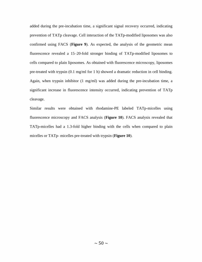
~ 50 ~
added during the pre-incubation time, a significant signal recovery occurred, indicating
prevention of TATp cleavage. Cell interaction of the TATp-modified liposomes was also
confirmed using FACS (Figure 9). As expected, the analysis of the geometric mean
fluorescence revealed a 15–20-fold stronger binding of TATp-modified liposomes to
cells compared to plain liposomes. As obtained with fluorescence microscopy, liposomes
pre-treated with trypsin (0.1 mg/ml for 1 h) showed a dramatic reduction in cell binding.
Again, when trypsin inhibitor (1 mg/ml) was added during the pre-incubation time, a
significant increase in fluorescence intensity occurred, indicating prevention of TATp
cleavage.
Similar results were obtained with rhodamine-PE labeled TATp-micelles using
fluorescence microscopy and FACS analysis (Figure 10). FACS analysis revealed that
TATp-micelles had a 1.3-fold higher binding with the cells when compared to plain
micelles or TATp- micelles pre-treated with trypsin (Figure 10).
~ 51 ~
Figure 9. Fluorescence microscopy (upper panel) and flow cytometry analysis (lower panel)
showing interaction of rhodamine-PE labeled TATp- liposomes upon pretreatment with trypsin /
trypsin inhibitor (0.1 mg/ml and 1 mg/ml respectively) for 1 hr at 370C followed by incubation
with B16F10 and HeLa cells for 1 hr. (*p≤ 0.05 compared to plain liposomes and TATp-
liposomes pre-treated with trypsin, n=3, mean ± SD)

~ 52 ~
Figure 10. Fluorescence microscopy (left panel) and flow cytometry analysis (right panel)
showing interaction of rhodamine-PE labeled TATp- micelles upon pretreatment with trypsin /
trypsin inhibitor (0.1 mg/ml and 1 mg/ml respectively) for 1 hr at 370C followed by incubation
with B16F10 cells for 1 hr. (*p≤ 0.05 compared to plain micelle and TATp-micelles pre-treated
with trypsin, n=3, mean ± SD)
These results suggest that proteolytic enzymes, like trypsin, will prevent or reduce the
interaction of TATp-modified liposomes with cells by degrading TATp moieties on their
surface and that those carriers should be shielded in order to maintain their cell transport
activity.

~ 53 ~
4.1.4. In vitro cytotoxicity of doxorubicin-loaded TATp liposomes.
The cytotoxicity of TATp-modified liposomes, loaded with doxorubicin (TATp-Doxil),
against B16-F10 and HeLa cells was studied. Liposomal formulations were pre-treated
with trypsin (0.1 mg/ml) or trypsin (0.1 mg/ml) with trypsin inhibitor (1 mg/ml) for 1 h
and diluted with media to neutralize trypsin before addition to cells. Cells were seeded
into 96-well microplates and grown to 50% confluency. Trypsin-treated formulations
containing doxorubicin were added to cells in triplicate to give the final concentration of
5μg/ml. After 24 h incubation at 37°C, cell viability was assessed using Cell-Titer Blue
solution (Figure 11).
Figure 11. In vitro cytotoxicity of TATp-Doxil formulations toward B16-F10 (Left) and HeLa
(Right) cells. The formulation was pre-treated with 0.1mg/ml trypsin or trypsin (0.1 mg/ml)+
1mg/ml trypsin inhibitor for 1 hr followed by 24 hrs incubation with cells at 370 C before the
viability assay. Data represented are at a 5 µg/ml final doxorubicin concentration. (*p≤0.05
TATp-Doxil + trypsin vs. TATp-Doxil, # p≤0.05 TATp-Doxil + trypsin vs.TATp-Doxil+ trypsin+
inhibitor (n= 3, mean± SD)

~ 54 ~
The interaction between TATp-modified liposomes and cells is necessary for increased
penetration of the liposomes in the cells. This results in an increased cytotoxic effect of
TATp-modified Doxil as seen above. Since pre-incubation with trypsin degrades TATp,
cell interaction is reduced and the cytotoxic effect is diminished. Trypsin inhibitor
inhibits trypsin activity, facilitates the interaction between the cells and TATp-Doxil, and
thus increases the cytotoxic effect of the drug-loaded formulation.
Degradation of CPPs before reaching their target, whether in the cytoplasm, nucleus, or
extracellular fluids, must be avoided for CPPs to serve as an efficient moiety that
promotes intracellular delivery of a carrier or cargo. Steric shielding of CPPs is essential
to ensure its effective in vivo function.
4.1.5. Characterization of non pH-sensitive steric shielding of TATp-modified
micelles.
Steric shielding of TATp from proteolytic degradation is essential to maintain its
biological activity. PEGylated TATp- micelles were prepared as described in materials
and methods. The percent cleavage of TATp produced by trypsin was evaluated by
following the formation of arginine-arginine dimer by FMOC-Cl labeling using an
HPLC method established previously in the lab [107] (Figure 12).

~ 55 ~
Figure 12. PEG shielding of TATp-micelles. TATp RR fragment analysis of TATp-modified
micelles, shielded with increasing mol% of long PEG2k-PE or PEG5k-PE blocks as followed by
HPLC. TATp enzymatic cleavage was measured following incubation of TATp-modified micelles
with trypsin (0.1mg/ml) and presented as percent of cleavage. Particles were incubated with
enzyme for 10 min in PBS, pH 7.4, at 370 C. (n=3, mean± SD)
In the design of TATp-PEGylated nanoparticles, it is necessary that the system provides
maximum protection to the peptide. Long chain polymers, such as PEG2000-PE and
PEG5000-PE were used here to represent polymers which show such shielding and the
cleavage of TATp produced by trypsin was quantified. Increasing the molar
concentration of PEG had an inverse linear relationship with enzymatic cleavage of
TATp indicating successful shielding with PEG.

~ 56 ~
These results suggest that stimuli-sensitive architecture of intelligent drug delivery
platforms should also take into consideration the need for the shielding of TATp
from enzymatic cleavage.
Since these particles are designed to be delivered to tumor cells, we took the
advantage of lowered pH, typical of inflamed and neoplastic tissues and shielded
TATp with pH-sensitive polymer, PEG2000-Hz-PE to enable the protection of the
peptide at a neutral pH and its exposure at the tumor cell surface after degradation
of the pH-sensitive polymer at lower pH.

~ 57 ~
4.2. TATp-MODIFIED pH-SENSITIVE MICELLES
4.2.1 Preparation and characterization of TATp-modified pH-sensitive micelles.
pH-sensitive micelles were prepared from PEG2000-Hz-PE, PEG1000-PE and TATp-
PEG1000-PE in the mol % 77.5:20:2.5. In order to mimic the acidic conditions of the
tumor extracellular environment, samples were briefly incubated at pH 5.0. For the
normal pH conditions, samples were incubated at pH 7.4. Zeta potential value for
micelles pre-incubated at pH 5 was less negative compared to micelles pre-incubated at
pH 7.4, but was comparable to those of TATp-micelles (Table 3). This result supports
our conclusion that pH-sensitive polymer degrades at acidic pH and “exposes” TATp.
The particle size of the micelles was in the range 7.4-20 nm.
TEM image shows uniform distribution of the TATp-modified pH-sensitive micelles
(Figure 13).
Table 3 Size range and zeta potential of TATp-modified pH-sensitive micelles at pH 5.0 and pH
7.4 (values of zeta potential are mean ± SD, n = 5, p < 0.05, as analyzed by Student’s T-test)
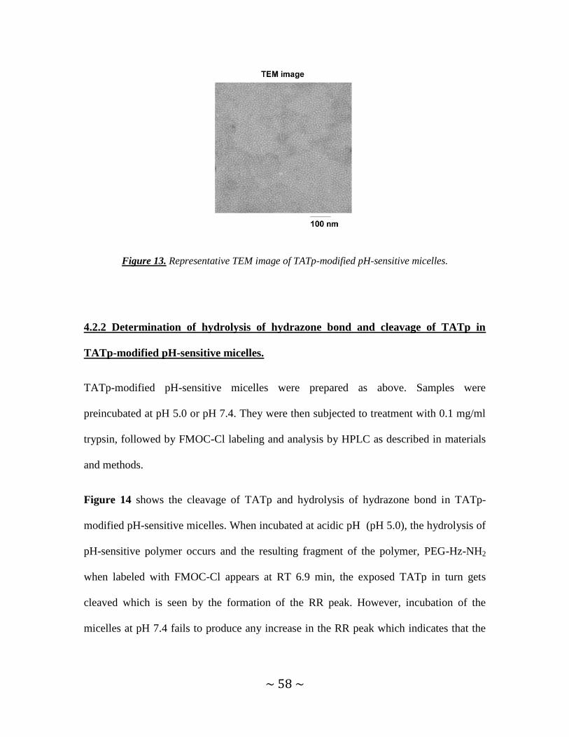
~ 58 ~
Figure 13. Representative TEM image of TATp-modified pH-sensitive micelles.
4.2.2 Determination of hydrolysis of hydrazone bond and cleavage of TATp in
TATp-modified pH-sensitive micelles.
TATp-modified pH-sensitive micelles were prepared as above. Samples were
preincubated at pH 5.0 or pH 7.4. They were then subjected to treatment with 0.1 mg/ml
trypsin, followed by FMOC-Cl labeling and analysis by HPLC as described in materials
and methods.
Figure 14 shows the cleavage of TATp and hydrolysis of hydrazone bond in TATp-
modified pH-sensitive micelles. When incubated at acidic pH (pH 5.0), the hydrolysis of
pH-sensitive polymer occurs and the resulting fragment of the polymer, PEG-Hz-NH2
when labeled with FMOC-Cl appears at RT 6.9 min, the exposed TATp in turn gets
cleaved which is seen by the formation of the RR peak. However, incubation of the
micelles at pH 7.4 fails to produce any increase in the RR peak which indicates that the

~ 59 ~
protective hydrazone polymer shields the TATp and prevents it from degradation by the
added trypsin.
(a)
MIN

~ 60 ~
(b)
Figure 14. HPLC analysis of the cleavage of TATp and hydrolysis of hydrazone bond in TATp-
modified pH-sensitive micelles, at pH 5.0 (a) and pH 7.4(b), 37 0C. The analysis was performed
following the exposure of TATp to trypsin digest and monitored by the RR fragment from its
appearance at 1.5 min and PEG-Hz-NH2 fragment at 6.9 min.
The kinetics of hydrolysis of hydrazone bond and degradation of TATp were followed
(Figure 15). It was observed that the hydrolysis of hydrazone bond and cleavage of
TATp follow the same kinetic pattern. With increasing time of incubation, hydrolysis of
pH-sensitive polymer increases along with increased exposure of TATp as seen by
MIN
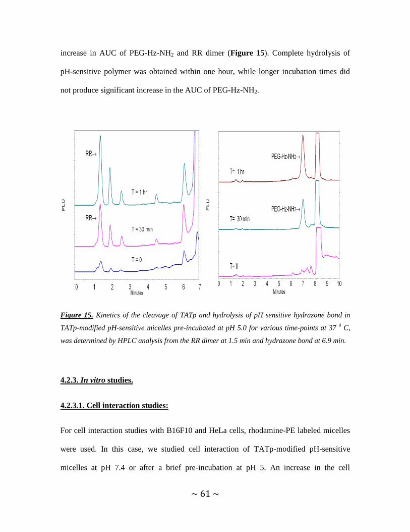
~ 61 ~
increase in AUC of PEG-Hz-NH2 and RR dimer (Figure 15). Complete hydrolysis of
pH-sensitive polymer was obtained within one hour, while longer incubation times did
not produce significant increase in the AUC of PEG-Hz-NH2.
Figure 15. Kinetics of the cleavage of TATp and hydrolysis of pH sensitive hydrazone bond in
TATp-modified pH-sensitive micelles pre-incubated at pH 5.0 for various time-points at 37 0 C,
was determined by HPLC analysis from the RR dimer at 1.5 min and hydrazone bond at 6.9 min.
4.2.3. In vitro studies.
4.2.3.1. Cell interaction studies:
For cell interaction studies with B16F10 and HeLa cells, rhodamine-PE labeled micelles
were used. In this case, we studied cell interaction of TATp-modified pH-sensitive
micelles at pH 7.4 or after a brief pre-incubation at pH 5. An increase in the cell

~ 62 ~
interaction was observed when the micelles were pre-incubated at pH 5 but not at pH 7.4
indicating that pH-sensitive PEG shielding was lost at pH 5. Figure 16 shows that plain
micelles and the TATp-modified pH-sensitive micelles pre-incubated at pH 7.4 showed
only a marginal interaction with the cells, while those pre-incubated at pH 5.0 showed a
significantly higher interaction with the cells, indicating exposure of the TATp.
Unshielded TATp-micelles used as a control also showed increased cell interaction.
In agreement with the fluorescence microscopy experiment, when analyzed by flow
cytometry, the fluorescence associated with B16F10 and HeLa cells was higher in case of
cells treated with TATp-micelles and TATp-modified pH-sensitive micelles pre-
incubated at pH 5 in comparison to those pre-incubated at pH 7.4. These studies again
demonstrate the loss of pH-sensitive polymer at acidic pH and availability of TATp for
cell interaction.
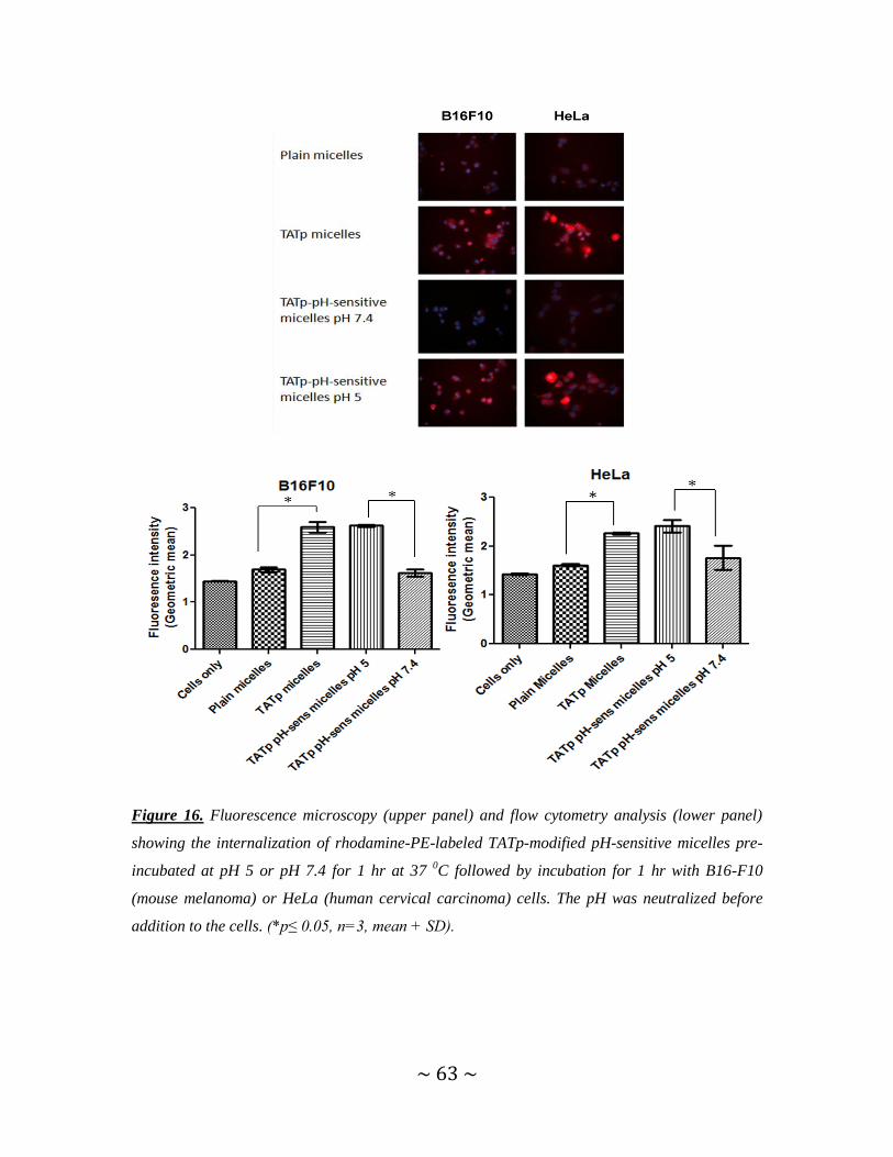
~ 63 ~
Figure 16. Fluorescence microscopy (upper panel) and flow cytometry analysis (lower panel)
showing the internalization of rhodamine-PE-labeled TATp-modified pH-sensitive micelles pre-
incubated at pH 5 or pH 7.4 for 1 hr at 37 0C followed by incubation for 1 hr with B16-F10
(mouse melanoma) or HeLa (human cervical carcinoma) cells. The pH was neutralized before
addition to the cells. (*p≤ 0.05, n=3, mean + SD).

~ 64 ~
4.2.3.2. Cell viability studies.
The cytotoxicity of TATp-modified pH-sensitive micelles loaded with paclitaxel against
B16F10 cells was studied. Micelles were pre-incubated at pH 5 or pH 7.4 for 1 hour.
After one hour, the pH was neutralized and the micelles were added to the cells in
triplicate. Plain, non-drug containing micelles, paclitaxel in methanol solution, paclitaxel-
loaded plain micelles and paclitaxel-loaded TATp micelles were used as controls.
Cytotoxicity was determined after 72 hours of incubation with the micelles. Micelles pre-
incubated at pH 5 and the non-shielded TATp micelles showed comparable cytotoxicity
which was significantly higher than that of the micelles pre-incubated at pH 7.4 as well as
other formulations (Figure 17). Since TATp was exposed, improved drug delivery and
enhanced cytotoxicity was produced.

~ 65 ~
Figure 17. In vitro cytotoxicity of PCT loaded TATp-modified pH-sensitive-micelles preincubated
at pH 5.0 or pH 7.4 at 37 0C, on B16F10 cells (*p <0.05, mean ± SD, n= 3).
4.2.4. In vivo studies- Determination of apoptosis using TUNEL assay.
The next question was, whether this deshielding of polymer and exposure of the TATp
would occur in vivo to improve drug delivery and enhance apoptosis. In other words, an
in vivo “proof-of-concept” study was conducted and hence, to prevent unwanted
distribution, different paclitaxel-loaded micelles were injected intratumorally. Tumors
were harvested after 72 hours. TUNEL assay was carried out using the DNA
fragmentation kit to confirm the presence of apoptosis in the tumor tissue. DAPI stained
the nuclei of the cells present in the tissue. The TUNEL method revealed apoptotic
bodies showing green fluorescence of FITC-labeled TdT. Since TUNEL method stains
0
20
40
60
80
100
120
12,5 25 50 100 200
Ce
ll V
iab
ility
(%
)
Paclitaxel (PCT) conc (ng/ml)
B16F10
Plain micelles
PCT solution
PCT plain micelles
PCT- TATp pH-sensitive micelles pH 7.4
PCT -TATp pH-sensitive micelles pH 5
PCT- TATp micelles
**
**
*

~ 66 ~
fragments produced by apoptosis alone, the cell nuclei showing green fluorescence were
apoptotic. TUNEL staining showed considerable apoptotic activity in the sections
obtained from tumors treated with paclitaxel-loaded TATp-modified pH-sensitive
micelles and TATp-modified micelles. Few apoptotic cells were seen in paclitaxel-
loaded, non-modified, plain micelles (Figure 18). These results show that TATp-
modification and pH-sensitive shielding work efficiently in vivo and the increased
delivery of drug on account of the presence of TATp promotes apoptosis. The data
obtained in this experiment is in line with the in vitro results.
Figure 18. Detection of apoptotic activity by fluorescence microscopy in B16F10 tumor sections
as shown by TUNEL staining. Intratumor injections of plain micelles, TATp-micelles and TATp-
pH-sensitive micelles loaded with paclitaxel (1 mg/kg) were given. Left panel shows DAPI
staining while right panel shows TUNEL staining.
PCT Plain micelles
PCT- TATp micelles
PCT- TATp-pH sensitive
micelles

~ 67 ~
So far we have seen how a cell penetrating peptide can be shielded at normal pH
and made to act at the typical acidic pH of a tumor cell environment after the pH-
sensitive shielding is removed.
This is a significant step towards the development of a multifunctional nanocarrier
containing TATp shielded by a pH-sensitive PEG-PE. The addition of mAb 2C5 for
enhanced targeting would potentially give a therapeutic benefit over the non-
modified counterpart. We will be modifying a commercial preparation Doxil, using
these targeting moieties and studying if the modified multifunctional Doxil provides
an enhanced therapeutic benefit over plain Doxil.

~ 68 ~
4.3. MULTIFUNCTIONAL TATp-CONTAINING, pH-SENSITIVE, mAb 2C5-
CONTAINING LIPOSOMES
4.3.1. Concept of multifunctional TATp-modified pH-sensitive immuno-liposomes.
In this study, a surface-modified multifunctional liposomal carrier with 2C5 monoclonal
antibody, the cell penetrating peptide TATp and pH-sensitive PEG-hydrazone-PE
polymer as an effective TATp steric shield was designed. mAb 2C5 was attached to a
long PEG chain (2C5-PEG3400-PE) . TATp was conjugated with a short PEG1000-PE
spacer and the “shielding” effect of TATp by long PEG2000 chains was optimized. This
nanocarrier was designed in such a way that during the first phase of its delivery, the 2C5
mAb moieties are available, targeting the carrier specific to cancer cells. At normal pH,
surface TATp moieties are “shielded” by the long PEG blocks. Upon the exposure to
lowered pH, degradation of the hydrazone bond and removal of the long PEG chains
occur, TATp moieties are exposed and enhanced penetration of TATp-modified carrier
through the cell membrane occurs.

~ 69 ~
Figure 19. Schematic of the effect of low pH on TATp-modified pH-sensitive immunoliposomes
composed of a pH-degradable PEG2000-Hz-PE with a long PE block, TATp-PEG1000-PE with a
short PEG block, and mAb2C5-PEG3400-PE.
4.3.2. Preparation and characterization of multifunctional liposomes.
In this study, the outer surface of liposomes was modified by incorporating PEG2000-Hz-
PE, TATp-PEG1000-PE and mAb 2C5-PEG3400-PE into pre-formed liposomes using a
post-insertion technique [132].
Previous studies have shown that the amount of PEG that can be incorporated into
vesicles depends upon the content of cholesterol [50]. Several groups have shown that 15
mol% of PEG did not increase the particle size nor their removal from the circulation
[51]. Based on this data reported earlier by various groups, it was decided that liposomes
with PEG2000-Hz-PE shielding up to 15 mol % would be tested for their particle size and
zeta potential to determine the optimal amount that could be used.

~ 70 ~
4.3.2.1. Determination of optimum PEG2000-Hz-PE shielding.
In this study we have modified the commercially-available liposomal preparations Doxil.
Lipodox (generic of Doxil) was used later in place of Doxil because of shortage in Doxil
supply and was modified similarly.
To determine the amount of PEG2000-Hz-PE that could efficiently shield TATp, zeta
potentials of the TATp-modified liposomal preparations with different mol % of longer
PEG blocks were measured. TATp was conjugated to short PEG1000-PE block. The longer
PEG2000-Hz-PE was used as the shielding polymer containing longer PEG chains. TATp-
Doxil and TATp-Lipodox were coated with increasing molar ratios of PEGs from 2.5
mol% to the highest shielding at 15 mol% (Figure 20).
Figure 20. Zeta potentials of TATp-modified liposomes shielded with increasing quantity of
PEG2000-Hz-PE. Plain liposomes ( ), TATp-modified liposomes ( ), PEG2000-Hz-PE shielded
liposomes ( ). n=5, results are mean ± SD.

~ 71 ~
The zeta potential parameter reflects the degree of exposure of TATp moieties on the
carrier‟s surface and allows determining the effective PEG shielding content needed.
Higher PEG shielding resulted in lower zeta potential values suggesting effective
shielding of TATp in a dose dependent manner (Figure 18). The zeta potential of
liposomes coated with 15mol% approached that of plain liposomes indicating optimum
shielding with 15 mol% PEG2000-Hz-PE. No significant change in zeta potentials was
observed for plain, unmodified liposomes that were PEGylated at increasing molar ratios
(up to 15 mol %).
The size of the liposomal formulations prepared in this study following the increase in the
molar ratios of PEG-PE block polymer was within 100nm interval as confirmed by
dynamic light scattering analysis (Figure 21).
Figure 21. Particle size of liposomes with increasing mol % of PEG coating. (n=3, mean ± SD)
Results obtained from particle size and zeta potential analysis suggest that different
liposomal formulations could be efficiently coated with additional PEG blocks that did
not drastically change the size of the liposomes and shielded TATp as well. For the
further experiments, we used PEG2000-Hz-PE (pH cleavable conjugate) at 15 mol%.
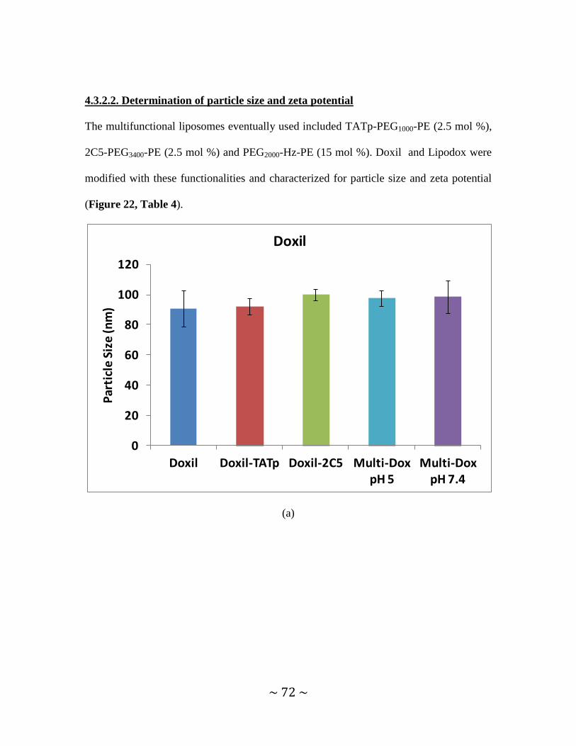
~ 72 ~
4.3.2.2. Determination of particle size and zeta potential
The multifunctional liposomes eventually used included TATp-PEG1000-PE (2.5 mol %),
2C5-PEG3400-PE (2.5 mol %) and PEG2000-Hz-PE (15 mol %). Doxil and Lipodox were
modified with these functionalities and characterized for particle size and zeta potential
(Figure 22, Table 4).
(a)
0
20
40
60
80
100
120
Doxil Doxil-TATp Doxil-2C5 Multi-Dox pH 5
Multi-Dox pH 7.4
Par
ticl
e S
ize
(nm
)
Doxil
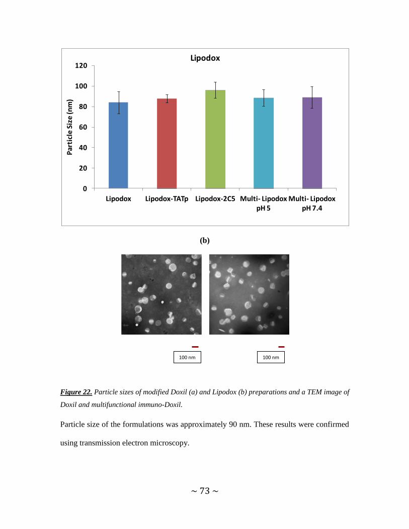
~ 73 ~
(b)
Figure 22. Particle sizes of modified Doxil (a) and Lipodox (b) preparations and a TEM image of
Doxil and multifunctional immuno-Doxil.
Particle size of the formulations was approximately 90 nm. These results were confirmed
using transmission electron microscopy.
0
20
40
60
80
100
120
Lipodox Lipodox-TATp Lipodox-2C5 Multi- Lipodox pH 5
Multi- Lipodox pH 7.4
Par
ticl
e S
ize
(nm
)
Lipodox
100 nm 100 nm

~ 74 ~
Table 4 presents zeta potentials of modified Doxil and Lipodox preparations
Table 4. Zeta potential of liposomal formulations.
TATp moieties on the carrier surfaces contributed to the positive charge of the carriers,
compared to plain Doxil and plain Lipodox (Table 4). While the original Doxil and
Lipodox had zeta potential values of -48.8±2.9 mV and -45.6±3.1mV, TATp-modified
liposomes had value of -8.5±2.2 mV and -8.3 ±1.3 mV respectively. These positive
values are due to the TATp‟s content of positively charged amino acids, arginine and
lysine. Multifunctional immuno-Doxil and Lipodox possess zeta potentials (mV) of -42
±3.5 mV and -40±1.9 for preparation incubated at pH 7.4. When pre-exposed to pH 5,
multifunctional nanocarriers showed more positive zeta value: -10± 1.5 mV and -7± 1.8
mV confirming our proposed mechanism for the TATp shielding and de-shielding effect.

~ 75 ~
4.3.2.3. Specific activity of modified mAb 2C5 by indirect ELISA
To confirm the presence and activity of mAb 2C5 on the surface of liposomes, an indirect
enzyme-linked immunosorbent assay (ELISA) was performed as described previously
[75]. A water-soluble fraction of calf thymus nucleohistone was used as the antigen and
horseradish peroxidase-linked goat anti-mouse IgG was used as secondary antibody to
confirm the presence of mAb 2C5 on Doxil and Lipodox. IgG alone and IgG-modified
liposomes were used as non-specific controls. mAb 2C5 at 2.5 mol% was conjugated to
these liposomes (Figure 23).
(a)
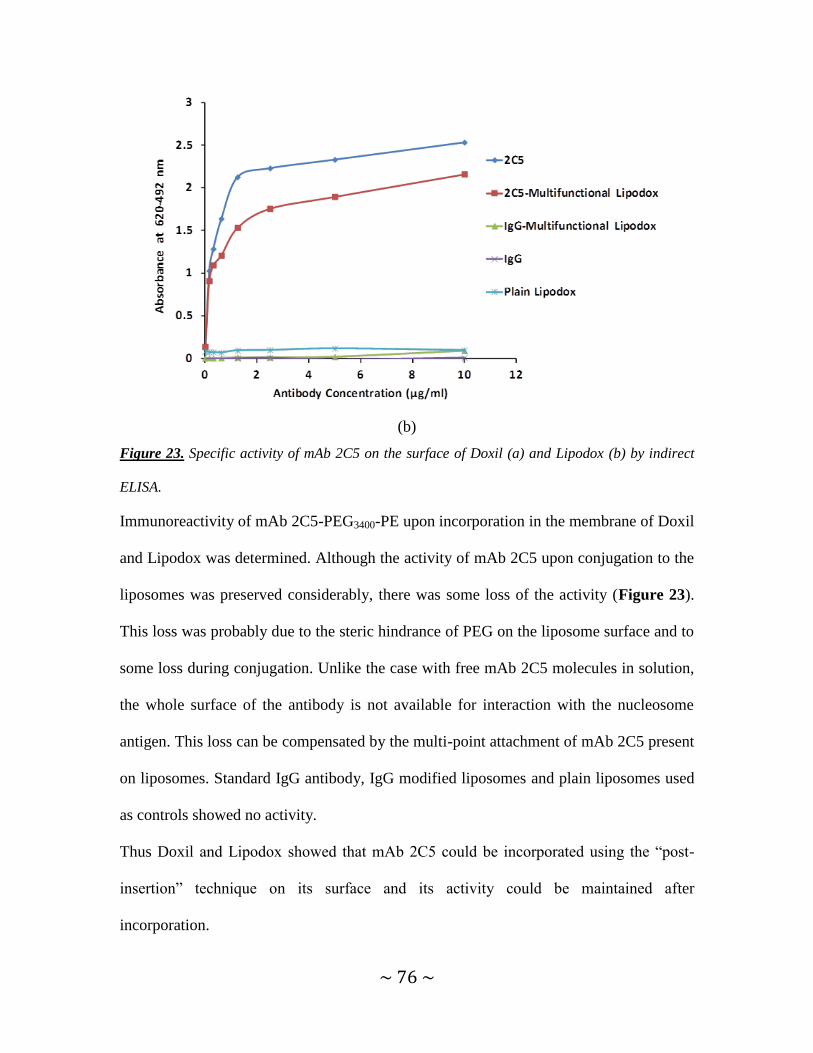
~ 76 ~
(b)
Figure 23. Specific activity of mAb 2C5 on the surface of Doxil (a) and Lipodox (b) by indirect
ELISA.
Immunoreactivity of mAb 2C5-PEG3400-PE upon incorporation in the membrane of Doxil
and Lipodox was determined. Although the activity of mAb 2C5 upon conjugation to the
liposomes was preserved considerably, there was some loss of the activity (Figure 23).
This loss was probably due to the steric hindrance of PEG on the liposome surface and to
some loss during conjugation. Unlike the case with free mAb 2C5 molecules in solution,
the whole surface of the antibody is not available for interaction with the nucleosome
antigen. This loss can be compensated by the multi-point attachment of mAb 2C5 present
on liposomes. Standard IgG antibody, IgG modified liposomes and plain liposomes used
as controls showed no activity.
Thus Doxil and Lipodox showed that mAb 2C5 could be incorporated using the “post-
insertion” technique on its surface and its activity could be maintained after
incorporation.

~ 77 ~
HSPC (fully hydrogenated soy Phosphatidylcholine): cholesterol: PEG liposomes were
also modified in a similar way and labeled with Rhodamine-PE for cell interaction
studies.
4.3.3 Cell interaction studies.
Fluorescence microscopy:
Cell interaction of multifunctional immuno-liposomes with NIH-3T3 (normal cells),
MCF-7 (human breast cancer cells), and HeLa (human cervical cancer cells) was studied
visually using fluorescence microscopy. Plain liposomes, TATp liposomes and 2C5
liposomes were used as controls. Rhodamine-PE (1 mol %) was used to fluorescently
label all liposomes. All liposomes were made from HSPC-Cholesterol-PEG.
Figure 24. Fluorescence microscopy of rhodamine-PE labeled plain liposomes, TATp liposomes,
mAb 2C5 liposomes or TATp-2C5-Hz liposomes pre- incubated at pH 5 or pH 7.4 with NIH-3T3,
MCF-7 and HeLa cells.

~ 78 ~
TATp-liposomes were internalized by all the cell lines (Figure 24). However, 2C5-
modified liposomes were taken up only by cancer cells and not by NIH-3T3 which are
normal cells and which do not over-express nucleosomes. The internalization of
multifunctional immuno-liposomes pre-incubated at pH 5 was significantly higher than
those pre-incubated at pH 7.4. These results indicate that mAb 2C5 on the surface of
liposomes targets the cancer cells and that the TATp exposed upon degradation of pH-
sensitive polymer at an acidic pH further increases the cellular interaction.
FACS analysis:
Quantitative analysis of cell interaction of liposomes was performed using Doxil, TATp-
modified Doxil, 2C5-modified Doxil and multifunctional immuno-Doxil (Multi-Dox,
pre-incubated at pH 5 or pH 7.4). The innate fluorescence of doxorubicin in these
liposomes was used to help determine the changes in geometric mean fluorescence
intensity upon binding of different liposomes to the cells.

~ 79 ~
Figure 25. Flow cytometry. Increase in the geometric mean fluorescence intensity (vs. non-
modified liposomes) of multifunctional immuno-Doxil incubated for 1 hour with NIH-3T3, MCF-
7and HeLa cells. Multifunctional carriers were incubated with cells following their pre-exposure
to normal or acidic conditions for 1hr [*p ≤ 0.05, by Student’s T-test] (n=3, mean ± SD).
A two-fold stronger binding of Doxil-2C5 was observed in MCF-7 and HeLa cell lines,
but not in NIH-3T3 cells which do not over-express nucleosomes. Doxil-TATp had a 6.5-
fold stronger binding with MCF-7 cells, a 6-fold stronger binding for HeLa cells and a
3.6-fold stronger binding with NIH-3T3 cells, in comparison to Doxil alone.
Multifunctional Doxil pre-incubated at pH 5 generated a 3.7 fold, 7-fold and 7.5 -fold
stronger binding with NIH-3T3, MCF-7 and HeLa cells respectively, while

~ 80 ~
multifunctional Doxil pre-incubated at pH 7.4 showed 2-fold stronger binding for MCF-7
and HeLa cells but not with NIH-3T3 (Figure 25).
Based on the results, it can be concluded that the deshielding of TATp at acidic pH and
increased cell association with mAb 2C5 both promote interaction of multifunctional
Doxil with MCF-7 cells and HeLa cells, while improved interaction with NIH-3T3 cells
occurs through the action of TATp alone, owing to the non-specific interaction of TATp
with all cells.
4.3.4. In vitro cytotoxicity studies.
Cytotoxicity of multifunctional immuno-Doxil and controls was determined against HeLa
cells.
0
20
40
60
80
100
0 20 40 60 80 100
Ce
ll V
iab
ility
%
Doxorubicin Concentration (µg/ml)
HeLaDoxil
Doxil-TATp
Doxil-2C5
Multi-Dox pH 7.4
Multi-Dox pH 5

~ 81 ~
Figure 26. In vitro cytotoxicity of plain Doxil, TATp-modified Doxil, mAb 2C5-modified Doxil
and multifunctional TATp-modified, pH-sensitive, immuno-Doxil (Multi-Dox) preincubated at pH
5.0 or pH 7.4 for 1 hr at 370C. Upper panel shows the cytotoxicity of the different preparations
over a range of concentrations of doxorubicin whereas lower panel compares survival of cells at
a representative concentration of 12.5μg/ml of doxorubicin (mid-concentration of all the
concentrations tested). [*p ≤ 0.05, Student’s t-test n=3, mean ± SD].
In vitro cytotoxicity revealed highest toxicity of TATp-Doxil in HeLa cells which is in
agreement with the highest cell interaction of TATp-Doxil with these cells (Figure 26).
The next most effective formulation showing significantly higher cytotoxicity was
multifunctional-Doxil pre-incubated at pH 5. The highest cell viability was observed in
plain, unmodified Doxil treated cells. Though TATp remains shielded in multifunctional
immuno-Doxil incubated at pH 7.4, reduction in cell viability can be attributed to the
presence of mAb 2C5 on the surface which imparts cell targeting property to Doxil, also
0
10
20
30
40
50
60
70
80
Doxil Doxil-TATp Doxil-2C5Multi-Dox pH 7.4Multi-Dox pH 5
Ce
ll v
iab
ility
(%
)
HeLa (at Dox conc 12.5 µg/ml)

~ 82 ~
seen in case of Doxil-2C5. Cytotoxicity of control, doxorubicin-free plain liposomes,
TATp-liposomes, 2C5-liposomes and multifunctional liposomes was conducted with a
lipid concentration similar to the Doxil modified liposomes. No significant difference
from untreated cells was observed and hence these formulations were not used for further
cytotoxicity studies.
Table 5 provides the IC50 values of Doxil ® modified with various functionalities. The
IC50 values of Doxil-TATp were the lowest, followed by multifunctional immuno-Doxil
pre-incubated at pH 5. HeLa cells showed similar IC50 values for Doxil-2C5 and
multifunctional immuno-Doxil pre-incubated at pH 7.4.
Table 5. IC50 values of unmodified and surface modified Doxil multifunctional immuno-liposomes
with HeLa cells.
In conclusion, multifunctional Doxil helps increasing the liposomal association due
to the presence of mAb 2C5. When this formulation is pre-incubated at an acidic
pH, the TATp is de-shielded and with the further help of mAb 2C5, improves
interaction with the cells and promotes increased tumor cell killing.

~ 83 ~
The next step was to determine the effectiveness of this formulation in multi drug
resistant cells.
4.3.5. Development of drug-resistance in sensitive cell lines.
SKOV3 cells were grown in a medium containing 50 ng/ml of paclitaxel for 11 passages.
IC50 values of sensitive and resistant strains were calculated periodically to determine the
extent of resistance.
P-gp efflux pump is one of the most widely characterized proteins responsible for
development of resistance. Cross resistance to structurally and functionally unrelated
compounds is a characteristic of P-gp. It has already been reported to efflux compounds
such as doxorubicin, paclitaxel, colchicine and etoposide. So, the resistant strains
developed from incubation with paclitaxel were used for the studies involving
doxorubicin. A 50-fold difference was obtained in the IC50 values calculated for
doxorubicin in sensitive vs. resistant SKOV3 cell line.
Additionally, P-gp expression studies were also carried out to confirm that this protein
was over-expressed in a higher concentration in the resistant cells as compared to
sensitive cells.

~ 84 ~
4.3.6. P-gp expression studies to determine/confirm drug-resistant and sensitive
cells.
To differentiate drug-sensitive cells from drug-resistant cells, the levels of P-gp were
measured. An assay of P-gp expression on the cell surface using FITC labeled P-gp
specific antibody (UIC2) was conducted.
P-gp expression levels:
Figure 27. Pgp expression in SKOV3 drug-sensitive and drug-resistant cells. Results are mean ±
SD, n=3, *p≤ 0.05 indicates significant difference.
P-gp expression in drug-resistant and sensitive cells was analyzed by flow cytometry.
SKOV-3 sensitive cells showed 8% increase in the fluorescence intensity whereas
resistant cells had 73 % increase in the fluorescence intensity when compared to
untreated cells, indicating the presence of higher amounts of P-gp in the resistant cells
(Figure 27).
Thus the presence of P-gp was confirmed as the basis for multi-drug resistance.

~ 85 ~
4.3.7. Cell interaction studies with SKOV3 drug-resistant and sensitive cells
Fluorescence microscopy:
Cell interaction was determined by the method described earlier.
Figure 28. Fluorescence microscopy of rhodamine-PE labeled (HSPC-Chol-PEG) plain
liposomes, TATp liposomes, mAb 2C5 liposomes or TATp-2C5-Hz liposomes pre- incubated at
pH 5 or pH 7.4 with SKOV3 drug-sensitive and resistant cells.
Figure 28 shows the uptake of various rhodamine-PE labeled HSPC: Cholesterol: PEG
liposomes by SKOV3 drug-resistant and drug-sensitive cells. TATp liposomes and
multifunctional immuno-liposomes pre-incubated at pH 5.0 show a higher interaction in
both sensitive as well as resistant cell lines. 2C5 liposomes also conferred cell-interaction
property to the liposomes in both the cell lines. These results indicate that mAb 2C5 on

~ 86 ~
the surface of liposomes helps in targeting the cancer cells and TATp exposed upon
degradation of pH-sensitive polymer at acidic pH, further increases the cellular
interaction in both cell lines.
FACS analysis:
Quantitative analysis of cell interaction of liposomes was performed with Lipodox ®,
TATp-modified Lipodox ®, 2C5-modified Lipodox ® and multifunctional immuno-
Lipodox ® as described earlier. The innate fluorescence of doxorubicin in these
liposomes was used to determine the changes in binding of different liposome
formulations to the cells.

~ 87 ~
Figure 29. Flow cytometry. Increase in the geometric mean fluorescence intensity of SKOV3
drug-sensitive (upper panel) and resistant (lower panel) cells incubated for 1 hour with various
formulations. Multifunctional carriers were incubated with cells following their pre-exposure to
normal/acidic conditions for 1hr. (*p ≤ 0.05, n=3, mean ± SD).
Results obtained in FACS analysis were similar to that obtained from fluorescence
microscopy. Lipodox-TATp and multifunctional immuno-lipodox pre-incubated at pH 5
had an overall increased fluorescence intensity compared to Lipodox alone (Figure 29).

~ 88 ~
The pattern of interaction of each formulation remained the same in both type of cells,
indicating that the TATp and 2C5 moieties attached to the surface of the liposomes
provide increased interaction of the liposomes with the resistant cells as well.
4.3.8. In vitro cytotoxicity studies.
Cytotoxicity was determined by the method described earlier.
Figure 30. In vitro cytotoxicity of plain Lipodox ®, TATp-modified Lipodox ®, mAb 2C5-
modified Lipodox® and multifunctional TATp-modified, pH-sensitive, immuno- Lipodox ®,
preincubated at pH 5.0 or pH 7.4 for 1 hr at 370C. Upper panel shows the cytotoxicity of the
different preparations over a range of concentrations of doxorubicin. The lower panel compares
survival of cells at a concentration of 12.5μg/ml of doxorubicin. (*p ≤ 0.05, n=3, mean ± SD).
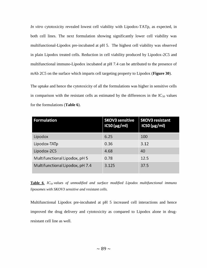
~ 89 ~
In vitro cytotoxicity revealed lowest cell viability with Lipodox-TATp, as expected, in
both cell lines. The next formulation showing significantly lower cell viability was
multifunctional-Lipodox pre-incubated at pH 5. The highest cell viability was observed
in plain Lipodox treated cells. Reduction in cell viability produced by Lipodox-2C5 and
multifunctional immuno-Lipodox incubated at pH 7.4 can be attributed to the presence of
mAb 2C5 on the surface which imparts cell targeting property to Lipodox (Figure 30).
The uptake and hence the cytotoxicity of all the formulations was higher in sensitive cells
in comparison with the resistant cells as estimated by the differences in the IC50 values
for the formulations (Table 6).
Table 6. IC50 values of unmodified and surface modified Lipodox multifunctional immuno
liposomes with SKOV3 sensitive and resistant cells.
Multifunctional Lipodox pre-incubated at pH 5 increased cell interactions and hence
improved the drug delivery and cytotoxicity as compared to Lipodox alone in drug-
resistant cell line as well.

~ 90 ~
4.3.9. In vivo studies of multifunctional liposomes in drug-resistant and drug-
sensitive tumors.
4.3.9.1.Tumor growth inhibition studies.
0
50
100
150
200
250
300
0 5 10 15 20 25
% c
ha
ng
e in
tu
mo
r vo
lum
e
Days post treatment
SKOV-3 Sensitive
***
****
*

~ 91 ~
Figure 31. Effect of i.v. administration of liposomal preparations on tumor growth in nude mice
bearing SKOV-3 drug sensitive (upper panel) and drug resistant (lower panel) tumors shown as
percentage change in tumor volume from initial, calculated as (Vn-V0)/V0*100, where Vo is the
tumor volume on day zero of drug administration and Vn is the volume on day “n” after drug
administration has begun.(n=5, mean + S.D.). *** p≤ 0.0001, **p≤ 0.001, *p≤ 0.05 calculated
from day 10 to day 20, by ANOVA using Tukey’s Post-Hoc test. For SKOV-3 sensitive tumors,*p
≤ 0.05 is from day 15 to day 20 between multifunctional Lipodox and Lipodox-2C5. Arrows
indicate day of treatment with the formulations given after tumor size of 100 mm3 was reached.
Multifunctional-Lipodox had an enhanced therapeutic efficacy in vivo in comparison to
all controls. The tumor volume in animals treated with this preparation was at least two-
fold smaller compared to Lipodox alone in drug-resistant as well as in drug-sensitive
SKOV3 tumors (Figure 31). At the end of the treatment, mice treated with
multifunctional-Lipodox showed 55 % reduction in tumor volume compared to the initial
0
50
100
150
200
250
300
0 5 10 15 20 25
% c
ha
ng
e in
tu
mo
r vo
lum
e
Days post treatment
SKOV3- Resistant
***
****

~ 92 ~
volume when the treatment was started on SKOV3 drug-sensitive tumors. This was the
maximum reduction in tumor volumes when compared to any other formulation
treatment. In mice with SKOV3 drug-resistant tumors, treatment with multifunctional-
Lipodox increased the tumor volume by only 12% which was the smallest increment
when compared to other treatment groups in drug-resistant tumor group. In both drug-
sensitive and resistant tumors, multifunctional Lipodox showed significantly reduced
tumor volume when compared to Lipodox alone.
Unlike the in vitro data where Lipodox-TATp showed the highest cell interaction and
cytotoxicity, this formulation showed no significant difference in reducing the tumor
volumes when compared to Lipodox alone. Previous in vitro studies have shown that the
peptide gets degraded by proteolytic enzymes in the body which affects its cell
interaction as well as cytotoxic effect. Other groups have noted that TATp when
conjugated to a drug or to the surface of nanoparticles doesn‟t show the expected in vivo
results, mainly improved tumor reduction. The effect of human plasma on TATp has
shown to reduce the stability of this peptide [107].TATp mediated drug delivery is also
hindered by metabolic degradation in body fluids [133-137].
On treatment with Lipodox-2C5, the tumor volumes were reduced by 3% when compared
to the initial tumor volume. Similarly, even though the tumor volumes in SKOV3
resistant tumors increased when treated with Lipodox-2C5, they were significantly
smaller as compared to Lipodox alone indicating higher activity of Lipodox-2C5 (Figure
31).

~ 93 ~
4.3.9.2. Analysis of changes in body weight of mice
An important part in the development of a drug formulation is to determine its safety.
Anticancer drugs are known to produce weight loss in patients. Formulations in the form
of nanoparticles have shown preferential toxicity to the tumor and low side-effects. As a
result it is expected that the body weight will not drop drastically when animals are
treated with these formulations.
Figure 32. Effect of intravenous administration of liposomal preparations on the body weight in
nude mice bearing SKOV-3 drug- sensitive and resistant tumors shown as percentage change in
body weight (from initial). (n=5, mean + S.D, *P ≤ 0.05)
Mice were weighed every 3 days (Figure 32). There was no significant change in the
body weight of mice in all treatment groups supporting the absence of gross toxicity.
50
60
70
80
90
100
110
120
0 5 10 15 20 25
% c
ha
ng
e in
bo
dy
we
igh
t o
f m
ice
Time (Days)
SKOV3 sensitive
50
60
70
80
90
100
110
120
0 5 10 15 20 25
% c
ha
ng
e in
bo
dy
we
igh
t o
f m
ice
Time (Days)
SKOV3 resistant
Saline
Lipodox
Lipodox-TATp
Lipodox-2C5
Multifunctional-Lipodox

~ 94 ~
4.3.9.3. Determination of tumor weight.
The study was ended when the tumors in the mice treated with saline started to exceed a
volume of 1000 mm3. Tumors were isolated from the body of the sacrificed mice and
weighed.
0
0,2
0,4
0,6
0,8
1
1,2
Saline Lipodox Lipodox-TATp Lipodox-2C5 Multifunctional Lipodox
Fin
al tu
mo
r w
eig
ht
(gm
s)
SKOV3 Sensitive
*

~ 95 ~
Figure 33. Final tumor weights in nude mice bearing SKOV-3 sensitive and resistant tumors
measured on day 20, following intravenous administration of liposomal preparations as
described previously. (n=5, mean + S.D., *P<0.05)
The final tumor weight determined at the end of the study confirmed that the
multifunctional-Lipodox® in both sensitive and resistant tumors served as the most
effective treatment (Figure 33). Lipodox treated tumors weighed significantly less than
saline treated group indicating activity of the drug. A further increase in the drug‟s
activity was imparted by the presence of multiple moieties, TATp and mAb 2C5.
4.3.9.4. Determination of apoptosis by TUNEL assay:
TUNEL assay was carried out using the DNA fragmentation kit to confirm the presence
of apoptosis in the tumor tissue. DAPI stained the nuclei of the cells present in the tissue.
The TUNEL method revealed apoptotic bodies showing green fluorescence of FITC-
0
0,2
0,4
0,6
0,8
1
1,2
Saline Lipodox Lipodox-TATp Lipodox-2C5 Multifunctional Lipodox
Fin
al tu
mo
r w
eig
ht
(gm
s)
SKOV3 Resistant
*

~ 96 ~
labeled TdT. Since TUNEL method stains fragments produced by apoptosis alone, the
cell nuclei showing green fluorescence were apoptotic. TUNEL staining showed
considerable apoptotic activity in the sections obtained from tumors treated with
MultiLipodox, Lipodox-2C5 and Lipodox-TATp. Few apoptotic cells were also seen in
non-modified, plain Lipodox. These results show that modification with mAb 2C5 and
TATp with pH-sensitive shielding work efficiently in vivo to promote apoptosis (Figure
34, 35).
Figure 34. Detection of apoptotic activity by fluorescence microscopy in SKOV-3 drug-sensitive
tumor sections as shown by TUNEL staining. Left panel shows DAPI staining while right panel
shows TUNEL staining.

~ 97 ~
Figure 35. Detection of apoptotic activity by fluorescence microscopy in SKOV-3 drug-resistant
tumor sections as shown by TUNEL staining. Left panel shows DAPI staining while right panel
shows TUNEL staining.
Thus drug-loaded multifunctional immuno-liposomes had an enhanced anti-cancer
effect in both drug-resistant and drug-sensitive tumors, indicating that modification
of liposomes with active targeting moieties can provide a more prominent
therapeutic effect than the conventional unmodified liposomes for the treatment of
cancer.

~ 98 ~
5. CONCLUSIONS
A. Presence of TATp on the surface of nanoparticles increases their interaction with
the cells.
B. Degradation of TATp occurs in the presence of proteolytic enzymes and can be
prevented by shielding it with a long PEG block.
C. Drug-loaded micelles modified with TATp on the surface shielded by a pH-
sensitive long PEG block show enhanced cell interaction and drug delivery when
the pH-sensitive bond breaks at the typical acidic pH of a tumor cell environment
and TATp is exposed. Enhanced in vivo tumor cell apoptosis is observed with this
system indicating that the system is effective in vivo.
D. Drug-loaded multifunctional TATp-modified, pH-sensitive, mAb 2C5 liposomes
show enhanced cell uptake and cytotoxicity in comparison to non-modified
liposomes in various tumor cell lines including doxorubicin-resistant and sensitive
cells.
E. In vivo studies with these liposomes show remarkable reduction of both drug-
resistant and sensitive tumors in mice confirming that our system is successful in
showing improved therapeutic efficacy over the commercially available
unmodified liposomes.
This effect is provided by the multiple functionalities. mAb 2C5 on the surface
specifically targets cancer cells. pH-sensitive PEG helps in longer circulation of

~ 99 ~
the liposome and at the extracellular acidic pH, degradation of this polymer
exposes TATp which increases cell penetration.
F. Thus nanoparticles modified with multiple functionalities enhance the effect of an
anticancer drug. There are numerous ways to modify the particles. The patho-
physiology of the organ should be taken into consideration while developing
multifunctional nanoparticles for treatment of the particular disease under
research.
~ 100 ~
6. REFERENCES
1. Torchilin, V.P., Strategies and Means for Drug Targeting: An Overview, in
Biomedical Aspects of Drug Targeting, 2002, Kluwer Academic Publishers. p. 3-
25.
2. Davis, M.E., Chen, Z., Shin D.M., Nanoparticle therapeutics: an emerging
treatment modality for cancer. Nature Reviews. Drug Discovery, 2008. 7: p. 771-
782.
3. Eiffler, A.C., Thaxton, C.S., Nanoparticle therapeutics: FDA approval, clinical
trials, regulatory pathways and case study., in Biomedical Nanotechnology:
Methods and Protocols, Methods in Molecular Biology, 2011.p. 42.
4. Iessi, E., Marino, M.L., Lozupone, F., Fais, S., DeMilito, A., Tumor acidity and
malignancy: novel aspects in the design of anti-tumor therapy. Cancer Therapy,
2008. 6: p. 55-66.
5. Tunggal, J.K., Cowan,.D., Shaikh, H., Tannock, I.F., Penetration of anticancer
drugs through solid tissue: A factor that limits the effectiveness of chemotherapy
for solid tumors. Clin. cancer research, 1999. 5: p. 1583-1586.
6. Folkman, J., Tumor angiogenesis: therapeutic implications. N Engl. J Med., 1971.
285: p. 1182-1186.
7. Jain, R.K., Vascular and interstitial barriers to delivery of therapeutic agents in
tumors. Cancer Metastasis Reviews, 1990. 9: p. 253-266.
8. Yuan, G., Dellian, M., Fukumara, D., Leunig, M., Berk, D.A., Torchilin, V.P.,
Jain R.K., Vascular permeability in a human tumor xenograft: molecular size
dependence and cutoff size. Cancer Res, 1995. 55: p. 3752-3756.

~ 101 ~
9. Maeda, H., Wu, J., Sawa, T., Matsumura, Y., Hori, K., Tumor vascular
permeability and the EPR effect in macromolecular therapeutics: a review. J.
Controlled Release, 2000. 65: p. (271-284).
10. Matsumura, Y., Maeda, H., A new concept for macromolecular therapeutics in
cancer chemotherapy: Mechanism of tumoritropic accumulation of proteins and
the antitumor agent smancs. Cancer Res, 1986. 46: p. 6387-6392.
11. Gao, Z., Lukyanov, A.N., Chakilam, A.R., Torchilin, V.P, PEG-
PE/phosphatidylcholine mixed immunomicelles specifically deliver encapsulated
taxol to tumor cells of different origin and promote their efficient killing. J Drug
Target, 2003. 11: p. 87-92.
12. Torchilin, V.P., Trubetskoy, V.S., Which polymers can make nanoparticulate
drug carriers long-circulating? Adv. Drug Deliv. Rev., 1995 a. 16: p. 141-155.
13. Torchilin, V.P., Trubetskoy, V.S., Whiteman, K.R., Caliceti, P., Ferruti, P.,
Veronese, F.M., New synthetic amphiphilic polymers for steric protection of
liposomes in vivo. J Pharm Sci, 1995 b. 84: p. 1049-1053.
14. Torchilin, V.P., Omelyanenko, V.G., Papisov, M.I., Bogdanov, A.A.Jr.,
Trubetskoy, V.S., Herron, J.N., Gentry, C.A., Poly(ethylene glycol) on the
liposome surface: on the mechanism of polymer-coated liposome longevity.
Biochim Biophys Acta, 1994. 1195: p. 11-20.
15. Lasic, D.D., Martin, F.J., Gabizon, A., Huang, S.K., Papahadjopoulos, D.,
Sterically stabilized liposomes: a hypothesis on the molecular origin of the
extended circulation times. Biochim Biophys Acta, 1991 a. 1070: p. 187-192.
16. Lasic, D.D., Martin, F.J., Stealth Liposomes. 1995, Boca Raton: CRC Press. 320.
~ 102 ~
17. Kommareddy, S., Tiwari, S.B., Amiji, M.M, Long-circulating polymeric
nanovectors for tumor-selective gene delivery. Technol.Cancer Res. Treat., 2005.
4(6): p. 615-625.
18. Lukyanov, A.N., Gao, Z., Mazzola, L., Torchilin, V.P., Polyethylene glycol-
diacyllipid micelles demonstrate increased accumulation in subcutaneous tumors
in mice. Pharm. Res, 2002. 19(10): p. 1424-1429.
19. Frankel, A.D., Pabo, C.O., Cellular uptake of the Tat Protein from Human
Immunodeficiency Virus. Cell, 1988. 55: p. 1189-1193.
20. Grunwald, J., Rejtar, T., Sawant, R., Zhouxi, W., Torchilin, V.P., TAT peptide
and its conjugates: proteolytic stability. Bioconjug Chem, 2009. 20(1531-1537).
21. Mendonca, L.S., Firmino, F., Moreira, J.N., Pedroso de Lima, M.C., Simoes, S.,
Transferrin receptor-targeted liposomes encapsulating anti-BCR-ABL siRNA or
asODN for chronic myeloid leukemia treatment. Bioconjug Chem, 2010. 21(1): p.
157-168.
22. Elbayoumi, T., Torchilin, V.P., Tumor-targeted immuno-liposomes for delivery of
therapeutics and diagnostics. Pharmaceutical Engineering, 2006. 26(5): p. 1-6.
23. Yamagata, M., Hasuda, K., Stamato, T., Tannock, I.F., The contribution of lactic
acid to acidification of tumours: Studies of variant cells lacking LDH. Br.J
Cancer, 1998. 77: p. 1726-1731.
24. Vaupel, P., Physiological properties of malignant tumors. NMR in biomedicine,
1992. 5: p. 220-225.

~ 103 ~
25. Engin, K., Leeper, D.B., Cater, J.R., Thistlethwaite, A.J., Tupchong, L.,
McFarlane, J.D., Extracellular pH distribution in human tumours.
Int.J.Hyperthermia, 1995. 11(2): p. 211-216.
26. Gref, R., Minamitake, Y., Peracchia, M.T., Trubetskoy, V., Torchilin, V.P.,
Langer, R., Biodegradable long-circulating polymeric nanospheres. Science,
1994. 263: p. 1600-1603.
27. Torchilin, V.P., Structure and design of polymeric surfactant based drug-delivery
systems. Journal of Controlled Release, 2001. 73: p. 137-172.
28. Torchilin, V.P., Weissig, V., Polymeric micelles for delivery of poorly soluble
drugs. Polym Prepr, 1999. 40: p. 320-321.
29. Gao, Z., Lukyanov, A.N., Singhal, A., Torchilin, V.P., Diacyllipid-polymer
micelles as nanocarriers for poorly soluble anticancer drugs. Nano Letters, 2002.
2(9): p. 979-982.
30. Torchilin, V.P., Passive and active drug targeting: drug delivery to tumors as an
example. Handbook of experimental pharmacology, 2010. 197: p. 3-53.
31. Torchilin, V.P., Drug Targeting. Eur J Pharm Sci, 2000. 11(Suppl 2): p. S81-S91.
32. Weissig, V., Whiteman, K.R., Torchilin, V.P., Accumulation of protein-loaded
long-circulating micelles and liposomes in subcutaneous Lewis lung carcinoma in
mice. Pharm. Res, 1998. 15: p. 1552-1556.
33. Torchilin, V.P., Recent advances with liposomes as pharmaceutical carriers.
Nature Reviews. Drug Discovery, 2005. 4: p. 145-160.

~ 104 ~
34. Klibanov, A.L., Maruyama, K., Torchilin, V.P.,Huang, L, Amphipathic
polyethyleneglycols effectively prolong the circulation time of liposomes. FEBS
Lett., 1990. 268: p. 235-238.
35. Gabizon, A., Catane, R., Uziely, B., Prolonged circulation time and enhanced
accumulation in malignant exudates of doxorubicin encapsulated in polyethylene-
glycol coated liposomes. Cancer Res, 1994. 54: p. 987-992.
36. Torchilin, V.P., Rammohan, R., Weissig, V., Levchenko, T.S., TAT peptide on the
surface of liposomes affords their efficient intracellular delivery even at low
temperatures and in the presence of metabolic inhibitors. Proc Natl Acad Sci
USA, 2001. 98: p. 8786-8791.
37. Sawant, R.R., Torchilin, V.P., Enhanced cytotoxicity of TATp-bearing paclitaxel-
loaded micelles. International Journal of Pharmaceutics, 2009. 374: p. 114-118.
38. Elbayoumi, T., Torchilin, V.P., Tumor-specific anti-nucleosome antibody
improves therapeutic efficacy of doxorubicin-loaded long-circulating liposomes
against primary and metastatic tumor in mice. Mol. Pharm., 2008. 6(1): p. 246-
254.
39. Ko, Y.T., Hartner, W.C., Kale, A., Torchilin, V.P., Gene delivery into ischemic
myocardium by double-targeted lipoplexes with anti-myosin antibody and TAT
peptide. Gene Ther., 2009. 16(1): p. 52-59.
40. Sawant, R.R., Sawant, R.M., Torchilin, V.P, Mixed PEG-PE/vitamin E tumor-
targeted immunomicelles as carriers for poorly soluble anti-cancer drugs:
improved drug solubilization and enhanced in vitro cytotoxicity. Eur J Pharm
Biopharm, 2008. 70(1): p. 51-57.

~ 105 ~
41. Torchilin V.P., Lukyanov, A., Gao, Z., Papahadjopaoulos-Sternberg, B.,
Immunomicelles: Targeted pharmaceutical carriers for poorly soluble drugs.
Proc Natl Acad Sci USA, 2003. 100(10): p. 6039-6044.
42. Elbayoumi, T.A., Torchilin, V.P., Enhanced anti-tumor efficacy in vivo of
doxorubicin-loaded long-circulating liposomes modified with cancer-specific
monoclonal antibody. Clin. cancer research, 2009. 15(6): p. 1973-1980.
43. Elbayoumi, T.A., Torchilin, V.P., Enhanced accumulation of long-circulating
liposomes modified with the nucleosome-specific monoclonal antibody 2C5 in
various tumours in mice: gamma-imaging studies. Eur J Nucl Med Mol Imaging,
2006. 33(10): p. 1196-205.
44. Elbayoumi, T.A., Torchilin ,V.P., Enhanced cytotoxicity of monoclonal
anticancer antibody 2C5-modified doxorubicin-loaded PEGylated liposomes
against various tumor cell lines. Eur J Pharm Sci, 2007. 32(3): p. 159-68.
45. Kale, A.A., Torchilin, V.P., Design, synthesis and characterization of pH-
sensitive PEG-PE conjugates for stimuli-sensitive pharmaceutical nanocarriers:
The effect of substitutes at the hydrazone linkage on the pH-stability of PEG-PE
conjugates. Bioconjug Chem, 2007 a. 18(2): p. 363-370.
46. Kale, A. A., Torchilin, V.P., Enhanced transfection of tumor cells in vivo using
"smart" pH-sensitive TAT-modified PEGylated liposomes. Journal of Drug
Targeting, 2007 b. 15: p. 538-545.
47. Sawant, R.M., Hurley, J.P., Salmaso S., Kale, A.A., Tolcheva, E., Levchenko, T.,
Torchilin, V.P., "Smart" drug delivery systems: double-targeted pH-responsive
pharmaceutical nanocarriers. Bioconjug Chem, 2006. 17: p. 943-949.

~ 106 ~
48. Allen, T.M., Hansen, C., Pharmacokinetics of stealth versus conventional
liposomes: effect of dose. Biochim Biophys Acta, 1991. 1237: p. 99-108.
49. Mayhew, E.G., Lasic, D., Babbar, S., Martin, F.J., Pharmacokinetics and
antitumor activity of epirubicin encapsulated in long-circulating liposomes
incorporating a polyethylene glycol- derivatized phospholipid. Int.J.Cancer, 1992.
51: p. 302-309.
50. Hristova, K., Needham, D., Phase behavior of a lipid/polymer-lipid mixture in
aqueous medium. Macromolecules, 1995. 28(4): p. 991-1002.
51. Khalid, M.N., et al., Long circulating poly(ethylene glycol)-decorated lipid
nanocapsules deliver docetaxel to solid tumors. Pharm Res, 2006. 23(4): p. 752-8.
52. Kenworthy, A.K., et al., Range and magnitude of the steric pressure between
bilayers containing phospholipids with covalently attached poly(ethylene glycol).
Biophys J, 1995. 68(5): p. 1921-36.
53. Lasic, D.D., Martin,F.J., Stealth liposomes. 1995, Boca Raton, FL: CRC Press.
54. Schubert, U.S., et al., Poly(ethylene glycol) in Drug Delivery: Pros and Cons as
Well as Potential Alternatives. Angewandte Chemie-International Edition, 2010.
49(36): p. 6288-6308.
55. Howard, M.D., et al., PEGylation of nanocarrier drug delivery systems: State of
the art. Journal of Biomedical Nanotechnology, 2008. 4(2): p. 133-148.
56. Photos, P.J., et al., Polymer vesicles in vivo: correlations with PEG molecular
weight. J Control Release, 2003. 90(3): p. 323-34.

~ 107 ~
57. Malmsten, M., Emoto, K., Van Alstine, J.M., Effect of Chain Density on
Inhibition of Protein Adsorption by Poly (ethylene glycol) Based Coatings* 1.
Journal of colloid and interface science, 1998. 202(2): p. 507-517.
58. Kingshott, P., Thissen, H., Griesser, H.J., Effects of cloud-point grafting, chain
length, and density of PEG layers on competitive adsorption of ocular proteins.
Biomaterials, 2002. 23(9): p. 2043-56.
59. Kenworthy, A.K., Simon, S.A., McIntosh, T.J., Structure and phase behavior of
lipid suspensions containing phospholipids with covalently attached poly(ethylene
glycol). Biophys J, 1995. 68(5): p. 1903-20.
60. Garbuzenko, O., Barenholz, Y., Priev, A., Effect of grafted PEG on liposome size
and on compressibility and packing of lipid bilayer. Chemistry and Physics of
Lipids, 2005. 135(2): p. 117-129.
61. Zhang, J.X., et al., Pharmaco attributes of
dioleoylphosphatidylethanolamine/cholesterylhemisuccinate liposomes containing
different types of cleavable lipopolymers. Pharmacol Res, 2004. 49(2): p. 185-98.
62. Lasic, D., et al., Phase behavior of stealth-lipid"-lecithin mixtures. Period. Biol,
1991. 93(2): p. 287-290.
63. Shimada, K., et al., Determination of incorporated amounts of poly(ethylene
glycol)-derivatized lipids in liposomes for the physicochemical characterization of
stealth liposomes. Int J Pharm, 2000. 203(1-2): p. 255-63.
64. Bedu-Addo, F.K., et al., Effects of polyethyleneglycol chain length and
phospholipid acyl chain composition on the interaction of polyethyleneglycol-

~ 108 ~
phospholipid conjugates with phospholipid: implications in liposomal drug
delivery. Pharm Res, 1996. 13(5): p. 710-7.
65. Sawant, R.M., et al., "SMART" drug delivery systems: double-targeted pH-
responsive pharmaceutical nanocarriers. Bioconjug Chem, 2006. 17(4): p. 943-9.
66. Roux, E., Francis, M., Winnik, F.M., Leroux J.C., Polymer based pH-sensitiv
carriers as a means to improve the cytoplasmic delivery of drugs. Int.J.Pharm,
2002 242: p. 25-36.
67. Guo, X., Szoka, F.C.Jr, Steric stabilization of fusogenic liposomes by a low-pH
sensitive PEG-diortho ester-lipid conjugate. Bioconjug Chem, 2001. 12: p. 291-
300.
68. Kratz, F., Beyer, U., Schutte, M.T., Drug-polymer conjugates containing acid-
cleavable bonds. Crit Rev Ther Drug Carrier Syst, 1999. 16: p. 245-288.
69. Kale, A.A., Torchilin, V.P., Enhanced transfection of tumor cells in vivo using
"smart" pH-sensitive TAT-modified PEGylated liposomes. J. Drug Target, 2007.
15: p. 538-545.
70. Kale, A.A, Torchlin, V.P., Design, synthesis and characterization of pH-sensitive
PEG-PE conjugates for stimuli-sensitive pharmaceutical nanocarriers: The effect
of substitutes at the hydrazone linkage on the pH-stability of PEG-PE conjugates.
Bioconjug Chem., 2007. 18(2): p. 363-370.
71. Iakoubov, L.Z., Torchilin, V.P., A novel class of antitumor antibodies:
nucleosome-restricted antinuclear autoantibodies (ANA) from healthy aged
nonautoimmune mice. Oncol Res, 1997. 9(8): p. 439-46.

~ 109 ~
72. Iakoubov, L.Z., Torchilin, V.P., Nucleosome-releasing treatment makes surviving
tumor cells better targets for nucleosome-specific anticancer antibodies. Cancer
Detect Prev, 1998. 22(5): p. 470-5.
73. Chakilam, A.R., Pabba, S., Mongayt, D., Iakoubov, L.Z., Torchilin, V.P., A single
monoclonal antinuclear autoantibody with nucleosome-restricted specificity
inhibits growth of diverse human tumors in nude mice. Cancer Therapy, 2004. 2:
p. 353-364.
74. Torchilin, V.P., et al., p-Nitrophenylcarbonyl-PEG-PE-liposomes: fast and simple
attachment of specific ligands, including monoclonal antibodies, to distal ends of
PEG chains via p-nitrophenylcarbonyl groups. Biochim Biophys Acta, 2001.
1511(2): p. 397-411.
75. Lukyanov, A.N., Elbayoumi, T.A., Chakilam, A.R., Torchilin, V.P., Tumor-
targeted liposomes: doxorubicin-loaded long-circulating liposomes modified with
anti-cancer antibody. J. Controlled Release, 2004. 100: p. 135-144.
76. Allen, T.M., Sapra, P., Moase, E., Use of the post-insertion method for the
formation of ligand-coupled liposomes. Cell Mol Biol Lett, 2002. 7(3): p. 889-94.
77. Lukyanov, A.N., et al., Tumor-targeted liposomes: doxorubicin-loaded long-
circulating liposomes modified with anti-cancer antibody. J Control Release,
2004. 100(1): p. 135-44.
78. Elbayoumi, T.A., Torchilin V.P., Enhanced anti-tumor efficacy in vivo of
doxorubicin-loaded long-circulating liposomes modified with cancer-specific
monoclonal antibody. Clin. cancer research, 2009. 15(6): p. 1973-1980.

~ 110 ~
79. Elbayoumi, T.A., Torchilin, V.P., Tumor-specific anti-nucleosome antibody
improves therapeutic efficacy of doxorubicin-loaded long-circulating liposomes
against primary and metastatic tumor in mice. Mol. Pharm., 2008. 6(1): p. 246-
254.
80. Gupta, B., Torchilin, V.P., Monoclonal antibody 2C5-modified doxorubicin-
loaded liposomes with significanty enhanced therapeutic activity against
intracranial human brain U-87 MG tumor xenografts in nude mice. Cancer
Immunol Immunother, 2007. 56: p. 1215-1223.
81. Torchilin, V.P., Lukyanov, A.N., Gao, Z., Papahadjopoulos-Sternberg, B,
Immunomicelles: Targeted pharmaceutical carriers for poorly soluble drugs.
PNAS, 2003. 100(10): p. 6039-6044.
82. Mamot, C., Ritschard, R., Kung, W., Park, J.W., Herrmann, R., Rochlitz, C.F.,
EGFR-targeted immunoliposomes derived from the monoclonal antibody
EMD72000 mediate specific and efficient drug delivery to a variety of colorectal
cancer cells. J Drug Target, 2006. 14(4): p. 215-23.
83. Mamot, C., Drummond, D.C., Noble, C.O., Kallab, V., Guo, Z., Hong, K.,
Kirpotin, D.B., Park, J.W, Epidermal growth factor receptor- targeted
immunoliposomes significantly enhance the efficacy of multiple anticancer drugs
in vivo. Cancer Res, 2005. 65(24): p. 11631-11638.
84. Gupta, Y., Jain, A., Jain, P., Jain, S.K., Design and development of folate
appended liposomes for enhanced delviery of 5-FU to tumor cells. J Drug Target,
2007. 15: p. 231-240.

~ 111 ~
85. Park, J.W., Kirpotin, D.B., Hong, K., Shalaby, R., Shao, Y., Nielsen, U.B., Marks,
J.D.,Paphadjopoulos, D., Benz, C.C, Tumor targeting using anti-Her2
immunoliposomes. J Control Release, 2001. 74: p. 95-113.
86. Sugano, M., Egilmez, N.K., Yokota, S.J., Chen, F.A., Harding, J., Huang, S.K.,
Bankert, B.B, Antibody targeting of doxorubicin-loaded liposomes suppresses the
growth and metastatic spread of established human lung tumor xenografts in
severe combined immunodeficient mice. Cancer Res, 2000. 60: p. 6942-6949.
87. Medina, O.P., et al., Liposomal tumor targeting in drug delivery utilizing MMP-2-
and MMP-9- binding ligands. Journal Of Drug Delivery, 2011: p. Epub 2010 Dec.
88. Frankel, A.D, Pabo, C.O., Cellular uptake of the Tat Protein from Human
Immunodeficiency Virus. Cell, 1988. 55: p. 1189-1193.
89. Vives, E., Brodin, P., Lebleu, B., A truncated HIV-1 Tat protein basic domain
rapidly translocates through the plasma membrane and accumulates in the cell
nucleus. J. Biol. Chem, 1997. 272: p. 16010-16017.
90. Derossi, D., Joliot, A.H., Chassaing, G., Prochiantz, A., The third helix of the
Antennapedia homeodomain translocates through biological membranes. J. Biol.
Chem, 1994. 269: p. 10444-10450.
91. Pooga, M., Hallbrink, M., Zorko, M., Langel, U., Cell penetration by transportan.
FASEB J, 1998. 12: p. 67-77.
92. Oehlke, J., Beyermann, M., Wiesner, B., Melzig, M., Berger, H., Krause, E.,
Bienert, M., Evidence for extensive and nonspecific translocation of oligopeptides
across plasma membrane of mammalian cells. Biochim. Biophys. Acta, 1997.
1330: p. 50-60.

~ 112 ~
93. Chaloin, L., Vidal, P., Lory, P., Lautredou, M., Divita, G., Heitz, F., Design of
carrier peptide-oligonucleotide conjugates with rapid membrane translocation
and nuclear localization. Biochem. Biophys. Res. Commun., 1998. 243: p. 601-
608.
94. Jones, S.W., Christison, R., Bundell, K., Voyce, C.J., Brockbank, S., Newham, P.,
Lindsay, M., Characterisation of cell-penetrating peptide-mediated peptide
delivery. British Journal of Pharmacology, 2005. 145: p. 1093-1102.
95. Silhol, M., T.M., Giacca, M., Lebleu, B., Vives, E., Different mechanisms for
cellular internalization of the HIV-1 Tat-derived cell penetrating peptide and
recombinant proteins fused to Tat. Eur. J. Biochem, 2002. 269: p. 494-501.
96. Ferrari, A., Arcangeli, C., Fittipaldi, A., Giacca, M., Beltram, F., Caveolae-
mediated internalization of extracellular HIV-1 Tat fusion proteins visualized in
real time. Molecular therapy, 2003. 8(2): p. 284-294.
97. Zorko, M., Lebleu, U., Cell-penetrating peptides: mechanism and kinetics of
cargo delivery. Adv. Drug Deliv. Rev., 2005. 57: p. 529-545.
98. Rothbard, J.B., Jessop, T.C., Lewis, R.S, Murray, B.A, Wender, P.A., Role of
membrane potential and hydrogen bonding in the mechanism of translocation of
guanidium-rich peptides into cells. J. Am. Chem. Soc., 2004. 126: p. 9506-9507.
99. Vives, E., Richard, J.P., Rispal, C., Lebleu, B., TAT peptide internalization:
seeking the mechanism of entry. Curr. Protein Pept. Sci., 2003. 4: p. 125-132.
100. Wadia, J.S., Stan, R.V., Dowdy, S.F., Transducible TAT-HA fusogenic peptide
enhances escape of TAT-fusion proteins after lipid raft macropinocytosis. Nat.
Med., 2004. 10: p. 310-315.

~ 113 ~
101. Astriab-Fisher, A., Sergueev, D., Fisher, M., Shaw, B.R., Juliano, R.I.,
Conjugates of antisense oligonucleotides with the Tat and antennaperdia cell-
penetrating peptides: effects on cellular uptake, binding to target sequences, and
biologic actions. Pharm. Res, 2002. 19: p. 744-754.
102. MacKay, J.A., Li, W., Huang, Z., Dy, E.E., Huynh, G., Tihan, T., Collins, R.,
Deen, D.F., Szoka, F.C Jr., HIV TAT peptide modifies the distribution of DNA
nanoparticles following convection-enhanced delivery. Mol. Ther, 2008. 16: p.
893-900.
103. Liu, L., Venkatraman, S.S., Yang, Y.Y, Guo, K., Lu, J., He, B., Moochhala, S.,
Kan, L., Polymeric micelles anchored with TAT for delivery of antibiotics across
the blood brain barrier. Biopolymers, 2008. 90: p. 617-623.
104. Torchilin, V.P., Levchenko, T.S., Rammohan, R., Volodina, N., Papahadjopoulos-
Sternberg, B., D'Souza, G.G., Cell transfection in vitro and in vivo with nontoxic
TAT peptide-liposome-DNA complexes. Proc Natl Acad Sci USA, 2003. 100: p.
1972-1977.
105. Levchenko, T.S., Rammohan, R., Volodina, N., Torchilin, V.P., Tat peptide-
mediated intracellular delivery of liposomes. Methods in enzymology, 2003. 372:
p. 339-349.
106. Gupta, B., Levchenko, T., Torchilin, V.P., TAT peptide-modified liposomes
provide enhanced gene delivery to intracranial human brain tumor xenografts in
nude mice. Oncol Res, 2007. 16: p. 351-359.
107. Grunwald, J., Reijtar, T., Sawant, R., Zhouxi, W., Torchilin, V.P., TAT peptide
and its conjugates: proteolytic stability. Bioconjug Chem, 2009. 20(1531-1537).

~ 114 ~
108. Chauhan, A., Tickoo, A., Kapur, A.K., Singh, M., The taming of the cell
penetrating domain of the HIV Tat: myths and realities. J.Controlled Release,
2007. 117: p. 148-162.
109. Torchilin, V.P., Multifunctional nanocarriers. Adv. Drug Deliv. Rev., 2006. 58:
p. 1532-1555.
110. Gottesman, M.M., Pastan, I., Mechanisms of drug resistance in cancer therapy.
Europ J Pharmacol, 1990. 183: p. 17-18.
111. Filipits, M., Mechanisms of cancer: multidrug resistance. Drug Discov Today:
Dis Mech, 2004. 1: p. 229-234.
112. Leslie, E.M., Deeley, R.G., Cole, S.P., Multidrug resistance proteins: role of p-
glycoprotein, MRP1, MRP2, and BCRP (ABCG2) in tissue defense. Toxicol Appl
Pharmacol, 2005. 204: p. 216-237.
113. Ford, J.M., Hait, W.N., Pharmacologic circumvention of multidrug resistance.
Cytotechnology, 1993. 12: p. 171-212.
114. Zhou, Y., Gottesman, M.M., Pastan, I., The extracellular loop between TM5 and
TM6 of p-glycoprotein is required for reactivity with monoclonal antibody UIC2.
Arch Biochem Biophys, 1999. 367: p. 74-80.
115. Thierry, A.R., Rahman, A., Dritschilo, A., Overcoming multidrug resistance in
human tumor cells using free and liposomally encapsulated antisense
oligodeoxynucleotides. Biochem. Biophys. Res. Commun., 1993. 190: p. 952-960.
116. Nagata, J., Kijima, H., Hatanaka, H., Asai, S., Miyachi, H., Abe, Y., et al,
Reversal of drug resistance using hammerhead ribozymes against multidrug

~ 115 ~
resistance-associated protein and multidrug resistance 1 gene. Int.J.Oncol, 2002.
21: p. 1021-1026.
117. Nieth, C., Priebsch, A., Stege, A., Lage, H, Modulation of the classical multidrug
resistance (MDR) phenotype by RNA interference (RNAi). FEBS Lett, 2003. 545:
p. 144-150.
118. Priebe, W., Perez-Soler, R., Design and tumor targeting of anthracyclines able to
overcome multidrug resistance: a double-advantage approach. Pharmacol Ther,
1993. 60: p. 215-234.
119. Kanzawa, F., Nishio, K., Ishida, T., Fukuda, M., Kurokawa, H., et.al, Anti-tumour
activities of a new benzo[c]phenanthridine agent, 2,3-(methylenedioxy)-5-methyl-
7-hydroxy-8-methoxybenzo[c]phenanthridinium hydrogensulphate dihydrate
(NK109), against several drug-resistant human tumour cell lines. Br.J Cancer,
1997. 76: p. 571-581.
120. Teodori, E., Dei, S., Scapecchi, S., Gualtieri, F., The medicinal chemistry of
multidrug resistance (MDR) reversing drugs. Farmaco, 2002. 57: p. 385-415.
121. Pratesi, G., Beretta, G.L., Zunino, F., Gimatecan, a novel camptothecin with a
promising preclinical profile. Anticancer Drugs, 2004. 15: p. 545-552.
122. Krishna, R., Mayer, L.D., Liposomal doxorubicin circumvents PSC 833-free drug
interactions, resulting in effective therapy of multidrug-resistant solid tumours.
Cancer Res, 1997. 57: p. 5246-5253.
123. Gaber, M.H., Modulation of doxorubicin resistance in multidrug-resistance cells
by targeted liposomes combined with hyperthermia. J Biochem Mol Biol Biophys,
2002. 6: p. 309-314.

~ 116 ~
124. Mamot, C., Drummond, D.C., Hong, K., Kirpotin, D.B., Park, J.W, Liposome-
based approaches to overcome anticancer drug resistance. Drug Resist Updat,
2003. 6: p. 271-279.
125. Barraud, l., Merle, P., Soma, E., Lefrancois, L., Guerret, S., Chevallier, M.,
Dubernet, C., Couvereur, P., Trepo, C., Vivitski, L, Increase of doxorubicin
sensitivity by doxorubicin-loading into nano-particles for hepatocellular
carcinoma cells in vitro and in vivo. J Hepatol, 2005. 42: p. 736-743.
126. van Vlerken L.E., Duan, Z., Little, S.R., Seiden, M.V., Amiji, M.M.,
Augmentation of therapeutic efficacy in drug-resistant tumor models using
ceramide coadministration in temporal-controlled polymer-blend nanoparticle
delivery systems. The AAPS journal, 2010. 12(2): p. 171-180.
127. Kopechek, J., Kopeckov,.P., Minko, T., Lu, Z., Peterson, C., Water soluble
polymers in tumor targeted delivery. J.Controlled Release, 2001. 74(1-3): p. 147-
158.
128. Kang, D., Kang, H., Gwak, H., Han, H., Lim, S., Liposome composition is
important for retention of liposomal rhodamine in P-glycoprotein-overexpressing
cancer cells. Drug Deliv, 2009. 16(5): p. 261-267.
129. Shen, F., Chu, S., Bence, A., Bailey, B., Xue, X., Erickson, P., Montrose, M.,
Beck, W., Erickson, L., Quantitation of doxorubicin uptake, efflux, and
modulation of multi-drug resistance (MDR) in MDR human cancer cells.
J.Pharmacol. Exp. Ther, 2008. 324(1): p. 95.
130. Kale, A.A., Torchilin, V.P., Design, synthesis, and characterization of pH-
sensitive PEG-PE conjugates for stimuli-sensitive pharmaceutical nanocarriers:

~ 117 ~
the effect of substitutes at the hydrazone linkage on the ph stability of PEG-PE
conjugates. Bioconjug Chem, 2007. 18(2): p. 363-70.
131. Devalapally, H., Duan, Z., Seiden, M.V., Modulation of drug resistance in
ovarian adenocarcinoma by enhancing intracellular ceramide using tamoxifen-
loaded biodegradable polymeric nanoparticles. Clin. cancer research, 2008. 14: p.
3193-3203.
132. Uster, P.S., et al., Insertion of poly(ethylene glycol) derivatized phospholipid into
pre-formed liposomes results in prolonged in vivo circulation time. FEBS Lett,
1996. 386(2-3): p. 243-6.
133. Niesner, U., Halin, C., Lozzi, L., Gunthert, M., Neri, P., Wunderli-Allenspach, H.,
Zardi, L., Neri, D., Quantitation of the tumor-targeting properties of antibody
fragments conjugaed to cell-permeating HIV-1 TAT peptides. Bioconjug Chem,
2002. 13: p. 729-736.
134. Trehin, R., Nielsen, H.M., Jahnke H.G., Krauss U, Beck-Sickinger A.G., Merkle
H.P, Metabolic cleavage of cell-penetrating peptides in contact with epithelial
models: human calcitonin (hCT)-derived peptides, Tat(47–57) and penetratin(43–
58). Biochem. J, 2004. 382: p. 945-956.
135. Elmquist, A., Lebleu, U., In vitro uptake and stability study of pVEC and its all-D
analog. Biol. Chem., 2003. 384: p. 387-393.
136. Foerg, C., Weller, K.M., Rechsteiner, H., Nielsen, H.M., Fernandez-Carneado, J.,
Brunisholz, R., Giralt, E., Merkle, H.P., Metabolic cleavage and translocation
efficiency of selected cell penetrating peptides: a comparative study with
epithelial cell cultures. AAPS J, 2008. 10: p. 349-359.

~ 118 ~
137. Lee, H.J., Pardridge, W.M., Pharmacokinetics and delivery of TAT and TAT-
protein conjugates to tissues in vivo. Bioconjug Chem, 2001. 12: p. 995-999.

~ 119 ~
7. APPENDIX
7.1. List of Publications
Some of the data in this thesis has been already published in the following publications:
1. Koren E, Apte A, Sawant R.R, Grunwald J, Torchilin V.P., (2011), Cell-penetrating
TAT peptide in drug delivery systems: Proteolytic stability requirements, Drug Delivery,
377-384.
2. Koren E, Apte A, Jani A, Torchilin V.P (2011). "Multifunctional PEGylated 2C5-
immunoliposomes containing pH-sensitive bonds and TAT peptide for enhanced tumor
cell internalization and cytotoxicity." Journal of Controlled Release., December 2011,
Epub, ahead of print.