multimodality imaging guidelines for patients with ... · tof is the most common cyanotic chd, with...
TRANSCRIPT
GUIDELINES AND STANDARDS
From the Dep
Pediatrics, Ha
Adult Conge
University of
Pediatric Ca
Fondazione G
of Pediatric C
Edward B. S
Hospital, Ba
Cardiovascula
Street Hospit
Cardiovascula
Minnesota (C
Pittsburgh, U
(J.K.).
The following
to this docum
MD, H. Helen
ole A. Warnes
Multimodality Imaging Guidelines for Patients withRepaired Tetralogy of Fallot: A Report from the
American Society of Echocardiography
Developed in Collaboration with the Society for Cardiovascular MagneticResonance and the Society for Pediatric Radiology
Anne Marie Valente, MD, FASE, Co-Chair, Stephen Cook, MD, Pierluigi Festa, MD, H. Helen Ko, BS, RDMS,RDCS, FASE, Rajesh Krishnamurthy, MD, Andrew M. Taylor, MD, Carole A. Warnes, MD,
Jacqueline Kreutzer, MD, and Tal Geva, MD, FASE, Co-Chair, Boston, Massachusetts; Pittsburgh, Pennsylvania;Massa, Italy; New York, New York; Houston, Texas, London, United Kingdom; Rochester, Minnesota
(J Am Soc Echocardiogr 2014;27:111-41.)
Keywords: Tetralogy of Fallot, Congenital heart disease, Imaging, Cardiac magnetic resonance, Echocardio-graphy, Computed tomography
TABLE OF CONTENTS
1. Executive Summary 112Goals of Imaging 112Imaging Modalities 112Echocardiography 113CMR 113Cardiovascular CT 113Nuclear Scintigraphy 113X-Ray Angiography 113Multimodality Imaging 113
2. Background 1133. General Considerations 1144. Goals of Imaging 1145. Echocardiography 114
a. Overview of Modality 114b. Strength and Limitations 115c. Assessment of Repaired TOF with Echocardiography 115
RVOT 115PAs 115
artment of Cardiology, Boston Children’s Hospital, Department of
rvard Medical School, Boston, Massachusetts (A.M.V., T.G.); The
nital Heart Disease Center, Children’s Hospital of Pittsburgh,
Pittsburgh Medical Center, Pittsburgh, Pennsylvania (S.C.);
rdiology Department, Ospedale del Cuore ‘‘G. Pasquinucci’’
. Monasterio CNR-Regione Toscana, Maasa, Italy (P.F.); Division
ardiology, Mount Sinai Hospital, New York, New York (H.H.K.);
ingleton Department of Pediatric Radiology, Texas Children’s
ylor College of Medicine, Houston, Texas (R.K.); Centre for
r MR, UCL Institute of Cardiovascular Sciences, Great Ormond
al for Children, London, United Kingdom (A.M.T.); Division of
r Diseases and Internal Medicine, Mayo Clinic, Rochester,
.A.W.); and Division of Pediatric Cardiology, Children’s Hospital of
niversity of Pittsburgh Medical Center, Pittsburgh, Pennsylvania
authors reported no actual or potential conflicts of interest in relation
ent: Anne Marie Valente, MD, Stephen Cook, MD, Pierluigi Festa,
Ko, BS, RDMS, RDCS, FASE, Rajesh Krishnamurthy, MD, and Car-
, MD. The following authors reported relationships with one or more
PR 115TV Morphology and Function 116Right Ventricle 117i. Size 117ii. Function 117iii. Pressure 119Right Atrium 119LV Size and Function 119Residual Shunts 119Aortic Valve, Root, and Ascending Aorta 119Other Cardiovascular Issues 120
d. Standard Protocol 120Patient Preparation 120Scanning Protocol 120
e. Reporting Elements and Measurements 120f. Transesophageal and Intracardiac Echocardiography 120g. Recommendations 120
6. Cardiovascular Magnetic Resonance Imaging 120
commercial interests: Jacqueline Kreutzer, MD, has received research support
fromMedtronic and St JudeMedical and has served as a consultant for Medtronic.
Andrew M. Taylor, MD, has a research agreement and PhD student funding in car-
diovascular magnetic resonance from Siemens Medical Solutions. Tal Geva, MD,
is a consultant to Medtronic.
Attention ASE Members:
The ASE has gone green! Visit www.aseuniversity.org to earn free continuing
medical education credit through an online activity related to this article.
Certificates are available for immediate access upon successful completion
of the activity. Nonmembers will need to join the ASE to access this great
member benefit!
Reprint requests: American Society of Echocardiography, 2100 Gateway Centre
Boulevard, Suite 310, Morrisville, NC 27560 (E-mail: [email protected]).
0894-7317/$36.00
Copyright 2014 by the American Society of Echocardiography.
http://dx.doi.org/10.1016/j.echo.2013.11.009
111
Abbreviations
AP = Anteroposterior
AR = Aortic regurgitation
ASE = American Society of
Echocardiography
BSA = Body surface area
CHD = Congenital heart
disease
CMR = Cardiovascular
magnetic resonance
CT = Computed tomography
EF = Ejection fraction
IVC = Inferior vena cava
LGE = Late gadolinium
enhancement
LPA = Left pulmonary artery
LV = Left ventricular
MDCT = Multidetectorcomputed tomography
MPA =Main pulmonary artery
MRA = Magnetic resonanceangiography
PA = Pulmonary artery
PC = Phase-contrast
PR = Pulmonary regurgitation
PV = Pulmonary valve
RA = Right atrial
RPA = Right pulmonary artery
RV = Right ventricular
RVOT = Right ventricularoutflow tract
SSFP = Steady-state freeprecession
TAPSE = Tricuspid annular
plane systolic excursion
TEE = Transesophageal
echocardiography
3D = Three-dimensional
TOF = Tetralogy of Fallot
TR = Tricuspid regurgitation
TSE = Turbo (fast) spin-echo
TV = Tricuspid valve
2D = Two-dimensional
VSD = Ventricular septal
defect
112 Valente et al Journal of the American Society of EchocardiographyFebruary 2014
a. Overview of Modal-ity 120
b. Strengths and Limita-tions 123
c. Assessment of RepairedTOF with CMR 123
RVOT 123PAs 123PR 123TV Morphology andFunction 124
Right Ventricle 124i. Size andFunction 124
ii. Viability 126Right Atrium 126LV Size andFunction 126
Residual Shunts 126Aortic Valve, AorticRoot, and AscendingAorta 126
Other CardiovascularIssues 126
NoncardiacFindings 127
d. Standard Protocol 127PatientPreparation 127
ScanningProtocol 127
e. Reporting Elements andMeasurements 127
f. Recommendations 1277. Cardiovascular ComputedTomography 127a. Overview of
Modality 127b. Strengths and
Limitations 129c. Assessment of Repaired
TOF with CT 129PAs 129RV and LV Size andFunction 130
Aortic Valve, AorticRoot, and AscendingAorta 130
Other CardiovascularIssues 130i. CoronaryArteries 130
NoncardiacFindings 130
d. Standard Protocol 130PatientPreparation 130
ScanningProtocol 131
ScanReconstruction 131
e. Recommendations 1318. Nuclear Scintigraphy 131
a. Overview ofModality 131
b. Strengths andLimitations 131
c. Assessment of Repaired TOF with Nuclear Angiography 131PAs 131Right Ventricle 132i. Size andFunction 132
ii. Viability 132Noncardiac Findings 132
d. Standard Protocol 132e. Recommendations 132
9. X-Ray Angiography 132a. Overview of Modality 132b. Strengths and Limitations 132c. Assessment of Repaired TOF with X-Ray Angiography 133
RVOT 133PAs 133PR 133Right Ventricle 133i. Size 133ii. Function 133
Aortic Valve, Root, and Ascending Aorta 134Other Cardiovascular Issues 134i. Coronary Arteries 134ii. Aortopulmonary Collateral Vessels 134
Noncardiac Findings 134d. Standard Protocol 134
Patient Preparation 134e. Recommendations 134
10. Multimodality Approach 134Notice and Disclaimer 136
References 136
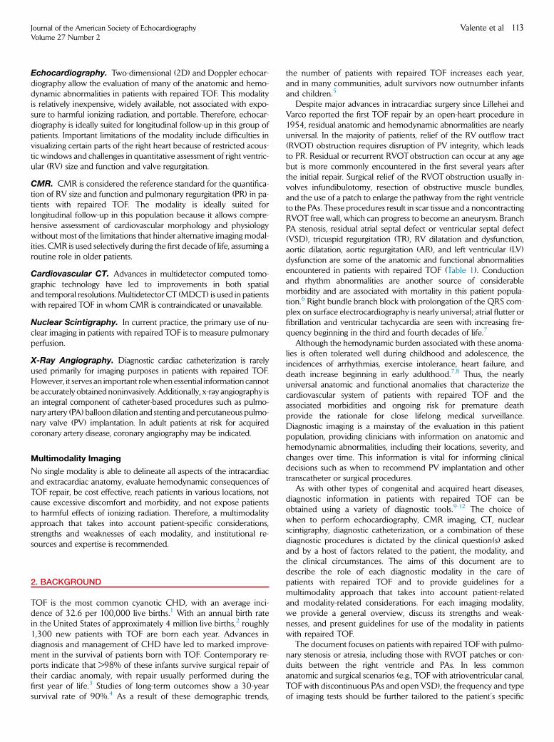
1. EXECUTIVE SUMMARY
Advances in the diagnosis and management of congenital heart dis-ease (CHD) have led to a marked improvement in the survival of pa-tients with tetralogy of Fallot (TOF). However, residual anatomic andhemodynamic abnormalities are common. As with other types ofcongenital and acquired heart diseases, diagnostic information in pa-tients with repaired TOF can be obtained using a variety of diagnostictools. The choice of when to perform echocardiography, cardiovascu-lar magnetic resonance (CMR) imaging, computed tomography (CT),nuclear scintigraphy, diagnostic catheterization, or a combination ofthese diagnostic procedures is dictated by the clinical question(s) askedand by a host of factors related to the patient, the modality, and theclinical circumstances. The aims of this document are to describe therole of each diagnostic modality in the care of patients with repairedTOF and to provide guidelines for amultimodality approach that takesinto account patient-related and modality-related considerations.
Goals of Imaging
The overarching goals of diagnostic imaging are to identify anatomicand functional abnormalities, assess their severity, and provide infor-mation that informs clinical decisions. A list of essential data elementsrequired for optimal management is summarized in section 4.
Imaging Modalities
In the following sections, each of the imaging modalities used for theevaluation of patients with repaired TOF is reviewed, and thestrengths, weaknesses, and clinical utility of each modality are dis-cussed. Finally, we propose an integrated multimodality imagingapproach in this group of patients.
Journal of the American Society of EchocardiographyVolume 27 Number 2
Valente et al 113
Echocardiography. Two-dimensional (2D) and Doppler echocar-diography allow the evaluation of many of the anatomic and hemo-dynamic abnormalities in patients with repaired TOF. This modalityis relatively inexpensive, widely available, not associated with expo-sure to harmful ionizing radiation, and portable. Therefore, echocar-diography is ideally suited for longitudinal follow-up in this group ofpatients. Important limitations of the modality include difficulties invisualizing certain parts of the right heart because of restricted acous-tic windows and challenges in quantitative assessment of right ventric-ular (RV) size and function and valve regurgitation.
CMR. CMR is considered the reference standard for the quantifica-tion of RV size and function and pulmonary regurgitation (PR) in pa-tients with repaired TOF. The modality is ideally suited forlongitudinal follow-up in this population because it allows compre-hensive assessment of cardiovascular morphology and physiologywithout most of the limitations that hinder alternative imaging modal-ities. CMR is used selectively during the first decade of life, assuming aroutine role in older patients.
Cardiovascular CT. Advances in multidetector computed tomo-graphic technology have led to improvements in both spatialand temporal resolutions.Multidetector CT (MDCT) is used in patientswith repaired TOF in whom CMR is contraindicated or unavailable.
Nuclear Scintigraphy. In current practice, the primary use of nu-clear imaging in patients with repaired TOF is to measure pulmonaryperfusion.
X-Ray Angiography. Diagnostic cardiac catheterization is rarelyused primarily for imaging purposes in patients with repaired TOF.However, it serves an important rolewhen essential information cannotbe accurately obtained noninvasively. Additionally, x-ray angiography isan integral component of catheter-based procedures such as pulmo-nary artery (PA) balloon dilation and stenting and percutaneous pulmo-nary valve (PV) implantation. In adult patients at risk for acquiredcoronary artery disease, coronary angiography may be indicated.
Multimodality Imaging
No single modality is able to delineate all aspects of the intracardiacand extracardiac anatomy, evaluate hemodynamic consequences ofTOF repair, be cost effective, reach patients in various locations, notcause excessive discomfort and morbidity, and not expose patientsto harmful effects of ionizing radiation. Therefore, a multimodalityapproach that takes into account patient-specific considerations,strengths and weaknesses of each modality, and institutional re-sources and expertise is recommended.
2. BACKGROUND
TOF is the most common cyanotic CHD, with an average inci-dence of 32.6 per 100,000 live births.1 With an annual birth ratein the United States of approximately 4 million live births,2 roughly1,300 new patients with TOF are born each year. Advances indiagnosis and management of CHD have led to marked improve-ment in the survival of patients born with TOF. Contemporary re-ports indicate that >98% of these infants survive surgical repair oftheir cardiac anomaly, with repair usually performed during thefirst year of life.3 Studies of long-term outcomes show a 30-yearsurvival rate of 90%.4 As a result of these demographic trends,
the number of patients with repaired TOF increases each year,and in many communities, adult survivors now outnumber infantsand children.5
Despite major advances in intracardiac surgery since Lillehei andVarco reported the first TOF repair by an open-heart procedure in1954, residual anatomic and hemodynamic abnormalities are nearlyuniversal. In the majority of patients, relief of the RV outflow tract(RVOT) obstruction requires disruption of PV integrity, which leadsto PR. Residual or recurrent RVOTobstruction can occur at any agebut is more commonly encountered in the first several years afterthe initial repair. Surgical relief of the RVOT obstruction usually in-volves infundibulotomy, resection of obstructive muscle bundles,and the use of a patch to enlarge the pathway from the right ventricleto the PAs. These procedures result in scar tissue and a noncontractingRVOT free wall, which can progress to become an aneurysm. BranchPA stenosis, residual atrial septal defect or ventricular septal defect(VSD), tricuspid regurgitation (TR), RV dilatation and dysfunction,aortic dilatation, aortic regurgitation (AR), and left ventricular (LV)dysfunction are some of the anatomic and functional abnormalitiesencountered in patients with repaired TOF (Table 1). Conductionand rhythm abnormalities are another source of considerablemorbidity and are associated with mortality in this patient popula-tion.6 Right bundle branch block with prolongation of the QRS com-plex on surface electrocardiography is nearly universal; atrial flutter orfibrillation and ventricular tachycardia are seen with increasing fre-quency beginning in the third and fourth decades of life.7
Although the hemodynamic burden associated with these anoma-lies is often tolerated well during childhood and adolescence, theincidences of arrhythmias, exercise intolerance, heart failure, anddeath increase beginning in early adulthood.7,8 Thus, the nearlyuniversal anatomic and functional anomalies that characterize thecardiovascular system of patients with repaired TOF and theassociated morbidities and ongoing risk for premature deathprovide the rationale for close lifelong medical surveillance.Diagnostic imaging is a mainstay of the evaluation in this patientpopulation, providing clinicians with information on anatomic andhemodynamic abnormalities, including their locations, severity, andchanges over time. This information is vital for informing clinicaldecisions such as when to recommend PV implantation and othertranscatheter or surgical procedures.
As with other types of congenital and acquired heart diseases,diagnostic information in patients with repaired TOF can beobtained using a variety of diagnostic tools.9-12 The choice ofwhen to perform echocardiography, CMR imaging, CT, nuclearscintigraphy, diagnostic catheterization, or a combination of thesediagnostic procedures is dictated by the clinical question(s) askedand by a host of factors related to the patient, the modality, andthe clinical circumstances. The aims of this document are todescribe the role of each diagnostic modality in the care ofpatients with repaired TOF and to provide guidelines for amultimodality approach that takes into account patient-relatedand modality-related considerations. For each imaging modality,we provide a general overview, discuss its strengths and weak-nesses, and present guidelines for use of the modality in patientswith repaired TOF.
The document focuses on patients with repaired TOF with pulmo-nary stenosis or atresia, including those with RVOT patches or con-duits between the right ventricle and PAs. In less commonanatomic and surgical scenarios (e.g., TOF with atrioventricular canal,TOF with discontinuous PAs and open VSD), the frequency and typeof imaging tests should be further tailored to the patient’s specific
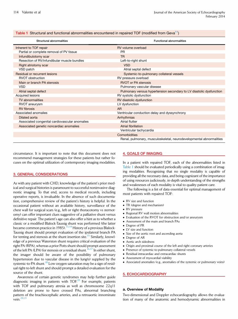
Table 1 Structural and functional abnormalities encountered in repaired TOF (modified from Geva11)
Structural abnormalities Functional abnormalities
Inherent to TOF repair RV volume overload
Partial or complete removal of PV tissue PR
Infundibulotomy scar TR
Resection of RV/infundibular muscle bundles Left-to-right shunt
Right atriotomy scar VSD
VSD patch Atrial septal defect
Residual or recurrent lesions Systemic-to-pulmonary collateral vessels
RVOT obstruction RV pressure overload
Main or branch PA stenosis RVOT or PA stenosis
VSD Pulmonary vascular disease
Atrial septal defect Pulmonary venous hypertension secondary to LV diastolic dysfunction
Acquired lesions RV systolic dysfunction
TV abnormalities RV diastolic dysfunction
RVOT aneurysm LV dysfunction
RV fibrosis AR
Associated anomalies Ventricular conduction delay and dyssynchrony
Dilated aorta Arrhythmias
Associated congenital cardiovascular anomalies Atrial flutter
Associated genetic noncardiac anomalies Atrial fibrillationVentricular tachycardia
ComorbiditiesRenal, pulmonary, musculoskeletal, neurodevelopmental abnormalities
114 Valente et al Journal of the American Society of EchocardiographyFebruary 2014
circumstance. It is important to note that this document does notrecommend management strategies for these patients but rather fo-cuses on the optimal utilization of contemporary imaging modalities.
3. GENERAL CONSIDERATIONS
As with any patient with CHD, knowledge of the patient’s prior med-ical and surgical histories is paramount to successful noninvasive diag-nostic imaging. To that end, access to medical records, includingoperative reports, is invaluable. In the absence of such documenta-tion, comprehensive review of the patient’s history is helpful. In theoccasional patient without an available history, surveillance of thechest wall for surgical scars (e.g., left or right thoracotomy vs sternot-omy) can offer important clues suggestive of a palliative shunt versusdefinitive repair. The patient’s age can also offer a hint as to whether aclassic or a modified Blalock-Taussig shunt was performed (the latterbecame common practice in 1985).13,14 History of a previous Blalock-Taussig shunt should prompt evaluation of the ipsilateral branch PAfor tenting and stenosis at the shunt insertion site.15 Similarly, knowl-edge of a previous Waterston shunt requires critical evaluation of theright PA (RPA), whereas a prior Potts shunt should prompt assessmentof the left PA (LPA) for stenosis or a residual shunt.16,17 In either shunt,the imager should be aware of the possibility of pulmonaryhypertension due to vascular disease in the lung(s) supplied by thesystemic-to-PA shunt.18 Low oxygen saturation may be a sign of resid-ual right-to-left shunt and should prompt a detailed evaluation for thesource of the shunt.
Awareness of certain genetic syndromes may help further guidediagnostic imaging in patients with TOF.19 For example, patientswith TOF and pulmonary atresia as well as chromosome 22q11deletion are prone to have crossed PAs, abnormal branchingpattern of the brachiocephalic arteries, and a retroaortic innominatevein.20,21
4. GOALS OF IMAGING
In a patient with repaired TOF, each of the abnormalities listed inTable 1 should be evaluated periodically using a combination of imag-ing modalities. Recognizing that no single modality is capable ofproviding all the necessary data, and being cognizant of the importanceof using resources judiciously, in-depth understanding of the strengthsand weaknesses of each modality is vital to quality patient care.
The following is a list of data essential for optimal management ofmost patients with repaired TOF:
� RV size and function� TR (degree and mechanism)� RV pressure� Regional RV wall motion abnormalities� Evaluation of the RVOT for obstruction and/or aneurysm� Assessment of the main and branch PAs� Degree of PR� LV size and function� Size of the aortic root and ascending aorta� Degree of AR� Aortic arch sidedness� Origin and proximal course of the left and right coronary arteries� Presence of systemic-to-pulmonary collateral vessels� Residual intracardiac and extracardiac shunts� Assessment of myocardial viability� Associated anomalies (e.g., anomalies of the systemic or pulmonary veins)
5. ECHOCARDIOGRAPHY
a. Overview of Modality
Two-dimensional and Doppler echocardiography allows the evalua-tion of many of the anatomic and hemodynamic abnormalities in
Journal of the American Society of EchocardiographyVolume 27 Number 2
Valente et al 115
patients with repaired TOF. As detailed in subsequent sections, 2Dimaging allows qualitative and quantitative assessments of the rightatrium, right ventricle, RVOT, PAs, tricuspid valve (TV), PV, and atrialand ventricular septa. Given the potential for LV dysfunction, dilata-tion of the proximal aorta, and AR, assessment of the left heart is in-tegral to the echocardiographic assessment of the patient withrepaired TOF. Doppler echocardiography is particularly importantin this population for noninvasive hemodynamic assessment of pa-rameters such as RVand PA pressures. Three-dimensional (3D) echo-cardiography can further aid in delineating anatomic pathology andbiventricular size and function. Transesophageal echocardiography(TEE) is useful in certain patients to guide interventional proceduresor evaluate valve anatomy when transthoracic imaging is challengingor when infective endocarditis is suspected. More recently, the role ofmyocardial deformation imaging for assessment of RV function is atopic of intense investigation in this patient population.22
b. Strength and Limitations
Echocardiography is the primary noninvasive imaging modality in pa-tients with CHD, including those with repaired TOF.9,12 This modalityis relatively inexpensive, widely available, portable, and not associatedwith exposure to harmful ionizing radiation. Importantly, experiencewith echocardiography is extensive, and clinicians caring for thesepatients are familiar with its application.
Important limitations of echocardiography in this group of patientsinclude difficulties in visualizing certain parts of the right heartbecause of restricted acoustic windows and challenges in quantitativeassessments of RV size and function and valve regurgitation. Theselimitations are usually of lesser concern during the first decade oflife because the acoustic windows are not as restricted as in older pa-tients with larger body sizes and because themajority of these patientstend to be clinically well. Once patients reach adolescence and adult-hood, acoustic windows tend to become more challenging, and theimportance of accurate quantitative assessment of RV size and func-tion requires the complementary use of echocardiography and othermodalities, most often CMR.
c. Assessment of Repaired TOF with Echocardiography
RVOT. In young patients with good subcostal windows, the RVOTcan be evaluated in the long-axis and short-axis planes and in theinflow-outflow view, analogous to the angiographic right anterior ob-lique plane (transducer mark at 1–2 o’clock). In the great majority ofpatients, the RVOT can be thoroughly evaluated from the parasternallong-axis and short-axis views, which facilitates assessment of theinfundibulum and any associated aneurysm, residual PV tissue, andmain and branch PAs. The RVOT dimension at the site of the formerPV is important when transcatheter valve implantation is beingconsidered. Attention is paid to the presence of RV hypertrophy,which is defined in adults as RV diastolic wall thickness > 5 mm.23
In most patients with repaired TOF, however, the infundibular freewall is composed of a patch, and its thickness might not reflect RV hy-pertrophy in other parts of the chamber.
When evaluating for residual RVOTobstruction, it is important toidentify the site of obstruction using color, pulsed-wave, andcontinuous-wave Doppler. In the case of an RV-to-PA conduit, theobstruction can be at any location along the entire length of theconduit, with or without involvement of the origins of the PAs. Thespectral Doppler flow profile across the RVOT can help differentiatebetween dynamic obstruction within the RV cavity and residual val-var or supravalvar stenosis. The former has late peaking of the
Doppler signal (‘‘lobster claw’’ shape), whereas the latter has midsys-tolic peaking. In patients with normal cardiac output, a peak instanta-neous gradient of >4 m/sec (>64 mm Hg) is considered severestenosis, 3 to 4 m/sec (36–64 mmHg) is considered moderate steno-sis, and <3 m/sec (<36 mm Hg) is considered mild stenosis.24
Multiple levels of obstructions in the RVOTand PAs pose a partic-ular challenge to differentiate the contribution at each level, especiallywhen they are in close proximity. In older patients or in patients withRV-to-PA conduits, the branch PAs are sometimes difficult to assessfor residual stenosis because of the high flow velocity within theconduit. A high TR jet velocity should prompt a careful search forRVOTor PA obstruction at some level.
PAs. The PAs are evaluated using a combination of 2D imaging andcolor and spectral Doppler. The suprasternal and high left and rightparasternal windows are used to image the mediastinal PAs, althoughimaging may be challenging in patients with larger body habitus. Themain PA (MPA) is measured at its midpoint during systole. Whensupravalvar PA stenosis is present, the smallest diameter is measuredas well. The diameters of the branch PAs are measured at the level ofthe origin, and the smallest dimension of any stenotic segment shouldbe reported. The subcostal long-axis, high left parasternal short-axis,suprasternal short-axis, and right parasternal short-axis views are help-ful in visualizing the length of the RPA. The high left parasternal short-axis and the suprasternal long-axis (near parasagittal plane angled tothe left) views are useful in visualizing the length of the LPA. The distalLPA is more difficult to image than the RPA because of interferencefrom air in the lung and the left bronchus.
PR. PR, occurring as a result of transannular patching, pulmonaryvalvotomy or valvectomy, or any other procedure that disrupts thevalve, is an important factor in the long-term outcomes of patientswith repaired TOF.25-27 It is a key initiating element in apathophysiologic cascade that leads to RV dilatation anddysfunction, which in turn has been linked to secondary TR,decreased exercise capacity, and increased risk for atrial andventricular arrhythmias, as well as sudden cardiac death.28-30
Therefore, evaluation of PR is an essential component of theechocardiographic examination in these patients.
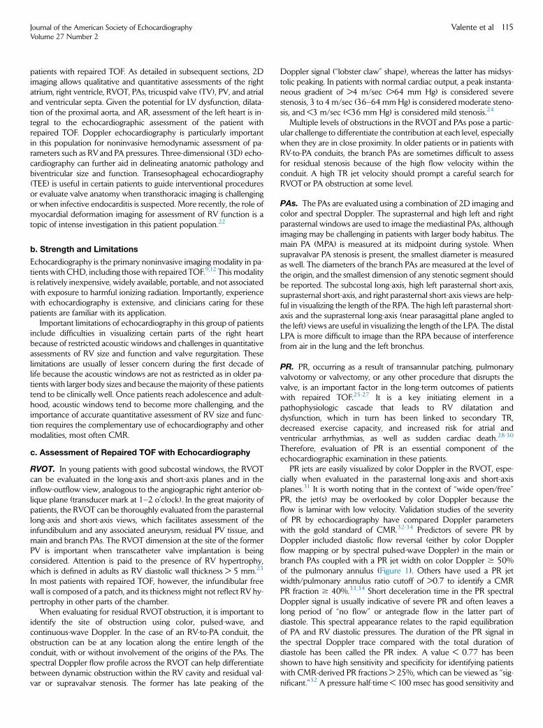
PR jets are easily visualized by color Doppler in the RVOT, espe-cially when evaluated in the parasternal long-axis and short-axisplanes.31 It is worth noting that in the context of ‘‘wide open/free’’PR, the jet(s) may be overlooked by color Doppler because theflow is laminar with low velocity. Validation studies of the severityof PR by echocardiography have compared Doppler parameterswith the gold standard of CMR.32-34 Predictors of severe PR byDoppler included diastolic flow reversal (either by color Dopplerflow mapping or by spectral pulsed-wave Doppler) in the main orbranch PAs coupled with a PR jet width on color Doppler $ 50%of the pulmonary annulus (Figure 1). Others have used a PR jetwidth/pulmonary annulus ratio cutoff of >0.7 to identify a CMRPR fraction $ 40%.33,34 Short deceleration time in the PR spectralDoppler signal is usually indicative of severe PR and often leaves along period of ‘‘no flow’’ or antegrade flow in the latter part ofdiastole. This spectral appearance relates to the rapid equilibrationof PA and RV diastolic pressures. The duration of the PR signal inthe spectral Doppler trace compared with the total duration ofdiastole has been called the PR index. A value < 0.77 has beenshown to have high sensitivity and specificity for identifying patientswith CMR-derived PR fractions > 25%, which can be viewed as ‘‘sig-nificant.’’32 A pressure half-time < 100 msec has good sensitivity and
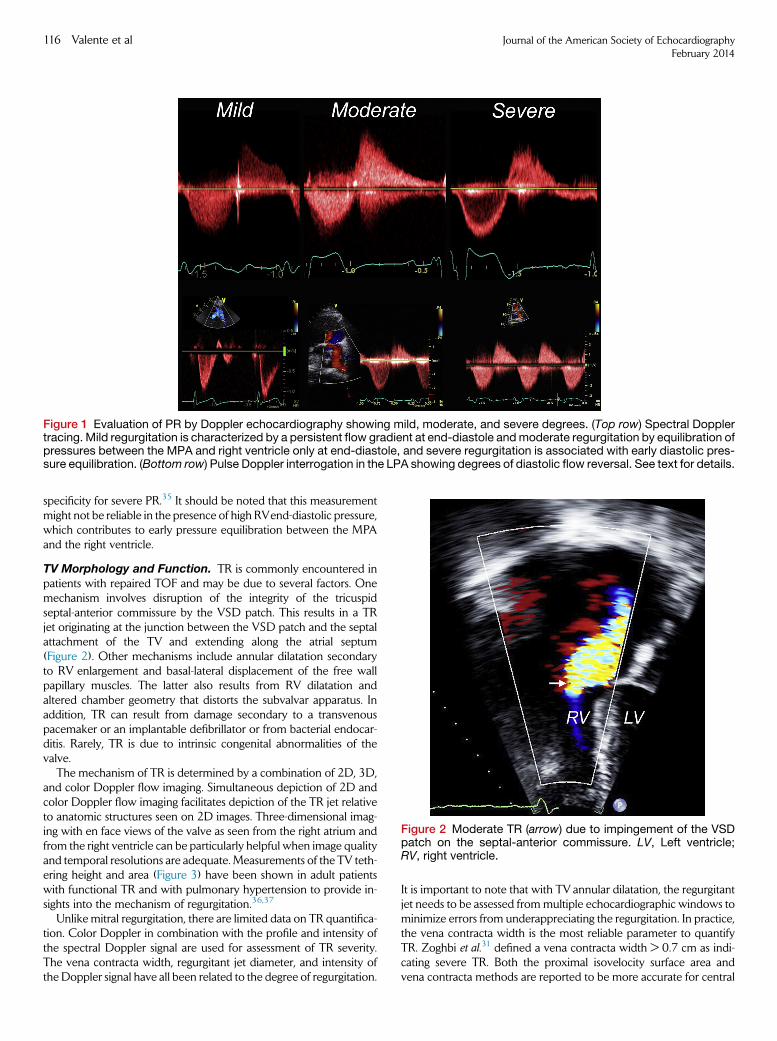
Figure 1 Evaluation of PR by Doppler echocardiography showing mild, moderate, and severe degrees. (Top row) Spectral Dopplertracing. Mild regurgitation is characterized by a persistent flow gradient at end-diastole andmoderate regurgitation by equilibration ofpressures between the MPA and right ventricle only at end-diastole, and severe regurgitation is associated with early diastolic pres-sure equilibration. (Bottom row) Pulse Doppler interrogation in the LPA showing degrees of diastolic flow reversal. See text for details.
Figure 2 Moderate TR (arrow) due to impingement of the VSDpatch on the septal-anterior commissure. LV, Left ventricle;RV, right ventricle.
116 Valente et al Journal of the American Society of EchocardiographyFebruary 2014
specificity for severe PR.35 It should be noted that this measurementmight not be reliable in the presence of high RVend-diastolic pressure,which contributes to early pressure equilibration between the MPAand the right ventricle.
TV Morphology and Function. TR is commonly encountered inpatients with repaired TOF and may be due to several factors. Onemechanism involves disruption of the integrity of the tricuspidseptal-anterior commissure by the VSD patch. This results in a TRjet originating at the junction between the VSD patch and the septalattachment of the TV and extending along the atrial septum(Figure 2). Other mechanisms include annular dilatation secondaryto RV enlargement and basal-lateral displacement of the free wallpapillary muscles. The latter also results from RV dilatation andaltered chamber geometry that distorts the subvalvar apparatus. Inaddition, TR can result from damage secondary to a transvenouspacemaker or an implantable defibrillator or from bacterial endocar-ditis. Rarely, TR is due to intrinsic congenital abnormalities of thevalve.
The mechanism of TR is determined by a combination of 2D, 3D,and color Doppler flow imaging. Simultaneous depiction of 2D andcolor Doppler flow imaging facilitates depiction of the TR jet relativeto anatomic structures seen on 2D images. Three-dimensional imag-ing with en face views of the valve as seen from the right atrium andfrom the right ventricle can be particularly helpful when image qualityand temporal resolutions are adequate. Measurements of the TV teth-ering height and area (Figure 3) have been shown in adult patientswith functional TR and with pulmonary hypertension to provide in-sights into the mechanism of regurgitation.36,37
Unlike mitral regurgitation, there are limited data on TR quantifica-tion. Color Doppler in combination with the profile and intensity ofthe spectral Doppler signal are used for assessment of TR severity.The vena contracta width, regurgitant jet diameter, and intensity ofthe Doppler signal have all been related to the degree of regurgitation.
It is important to note that with TV annular dilatation, the regurgitantjet needs to be assessed frommultiple echocardiographic windows tominimize errors from underappreciating the regurgitation. In practice,the vena contracta width is the most reliable parameter to quantifyTR. Zoghbi et al.31 defined a vena contracta width > 0.7 cm as indi-cating severe TR. Both the proximal isovelocity surface area andvena contracta methods are reported to be more accurate for central
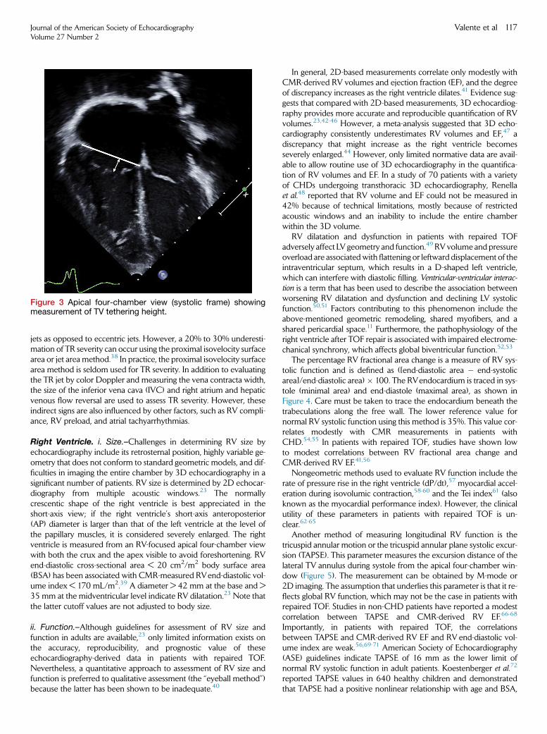
Figure 3 Apical four-chamber view (systolic frame) showingmeasurement of TV tethering height.
Journal of the American Society of EchocardiographyVolume 27 Number 2
Valente et al 117
jets as opposed to eccentric jets. However, a 20% to 30% underesti-mation of TR severity can occur using the proximal isovelocity surfacearea or jet area method.38 In practice, the proximal isovelocity surfacearea method is seldom used for TR severity. In addition to evaluatingthe TR jet by color Doppler and measuring the vena contracta width,the size of the inferior vena cava (IVC) and right atrium and hepaticvenous flow reversal are used to assess TR severity. However, theseindirect signs are also influenced by other factors, such as RV compli-ance, RV preload, and atrial tachyarrhythmias.
Right Ventricle. i. Size.–Challenges in determining RV size byechocardiography include its retrosternal position, highly variable ge-ometry that does not conform to standard geometric models, and dif-ficulties in imaging the entire chamber by 3D echocardiography in asignificant number of patients. RV size is determined by 2D echocar-diography from multiple acoustic windows.23 The normallycrescentic shape of the right ventricle is best appreciated in theshort-axis view; if the right ventricle’s short-axis anteroposterior(AP) diameter is larger than that of the left ventricle at the level ofthe papillary muscles, it is considered severely enlarged. The rightventricle is measured from an RV-focused apical four-chamber viewwith both the crux and the apex visible to avoid foreshortening. RVend-diastolic cross-sectional area < 20 cm2/m2 body surface area(BSA) has been associated with CMR-measured RVend-diastolic vol-ume index < 170 mL/m2.39 A diameter > 42 mm at the base and >35 mm at the midventricular level indicate RV dilatation.23 Note thatthe latter cutoff values are not adjusted to body size.
ii. Function.–Although guidelines for assessment of RV size andfunction in adults are available,23 only limited information exists onthe accuracy, reproducibility, and prognostic value of theseechocardiography-derived data in patients with repaired TOF.Nevertheless, a quantitative approach to assessment of RV size andfunction is preferred to qualitative assessment (the ‘‘eyeball method’’)because the latter has been shown to be inadequate.40
In general, 2D-based measurements correlate only modestly withCMR-derived RV volumes and ejection fraction (EF), and the degreeof discrepancy increases as the right ventricle dilates.41 Evidence sug-gests that compared with 2D-based measurements, 3D echocardiog-raphy provides more accurate and reproducible quantification of RVvolumes.23,42-46 However, a meta-analysis suggested that 3D echo-cardiography consistently underestimates RV volumes and EF,47 adiscrepancy that might increase as the right ventricle becomesseverely enlarged.44 However, only limited normative data are avail-able to allow routine use of 3D echocardiography in the quantifica-tion of RV volumes and EF. In a study of 70 patients with a varietyof CHDs undergoing transthoracic 3D echocardiography, Renellaet al.48 reported that RV volume and EF could not be measured in42% because of technical limitations, mostly because of restrictedacoustic windows and an inability to include the entire chamberwithin the 3D volume.
RV dilatation and dysfunction in patients with repaired TOFadversely affect LV geometry and function.49 RVvolume and pressureoverload are associatedwith flattening or leftward displacement of theintraventricular septum, which results in a D-shaped left ventricle,which can interfere with diastolic filling. Ventricular-ventricular interac-tion is a term that has been used to describe the association betweenworsening RV dilatation and dysfunction and declining LV systolicfunction.50,51 Factors contributing to this phenomenon include theabove-mentioned geometric remodeling, shared myofibers, and ashared pericardial space.11 Furthermore, the pathophysiology of theright ventricle after TOF repair is associated with impaired electrome-chanical synchrony, which affects global biventricular function.52,53
The percentage RV fractional area change is a measure of RV sys-tolic function and is defined as ([end-diastolic area � end-systolicarea]/end-diastolic area)� 100. The RVendocardium is traced in sys-tole (minimal area) and end-diastole (maximal area), as shown inFigure 4. Care must be taken to trace the endocardium beneath thetrabeculations along the free wall. The lower reference value fornormal RV systolic function using this method is 35%. This value cor-relates modestly with CMR measurements in patients withCHD.54,55 In patients with repaired TOF, studies have shown lowto modest correlations between RV fractional area change andCMR-derived RV EF.41,56
Nongeometric methods used to evaluate RV function include therate of pressure rise in the right ventricle (dP/dt),57 myocardial accel-eration during isovolumic contraction,58-60 and the Tei index61 (alsoknown as the myocardial performance index). However, the clinicalutility of these parameters in patients with repaired TOF is un-clear.62-65
Another method of measuring longitudinal RV function is thetricuspid annular motion or the tricuspid annular plane systolic excur-sion (TAPSE). This parameter measures the excursion distance of thelateral TV annulus during systole from the apical four-chamber win-dow (Figure 5). The measurement can be obtained by M-mode or2D imaging. The assumption that underlies this parameter is that it re-flects global RV function, which may not be the case in patients withrepaired TOF. Studies in non-CHD patients have reported a modestcorrelation between TAPSE and CMR-derived RV EF.66-68
Importantly, in patients with repaired TOF, the correlationsbetween TAPSE and CMR-derived RV EF and RV end-diastolic vol-ume index are weak.56,69-71 American Society of Echocardiography(ASE) guidelines indicate TAPSE of 16 mm as the lower limit ofnormal RV systolic function in adult patients. Koestenberger et al.72
reported TAPSE values in 640 healthy children and demonstratedthat TAPSE had a positive nonlinear relationship with age and BSA,
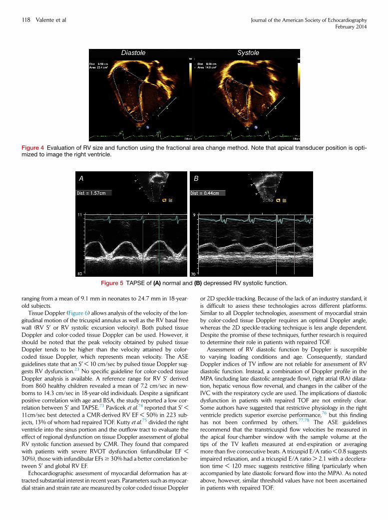
Figure 4 Evaluation of RV size and function using the fractional area change method. Note that apical transducer position is opti-mized to image the right ventricle.
Figure 5 TAPSE of (A) normal and (B) depressed RV systolic function.
118 Valente et al Journal of the American Society of EchocardiographyFebruary 2014
ranging from a mean of 9.1 mm in neonates to 24.7 mm in 18-year-old subjects.
Tissue Doppler (Figure 6) allows analysis of the velocity of the lon-gitudinal motion of the tricuspid annulus as well as the RV basal freewall (RV S0 or RV systolic excursion velocity). Both pulsed tissueDoppler and color-coded tissue Doppler can be used. However, itshould be noted that the peak velocity obtained by pulsed tissueDoppler tends to be higher than the velocity attained by color-coded tissue Doppler, which represents mean velocity. The ASEguidelines state that an S0 < 10 cm/sec by pulsed tissue Doppler sug-gests RV dysfunction.23 No specific guideline for color-coded tissueDoppler analysis is available. A reference range for RV S0 derivedfrom 860 healthy children revealed a mean of 7.2 cm/sec in new-borns to 14.3 cm/sec in 18-year-old individuals. Despite a significantpositive correlation with age and BSA, the study reported a low cor-relation between S0 and TAPSE.73 Pavlicek et al.74 reported that S0 <11cm/sec best detected a CMR-derived RV EF < 50% in 223 sub-jects, 13% of whom had repaired TOF. Kutty et al.75 divided the rightventricle into the sinus portion and the outflow tract to evaluate theeffect of regional dysfunction on tissue Doppler assessment of globalRV systolic function assessed by CMR. They found that comparedwith patients with severe RVOT dysfunction (infundibular EF <30%), those with infundibular EFs$ 30% had a better correlation be-tween S0 and global RV EF.
Echocardiographic assessment of myocardial deformation has at-tracted substantial interest in recent years. Parameters such asmyocar-dial strain and strain rate are measured by color-coded tissue Doppler
or 2D speckle-tracking. Because of the lack of an industry standard, itis difficult to assess these technologies across different platforms.Similar to all Doppler technologies, assessment of myocardial strainby color-coded tissue Doppler requires an optimal Doppler angle,whereas the 2D speckle-tracking technique is less angle dependent.Despite the promise of these techniques, further research is requiredto determine their role in patients with repaired TOF.
Assessment of RV diastolic function by Doppler is susceptibleto varying loading conditions and age. Consequently, standardDoppler indices of TV inflow are not reliable for assessment of RVdiastolic function. Instead, a combination of Doppler profile in theMPA (including late diastolic antegrade flow), right atrial (RA) dilata-tion, hepatic venous flow reversal, and changes in the caliber of theIVC with the respiratory cycle are used. The implications of diastolicdysfunction in patients with repaired TOF are not entirely clear.Some authors have suggested that restrictive physiology in the rightventricle predicts superior exercise performance,76 but this findinghas not been confirmed by others.77,78 The ASE guidelinesrecommend that the transtricuspid flow velocities be measured inthe apical four-chamber window with the sample volume at thetips of the TV leaflets measured at end-expiration or averagingmore than five consecutive beats. A tricuspid E/A ratio < 0.8 suggestsimpaired relaxation, and a tricuspid E/A ratio > 2.1 with a decelera-tion time < 120 msec suggests restrictive filling (particularly whenaccompanied by late diastolic forward flow into the MPA). As notedabove, however, similar threshold values have not been ascertainedin patients with repaired TOF.
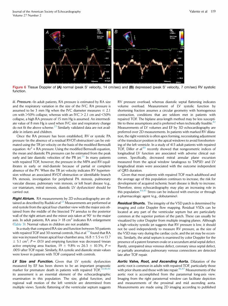
Figure 6 Tissue Doppler of (A) normal (peak S0 velocity, 14 cm/sec) and (B) depressed (peak S0 velocity, 7 cm/sec) RV systolicfunction.
Journal of the American Society of EchocardiographyVolume 27 Number 2
Valente et al 119
iii. Pressure.–In adult patients, RA pressure is estimated by RA sizeand the respiratory variation in the size of the IVC. RA pressure isassumed to be 3 mm Hg when the IVC diameter measures # 2.1cm with >50% collapse, whereas with an IVC > 2.1 cm and <50%collapse, a high RA pressure of 15 mm Hg is assumed. An intermedi-ate value of 8 mm Hg is used when IVC size and respiratory changedo not fit the above scheme.23 Similarly validated data are not avail-able in infants and children.
Once the RA pressure has been established, RV or systolic PApressure (in the absence of a residual RVOTobstruction) can be esti-mated using the TR jet velocity on the basis of the modified Bernoulliequation: 4v2 + RA pressure. Using the modified Bernoulli equation,the mean and diastolic PA pressures can be estimated from the peakearly and late diastolic velocities of the PR jet.23 In many patientswith repaired TOF, however, the pressure in the MPA and RVequil-ibrates in early or mid-diastole because of partial or completeabsence of the PV. When the TR jet velocity indicates RV hyperten-sion without an associated RVOT obstruction or identifiable branchPA stenosis, investigation for peripheral PA stenosis, pulmonaryvascular disease, pulmonary vein stenosis, or left heart disease (e.g.,cor triatriatum, mitral stenosis, diastolic LV dysfunction) should becarried out.
Right Atrium. RA measurements by 2D echocardiography are ob-tained as described by Rudski et al.23 Measurements are performed atend-systole from the apical four-chamber viewwith themajor axis ob-tained from the middle of the bisected TV annulus to the posteriorwall of the right atrium and the minor axis taken at 90� to the majoraxis. In adult patients, RA area > 18 cm2 indicates RA enlargement(Table 3). Normal values in children are not available.
In a study that compared RA size and function between 50 patientswith repaired TOF and 30 normal controls, Hui et al.79 found that RAsize was increased (mean apical four-chamber area, 16.86 5.5 vs 13.76 5.1 cm2; P = .013) and emptying function was decreased (meanactive emptying area fraction, 19 6 9.8% vs 26.3 6 10.3%, P =.005) after TOF repair. Similarly, RA systolic and diastolic strain valueswere lower in patients with TOF compared with controls.
LV Size and Function. Given that LV systolic dysfunctionmeasured by EF has been shown to be an important prognosticmarker for premature death in patients with repaired TOF,50,80,81
its assessment is an essential element of the echocardiographicexamination in this population. The size, global function, andregional wall motion of the left ventricle are determined frommultiple views. Systolic flattening of the ventricular septum suggests
RV pressure overload, whereas diastolic septal flattening indicatesvolume overload. Measurement of LV systolic function byshortening fraction assumes a circular geometry with homogenouscontraction, conditions that are seldom met in patients withrepaired TOF. The biplane area-length method may be less suscepti-ble to these assumptions and is preferred when technically feasible.82
Measurements of LV volumes and EF by 3D echocardiography arepreferred over 2D measurements. In patients with marked RV dilata-tion, the right ventricle is often apex forming, necessitating adjustmentof the transducer position in the apical windows to avoid foreshorten-ing of the left ventricle. In a study of 413 adult patients with repairedTOF, Diller et al.83 recently showed that nongeometric indices oflongitudinal LV function are associated with adverse clinical out-comes. Specifically, decreased mitral annular plane excursionmeasured from the apical window (analogous to TAPSE) and LVlongitudinal strain were associated with the outcome independentof QRS duration.
Given that most patients with repaired TOF reach adulthood andthe average age of this population continues to increase, the risk fordevelopment of acquired ischemic heart disease is likely to increase.Therefore, stress echocardiography may play an increasing role inthis population.84,85 Stress can be induced with exercise or througha pharmacologic agent (e.g., dobutamine).
Residual Shunts. The integrity of the VSD patch is determined byimaging and color Doppler flow mapping. Residual VSDs can belocated at any part of the ventricular septum but are particularlycommon at the superior portion of the patch. These can usually bevisualized by color Doppler from multiple imaging planes. Althougha high-velocity systolic jet suggests that RV pressure is low, it shouldnot be used independently to measure RV pressure, as the size ofthe VSDmay vary during the cardiac cycle, and the jet may be eccen-tric. Similarly, the atrial septum is examined by color Doppler for thepresence of a patent foramen ovale or a secundum atrial septal defect.Rarely, unrepaired sinus venosus defect, coronary sinus septal defect,or partially anomalous pulmonary venous connections are detectedlate after TOF repair.
Aortic Valve, Root, and Ascending Aorta. Dilatation of theaortic root is common in adults with repaired TOF, particularly thosewith prior shunts and those with late repair.86,87 Measurements of theaortic root is accomplished from the parasternal long-axis view.Imaging from the right parasternal window can facilitate depictionand measurements of the proximal and mid ascending aorta.Measurements are made using 2D imaging according to published
120 Valente et al Journal of the American Society of EchocardiographyFebruary 2014
guidelines in CHD during maximal expansion (mid to late systole).88
Measurement of internal diameter is the current standard.88,89
Other Cardiovascular Issues. Confirmation of aortic arch sided-ness is accomplished by imaging from the suprasternal short-axisaortic arch plane using both anterior and posterior tilt of the trans-ducer to display the ascending and descending aorta. When the aorticarch is right-sided (as it is in about 25% of patients with TOF), the de-scending aorta may be seen coursing to the right. Additionally, iden-tification of the innominate artery and its bifurcation into the leftsubclavian and left common carotid arteries is possible from thesuprasternal window.
Evaluation of the origin and proximal course of the left and rightcoronary arteries can be clinically relevant in patients with repairedTOF. Although in many patients, this information is known before orduring reparative surgery, previously undetected abnormal coronaryanatomy can complicate reoperation or catheter intervention. Themajor challenge in echocardiographic delineation of coronary anatomylate after TOF repair relates to suboptimal acoustic windows.
d. Standard Protocol
Patient Preparation. Review of the patient’s medical and surgicalhistory, current medical condition, the specific questions for the echo-cardiographic study, and the patient’s ability to cooperate with the ex-amination should be ascertained before the scan commences. Thepatient’s weight and height must be measured and recorded alongwith calculation of the BSA using an appropriate formula, such asthe Haycock formula.90 It is worth noting that the formula ofDuBois and DuBois91 does not cover the ranges of ages and bodysizes encountered in patients with CHD and should not be used.Documentation of the body mass index may also be helpful.Additional steps taken before imaging begins include placement ofelectrocardiographic leads that avoid the standard echocardiographicimaging sites and recording the blood pressure and the heart rate.When pertinent, the blood pressure should be recorded on the oppo-site side of a previous Blalock-Taussig shunt.
Scanning Protocol. The ASE guidelines published in 2006 detailscanning protocols for the performance of pediatric echocardiogra-phy in patients with CHD,92 and guidelines published in 2010 specifymeasurement techniques.88 Importantly, each laboratory shouldfollow a consistent detailed protocol that addresses the pertinent clin-ical issues in patients with repaired TOF. A sample echocardiographicprotocol for patients with repaired TOF is listed in Table 2, and refer-ence values for selected measurements are shown in Table 3.
e. Reporting Elements and Measurements
� RVOT and MPA◦ Dimensions◦ Location and mechanism of obstruction by 2D, color, and spectral
Doppler◦ Presence of an aneurysm◦ Peak and mean gradients
� RV-to-PA conduit: peak and mean gradients� Degree of PR on the basis of (1) regurgitation jet width by color Doppler, (2)spectral Doppler noting the duration and the slope (or pressure half-time) ofthe regurgitation jet, (3) presence and degree of flow reversal in the branchPAs, and (4) RPA pulsatility (systolic-diastolic diameter ratio)93
� Branch PAs◦ Dimensions of the narrowest and maximal segments◦ Location and severity of obstruction by 2D, color, and spectral Doppler
� Degree and mechanism of TR; if more than mild, measure (1) vena con-tracta width and (2) tethering height and area (optional)
� RV pressure on the basis of (1) TR jet velocity, (2) trans-VSD gradient, (3)systolic septal configuration (note the presence of right bundle branch blockor septal dyskinesis)
� RV size and volume load on the basis of (1) TV annular diameter, (2) dia-stolic septal flattening, and (3) measurements of RV size as detailed above
� RV function on the basis of the parameters detailed above� Residual VSDs: location, size, direction of flow, and peak transseptalgradient
� Residual atrial septal defects: location, size, and direction of flow� Aortic dimensions: annulus, root, and ascending aorta at the level of the RPA� AR� Systemic-to-pulmonary collateral vessels on the basis of color Doppler inter-rogation and spectral Doppler evaluation of the abdominal aorta for dia-stolic runoff
� LV size and function
f. Transesophageal and Intracardiac Echocardiography
The primary role of TEE in patients with repaired TOF is for intraoper-ative assessment in conjunction with late interventions such as PVreplacement and TV repair. Pre–cardiopulmonary bypass TEE maybe performed to evaluate the atrial septum for the presence of a pat-ent foramen ovale and the TV for the degree and mechanism of TR.TEE may be especially instrumental in the evaluation of the TV andPVas well as RV-to-PA conduits or the implanted PV for the presenceof vegetations that were not detected by transthoracic imaging. Aftercardiopulmonary bypass, TEE is used to evaluate the repair and forfunctional assessment of the ventricles and valves. TEE or intracardiacechocardiography may also be used for guidance of transcatheter PVimplantation94 and device closure of residual atrial septal defects.
g. Recommendations
The committee recommends comprehensive echocardiographicevaluation for longitudinal follow-up of patients with repaired TOFthrough the use of a standardized examination protocol. Given thatthe management of chronic PR is a subject of intense research withongoing evolution of therapies such as transcatheter PV implantation,the echocardiographic imaging protocol should be updated periodi-cally as new information emerges. Integration of echocardiographicdata with information from other modalities is imperative for optimalmanagement of these patients, particularly given limitations of acous-tic windows and assumptions of RV geometry that may well fail.
6. CARDIOVASCULAR MAGNETIC RESONANCE IMAGING
a. Overview of Modality
CMR is considered the reference standard for the quantification ofRV size, RV function, and PR in patients with repaired TOF.10
Through the use of multiple CMR techniques, different morphologicand hemodynamic aspects of the relevant pathophysiology are eval-uated. In the context of this discussion, the most widely used imagingsequence is steady-state free precession (SSFP), which is a type ofgradient-echo technique characterized by high signal-to-noise ratio,high T2/T1 contrast ratio, and sharp borders between the bloodpool and the myocardium.95 Electrocardiographically gated SSFPcan be used as a cine magnetic resonance sequence, which is typicallyused for assessment of ventricular size and function, valve function,and intracardiac and extracardiac anatomy. Alternatively, an
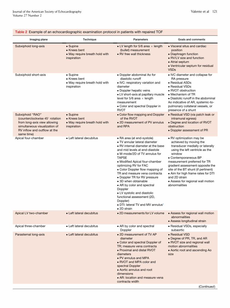
Table 2 Example of an echocardiographic examination protocol in patients with repaired TOF
Imaging plane Technique Parameters Goals and comments
Subxiphoid long-axis � Supine
� Knees bent
� May require breath hold withinspiration
� LV length for 5/6 area � length
(bullet) measurement
� RV free wall thickness
� Visceral situs and cardiac
position
� Diaphragm function� RV/LV size and function
� Atrial septum
� Ventricular septum for residual
VSDs
Subxiphoid short-axis � Supine
� Knees bent
� May require breath hold withinspiration
� Doppler abdominal Ao for
diastolic runoff
� IVC: respiratory variation anddiameter
� Doppler hepatic veins
� LV short-axis at papillary musclelevel for 5/6 area � length
measurement
� Color and spectral Doppler in
RVOT
� IVC diameter and collapse for
RA pressure
� Residual ASDs� Residual VSDs
� RVOT obstruction
� Mechanism of TR� Diastolic runoff in the abdominal
Ao indicative of AR, systemic-to-
pulmonary collateral vessels, or
presence of a shunt
Subxiphoid ‘‘RAO’’
(counterclockwise 45� rotationfrom long-axis view allowing
simultaneous visualization of
RV inflow and outflow at the
same time)
� Supine
� Knees bent� May require breath hold with
inspiration
�Color flowmapping and Doppler
of the RVOT� 2D measurement of PV annulus
and RPA
� Residual VSD (via patch leak or
intramural egress)� Degree and location of RVOT
obstruction
� Doppler assessment of PR
Apical four-chamber � Left lateral decubitus � RA area (at end-systole)
� TV annular lateral diameter
� RV internal diameter at the baseand mid levels at end-diastole
� M-mode/2D of TV annulus for
TAPSE
� Modified Apical four-chamberoptimizing RV for FAC
� Color Doppler flow mapping of
TR and measure vena contracta
� Doppler TR for RV pressure� 3D when obtainable
� AR by color and spectral
Doppler� LV systolic and diastolic
functional assessment (2D,
Doppler)
� DTI: lateral TV and MV annulus*� 2D strain
� RV optimization may be
achieved by moving the
transducer medially or laterallyusing the left ventricle as the
window
� Contemporaneous BP
measurement preferred for TRgradient assessment opposite the
site of the BT shunt (if pertinent)
� Aim for high frame rates for DTI
and 2D strain� Assess for regional wall motion
abnormalities
Apical LV two-chamber � Left lateral decubitus � 2Dmeasurements for LV volume � Assess for regional wall motionabnormalities
� Assess longitudinal strain
Apical three-chamber � Left lateral decubitus � AR by color and spectralDoppler
� Residual VSDs, especiallysubaortic
Parasternal long-axis � Left lateral decubitus � 2D measurement of TV AP
diameter
� Color and spectral Doppler of
TR; measure vena contracta� Proximal and distal RVOT
diameters
� PV annulus and MPA
� RVOT and MPA color andspectral Doppler
� Aortic annulus and root
dimensions� AR: location and measure vena
contracta width
� Residual VSD
� Degree of PR, TR, and AR
� RVOT size and regional wall
motion abnormalities� Aortic root and ascending Ao
size
(Continued )
Journal of the American Society of EchocardiographyVolume 27 Number 2
Valente et al 121
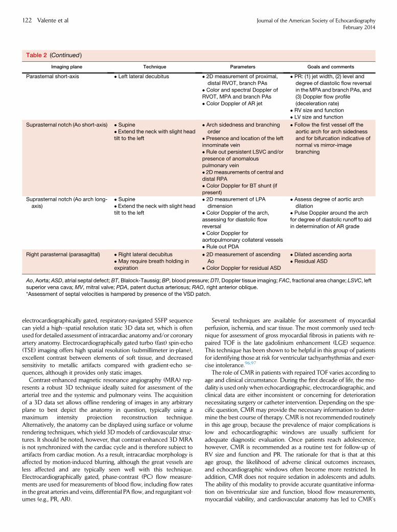
Table 2 (Continued )
Imaging plane Technique Parameters Goals and comments
Parasternal short-axis � Left lateral decubitus � 2D measurement of proximal,
distal RVOT, branch PAs
� Color and spectral Doppler ofRVOT, MPA and branch PAs
� Color Doppler of AR jet
� PR: (1) jet width, (2) level and
degree of diastolic flow reversal
in theMPA and branch PAs, and(3) Doppler flow profile
(deceleration rate)
� RV size and function
� LV size and function
Suprasternal notch (Ao short-axis) � Supine
� Extend the neck with slight headtilt to the left
� Arch sidedness and branching
order� Presence and location of the left
innominate vein
� Rule out persistent LSVC and/or
presence of anomalouspulmonary vein
� 2Dmeasurements of central and
distal RPA� Color Doppler for BT shunt (if
present)
� Follow the first vessel off the
aortic arch for arch sidednessand for bifurcation indicative of
normal vs mirror-image
branching
Suprasternal notch (Ao arch long-
axis)
� Supine
� Extend the neck with slight headtilt to the left
� 2D measurement of LPA
dimension� Color Doppler of the arch,
assessing for diastolic flow
reversal
� Color Doppler foraortopulmonary collateral vessels
� Rule out PDA
� Assess degree of aortic arch
dilation� Pulse Doppler around the arch
for degree of diastolic runoff to aid
in determination of AR grade
Right parasternal (parasagittal) � Right lateral decubitus
� May require breath holding in
expiration
� 2D measurement of ascending
Ao
� Color Doppler for residual ASD
� Dilated ascending aorta
� Residual ASD
Ao, Aorta; ASD, atrial septal defect; BT, Blalock-Taussig; BP, blood pressure;DTI, Doppler tissue imaging; FAC, fractional area change; LSVC, leftsuperior vena cava; MV, mitral valve; PDA, patent ductus arteriosus; RAO, right anterior oblique.
*Assessment of septal velocities is hampered by presence of the VSD patch.
122 Valente et al Journal of the American Society of EchocardiographyFebruary 2014
electrocardiographically gated, respiratory-navigated SSFP sequencecan yield a high–spatial resolution static 3D data set, which is oftenused for detailed assessment of intracardiac anatomy and/or coronaryartery anatomy. Electrocardiographically gated turbo (fast) spin-echo(TSE) imaging offers high spatial resolution (submillimeter in-plane),excellent contrast between elements of soft tissue, and decreasedsensitivity to metallic artifacts compared with gradient-echo se-quences, although it provides only static images.
Contrast-enhanced magnetic resonance angiography (MRA) rep-resents a robust 3D technique ideally suited for assessment of thearterial tree and the systemic and pulmonary veins. The acquisitionof a 3D data set allows offline rendering of images in any arbitraryplane to best depict the anatomy in question, typically using amaximum intensity projection reconstruction technique.Alternatively, the anatomy can be displayed using surface or volumerendering techniques, which yield 3D models of cardiovascular struc-tures. It should be noted, however, that contrast-enhanced 3D MRAis not synchronized with the cardiac cycle and is therefore subject toartifacts from cardiac motion. As a result, intracardiac morphology isaffected by motion-induced blurring, although the great vessels areless affected and are typically seen well with this technique.Electrocardiographically gated, phase-contrast (PC) flow measure-ments are used for measurements of blood flow, including flow ratesin the great arteries and veins, differential PA flow, and regurgitant vol-umes (e.g., PR, AR).
Several techniques are available for assessment of myocardialperfusion, ischemia, and scar tissue. The most commonly used tech-nique for assessment of gross myocardial fibrosis in patients with re-paired TOF is the late gadolinium enhancement (LGE) sequence.This technique has been shown to be helpful in this group of patientsfor identifying those at risk for ventricular tachyarrhythmias and exer-cise intolerance.96,97
The role of CMR in patients with repaired TOF varies according toage and clinical circumstance. During the first decade of life, the mo-dality is used only when echocardiographic, electrocardiographic, andclinical data are either inconsistent or concerning for deteriorationnecessitating surgery or catheter intervention. Depending on the spe-cific question, CMR may provide the necessary information to deter-mine the best course of therapy. CMR is not recommended routinelyin this age group, because the prevalence of major complications islow and echocardiographic windows are usually sufficient foradequate diagnostic evaluation. Once patients reach adolescence,however, CMR is recommended as a routine test for follow-up ofRV size and function and PR. The rationale for that is that at thisage group, the likelihood of adverse clinical outcomes increases,and echocardiographic windows often become more restricted. Inaddition, CMR does not require sedation in adolescents and adults.The ability of this modality to provide accurate quantitative informa-tion on biventricular size and function, blood flow measurements,myocardial viability, and cardiovascular anatomy has led to CMR’s
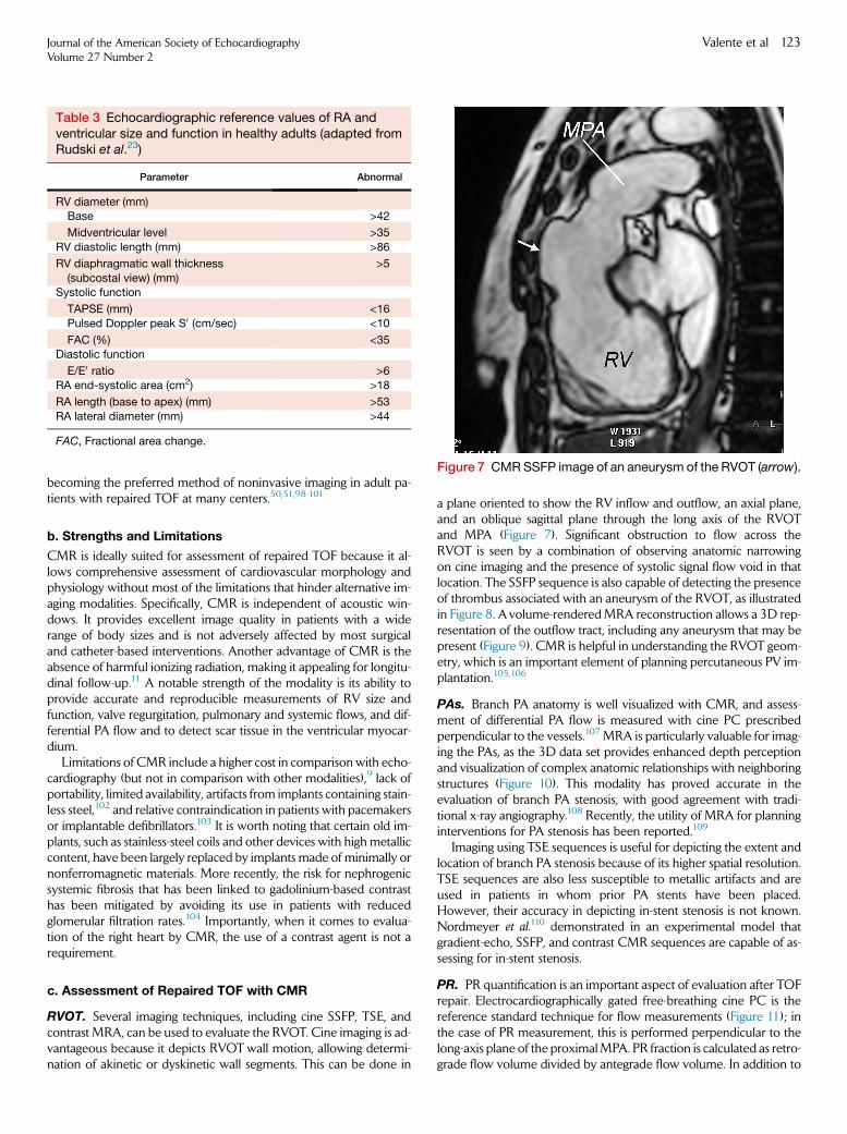
Figure 7 CMRSSFP image of an aneurysm of the RVOT (arrow).
Table 3 Echocardiographic reference values of RA andventricular size and function in healthy adults (adapted fromRudski et al.23)
Parameter Abnormal
RV diameter (mm)Base >42
Midventricular level >35
RV diastolic length (mm) >86
RV diaphragmatic wall thickness
(subcostal view) (mm)
>5
Systolic function
TAPSE (mm) <16Pulsed Doppler peak S0 (cm/sec) <10
FAC (%) <35Diastolic function
E/E0 ratio >6RA end-systolic area (cm2) >18
RA length (base to apex) (mm) >53
RA lateral diameter (mm) >44
FAC, Fractional area change.
Journal of the American Society of EchocardiographyVolume 27 Number 2
Valente et al 123
becoming the preferred method of noninvasive imaging in adult pa-tients with repaired TOF at many centers.50,51,98-101
b. Strengths and Limitations
CMR is ideally suited for assessment of repaired TOF because it al-lows comprehensive assessment of cardiovascular morphology andphysiology without most of the limitations that hinder alternative im-aging modalities. Specifically, CMR is independent of acoustic win-dows. It provides excellent image quality in patients with a widerange of body sizes and is not adversely affected by most surgicaland catheter-based interventions. Another advantage of CMR is theabsence of harmful ionizing radiation, making it appealing for longitu-dinal follow-up.11 A notable strength of the modality is its ability toprovide accurate and reproducible measurements of RV size andfunction, valve regurgitation, pulmonary and systemic flows, and dif-ferential PA flow and to detect scar tissue in the ventricular myocar-dium.
Limitations of CMR include a higher cost in comparison with echo-cardiography (but not in comparison with other modalities),9 lack ofportability, limited availability, artifacts from implants containing stain-less steel,102 and relative contraindication in patients with pacemakersor implantable defibrillators.103 It is worth noting that certain old im-plants, such as stainless-steel coils and other devices with high metalliccontent, have been largely replaced by implants made of minimally ornonferromagnetic materials. More recently, the risk for nephrogenicsystemic fibrosis that has been linked to gadolinium-based contrasthas been mitigated by avoiding its use in patients with reducedglomerular filtration rates.104 Importantly, when it comes to evalua-tion of the right heart by CMR, the use of a contrast agent is not arequirement.
c. Assessment of Repaired TOF with CMR
RVOT. Several imaging techniques, including cine SSFP, TSE, andcontrast MRA, can be used to evaluate the RVOT. Cine imaging is ad-vantageous because it depicts RVOTwall motion, allowing determi-nation of akinetic or dyskinetic wall segments. This can be done in
a plane oriented to show the RV inflow and outflow, an axial plane,and an oblique sagittal plane through the long axis of the RVOTand MPA (Figure 7). Significant obstruction to flow across theRVOT is seen by a combination of observing anatomic narrowingon cine imaging and the presence of systolic signal flow void in thatlocation. The SSFP sequence is also capable of detecting the presenceof thrombus associated with an aneurysm of the RVOT, as illustratedin Figure 8. A volume-renderedMRA reconstruction allows a 3D rep-resentation of the outflow tract, including any aneurysm that may bepresent (Figure 9). CMR is helpful in understanding the RVOT geom-etry, which is an important element of planning percutaneous PV im-plantation.105,106
PAs. Branch PA anatomy is well visualized with CMR, and assess-ment of differential PA flow is measured with cine PC prescribedperpendicular to the vessels.107 MRA is particularly valuable for imag-ing the PAs, as the 3D data set provides enhanced depth perceptionand visualization of complex anatomic relationships with neighboringstructures (Figure 10). This modality has proved accurate in theevaluation of branch PA stenosis, with good agreement with tradi-tional x-ray angiography.108 Recently, the utility of MRA for planninginterventions for PA stenosis has been reported.109
Imaging using TSE sequences is useful for depicting the extent andlocation of branch PA stenosis because of its higher spatial resolution.TSE sequences are also less susceptible to metallic artifacts and areused in patients in whom prior PA stents have been placed.However, their accuracy in depicting in-stent stenosis is not known.Nordmeyer et al.110 demonstrated in an experimental model thatgradient-echo, SSFP, and contrast CMR sequences are capable of as-sessing for in-stent stenosis.
PR. PR quantification is an important aspect of evaluation after TOFrepair. Electrocardiographically gated free-breathing cine PC is thereference standard technique for flow measurements (Figure 11); inthe case of PR measurement, this is performed perpendicular to thelong-axis plane of the proximalMPA. PR fraction is calculated as retro-grade flow volume divided by antegrade flow volume. In addition to
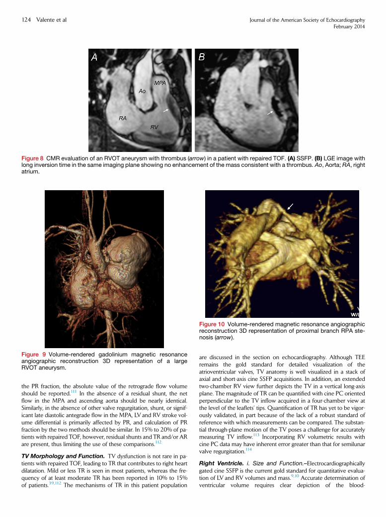
Figure 8 CMR evaluation of an RVOT aneurysm with thrombus (arrow) in a patient with repaired TOF. (A) SSFP. (B) LGE image withlong inversion time in the same imaging plane showing no enhancement of the mass consistent with a thrombus. Ao, Aorta; RA, rightatrium.
Figure 10 Volume-rendered magnetic resonance angiographicreconstruction 3D representation of proximal branch RPA ste-nosis (arrow).
Figure 9 Volume-rendered gadolinium magnetic resonanceangiographic reconstruction 3D representation of a largeRVOT aneurysm.
124 Valente et al Journal of the American Society of EchocardiographyFebruary 2014
the PR fraction, the absolute value of the retrograde flow volumeshould be reported.111 In the absence of a residual shunt, the netflow in the MPA and ascending aorta should be nearly identical.Similarly, in the absence of other valve regurgitation, shunt, or signif-icant late diastolic antegrade flow in the MPA, LV and RV stroke vol-ume differential is primarily affected by PR, and calculation of PRfraction by the two methods should be similar. In 15% to 20% of pa-tients with repaired TOF, however, residual shunts and TR and/or ARare present, thus limiting the use of these comparisons.112
TV Morphology and Function. TV dysfunction is not rare in pa-tients with repaired TOF, leading to TR that contributes to right heartdilatation. Mild or less TR is seen in most patients, whereas the fre-quency of at least moderate TR has been reported in 10% to 15%of patients.39,112 The mechanisms of TR in this patient population
are discussed in the section on echocardiography. Although TEEremains the gold standard for detailed visualization of theatrioventricular valves, TV anatomy is well visualized in a stack ofaxial and short-axis cine SSFP acquisitions. In addition, an extendedtwo-chamber RV view further depicts the TV in a vertical long-axisplane. The magnitude of TR can be quantified with cine PC orientedperpendicular to the TV inflow acquired in a four-chamber view atthe level of the leaflets’ tips. Quantification of TR has yet to be vigor-ously validated, in part because of the lack of a robust standard ofreference with which measurements can be compared. The substan-tial through-plane motion of the TV poses a challenge for accuratelymeasuring TV inflow.113 Incorporating RV volumetric results withcine PC data may have inherent error greater than that for semilunarvalve regurgitation.114
Right Ventricle. i. Size and Function.–Electrocardiographicallygated cine SSFP is the current gold standard for quantitative evalua-tion of LV and RV volumes and mass.9,10 Accurate determination ofventricular volume requires clear depiction of the blood-
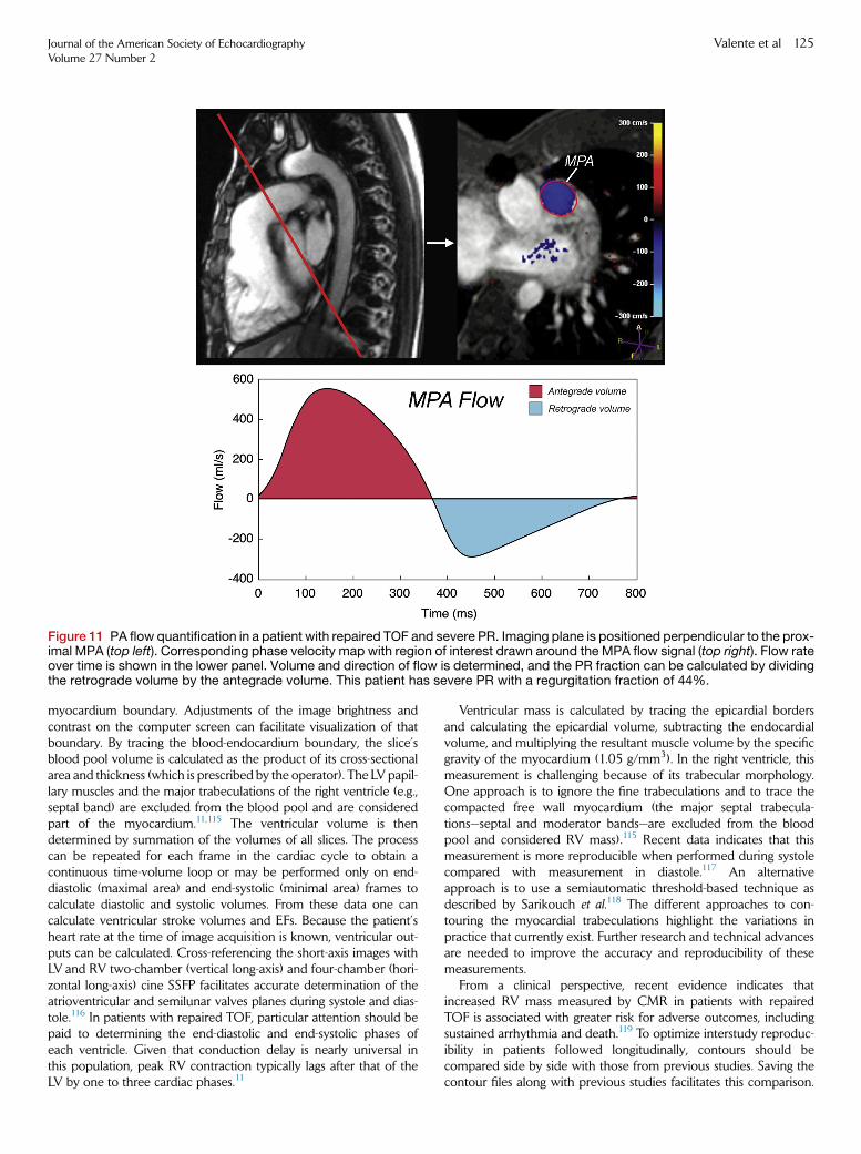
Figure 11 PA flow quantification in a patient with repaired TOF and severe PR. Imaging plane is positioned perpendicular to the prox-imal MPA (top left). Corresponding phase velocity map with region of interest drawn around the MPA flow signal (top right). Flow rateover time is shown in the lower panel. Volume and direction of flow is determined, and the PR fraction can be calculated by dividingthe retrograde volume by the antegrade volume. This patient has severe PR with a regurgitation fraction of 44%.
Journal of the American Society of EchocardiographyVolume 27 Number 2
Valente et al 125
myocardium boundary. Adjustments of the image brightness andcontrast on the computer screen can facilitate visualization of thatboundary. By tracing the blood-endocardium boundary, the slice’sblood pool volume is calculated as the product of its cross-sectionalarea and thickness (which is prescribed by the operator). The LV papil-lary muscles and the major trabeculations of the right ventricle (e.g.,septal band) are excluded from the blood pool and are consideredpart of the myocardium.11,115 The ventricular volume is thendetermined by summation of the volumes of all slices. The processcan be repeated for each frame in the cardiac cycle to obtain acontinuous time-volume loop or may be performed only on end-diastolic (maximal area) and end-systolic (minimal area) frames tocalculate diastolic and systolic volumes. From these data one cancalculate ventricular stroke volumes and EFs. Because the patient’sheart rate at the time of image acquisition is known, ventricular out-puts can be calculated. Cross-referencing the short-axis images withLV and RV two-chamber (vertical long-axis) and four-chamber (hori-zontal long-axis) cine SSFP facilitates accurate determination of theatrioventricular and semilunar valves planes during systole and dias-tole.116 In patients with repaired TOF, particular attention should bepaid to determining the end-diastolic and end-systolic phases ofeach ventricle. Given that conduction delay is nearly universal inthis population, peak RV contraction typically lags after that of theLV by one to three cardiac phases.11
Ventricular mass is calculated by tracing the epicardial bordersand calculating the epicardial volume, subtracting the endocardialvolume, and multiplying the resultant muscle volume by the specificgravity of the myocardium (1.05 g/mm3). In the right ventricle, thismeasurement is challenging because of its trabecular morphology.One approach is to ignore the fine trabeculations and to trace thecompacted free wall myocardium (the major septal trabecula-tions—septal and moderator bands—are excluded from the bloodpool and considered RV mass).115 Recent data indicates that thismeasurement is more reproducible when performed during systolecompared with measurement in diastole.117 An alternativeapproach is to use a semiautomatic threshold-based technique asdescribed by Sarikouch et al.118 The different approaches to con-touring the myocardial trabeculations highlight the variations inpractice that currently exist. Further research and technical advancesare needed to improve the accuracy and reproducibility of thesemeasurements.
From a clinical perspective, recent evidence indicates thatincreased RV mass measured by CMR in patients with repairedTOF is associated with greater risk for adverse outcomes, includingsustained arrhythmia and death.119 To optimize interstudy reproduc-ibility in patients followed longitudinally, contours should becompared side by side with those from previous studies. Saving thecontour files along with previous studies facilitates this comparison.
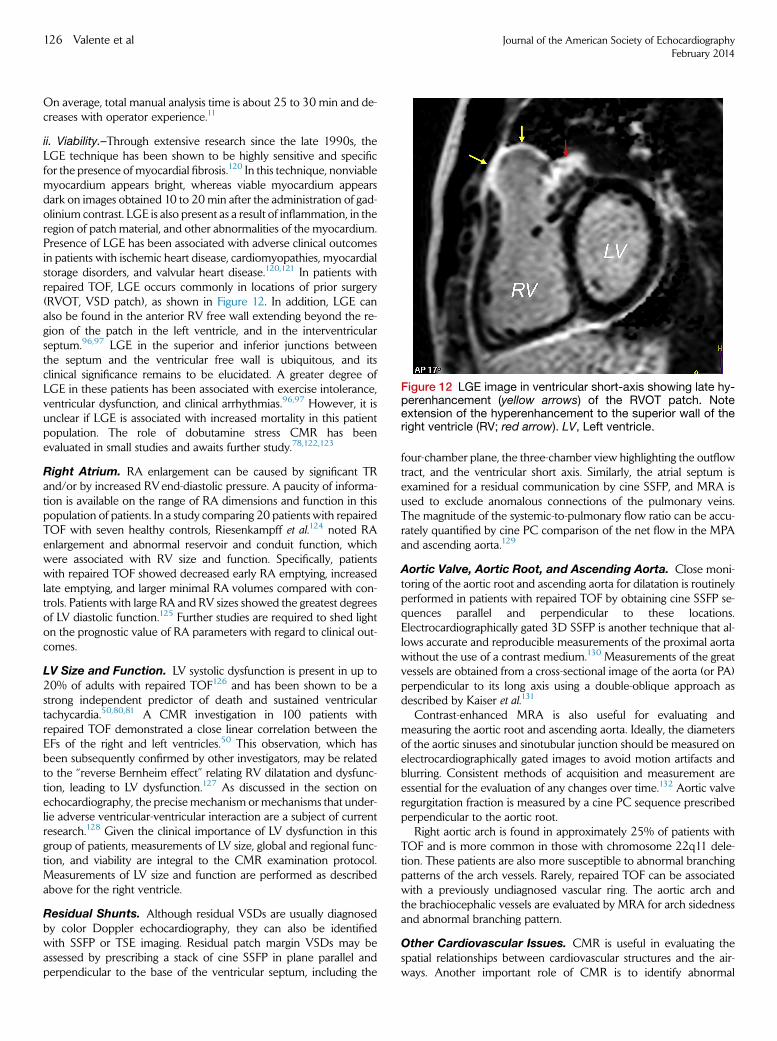
Figure 12 LGE image in ventricular short-axis showing late hy-perenhancement (yellow arrows) of the RVOT patch. Noteextension of the hyperenhancement to the superior wall of theright ventricle (RV; red arrow). LV, Left ventricle.
126 Valente et al Journal of the American Society of EchocardiographyFebruary 2014
On average, total manual analysis time is about 25 to 30 min and de-creases with operator experience.11
ii. Viability.–Through extensive research since the late 1990s, theLGE technique has been shown to be highly sensitive and specificfor the presence of myocardial fibrosis.120 In this technique, nonviablemyocardium appears bright, whereas viable myocardium appearsdark on images obtained 10 to 20min after the administration of gad-olinium contrast. LGE is also present as a result of inflammation, in theregion of patch material, and other abnormalities of the myocardium.Presence of LGE has been associated with adverse clinical outcomesin patients with ischemic heart disease, cardiomyopathies, myocardialstorage disorders, and valvular heart disease.120,121 In patients withrepaired TOF, LGE occurs commonly in locations of prior surgery(RVOT, VSD patch), as shown in Figure 12. In addition, LGE canalso be found in the anterior RV free wall extending beyond the re-gion of the patch in the left ventricle, and in the interventricularseptum.96,97 LGE in the superior and inferior junctions betweenthe septum and the ventricular free wall is ubiquitous, and itsclinical significance remains to be elucidated. A greater degree ofLGE in these patients has been associated with exercise intolerance,ventricular dysfunction, and clinical arrhythmias.96,97 However, it isunclear if LGE is associated with increased mortality in this patientpopulation. The role of dobutamine stress CMR has beenevaluated in small studies and awaits further study.78,122,123
Right Atrium. RA enlargement can be caused by significant TRand/or by increased RVend-diastolic pressure. A paucity of informa-tion is available on the range of RA dimensions and function in thispopulation of patients. In a study comparing 20 patients with repairedTOF with seven healthy controls, Riesenkampff et al.124 noted RAenlargement and abnormal reservoir and conduit function, whichwere associated with RV size and function. Specifically, patientswith repaired TOF showed decreased early RA emptying, increasedlate emptying, and larger minimal RA volumes compared with con-trols. Patients with large RA and RV sizes showed the greatest degreesof LV diastolic function.125 Further studies are required to shed lighton the prognostic value of RA parameters with regard to clinical out-comes.
LV Size and Function. LV systolic dysfunction is present in up to20% of adults with repaired TOF126 and has been shown to be astrong independent predictor of death and sustained ventriculartachycardia.50,80,81 A CMR investigation in 100 patients withrepaired TOF demonstrated a close linear correlation between theEFs of the right and left ventricles.50 This observation, which hasbeen subsequently confirmed by other investigators, may be relatedto the ‘‘reverse Bernheim effect’’ relating RV dilatation and dysfunc-tion, leading to LV dysfunction.127 As discussed in the section onechocardiography, the precise mechanism ormechanisms that under-lie adverse ventricular-ventricular interaction are a subject of currentresearch.128 Given the clinical importance of LV dysfunction in thisgroup of patients, measurements of LV size, global and regional func-tion, and viability are integral to the CMR examination protocol.Measurements of LV size and function are performed as describedabove for the right ventricle.
Residual Shunts. Although residual VSDs are usually diagnosedby color Doppler echocardiography, they can also be identifiedwith SSFP or TSE imaging. Residual patch margin VSDs may beassessed by prescribing a stack of cine SSFP in plane parallel andperpendicular to the base of the ventricular septum, including the
four-chamber plane, the three-chamber view highlighting the outflowtract, and the ventricular short axis. Similarly, the atrial septum isexamined for a residual communication by cine SSFP, and MRA isused to exclude anomalous connections of the pulmonary veins.The magnitude of the systemic-to-pulmonary flow ratio can be accu-rately quantified by cine PC comparison of the net flow in the MPAand ascending aorta.129
Aortic Valve, Aortic Root, and Ascending Aorta. Close moni-toring of the aortic root and ascending aorta for dilatation is routinelyperformed in patients with repaired TOF by obtaining cine SSFP se-quences parallel and perpendicular to these locations.Electrocardiographically gated 3D SSFP is another technique that al-lows accurate and reproducible measurements of the proximal aortawithout the use of a contrast medium.130 Measurements of the greatvessels are obtained from a cross-sectional image of the aorta (or PA)perpendicular to its long axis using a double-oblique approach asdescribed by Kaiser et al.131
Contrast-enhanced MRA is also useful for evaluating andmeasuring the aortic root and ascending aorta. Ideally, the diametersof the aortic sinuses and sinotubular junction should be measured onelectrocardiographically gated images to avoid motion artifacts andblurring. Consistent methods of acquisition and measurement areessential for the evaluation of any changes over time.132 Aortic valveregurgitation fraction is measured by a cine PC sequence prescribedperpendicular to the aortic root.
Right aortic arch is found in approximately 25% of patients withTOF and is more common in those with chromosome 22q11 dele-tion. These patients are also more susceptible to abnormal branchingpatterns of the arch vessels. Rarely, repaired TOF can be associatedwith a previously undiagnosed vascular ring. The aortic arch andthe brachiocephalic vessels are evaluated by MRA for arch sidednessand abnormal branching pattern.
Other Cardiovascular Issues. CMR is useful in evaluating thespatial relationships between cardiovascular structures and the air-ways. Another important role of CMR is to identify abnormal
Journal of the American Society of EchocardiographyVolume 27 Number 2
Valente et al 127
coronary artery origins and proximal courses. This is particularlyimportant in patients with prior repair who are being consideredfor percutaneous PV implantation.133 The current adult CHD man-agement guidelines recommend that coronary anatomy should bedetermined before any intervention on the RVOT (class I, level ofevidence C).12 Another consideration before reoperation in thepatient with repaired TOF (e.g., PV replacement) is RV anteriorwall and/or ascending aorta adherent to the posterior sternal tablebecause these structures are at risk for accidental injury duringsternotomy.
Noncardiac Findings. As patients with repaired TOF survive to anolder age, one can anticipate a higher potential for identifying extrac-ardiac findings by CMR. In a study of 240CMR examinations done innon-CHD patients, 27% of studies were found to have previously un-recognized incidental findings, including pleural effusions, atelectasis,and adenopathy, as well as five cases of newly diagnosed malig-nancy.134
d. Standard Protocol
Patient Preparation. The importance of careful attention to de-tails of patient preparation and placement in the scanner cannot beoveremphasized. Optimal placement of electrocardiographic leadsis paramount to quality gating. A peripheral intravenous cannula forinjection of gadolinium-based contrast is placed in the following cir-cumstances (subject to practice variations):
� First CMR examination� >3 years since last LGE evaluation� Deterioration in clinical status� Regional or global ventricular function has worsened
Placement of an intravenous cannula in young patients may be de-ferred in settings in which attempts at intravenous cannula insertionmight result in loss of patient cooperation.11
Scanning Protocol. An example of a CMR examination protocolfor repaired TOF patients is listed in Table 4, and reference normalvalues for measurements are provided in Table 5. The examinationprotocol is designed to accomplish the following objectives:
� Quantitative assessment of RV and LV volumes, mass, stroke volumes, andEFs
� Evaluation of regional wall motion abnormalities� Imaging the anatomy of the RVOT, PAs, aorta, and aortopulmonary collat-eral vessels
� Quantification of PR, TR, cardiac output, and pulmonary-to-systemic flowratio
� Assessment of myocardial viability, with particular attention to scar tissue inthe ventricular myocardium aside from sites of previous surgery (e.g., VSDand RVOT patches)
� Relationship of cardiovascular structures to the sternum
e. Reporting Elements and Measurements
� RVOTobstruction and or aneurysm; length of dyskinetic segment� Branch PA stenosis� RV and LV size and function (absolute and BSA-adjusted values): end-diastolic volume, end-systolic volume, stroke volume, EF, mass, mass-to-volume ratio
� Vessel dimensions: aortic root, ascending aorta, MPA, RPA, LPA� RA size by the area-length method135
� Flow measurements
◦ Ascending aorta, MPA, RPA, LPA◦ PR fraction and volume◦ Other valve regurgitation
� LGE presence and extent
f. Recommendations
CMR is recommended in young patients (usually #10 years) whenthe echocardiographic and clinical data are concerning for unfavor-able disease progression (e.g., ventricular dysfunction, right heartfailure symptoms). Once patients reach adolescence, CMR is per-formed routinely using a comprehensive imaging protocol.Quantitative assessment of ventricular size and function and PR,for which CMR is the reference standard, are essential elementsof the imaging protocol.
7. CARDIOVASCULAR COMPUTED TOMOGRAPHY
a. Overview of Modality
Widespread availability of CT has lead to increased utilization of thisnoninvasive modality in the general population.136 Early challengeswith motion artifacts encountered with imaging complex congenitalanatomy throughout the cardiac cycle limited the evolution of thisapplication in the assessment of this patient population. However, ad-vances in multidetector computed tomographic technology have ledto improvements in both spatial and temporal resolutions, allowingthe acquisition of a volume of data during a single breath-hold exam-ination.137 Subsequently, the ability to produce static images of theheart and great vessels as well as 3D reconstructions has revolution-ized this technique and propelled cardiac CT into the arena ofanatomic imaging of patients with complex CHD.
In general with MDCT, the x-ray point source(s) and the detectorarray are located on opposite sides of the patient on a ringlike struc-ture called a gantry. The gantry rotates around the patient, locatedat the table center. The table then moves at a constant speed as x-rays penetrate the patient and are captured by one or more of the de-tectors. Once complete, a series of helical projections are generated.From these data, images representing x-ray attenuation from eachpoint in the volume are mathematically reconstructed.138 Certainscanners are capable of volume scanning without table movement.139
Scanners with fewer detectors create an image set from data ac-quired from several heartbeats. To improve overall quality ofcomputed tomographic images and ensure a longer diastolic windowwith minimal cardiac motion, pharmacologic heart rate control isfrequently used. Successful prescan heart rate control facilitates post-processing that requires image reconstruction of the data set in a fixedinterval preceding the R wave. Furthermore, heart rate optimizationavoids gating errors associated with heart rate changes that may occurduring image acquisition and that lead to image artifacts. These strate-gies are particularly important in the evaluation of the coronary ar-teries and ascending aorta to avoid motion artifacts that occurthroughout the cardiac cycle. Therefore, heart rate control with oralor intravenous b-blockade is advised before CT for patients withoutcontraindications.140
Data postprocessing commences after image acquisition is com-plete. Postprocessing includes (1) electrocardiographic review forectopic beats or arrhythmias, (2) selection of the optimal phase forreconstruction, (3) selection of appropriate reconstruction parame-ters and kernels, (4) review of the axial (source) data set, and (5)
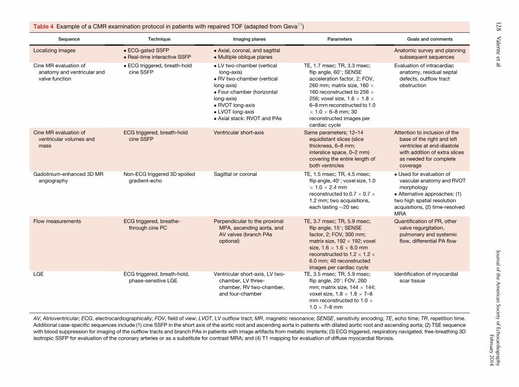
Table 4 Example of a CMR examination protocol in patients with repaired TOF (adapted from Geva11)
Sequence Technique Imaging planes Parameters Goals and comments
Localizing images � ECG-gated SSFP� Real-time interactive SSFP
� Axial, coronal, and sagittal� Multiple oblique planes
Anatomic survey and planningsubsequent sequences
Cine MR evaluation ofanatomy and ventricular and
valve function
� ECG triggered, breath-holdcine SSFP
� LV two-chamber (verticallong-axis)
� RV two-chamber (vertical
long-axis)
� Four-chamber (horizontallong-axis)
� RVOT long-axis
� LVOT long-axis
� Axial stack: RVOT and PAs
TE, 1.7 msec; TR, 3.3 msec;flip angle, 60�; SENSEacceleration factor, 2; FOV,
260 mm; matrix size, 160 �160 reconstructed to 256 �256; voxel size, 1.6 � 1.8 �6–8mm reconstructed to 1.0
� 1.0 � 6–8 mm; 30
reconstructed images percardiac cycle
Evaluation of intracardiacanatomy, residual septal
defects, outflow tract
obstruction
Cine MR evaluation ofventricular volumes and
mass
ECG triggered, breath-holdcine SSFP
Ventricular short-axis Same parameters; 12–14equidistant slices (slice
thickness, 6–8 mm;
interslice space, 0–2 mm)covering the entire length of
both ventricles
Attention to inclusion of thebase of the right and left
ventricles at end-diastole
with addition of extra slicesas needed for complete
coverage
Gadolinium-enhanced 3D MRangiography
Non-ECG triggered 3D spoiledgradient-echo
Sagittal or coronal TE, 1.5 msec; TR, 4.5 msec;flip angle, 40�; voxel size, 1.0� 1.0 � 2.4 mm
reconstructed to 0.7� 0.7�1.2 mm; two acquisitions,each lasting �20 sec
� Used for evaluation ofvascular anatomy and RVOT
morphology
� Alternative approaches: (1)
two high spatial resolutionacquisitions, (2) time-resolved
MRA
Flow measurements ECG triggered, breathe-
through cine PC
Perpendicular to the proximal
MPA, ascending aorta, and
AV valves (branch PAsoptional)
TE, 3.7 msec; TR, 5.9 msec;
flip angle, 15�; SENSEfactor, 2; FOV, 300 mm;matrix size, 192� 192; voxel
size, 1.6 � 1.6 � 6.0 mm
reconstructed to 1.2� 1.2�6.0 mm; 40 reconstructedimages per cardiac cycle
Quantification of PR, other
valve regurgitation,
pulmonary and systemicflow, differential PA flow
LGE ECG triggered, breath-hold,
phase-sensitive LGE
Ventricular short-axis, LV two-
chamber, LV three-
chamber, RV two-chamber,and four-chamber
TE, 3.5 msec; TR, 5.9 msec;
flip angle, 20�; FOV, 260
mm; matrix size, 144 � 144;voxel size, 1.8 � 1.8 � 7–8
mm reconstructed to 1.0 �1.0 � 7–8 mm
Identification of myocardial
scar tissue
AV, Atrioventricular; ECG, electrocardiographically; FOV, field of view; LVOT, LV outflow tract; MR, magnetic resonance; SENSE, sensitivity encoding; TE, echo time; TR, repetition time.
Additional case-specific sequences include (1) cine SSFP in the short axis of the aortic root and ascending aorta in patients with dilated aortic root and ascending aorta; (2) TSE sequencewith blood suppression for imaging of the outflow tracts and branch PAs in patients with image artifacts from metallic implants; (3) ECG triggered, respiratory navigated, free-breathing 3D
isotropic SSFP for evaluation of the coronary arteries or as a substitute for contrast MRA; and (4) T1 mapping for evaluation of diffuse myocardial fibrosis.
128
Valen
teet
alJournalo
ftheAmerican
Society
ofEchocard
iograp
hy
February
2014
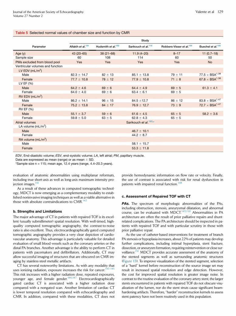
Table 5 Selected normal values of chamber size and function by CMR
Parameter
Study
Alfakih et al.191 Hudsmith et al.192 Sarikouch et al.118 Robbers-Visser et al.194 Buechel et al.195
Age (y) 43 (20–65) 38 (21–68) 11.9 (4–20) 8–17 11 (0.7–18)
Sample size 60 108 114 60 50
PMs excluded from blood pool Yes Yes Yes Yes No
Ventricular volumes and function
LV EDV (mL/m2)Male 82.3 6 14.7 82 6 13 85.1 6 13.8 79 6 11 77.5 � BSA1.38
Female 77.7 6 10.8 78 6 12 77.9 6 10.8 71 6 8 67.8 � BSA1.38
LV EF (%)
Male 64.2 6 4.6 69 6 6 64.4 6 4.9 69 6 5 61.3 6 4.1
Female 64.0 6 4.0 69 6 6 63.4 6 6.1 69 6 5
RV EDV (mL/m2)Male 86.2 6 14.1 96 6 15 84.5 612.7 86 6 12 83.8 � BSA1.47
Female 75.2 6 13.8 84 6 17 76.9 6 12.7 73 6 9 72.7 � BSA1.47
RV EF (%)
Male 55.1 6 3.7 59 6 6 61.6 6 4.5 65 6 5 58.2 6 3.6
Female 59.8 6 5.0 63 6 5 62.8 6 4.3 65 6 5
Atrial volumes Sarikouch et al.193,*LA volume (mL/m2)
Male 46.7 6 10.1Female 44.2 6 8.7
RA volume (mL/m2)Male 58.1 6 15.7
Female 53.3 6 11.8
EDV, End-diastolic volume; ESV, end-systolic volume; LA, left atrial; PM, papillary muscle.
Data are expressed as mean (range) or as mean 6 SD.*Sample size n = 115; mean age, 12.4 years (range, 4.4–20.3 years).
Journal of the American Society of EchocardiographyVolume 27 Number 2
Valente et al 129
evaluation of anatomic abnormalities using multiplanar reformats,including true short-axis as well as long-axis maximum intensity pro-jection images.141
As a result of these advances in computed tomographic technol-ogy, MDCT is now emerging as a complementary modality to estab-lished noninvasive imaging techniques as well as a viable alternative inthose with absolute contraindications to CMR.142
b. Strengths and Limitations
The major advantage of CT in patients with repaired TOF is its excel-lent (usually submillimeter) spatial resolution. With well-timed, high-quality computed tomographic angiography, the contrast-to-noiseratio is also excellent. Thus, electrocardiographically gated computedtomographic angiography provides a very clear depiction of cardio-vascular anatomy. This advantage is particularly valuable for detailedevaluation of small blood vessels such as the coronary arteries or thedistal PA branches. Another advantage is the ability to perform CT inpatients with pacemakers and defibrillators. Additionally, CT mayallow successful imaging of structures that are obscured on CMR im-aging by stainless-steel metallic artifacts.
CT has several noteworthy limitations. As with any modality thatuses ionizing radiation, exposure increases the risk for cancer.143-145
This risk increases with a higher radiation dose, repeated exposures,younger age, and female gender.146,147 Electrocardiographicallygated cardiac CT is associated with a higher radiation dosecompared with a nongated scan. Another limitation of cardiac CTis lower temporal resolution compared with echocardiography andCMR. In addition, compared with these modalities, CT does not
provide hemodynamic information on flow rate or velocity. Finally,the use of contrast is associated with risk for renal dysfunction inpatients with impaired renal function.148
c. Assessment of Repaired TOF with CT
PAs. The spectrum of morphologic abnormalities of the PAs,including obstruction, stenosis, aneurysmal dilatation, and abnormalcourse, can be evaluated with MDCT.149,150 Abnormalities in PAarchitecture are often the result of prior palliative repairs and shunt-related complications. The PA architecture should be inspected in pa-tients with repaired TOF and with particular scrutiny in those withprior palliative repair.
As the use of catheter-based interventions for treatment of branchPA stenosis or hypoplasia increases, about 22%of patientsmay developfurther complications, including intimal hyperplasia, stent fracture,dissection, or aneurysm formation, requiring reinterventionor close sur-veillance.151 MDCT provides accurate assessment of the anatomy ofthe stented segments as well as surrounding anatomic structures(Figure 13). To improve visualization of the stented segment, selectionof a ‘‘hard’’ kernel before reconstruction of the source image set mayresult in increased spatial resolution and edge detection. However,the cost for improved spatial resolution is greater image noise. Incontrast to the routine evaluation of the coronary artery stent, the largerstents encountered in patients with repaired TOF do not obscure visu-alization of the lumen, nor do the stent struts cause significant beam-hardening artifacts. Therefore, ‘‘harder’’ reconstruction kernels to assessstent patency have not been routinely used in this population.
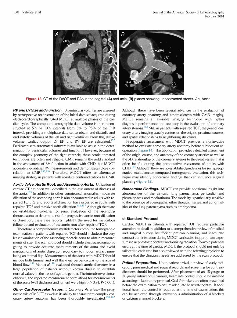
Figure 13 CT of the RVOT and PAs in the sagittal (A) and axial (B) planes showing unobstructed stents. Ao, Aorta.
130 Valente et al Journal of the American Society of EchocardiographyFebruary 2014
RV and LV Size and Function. Biventricular volumes are assessedby retrospective reconstruction of the initial data set acquired duringelectrocardiographically gated MDCT at multiple phases of the car-diac cycle. The computed tomographic data volume is then recon-structed at 5% or 10% intervals from 5% to 95% of the R-Rinterval, providing a multiphase data set to obtain end-diastolic andend-systolic volumes of the left and right ventricles. From this, strokevolume, cardiac output, LV EF, and RV EF are calculated.152
Dedicated semiautomated software is available to assist in the deter-mination of ventricular volumes and function. However, because ofthe complex geometry of the right ventricle, these semiautomatedtechniques are often not reliable. CMR remains the gold standardin the assessment of RV function in adults with CHD, but MDCTaccurately quantifies RV measurements and demonstrates close cor-relation to CMR.153,154 Therefore, MDCT offers an alternativeimaging strategy in patients with absolute contraindications to CMR.
Aortic Valve, Aortic Root, and Ascending Aorta. Utilization ofcardiac CT has been well described in the assessment of diseases ofthe aorta.155 In addition to other conotruncal anomalies, moderatedilatation of the ascending aorta is also encountered in adults with re-paired TOF. Rarely, reports of dissection have occurred in adults withrepaired TOF and massive aortic dilatation.156,157 Although there areno established guidelines for serial evaluation of the ascendingthoracic aorta to determine risk for progressive aortic root dilatationor dissection, these case reports highlight the need for meticulousfollow-up and evaluation of the aortic root after repair of TOF.
Therefore, a comprehensivemultidetector computed tomographicexamination in patients with repaired TOF should include at the veryleast examination of the ascending thoracic aorta to obtain measure-ments of size. The scan protocol should include electrocardiographicgating to provide accurate measurements of the aorta and avoidmisdiagnosis of aortic dissection secondary to motion artifact simu-lating an intimal flap. Measurements of the aorta with MDCT shouldinclude both luminal and wall thickness perpendicular to the axis ofblood flow.158 Mao et al.159 evaluated thoracic aortic diameters in alarge population of patients without known disease to establishnormal values on the basis of age and gender. The interobserver, intra-observer, and repeated measurement correlations for measurementsof the aorta (wall thickness and lumen) were high (r > 0.91, P < .001).
Other Cardiovascular Issues. i. Coronary Arteries.–The prog-nostic role of MDCTas well as its ability to characterize complex cor-onary artery anatomy has been thoroughly investigated.160-162
Although there have been several advances in the evaluation ofcoronary artery anatomy and atherosclerosis with CMR imaging,MDCT remains a favorable imaging technique with higherdiagnostic performance and accuracy in the evaluation of coronaryartery stenosis.163 Still, in patients with repaired TOF, the goal of cor-onary artery imaging usually centers on the origins, proximal courses,and spatial relationships to neighboring structures.
Preoperative assessment with MDCT provides a noninvasivemethod to evaluate coronary artery anatomy before subsequent re-operation (Figure 14). This application provides a detailed assessmentof the origin, course, and anatomy of the coronary arteries as well asthe 3D relationship of the coronary arteries to the great vessels that isoften helpful during the preoperative assessment of adults withCHD.164 Although there are no established guidelines for such preop-erative multidetector computed tomographic evaluation, this tech-nique may identify concerning findings that can influence surgicalplanning (Figure 15).
Noncardiac Findings. MDCT can provide additional insight intoabnormalities of the airways, lung parenchyma, pericardial andpleural spaces, andmediastinum. Themodality is particularly sensitiveto the presence of adenopathy, other thoracic masses, and abnormal-ities of the lung parenchyma such as emphysema.
d. Standard Protocol
Cardiac MDCT in patients with repaired TOF requires particularattention to detail in addition to a comprehensive review of medicaland surgical history. Insufficient prescan planning and inaccuratecontrast administration duringMDCT can lead to inappropriate expo-sures to nephrotoxic contrast and ionizing radiation. To avoid potentialerrors at the time of cardiac MDCT, the protocol should not only betailored to each case but also reviewed with the referring physician toensure that the clinician’s needs are addressed by the scan protocol.
Patient Preparation. Upon patient arrival, a review of study indi-cation, prior medical and surgical records, and screening for contrain-dications should be performed. After placement of an 18-gauge or20-gauge intravenous cannula, heart rate control should be initiatedaccording to laboratory protocol. Oral b-blockers are often prescribedbefore the examination to ensure adequate heart rate control. If addi-tional heart rate control is required at the time of examination, thiscan be achieved through intravenous administration of b-blockersor calcium channel blockers.
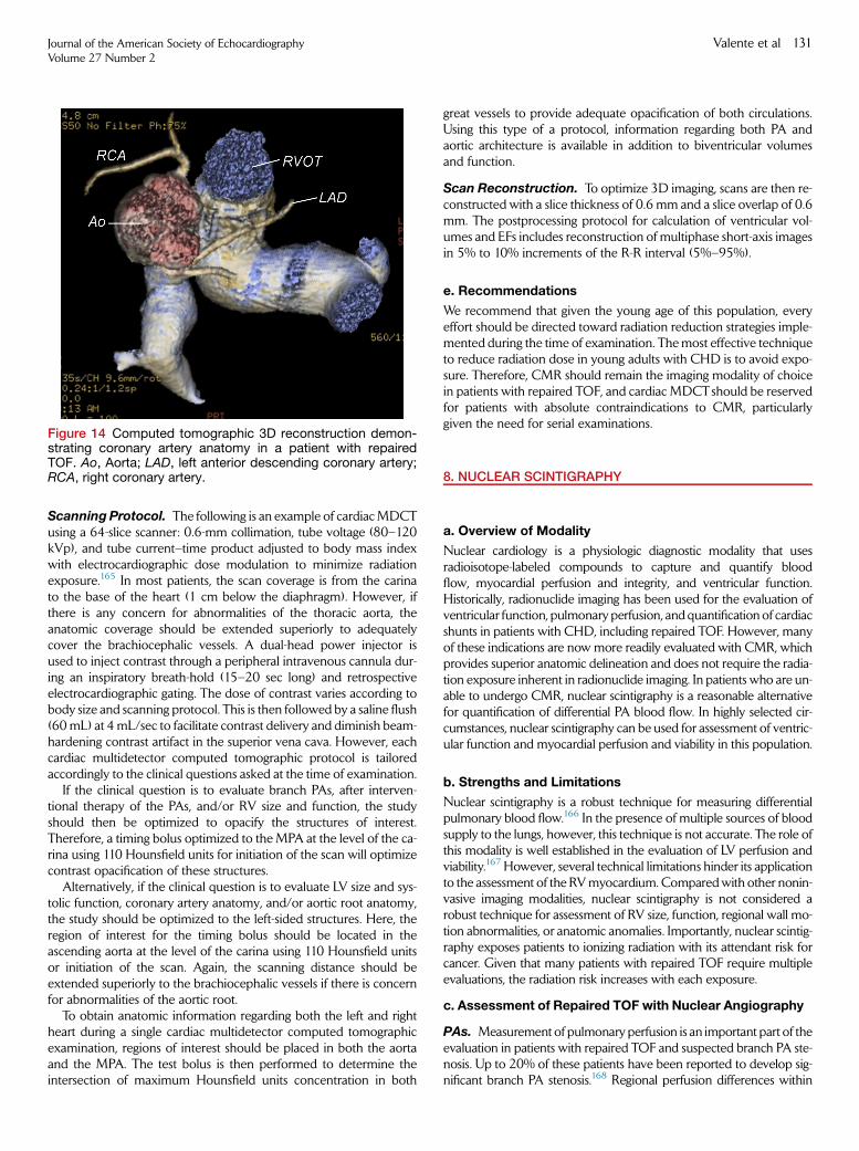
Figure 14 Computed tomographic 3D reconstruction demon-strating coronary artery anatomy in a patient with repairedTOF. Ao, Aorta; LAD, left anterior descending coronary artery;RCA, right coronary artery.
Journal of the American Society of EchocardiographyVolume 27 Number 2
Valente et al 131
Scanning Protocol. The following is an example of cardiacMDCTusing a 64-slice scanner: 0.6-mm collimation, tube voltage (80–120kVp), and tube current–time product adjusted to body mass indexwith electrocardiographic dose modulation to minimize radiationexposure.165 In most patients, the scan coverage is from the carinato the base of the heart (1 cm below the diaphragm). However, ifthere is any concern for abnormalities of the thoracic aorta, theanatomic coverage should be extended superiorly to adequatelycover the brachiocephalic vessels. A dual-head power injector isused to inject contrast through a peripheral intravenous cannula dur-ing an inspiratory breath-hold (15–20 sec long) and retrospectiveelectrocardiographic gating. The dose of contrast varies according tobody size and scanning protocol. This is then followed by a saline flush(60mL) at 4mL/sec to facilitate contrast delivery and diminish beam-hardening contrast artifact in the superior vena cava. However, eachcardiac multidetector computed tomographic protocol is tailoredaccordingly to the clinical questions asked at the time of examination.
If the clinical question is to evaluate branch PAs, after interven-tional therapy of the PAs, and/or RV size and function, the studyshould then be optimized to opacify the structures of interest.Therefore, a timing bolus optimized to the MPA at the level of the ca-rina using 110 Hounsfield units for initiation of the scan will optimizecontrast opacification of these structures.
Alternatively, if the clinical question is to evaluate LV size and sys-tolic function, coronary artery anatomy, and/or aortic root anatomy,the study should be optimized to the left-sided structures. Here, theregion of interest for the timing bolus should be located in theascending aorta at the level of the carina using 110 Hounsfield unitsor initiation of the scan. Again, the scanning distance should beextended superiorly to the brachiocephalic vessels if there is concernfor abnormalities of the aortic root.
To obtain anatomic information regarding both the left and rightheart during a single cardiac multidetector computed tomographicexamination, regions of interest should be placed in both the aortaand the MPA. The test bolus is then performed to determine theintersection of maximum Hounsfield units concentration in both
great vessels to provide adequate opacification of both circulations.Using this type of a protocol, information regarding both PA andaortic architecture is available in addition to biventricular volumesand function.
Scan Reconstruction. To optimize 3D imaging, scans are then re-constructed with a slice thickness of 0.6 mm and a slice overlap of 0.6mm. The postprocessing protocol for calculation of ventricular vol-umes and EFs includes reconstruction of multiphase short-axis imagesin 5% to 10% increments of the R-R interval (5%–95%).
e. Recommendations
We recommend that given the young age of this population, everyeffort should be directed toward radiation reduction strategies imple-mented during the time of examination. Themost effective techniqueto reduce radiation dose in young adults with CHD is to avoid expo-sure. Therefore, CMR should remain the imaging modality of choicein patients with repaired TOF, and cardiac MDCTshould be reservedfor patients with absolute contraindications to CMR, particularlygiven the need for serial examinations.
8. NUCLEAR SCINTIGRAPHY
a. Overview of Modality
Nuclear cardiology is a physiologic diagnostic modality that usesradioisotope-labeled compounds to capture and quantify bloodflow, myocardial perfusion and integrity, and ventricular function.Historically, radionuclide imaging has been used for the evaluation ofventricular function, pulmonary perfusion, and quantification of cardiacshunts in patients with CHD, including repaired TOF. However, manyof these indications are now more readily evaluated with CMR, whichprovides superior anatomic delineation and does not require the radia-tion exposure inherent in radionuclide imaging. In patients who are un-able to undergo CMR, nuclear scintigraphy is a reasonable alternativefor quantification of differential PA blood flow. In highly selected cir-cumstances, nuclear scintigraphy can be used for assessment of ventric-ular function and myocardial perfusion and viability in this population.
b. Strengths and Limitations
Nuclear scintigraphy is a robust technique for measuring differentialpulmonary blood flow.166 In the presence of multiple sources of bloodsupply to the lungs, however, this technique is not accurate. The role ofthis modality is well established in the evaluation of LV perfusion andviability.167 However, several technical limitations hinder its applicationto the assessment of the RVmyocardium. Comparedwith other nonin-vasive imaging modalities, nuclear scintigraphy is not considered arobust technique for assessment of RV size, function, regional wall mo-tion abnormalities, or anatomic anomalies. Importantly, nuclear scintig-raphy exposes patients to ionizing radiation with its attendant risk forcancer. Given that many patients with repaired TOF require multipleevaluations, the radiation risk increases with each exposure.
c. Assessment of Repaired TOF with Nuclear Angiography
PAs. Measurement of pulmonary perfusion is an important part of theevaluation in patients with repaired TOF and suspected branch PA ste-nosis. Up to 20% of these patients have been reported to develop sig-nificant branch PA stenosis.168 Regional perfusion differences within
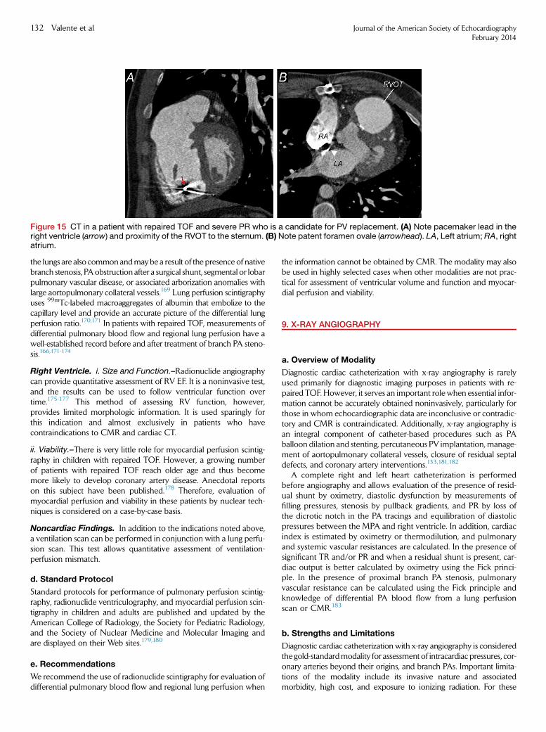
Figure 15 CT in a patient with repaired TOF and severe PR who is a candidate for PV replacement. (A) Note pacemaker lead in theright ventricle (arrow) and proximity of the RVOT to the sternum. (B)Note patent foramen ovale (arrowhead). LA, Left atrium; RA, rightatrium.
132 Valente et al Journal of the American Society of EchocardiographyFebruary 2014
the lungs are also commonandmay be a result of the presence of nativebranch stenosis, PAobstruction after a surgical shunt, segmental or lobarpulmonary vascular disease, or associated arborization anomalies withlarge aortopulmonary collateral vessels.169 Lung perfusion scintigraphyuses 99mTc-labeled macroaggregates of albumin that embolize to thecapillary level and provide an accurate picture of the differential lungperfusion ratio.170,171 In patients with repaired TOF, measurements ofdifferential pulmonary blood flow and regional lung perfusion have awell-established record before and after treatment of branch PA steno-sis.166,171-174
Right Ventricle. i. Size and Function.–Radionuclide angiographycan provide quantitative assessment of RV EF. It is a noninvasive test,and the results can be used to follow ventricular function overtime.175-177 This method of assessing RV function, however,provides limited morphologic information. It is used sparingly forthis indication and almost exclusively in patients who havecontraindications to CMR and cardiac CT.
ii. Viability.–There is very little role for myocardial perfusion scintig-raphy in children with repaired TOF. However, a growing numberof patients with repaired TOF reach older age and thus becomemore likely to develop coronary artery disease. Anecdotal reportson this subject have been published.178 Therefore, evaluation ofmyocardial perfusion and viability in these patients by nuclear tech-niques is considered on a case-by-case basis.
Noncardiac Findings. In addition to the indications noted above,a ventilation scan can be performed in conjunction with a lung perfu-sion scan. This test allows quantitative assessment of ventilation-perfusion mismatch.
d. Standard Protocol
Standard protocols for performance of pulmonary perfusion scintig-raphy, radionuclide ventriculography, and myocardial perfusion scin-tigraphy in children and adults are published and updated by theAmerican College of Radiology, the Society for Pediatric Radiology,and the Society of Nuclear Medicine and Molecular Imaging andare displayed on their Web sites.179,180
e. Recommendations
We recommend the use of radionuclide scintigraphy for evaluation ofdifferential pulmonary blood flow and regional lung perfusion when
the information cannot be obtained by CMR. The modality may alsobe used in highly selected cases when other modalities are not prac-tical for assessment of ventricular volume and function and myocar-dial perfusion and viability.
9. X-RAY ANGIOGRAPHY
a. Overview of Modality
Diagnostic cardiac catheterization with x-ray angiography is rarelyused primarily for diagnostic imaging purposes in patients with re-paired TOF. However, it serves an important role when essential infor-mation cannot be accurately obtained noninvasively, particularly forthose in whom echocardiographic data are inconclusive or contradic-tory and CMR is contraindicated. Additionally, x-ray angiography isan integral component of catheter-based procedures such as PAballoon dilation and stenting, percutaneous PV implantation, manage-ment of aortopulmonary collateral vessels, closure of residual septaldefects, and coronary artery interventions.133,181,182
A complete right and left heart catheterization is performedbefore angiography and allows evaluation of the presence of resid-ual shunt by oximetry, diastolic dysfunction by measurements offilling pressures, stenosis by pullback gradients, and PR by loss ofthe dicrotic notch in the PA tracings and equilibration of diastolicpressures between the MPA and right ventricle. In addition, cardiacindex is estimated by oximetry or thermodilution, and pulmonaryand systemic vascular resistances are calculated. In the presence ofsignificant TR and/or PR and when a residual shunt is present, car-diac output is better calculated by oximetry using the Fick princi-ple. In the presence of proximal branch PA stenosis, pulmonaryvascular resistance can be calculated using the Fick principle andknowledge of differential PA blood flow from a lung perfusionscan or CMR.183
b. Strengths and Limitations
Diagnostic cardiac catheterization with x-ray angiography is consideredthe gold-standardmodality for assessment of intracardiac pressures, cor-onary arteries beyond their origins, and branch PAs. Important limita-tions of the modality include its invasive nature and associatedmorbidity, high cost, and exposure to ionizing radiation. For these
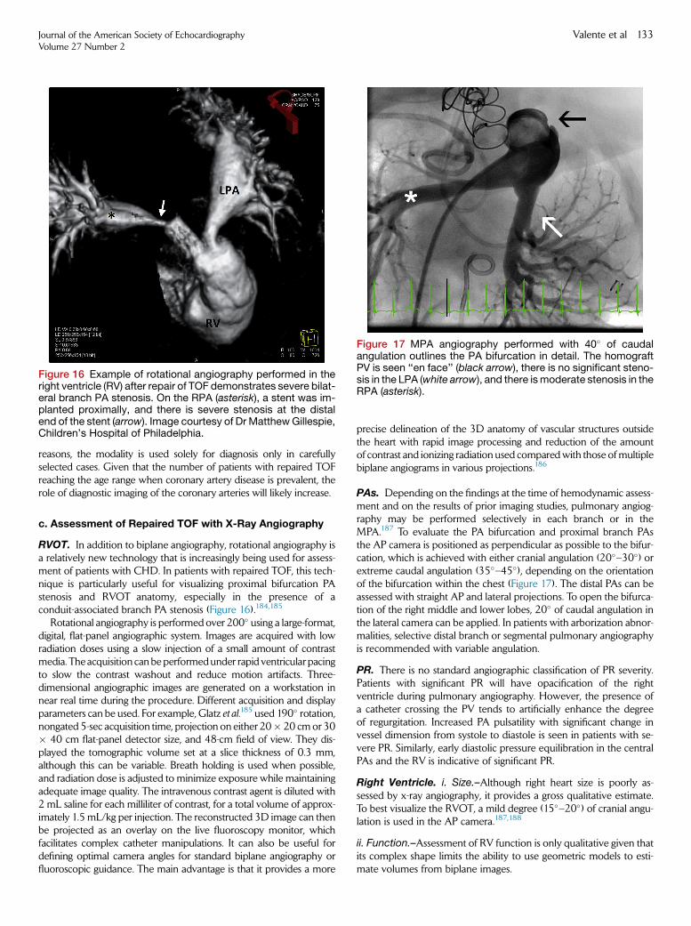
Figure 16 Example of rotational angiography performed in theright ventricle (RV) after repair of TOF demonstrates severe bilat-eral branch PA stenosis. On the RPA (asterisk), a stent was im-planted proximally, and there is severe stenosis at the distalend of the stent (arrow). Image courtesy of Dr MatthewGillespie,Children’s Hospital of Philadelphia.
Figure 17 MPA angiography performed with 40� of caudalangulation outlines the PA bifurcation in detail. The homograftPV is seen ‘‘en face’’ (black arrow), there is no significant steno-sis in the LPA (white arrow), and there ismoderate stenosis in theRPA (asterisk).
Journal of the American Society of EchocardiographyVolume 27 Number 2
Valente et al 133
reasons, the modality is used solely for diagnosis only in carefullyselected cases. Given that the number of patients with repaired TOFreaching the age range when coronary artery disease is prevalent, therole of diagnostic imaging of the coronary arteries will likely increase.
c. Assessment of Repaired TOF with X-Ray Angiography
RVOT. In addition to biplane angiography, rotational angiography isa relatively new technology that is increasingly being used for assess-ment of patients with CHD. In patients with repaired TOF, this tech-nique is particularly useful for visualizing proximal bifurcation PAstenosis and RVOT anatomy, especially in the presence of aconduit-associated branch PA stenosis (Figure 16).184,185
Rotational angiography is performed over 200� using a large-format,digital, flat-panel angiographic system. Images are acquired with lowradiation doses using a slow injection of a small amount of contrastmedia. The acquisition canbe performedunder rapid ventricular pacingto slow the contrast washout and reduce motion artifacts. Three-dimensional angiographic images are generated on a workstation innear real time during the procedure. Different acquisition and displayparameters can be used. For example, Glatz et al.185 used 190� rotation,nongated 5-sec acquisition time, projection on either 20� 20 cmor 30� 40 cm flat-panel detector size, and 48-cm field of view. They dis-played the tomographic volume set at a slice thickness of 0.3 mm,although this can be variable. Breath holding is used when possible,and radiation dose is adjusted to minimize exposure while maintainingadequate image quality. The intravenous contrast agent is diluted with2 mL saline for each milliliter of contrast, for a total volume of approx-imately 1.5 mL/kg per injection. The reconstructed 3D image can thenbe projected as an overlay on the live fluoroscopy monitor, whichfacilitates complex catheter manipulations. It can also be useful fordefining optimal camera angles for standard biplane angiography orfluoroscopic guidance. The main advantage is that it provides a more
precise delineation of the 3D anatomy of vascular structures outsidethe heart with rapid image processing and reduction of the amountof contrast and ionizing radiation used comparedwith those ofmultiplebiplane angiograms in various projections.186
PAs. Depending on the findings at the time of hemodynamic assess-ment and on the results of prior imaging studies, pulmonary angiog-raphy may be performed selectively in each branch or in theMPA.187 To evaluate the PA bifurcation and proximal branch PAsthe AP camera is positioned as perpendicular as possible to the bifur-cation, which is achieved with either cranial angulation (20�–30�) orextreme caudal angulation (35�–45�), depending on the orientationof the bifurcation within the chest (Figure 17). The distal PAs can beassessed with straight AP and lateral projections. To open the bifurca-tion of the right middle and lower lobes, 20� of caudal angulation inthe lateral camera can be applied. In patients with arborization abnor-malities, selective distal branch or segmental pulmonary angiographyis recommended with variable angulation.
PR. There is no standard angiographic classification of PR severity.Patients with significant PR will have opacification of the rightventricle during pulmonary angiography. However, the presence ofa catheter crossing the PV tends to artificially enhance the degreeof regurgitation. Increased PA pulsatility with significant change invessel dimension from systole to diastole is seen in patients with se-vere PR. Similarly, early diastolic pressure equilibration in the centralPAs and the RV is indicative of significant PR.
Right Ventricle. i. Size.–Although right heart size is poorly as-sessed by x-ray angiography, it provides a gross qualitative estimate.To best visualize the RVOT, a mild degree (15�–20�) of cranial angu-lation is used in the AP camera.187,188
ii. Function.–Assessment of RV function is only qualitative given thatits complex shape limits the ability to use geometric models to esti-mate volumes from biplane images.
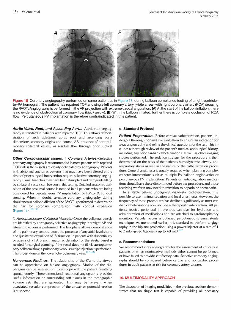
Figure 18 Coronary angiography performed on same patient as in Figure 17, during balloon compliance testing of a right ventricle–to–PA homograft. The patient has repaired TOF and single left coronary artery (white arrow) with right coronary artery (RCA) crossingthe RVOT. Angiography is performed in the AP projection with extreme caudal angulation. (A) At the start of the balloon inflation, thereis no evidence of obstruction of coronary flow (black arrow). (B)With the balloon inflated, further there is complete occlusion of RCAflow. Percutaneous PV implantation is therefore contraindicated in this patient.
134 Valente et al Journal of the American Society of EchocardiographyFebruary 2014
Aortic Valve, Root, and Ascending Aorta. Aortic root angiog-raphy is standard in patients with repaired TOF. This allows demon-stration of arch sidedness, aortic root and ascending aortadimensions, coronary origins and course, AR, presence of aortopul-monary collateral vessels, or residual flow through prior surgicalshunts.
Other Cardiovascular Issues. i. Coronary Arteries.–Selectivecoronary angiography is recommended inmost patients with repairedTOF unless the vessels are clearly delineated by aortography. Patientswith abnormal anatomic patterns that may have been altered at thetime of prior surgical intervention require selective coronary angiog-raphy. Conal branches may have been disrupted and retrograde fillingby collateral vessels can be seen in this setting. Detailed anatomic defi-nition of the proximal course is needed in all patients who are beingconsidered for percutaneous PV implantation or RV-to-PA conduitstenting. When in doubt, selective coronary angiography duringsimultaneous balloon dilation of the RVOT is performed to determinethe risk for coronary compression with conduit expansion(Figure 18).189,190
ii. Aortopulmonary Collateral Vessels.–Once the collateral vesselsare identified by aortography selective angiography in straight AP andlateral projections is performed. The levophase allows demonstrationof the pulmonary venous return, the presence of any atrial-level shunt,and qualitative evaluation of LV function. In patients with discontinuityor atresia of a PA branch, anatomic definition of the atretic vessel isneeded for surgical planning. If the vessel does not fill via aortopulmo-nary collateral flow, a pulmonary venouswedge injection is performed.This is best done in the lower lobe pulmonary vein.187,188
Noncardiac Findings. The relationship of the PAs to the airwaycan be appreciated on biplane angiography. Motion of the dia-phragms can be assessed on fluoroscopy with the patient breathingspontaneously. Three-dimensional rotational angiography providesuseful information on surrounding soft tissues in the tomographicvolume sets that are generated. This may be relevant whenassociated vascular compression of the airway or potential erosionis suspected.
d. Standard Protocol
Patient Preparation. Before cardiac catheterization, patients un-dergo a thorough noninvasive evaluation to ensure an indication forx-ray angiography and refine the clinical questions for the test. This in-cludes a thorough review of the patient’s medical and surgical history,including any prior cardiac catheterizations, as well as other imagingstudies performed. The sedation strategy for the procedure is thendetermined on the basis of the patient’s hemodynamic, airway, andrespiratory status as well as the nature of the catheterization proce-dure. General anesthesia is usually required when planning complexcatheter interventions such as multiple PA balloon angioplasties orpercutaneous PV implantation. Patients on anticoagulation medica-tions should have these discontinued before the procedure, and thosereceiving warfarin may need to transition to heparin or enoxaparin.
In a stable patient undergoing diagnostic catheterization, it ispossible to use minimal sedation and local anesthesia. However, thefrequency of these procedures has declined significantly as most car-diac catheterizations now include a therapeutic intervention. All pa-tients receive peripheral intravenous cannulas for hydration andadministration of medications and are attached to cardiorespiratorymonitors. Vascular access is obtained percutaneously using steriletechnique. As mentioned earlier, it is standard to perform angiog-raphy in the biplane projection using a power injector at a rate of 1to 2 mL/kg/sec (generally up to 40 mL).187
e. Recommendations
We recommend x-ray angiography for the assessment of critically illpatients or when noninvasive methods either cannot be performedor have failed to provide satisfactory data. Selective coronary angiog-raphy should be considered before cardiac and noncardiac proce-dures in adult patients at risk for coronary artery disease.
10. MULTIMODALITY APPROACH
The discussion of imaging modalities in the previous sections demon-strates that no single test is capable of providing all necessary
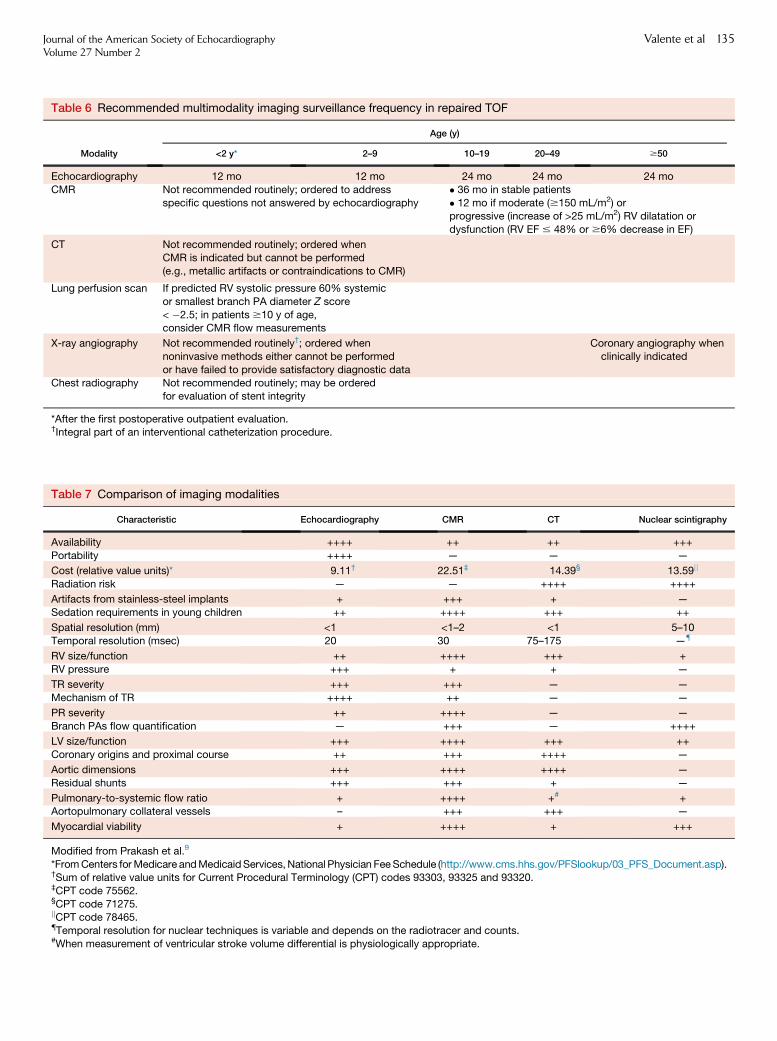
Table 6 Recommended multimodality imaging surveillance frequency in repaired TOF
Modality
Age (y)
<2 y* 2–9 10–19 20–49 $50
Echocardiography 12 mo 12 mo 24 mo 24 mo 24 mo
CMR Not recommended routinely; ordered to address
specific questions not answered by echocardiography
� 36 mo in stable patients
� 12 mo if moderate ($150 mL/m2) orprogressive (increase of >25 mL/m2) RV dilatation or
dysfunction (RV EF # 48% or $6% decrease in EF)
CT Not recommended routinely; ordered when
CMR is indicated but cannot be performed
(e.g., metallic artifacts or contraindications to CMR)
Lung perfusion scan If predicted RV systolic pressure 60% systemic
or smallest branch PA diameter Z score
< �2.5; in patients $10 y of age,consider CMR flow measurements
X-ray angiography Not recommended routinely†; ordered whennoninvasive methods either cannot be performed
or have failed to provide satisfactory diagnostic data
Coronary angiography whenclinically indicated
Chest radiography Not recommended routinely; may be orderedfor evaluation of stent integrity
*After the first postoperative outpatient evaluation.†Integral part of an interventional catheterization procedure.
Table 7 Comparison of imaging modalities
Characteristic Echocardiography CMR CT Nuclear scintigraphy
Availability ++++ ++ ++ +++
Portability ++++ — — —
Cost (relative value units)* 9.11† 22.51‡ 14.39§ 13.59jj
Radiation risk — — ++++ ++++
Artifacts from stainless-steel implants + +++ + —
Sedation requirements in young children ++ ++++ +++ ++
Spatial resolution (mm) <1 <1–2 <1 5–10
Temporal resolution (msec) 20 30 75–175 —{
RV size/function ++ ++++ +++ +
RV pressure +++ + + —
TR severity +++ +++ — —
Mechanism of TR ++++ ++ — —
PR severity ++ ++++ — —
Branch PAs flow quantification — +++ — ++++
LV size/function +++ ++++ +++ ++
Coronary origins and proximal course ++ +++ ++++ —
Aortic dimensions +++ ++++ ++++ —
Residual shunts +++ +++ + —
Pulmonary-to-systemic flow ratio + ++++ +# +Aortopulmonary collateral vessels – +++ +++ —
Myocardial viability + ++++ + +++
Modified from Prakash et al.9
*FromCenters forMedicare andMedicaidServices, National Physician FeeSchedule (http://www.cms.hhs.gov/PFSlookup/03_PFS_Document.asp).†Sum of relative value units for Current Procedural Terminology (CPT) codes 93303, 93325 and 93320.‡CPT code 75562.§CPT code 71275.jjCPT code 78465.{Temporal resolution for nuclear techniques is variable and depends on the radiotracer and counts.#When measurement of ventricular stroke volume differential is physiologically appropriate.
Journal of the American Society of EchocardiographyVolume 27 Number 2
Valente et al 135
136 Valente et al Journal of the American Society of EchocardiographyFebruary 2014
diagnostic information in all clinical circumstances relevant to patientswith repaired TOF. Therefore, a multimodality approach that takesinto account patient-specific considerations, the strengths and weak-nesses of each modality (Table 7), and institutional resources andexpertise is recommended.9
Echocardiography is the primary imagingmodality used in the eval-uation of patients with repaired TOF during the first decade of life andshould always be performed before othermodalities are used. CMR isperformed in this age group only when clinical or echocardiographicfindings are concerning for important anatomic or hemodynamic ab-normalities requiring therapeutic intervention. Reasons for notroutinely using CMR in patients <8 to 10 years of age include thefollowing: (1) most patients are clinically well during the first severalyears after repair, and RV size and function uncommonly reach severelevels of abnormalities, and (2) most young patients cannot cooperatewith the test without sedation. As noted in the section on echocardi-ography, several recent publications have provided echocardio-graphic thresholds that can be used to identify young patientsrequiring further evaluation by CMR. Cardiovascular CT is rarelyindicated in this age group except in patients with pacemakers or de-fibrillators who require quantification of RV size and function.Nuclear lung perfusion scanning is performed to quantify pulmonaryperfusion, but the number of repeat scans should be kept to a mini-mum given the risk for cancer related to radiation exposure.Cardiac catheterization is typically performed for therapeutic inter-ventions such as branch PA dilation or stenting, PV insertion, or occlu-sion of a residual shunt.
Once patients reach 8 to 10 years of age, the role of CMR in-creases, and the test becomes routine for surveillance of PR, RVsize and function, differential PA flow, anatomic abnormalities suchas RVOT aneurysm and aortic dilatation, and myocardial viability.Echocardiography continues to play an important role and is a vitaladjunct to CMR for assessing TR and estimating RVand PA pressures.However, the diagnostic capabilities of echocardiography becomemore constrained by declining acoustic windows as patients’ bodysizes increase. The roles of other modalities also evolve with age.Cardiovascular CT continues to be used only when CMR is contrain-dicated, nuclear lung perfusion scan can be replaced by CMR flowmeasurements, and cardiac catheterization is predominantly usedfor therapy.
As patient age advances, clinical concerns become more complex,with increased prevalence of coronary artery disease and noncardiaccomorbidities. These concerns influence the multimodality approachin this age group with an emphasis on assessment of the coronary cir-culation for acquired disease in addition to the other abnormalitiesrelated to TOF repair. As such, coronary CTA and x-ray angiographyassume an important role in anatomic evaluation of the coronary ar-teries, nuclear imaging is used for assessment of myocardial ischemia,and CMR is used for evaluation of myocardial perfusion and viability.
A rational multimodality approach to outpatient surveillance of pa-tients with repaired TOF takes into account all of the aforementionedconsiderations. A particular challenge in this population is the paucityof data on rates of progression of specific abnormalities such as RVdilatation and dysfunction. Specifically, we lack knowledge of predic-tors of rapid deterioration versus stable course of clinically importantparameters. Therefore, little information exists to inform the fre-quency of testing. This gap in knowledge is particularly glaring in anera of increased awareness of resource utilization. With this back-ground in mind, Table 6 represents a consensus recommendationfor multimodality imaging in clinically stable patients with repairedTOF (level of evidence C).
NOTICE AND DISCLAIMER
This report is made available by the ASE as a courtesy referencesource for its members. This report contains recommendations onlyand should not be used as the sole basis to make medical practice de-cisions or for disciplinary action against any employee. The statementsand recommendations contained in this report are based primarily onthe opinions of experts, rather than on scientifically verified data. TheASE makes no express or implied warranties regarding the complete-ness or accuracy of the information in this report, including the war-ranty of merchantability or fitness for a particular purpose. In no eventshall the ASE be liable to you, your patients, or any other third partiesfor any decision made or action taken by you or such other parties inreliance on this information. Nor does your use of this informationconstitute the offering of medical advice by the ASE or create anyphysician-patient relationship between the ASE and your patientsor anyone else.
REFERENCES
1. Hoffman JI, Kaplan S. The incidence of congenital heart disease. J AmColl Cardiol 2002;39:1890-900.
2. Hamilton BE, Hoyert DL,Martin JA, StrobinoDM,Guyer B. Annual sum-mary of vital statistics: 2010-2011. Pediatrics 2013;131:548-58.
3. Al Habib HF, Jacobs JP, Mavroudis C, Tchervenkov CI, O’Brien SM,Mohammadi S, et al. Contemporary patterns of management of tetralogyof Fallot: data from the Society of Thoracic Surgeons database. AnnThorac Surg 2010;90:813-9.
4. Chiu SN,Wang JK, ChenHC, LinMT,Wu ET, ChenCA, et al. Long-termsurvival and unnatural deaths of patients with repaired tetralogy of Fallotin an Asian cohort. Circ Cardiovasc Qual Outcomes 2012;5:120-5.
5. Marelli AJ, Mackie AS, Ionescu-Ittu R, Rahme E, Pilote L. Congenitalheart disease in the general population: changing prevalence and age dis-tribution. Circulation 2007;115:163-72.
6. Le Gloan L, Guerin P, Mercier LA, Abbey S, Dore A, Marcotte F, et al.Clinical assessment of arrhythmias in tetralogy of Fallot. Expert Rev Car-diovasc Ther 2010;8:189-97.
7. Khairy P, Aboulhosn J, Gurvitz MZ, Opotowsky AR, Mongeon FP, Kay J,et al. Arrhythmia burden in adults with surgically repaired tetralogy of Fal-lot: a multi-institutional study. Circulation 2010;122:868-75.
8. Nollert G, Fischlein T, Bouterwek S, Bohmer C, Dewald O, Kreuzer E,et al. Long-term results of total repair of tetralogy of Fallot in adulthood:35 years follow-up in 104 patients corrected at the age of 18 or older.Thorac Cardiovasc Surg 1997;45:178-81.
9. Prakash A, Powell AJ, Geva T. Multimodality noninvasive imaging forassessment of congenital heart disease. Circ Cardiovasc Imaging 2010;3:112-25.
10. Kilner PJ, Geva T, Kaemmerer H, Trindade PT, Schwitter J, Webb GD.Recommendations for cardiovascular magnetic resonance in adultswith congenital heart disease from the respective working groups ofthe European Society of Cardiology. Eur Heart J 2010;31:794-805.
11. Geva T. Repaired tetralogy of Fallot: the roles of cardiovascular magneticresonance in evaluating pathophysiology and for pulmonary valvereplacement decision support. J Cardiovasc Magn Reson 2011;13:9.
12. Warnes CA, Williams RG, Bashore TM, Child JS, Connolly HM,Dearani JA, et al. ACC/AHA 2008 guidelines for the management ofadults with congenital heart disease: a report of the American Collegeof Cardiology/American Heart Association Task Force on PracticeGuidelines (Writing Committee to Develop Guidelines on the Manage-ment of Adults with Congenital Heart Disease). Circulation 2008;118:e714-833.
13. Blalock A, Taussig HB. Landmark article May 191945: the surgical treat-ment of malformations of the heart in which there is pulmonary stenosisor pulmonary atresia. JAMA 1984;251:2123-38.
Journal of the American Society of EchocardiographyVolume 27 Number 2
Valente et al 137
14. Karpawich PP, Bush CP, Antillon JR, Amato JJ, Marbey ML, Agarwal KC.Modified Blalock-Taussig shunt in infants and young children. Clinicaland catheterization assessment. J Thorac Cardiovasc Surg 1985;89:275-9.
15. Sabri MR, Sholler G, Hawker R, Nunn G. Branch pulmonary arterygrowth after Blalock-Taussig shunts in tetralogy of Fallot and pulmonaryatresia with ventricular septal defect: a retrospective, echocardiographicstudy. Pediatr Cardiol 1999;20:358-63.
16. Alfieri O, Locatelli G, Bianchi T, Vanini V, Parenzan L. Repair of tetralogyof Fallot afterWaterston anastomosis. J Thorac Cardiovasc Surg 1979;77:826-31.
17. Daniel FJ, Clarke CP, Richardson JP, Westlake GW, Jones PG. An evalu-ation of Potts’ aortopulmonary shunt for palliation of cyanotic heart dis-ease. Thorax 1976;31:394-7.
18. Yamaki S. Pulmonary vascular disease in shunted and nonshunted pa-tients with tetralogy of Fallot. Tohoku J Exp Med 1990;162:109-19.
19. Gelb BD. Molecular genetics of congenital heart disease. Curr Opin Car-diol 1997;12:321-8.
20. Ito M, Kikuchi S, Hachiro Y, Abe T. Anomalous subaortic position of thebrachiocephalic vein associated with tetralogy of Fallot. Ann Thorac Car-diovasc Surg 2011;7:106-8.
21. Chaturvedi R, Mikailian H, Freedom RM. Crossed pulmonary arteries intetralogy of Fallot. Cardiol Young 2005;15:537.
22. Eyskens B, Brown SC, Claus P, Dymarkowski S, Gewillig M, Bogaert J,et al. The influence of pulmonary regurgitation on regional right ventric-ular function in children after surgical repair of tetralogy of Fallot. Eur JEchocardiogr 2010;11:341-5.
23. Rudski LG, Lai WW, Afilalo J, Hua L, Handschumacher MD,Chandrasekaran K, et al. Guidelines for the echocardiographic assess-ment of the right heart in adults: a report from the American Societyof Echocardiography endorsed by the European Association of Echocar-diography, a registered branch of the European Society of Cardiology,and the Canadian Society of Echocardiography. J Am Soc Echocardiogr2010;23:685-713.
24. Baumgartner H, Hung J, Bermejo J, Chambers JB, Evangelista A,Griffin BP, et al. Echocardiographic assessment of valve stenosis: EAE/ASE recommendations for clinical practice. J Am Soc Echocardiogr2009;22:1-23.
25. RedingtonAN.Determinants and assessment of pulmonary regurgitationin tetralogy of Fallot: practice and pitfalls. Cardiol Clin 2006;24:631-9.
26. Gatzoulis MA, Balaji S, Webber SA, Siu SC, Hokanson JS, Poile C, et al.Risk factors for arrhythmia and sudden cardiac death late after repair oftetralogy of Fallot: a multicentre study. Lancet 2000;356:975-81.
27. Lu JC, Cotts TB, Agarwal PP, Attili AK, Dorfman AL. Relation of rightventricular dilation, age of repair, and restrictive right ventricular physi-ology with patient-reported quality of life in adolescents and adultswith repaired tetralogy of Fallot. Am J Cardiol 2010;106:1798-802.
28. Therrien J, Siu SC, McLaughlin PR, Liu PP, WilliamsWG,Webb GD. Pul-monary valve replacement in adults late after repair of tetralogy of Fallot:are we operating too late? J Am Coll Cardiol 2000;36:1670-5.
29. Warnes CA. Adult congenital heart disease importance of the rightventricle. J Am Coll Cardiol 2009;54:1903-10.
30. Kilner PJ, Balossino R, Dubini G, Babu-Narayan SV, Taylor AM,Pennati G, et al. Pulmonary regurgitation: the effects of varying pulmo-nary artery compliance, and of increased resistance proximal or distalto the compliance. Int J Cardiol 2009;133:157-66.
31. Zoghbi WA, Enriquez-Sarano M, Foster E, Grayburn PA, Kraft CD,Levine RA, et al. Recommendations for evaluation of the severity ofnative valvular regurgitation with two-dimensional and Doppler echocar-diography. J Am Soc Echocardiogr 2003;16:777-802.
32. Li W, Davlouros PA, Kilner PJ, Pennell DJ, Gibson D, Henein MY, et al.Doppler-echocardiographic assessment of pulmonary regurgitation inadults with repaired tetralogy of Fallot: comparison with cardiovascularmagnetic resonance imaging. Am Heart J 2004;147:165-72.
33. Puchalski MD, Askovich B, Sower CT, Williams RV, Minich LL, Tani LY.Pulmonary regurgitation: determining severity by echocardiography andmagnetic resonance imaging. Congenit Heart Dis 2008;3:168-75.
34. Renella P, Aboulhosn J, Lohan DG, Jonnala P, Finn JP, Satou GM, et al.Two-dimensional and Doppler echocardiography reliably predict severepulmonary regurgitation as quantified by cardiac magnetic resonance. JAm Soc Echocardiogr 2010;23:880-6.
35. Silversides CK, Veldtman GR, Crossin J, Merchant N, Webb GD,McCrindle BW, et al. Pressure half-time predicts hemodynamically signif-icant pulmonary regurgitation in adult patients with repaired tetralogy ofFallot. J Am Soc Echocardiogr 2003;16:1057-62.
36. Fukuda S, Song JM, Gillinov AM, McCarthy PM, Daimon M,KongsaerepongV, et al. Tricuspid valve tethering predicts residual tricuspidregurgitation after tricuspid annuloplasty. Circulation 2005;111:975-9.
37. Fukuda S, Gillinov AM, Song JM, Daimon M, Kongsaerepong V,Thomas JD, et al. Echocardiographic insights into atrial and ventricularmechanisms of functional tricuspid regurgitation. Am Heart J 2006;152:1208-14.
38. GrossmannG, SteinM, KochsM, HoherM, KoenigW,HombachV, et al.Comparison of the proximal flow convergence method and the jet areamethod for the assessment of the severity of tricuspid regurgitation. EurHeart J 1998;19:652-9.
39. Alghamdi MH, Grosse-Wortmann L, Ahmad N, Mertens L,FriedbergMK. Can simple echocardiographicmeasures reduce the num-ber of cardiac magnetic resonance imaging studies to diagnose right ven-tricular enlargement in congenital heart disease? J Am Soc Echocardiogr2012;25:518-23.
40. Puchalski MD, Williams RV, Askovich B, Minich LL, Mart C, Tani LY.Assessment of right ventricular size and function: echo versus magneticresonance imaging. Congenit Heart Dis 2007;2:27-31.
41. LaiWW,Gauvreau K, Rivera ES, Saleeb S, Powell AJ, Geva T. Accuracy ofguideline recommendations for two-dimensional quantification of theright ventricle by echocardiography. Int J Cardiovasc Imaging 2008;24:691-8.
42. Jenkins C, Chan J, Bricknell K, Strudwick M, Marwick TH. Reproduc-ibility of right ventricular volumes and ejection fraction using real-timethree-dimensional echocardiography: comparison with cardiac MRI.Chest 2007;131:1844-51.
43. Lu X, Nadvoretskiy V, Bu L, Stolpen A, Ayres N, Pignatelli RH,Kovalchin JP, Grenier M, Klas B, Ge S. Accuracy and reproducibility ofreal-time three-dimensional echocardiography for assessment of rightventricular volumes and ejection fraction in children. J Am Soc Echocar-diogr 2008;21:84-9.
44. Grewal J, MajdalanyD, Syed I, Pellikka P,Warnes CA. Three-dimensionalechocardiographic assessment of right ventricular volume and functionin adult patients with congenital heart disease: comparisonwithmagneticresonance imaging. J Am Soc Echocardiogr 2010;23:127-33.
45. Dragulescu A, Grosse-Wortmann L, Fackoury C, Riffle S, Waiss M,Jaeggi E, et al. Echocardiographic assessment of right ventricular volumesafter surgical repair of tetralogy of Fallot: clinical validation of a new echo-cardiographic method. J Am Soc Echocardiogr 2011;24:1191-8.
46. Helbing WA, Bosch HG, Maliepaard C, Rebergen SA, van der Geest RJ,Hansen B, et al. Comparison of echocardiographic methods with mag-netic resonance imaging for assessment of right ventricular function inchildren. Am J Cardiol 1995;76:589-94.
47. Shimada YJ, Shiota M, Siegel RJ, Shiota T. Accuracy of right ventricularvolumes and function determined by three-dimensional echocardiogra-phy in comparison with magnetic resonance imaging: a meta-analysisstudy. J Am Soc Echocardiogr 2010;23:943-53.
48. Renella P, Marx GR, Gauvreau K, Zhou J, Geva T. 3D echocardiographicassessment of right ventricular size and function in children: feasibilityand reproducibility. J Am Soc Echocardiogr 2011;24.
49. Muzzarelli S, Ordovas KG, Cannavale G, Meadows AK, Higgins CB. Te-tralogy of Fallot: impact of the excursion of the interventricular septumon left ventricular systolic function and fibrosis after surgical repair. Radi-ology 2011;259:375-83.
50. Geva T, Sandweiss BM, Gauvreau K, Lock JE, Powell AJ. Factors associ-ated with impaired clinical status in long-term survivors of tetralogy ofFallot repair evaluated by magnetic resonance imaging. J Am Coll Car-diol 2004;43:1068-74.
138 Valente et al Journal of the American Society of EchocardiographyFebruary 2014
51. Davlouros PA, Kilner PJ, Hornung TS, Li W, Francis JM, Moon JC, et al.Right ventricular function in adults with repaired tetralogy of Fallot as-sessed with cardiovascular magnetic resonance imaging: detrimentalrole of right ventricular outflow aneurysms or akinesia and adverseright-to-left ventricular interaction. J Am Coll Cardiol 2002;40:2044-52.
52. Thambo JB, De Guillebon M, Dos Santos P, Xhaet O, Ploux S, Iriart X,et al. Electrical dyssynchrony and resynchronization in tetralogy of Fallot.Heart Rhythm 2011;8:909-14.
53. Mueller M, Rentzsch A, Hoetzer K, Raedle-Hurst T, Boettler P, Stiller B,et al. Assessment of interventricular and right-intraventriculardyssynchrony in patients with surgically repaired tetralogy of Fallotby two-dimensional speckle tracking. Eur J Echocardiogr 2010;11:786-92.
54. Anavekar NS, Gerson D, Skali H, Kwong RY, Yucel EK, Solomon SD.Two-dimensional assessment of right ventricular function: anechocardiographic-MRI correlative study. Echocardiography 2007;24:452-6.
55. Wang J, Prakasa K, Bomma C, Tandri H, Dalal D, James C, et al. Compar-ison of novel echocardiographic parameters of right ventricular functionwith ejection fraction by cardiac magnetic resonance. J Am Soc Echocar-diogr 2007;20:1058-64.
56. Srinivasan C, Sachdeva R, MorrowWR, Greenberg SB, Vyas HV. Limita-tions of standard echocardiographic methods for quantification of rightventricular size and function in children and young adults. J UltrasoundMed 2011;30:487-93.
57. Gleason WL, Braunwald E. Studies on the first derivative of the ventric-ular pressure pulse in man. J Clin Invest 1962;41:80-91.
58. Duan YY, Harada K, Toyono M, Ishii H, Tamura M, Takada G. Effects ofacute preload reduction on myocardial velocity during isovolumiccontraction and myocardial acceleration in pediatric patients. PediatrCardiol 2006;27:32-6.
59. Kjaergaard J, Snyder EM, Hassager C, Oh JK, Johnson BD. Impact of pre-load and afterload on global and regional right ventricular function andpressure: a quantitative echocardiography study. J Am Soc Echocardiogr2006;19:515-21.
60. VogelM, SchmidtMR, Kristiansen SB, CheungM,White PA, Sorensen K,et al. Validation of myocardial acceleration during isovolumic contractionas a novel noninvasive index of right ventricular contractility: comparisonwith ventricular pressure-volume relations in an animal model. Circula-tion 2002;105:1693-9.
61. Tei C, Dujardin KS, Hodge DO, Bailey KR, McGoon MD, Tajik AJ, et al.Doppler echocardiographic index for assessment of global right ventric-ular function. J Am Soc Echocardiogr 1996;9:838-47.
62. Cetin I, Tokel K, Varan B, Orun U, Aslamaci S. Evaluation of right ventric-ular function by using tissue Doppler imaging in patients after repair oftetralogy of Fallot. Echocardiography 2009;26:950-7.
63. Yasuoka K, Harada K, Toyono M, Tamura M, Yamamoto F. Tei indexdetermined by tissue Doppler imaging in patients with pulmonary regur-gitation after repair of tetralogy of Fallot. Pediatr Cardiol 2004;25:131-6.
64. Abd El Rahman MY, Abdul-Khaliq H, Vogel M, Alexi-Meskischvili V,GutberletM,Hetzer R, et al. Value of the newDoppler-derivedmyocardialperformance index for the evaluation of right and left ventricular functionfollowing repair of tetralogy of Fallot. Pediatr Cardiol 2002;23:502-7.
65. Schwerzmann M, Samman AM, Salehian O, Holm J, Provost Y,Webb GD, et al. Comparison of echocardiographic and cardiac magneticresonance imaging for assessing right ventricular function in adults withrepaired tetralogy of Fallot. Am J Cardiol 2007;99:1593-7.
66. Kjaergaard J, Petersen CL, Kjaer A, Schaadt BK, Oh JK, Hassager C. Eval-uation of right ventricular volume and function by 2D and 3D echocar-diography compared to MRI. Eur J Echocardiogr 2006;7:430-8.
67. Kaul S, Tei C, Hopkins JM, Shah PM. Assessment of right ventricular func-tion using two-dimensional echocardiography. Am Heart J 1984;107:526-31.
68. Urheim S, Cauduro S, Frantz R, McGoon M, Belohlavek M, Green T,et al. Relation of tissue displacement and strain to invasively determinedright ventricular stroke volume. Am J Cardiol 2005;96:1173-8.
69. Bonnemains L, Stos B, Vaugrenard T, Marie PY, Odille F, Boudjemline Y.Echocardiographic right ventricle longitudinal contraction indices cannotpredict ejection fraction in post-operative Fallot children. Eur Heart J Car-diovasc Imaging 2012;13:235-42.
70. Koestenberger M, Nagel B, Ravekes W, Everett AD, Stueger HP,Heinzl B, et al. Tricuspid annular plane systolic excursion and right ven-tricular ejection fraction in pediatric and adolescent patients with tetral-ogy of Fallot, patients with atrial septal defect, and age-matched normalsubjects. Clin Res Cardiol 2011;100:67-75.
71. Morcos P, Vick GW III, Sahn DJ, Jerosch-Herold M, Shurman A,Sheehan FH. Correlation of right ventricular ejection fraction andtricuspid annular plane systolic excursion in tetralogy of Fallot by mag-netic resonance imaging. Int J Cardiovasc Imaging 2009;25:263-70.
72. Koestenberger M, Ravekes W, Everett AD, Stueger HP, Heinzl B,Gamillscheg A, et al. Right ventricular function in infants, children andadolescents: reference values of the tricuspid annular plane systolicexcursion (TAPSE) in 640 healthy patients and calculation of Z scorevalues. J Am Soc Echocardiogr 2009;22:715-9.
73. Koestenberger M, Nagel B, Ravekes W, Everett AD, Stueger HP,Heinzl B, et al. Systolic right ventricular function in pediatric and adoles-cent patients with tetralogy of Fallot: echocardiography versus magneticresonance imaging. J Am Soc Echocardiogr 2011;24:45-52.
74. Pavlicek M, Wahl A, Rutz T, de Marchi SF, Hille R, Wustmann K, et al.Right ventricular systolic function assessment: rank of echocardiographicmethods vs. cardiac magnetic resonance imaging. Eur J Echocardiogr2011;12:871-80.
75. Kutty S, Zhou J, Gauvreau K, Trincado C, Powell AJ, Geva T. Regionaldysfunction of the right ventricular outflow tract reduces the accuracyof Doppler tissue imaging assessment of global right ventricular systolicfunction in patients with repaired tetralogy of Fallot. J Am Soc Echocar-diogr 2011;24:637-43.
76. Gatzoulis MA, Clark AL, Cullen S, Newman CG, Redington AN. Rightventricular diastolic function 15 to 35 years after repair of tetralogy of Fal-lot. Restrictive physiology predicts superior exercise performance. Circu-lation 1995;91:1775-81.
77. Cullen S, Shore D, Redington A. Characterization of right ventricular dia-stolic performance after complete repair of tetralogy of Fallot. Restrictivephysiology predicts slow postoperative recovery. Circulation 1995;91:1782-9.
78. van den Berg J, Wielopolski PA, Meijboom FJ, Witsenburg M, Bogers AJ,Pattynama PM, et al. Diastolic function in repaired tetralogy of Fallot atrest and during stress: assessment with MR imaging. Radiology 2007;243:212-9.
79. Hui W, Abd El Rahman MY, Dsebissowa F, Rentzsch A, Gutberlet M,Alexi-Meskishvili V, et al. Quantitative analysis of right atrial perfor-mance after surgical repair of tetralogy of Fallot. Cardiol Young 2004;14:520-6.
80. Ghai A, Silversides C, Harris L,WebbGD, Siu SC, Therrien J. Left ventric-ular dysfunction is a risk factor for sudden cardiac death in adults late af-ter repair of tetralogy of Fallot. J Am Coll Cardiol 2002;40:1675-80.
81. Knauth AL, Gauvreau K, Powell AJ, Landzberg MJ, Walsh EP, Lock JE,et al. Ventricular size and function assessed by cardiac MRI predict majoradverse clinical outcomes late after tetralogy of Fallot repair. Heart 2008;94:211-6.
82. Colan SD, Shirali G, Margossian R, Gallagher D, Altmann K, Canter C,et al. The Ventricular Volume Variability Study of the Pediatric HeartNetwork: study design and impact of beat averaging and variable typeon the reproducibility of echocardiographic measurements in childrenwith chronic dilated cardiomyopathy. J Am Soc Echocardiogr 2012;25:842-54.
83. Diller GP, Kempny A, Liodakis E, Alonso-Gonzalez R, Inuzuka R,Uebing A, et al. Left ventricular longitudinal function predicts life-threatening ventricular arrhythmia and death in adults with repaired te-tralogy of Fallot. Circulation 2012;125:2440-6.
84. Brili S, Stamatopoulos I, Barbetseas J, Chrysohoou C, Alexopoulos N,Misailidou M, et al. Usefulness of dobutamine stress echocardiographywith tissue Doppler imaging for the evaluation and follow-up of patients

Journal of the American Society of EchocardiographyVolume 27 Number 2
Valente et al 139
with repaired tetralogy of Fallot. J Am Soc Echocardiogr 2008;21:1093-8.
85. Baspinar O, Alehan D. Dobutamine stress echocardiography in the eval-uation of cardiac haemodynamics after repair of tetralogy of Fallot in chil-dren: negative effects of pulmonary regurgitation. Acta Cardiol 2006;61:279-83.
86. Dodds GA, Warnes CA, Danielson GK. Aortic valve replacement afterrepair of pulmonary atresia and ventricular septal defect or tetralogy ofFallot. J Thorac Cardiovasc Surg 1997;113:736-41.
87. Niwa K. Aortic root dilatation in tetralogy of Fallot long-term afterrepair—histology of the aorta in tetralogy of Fallot: evidence of intrinsicaortopathy. Int J Cardiol 2005;103:117-9.
88. Lopez L, Colan SD, Frommelt PC, Ensing GJ, Kendall K, Younoszai AK,et al. Recommendations for quantification methods during the perfor-mance of a pediatric echocardiogram: a report from the Pediatric Mea-surements Writing Group of the American Society ofEchocardiography Pediatric and Congenital Heart Disease Council. JAm Soc Echocardiogr 2010;23:465-95.
89. Hiratzka LF, Bakris GL, Beckman JA, Bersin RM, Carr VF, CaseyDE, et al.2010 ACCF/AHA/AATS/ACR/ASA/SCA/SCAI/SIR/STS/SVMguidelines for the diagnosis and management of patients with thoracicaortic disease. A report of the American College of Cardiology Founda-tion/American Heart Association Task Force on Practice Guidelines,American Association for Thoracic Surgery, American College of Radi-ology, American Stroke Association, Society of Cardiovascular Anesthe-siologists, Society for Cardiovascular Angiography and Interventions,Society of Interventional Radiology, Society of Thoracic Surgeons, andSociety for Vascular Medicine. J Am Coll Cardiol 2010;55:e27-129.
90. Haycock GB, Schwartz GJ, Wisotsky DH. Geometric method formeasuring body surface area: a height-weight formula validated in in-fants, children, and adults. J Pediatr 1978;93:62-6.
91. DuBois D, DuBois E. A formula to estimate the approximate surface areaif height and weight be known. Arch Intern Med 1916;17:863-71.
92. Lai WW, Geva T, Shirali GS, Frommelt PC, Humes RA, BrookMM, et al.Guidelines and standards for performance of a pediatric echocardio-gram: a report from the Task Force of the Pediatric Council of the Amer-ican Society of Echocardiography. J Am Soc Echocardiogr 2006;19:1413-30.
93. Festa P, Ait-Ali L, Minichilli F, Kristo I, Deiana M, Picano E. A new simplemethod to estimate pulmonary regurgitation by echocardiography inoperated Fallot: comparison with magnetic resonance imaging and per-formance test evaluation. J Am Soc Echocardiogr 2010;23:496-503.
94. ChessaM, Butera G, Carminati M. Intracardiac echocardiography duringpercutaneous pulmonary valve replacement. Eur Heart J 2008;29:2908.
95. Plein S, Bloomer TN, Ridgway JP, Jones TR, Bainbridge GJ,Sivananthan MU. Steady-state free precession magnetic resonance imag-ing of the heart: comparison with segmented k-space gradient-echo im-aging. J Magn Reson Imaging 2011;14:230-6.
96. Babu-Narayan SV, Kilner PJ, Li W, Moon JC, Goktekin O, Davlouros PA,et al. Ventricular fibrosis suggested by cardiovascular magnetic resonancein adults with repaired tetralogy of Fallot and its relationship to adversemarkers of clinical outcome. Circulation 2006;113:405-13.
97. Wald RM, Haber I, Wald R, Valente AM, Powell AJ, Geva T. Effects ofregional dysfunction and late gadolinium enhancement on global rightventricular function and exercise capacity in patients with repaired tetral-ogy of Fallot. Circulation 2009;119:1370-7.
98. HelbingWA, deRoosA.Clinical applications of cardiacmagnetic resonanceimaging after repair of tetralogy of Fallot. Pediatr Cardiol 2000;21:70-9.
99. van Straten A, Vliegen HW, Hazekamp MG, Bax JJ, Schoof PH,Ottenkamp J, et al. Right ventricular function after pulmonary valvereplacement in patients with tetralogy of Fallot. Radiology 2004;233:824-9.
100. Vliegen HW, Van Straten A, De Roos A, Roest AA, Schoof PH,Zwinderman AH, et al. Magnetic resonance imaging to assess the hemo-dynamic effects of pulmonary valve replacement in adults late afterrepair of tetralogy of Fallot. Circulation 2002;106:1703-7.
101. Babu-Narayan SV, Gatzoulis MA. Management of adults with operatedtetralogy of Fallot. Curr Treat Options Cardiovasc Med 2003;5:389-98.
102. Garg R, Powell AJ, Sena L, Marshall AC, Geva T. Effects of metallic im-plants on magnetic resonance imaging evaluation of Fontan palliation.Am J Cardiol 2005;95:688-91.
103. Sorrentino RA. A novel MRI-safe dual-chamber pacemaker system: itstime has come. Heart Rhythm 2011;8:74-5.
104. Reiter T, Ritter O, Prince MR, Nordbeck P, Wanner C, Nagel E, et al.Minimizing risk of nephrogenic systemic fibrosis in cardiovascular mag-netic resonance. J Cardiovasc Magn Reson 2012;14:31.
105. Sridharan S, Derrick G, Deanfield J, Taylor AM. Assessment of differentialbranch pulmonary blood flow: a comparative study of phase contrastmagnetic resonance imaging and radionuclide lung perfusion imaging.Heart 2006;92:963-8.
106. Schievano S, Coats L, Migliavacca F, Norman W, Frigiola A, Deanfield J,et al. Variations in right ventricular outflow tract morphology followingrepair of congenital heart disease: implications for percutaneous pulmo-nary valve implantation. J Cardiovasc Magn Reson 2007;9:687-95.
107. Weber OM, Higgins CB. MR evaluation of cardiovascular physiology incongenital heart disease: flow and function. J Cardiovasc Magn Reson2006;8:607-17.
108. Greil GF, Stuber M, Botnar RM, Kissinger KV, Geva T, Newburger JW,et al. Coronary magnetic resonance angiography in adolescents andyoung adults with Kawasaki disease. Circulation 2002;105:908-11.
109. Valverde I, Parish V, Hussain T, Rosenthal E, Beerbaum P, Krasemann T.Planning of catheter interventions for pulmonary artery stenosis:improved measurement agreement with magnetic resonance angiog-raphy using identical angulations. Catheter Cardiovasc Interv 2011;77:400-8.
110. Nordmeyer J, Gaudin R, Tann OR, Lurz PC, Bonhoeffer P, Taylor AM,et al. MRI may be sufficient for noninvasive assessment of great vesselstents: an in vitro comparison ofMRI, CT, and conventional angiography.AJR Am J Roentgenol 2010;195:865-71.
111. Wald RM, Redington AN, Pereira A, Provost YL, Paul NS, Oechslin EN,et al. Refining the assessment of pulmonary regurgitation in adults aftertetralogy of Fallot repair: should we be measuring regurgitant fractionor regurgitant volume? Eur Heart J 2009;30:356-61.
112. Geva T, Gauvreau K, Powell AJ, Cecchin F, Rhodes J, Geva J, et al. Ran-domized trial of pulmonary valve replacement with and without rightventricular remodeling surgery. Circulation 2010;122:S201-8.
113. van der Hulst AE, Westenberg JJ, Kroft LJ, Bax JJ, Blom NA, de Roos A,et al. Tetralogy of Fallot: 3D velocity-encoded MR imaging for evaluationof right ventricular valve flow and diastolic function in patients aftercorrection. Radiology 2010;256:724-34.
114. Norton KI, TongC, Glass RB, Nielsen JC. CardiacMR imaging assessmentfollowing tetralogy of Fallot repair. Radiographics 2006;26:197-211.
115. Samyn MM, Powell AJ, Garg R, Sena L, Geva T. Range of ventricular di-mensions and function by steady-state free precession cine MRI in re-paired tetralogy of Fallot: right ventricular outflow tract patch vs.conduit repair. J Magn Reson Imaging 2007;26:934-40.
116. Mooij CF, de Wit CJ, Graham DA, Powell AJ, Geva T. Reproducibility ofMRI measurements of right ventricular size and function in patients withnormal and dilated ventricles. J Magn Reson Imaging 2008;28:67-73.
117. Blalock SE, Banka P, Geva T, Powell AJ, Zhou J, Prakash A. Interstudy vari-ability in cardiac magnetic resonance imaging measurements of ventricu-lar volume, mass, and ejection fraction in repaired tetralogy of Fallot: aprospective observational study. J Magn Reson Imaging 2013;38:829-35.
118. Sarikouch S, Peters B, Gutberlet M, Leismann B, Kelter-Kloepping A,Koerperich H, et al. Sex-specific pediatric percentiles for ventricular sizeand mass as reference values for cardiac MRI: assessment by steady-state free-precession and phase-contrast MRI flow. Circ Cardiovasc Imag-ing 2010;3:65-76.
119. Valente AM, Gauvreau K, Assenza GE, Babu-Narayan SV, Schreier J, Gat-zoulis M, et al. Contemporary predictors of death and sustained ventric-ular tachycardia in patients with repaired tetralogy of Fallot enrolled inthe INDICATOR cohort. Heart. In press.
140 Valente et al Journal of the American Society of EchocardiographyFebruary 2014
120. Kim RJ, Fieno DS, Parrish TB, Harris K, Chen EL, Simonetti O, et al. Rela-tionship of MRI delayed contrast enhancement to irreversible injury,infarct age, and contractile function. Circulation 1999;100:1992-2002.
121. Gulati A, Jabbour A, Ismail TF, GuhaK, Khwaja J, Raza S, et al. Associationof fibrosis with mortality and sudden cardiac death in patients with non-ischemic dilated cardiomyopathy. JAMA 2013;309:896-908.
122. Valverde I, Parish V, Tzifa A, Head C, Sarikouch S, Greil G, et al. Cardio-vascular MR dobutamine stress in adult tetralogy of Fallot: disparity be-tween CMR volumetry and flow for cardiovascular function. J MagnReson Imaging 2011;33:1341-50.
123. Parish V, Valverde I, Kutty S, Head C, Qureshi SA, Sarikouch S, et al. Do-butamine stress MRI in repaired tetralogy of Fallot with chronic pulmo-nary regurgitation a comparison with healthy volunteers. Int J Cardiol2013;166:96-105.
124. Riesenkampff E, Mengelkamp L, Mueller M, Kropf S, Abdul-Khaliq H,Sarikouch S, et al. Integrated analysis of atrioventricular interactions intetralogy of Fallot. Am J Physiol Heart Circ Physiol 2010;299:H364-71.
125. Luijnenburg SE, Peters RE, van der Geest RJ, Moelker A, Roos-Hesselink JW, de Rijke YB, et al. Abnormal right atrial and right ventric-ular diastolic function relate to impaired clinical condition in patientsoperated for tetralogy of Fallot. Int J Cardiol 2013;167:833-9.
126. Broberg CS, Aboulhosn J, Mongeon FP, Kay J, Valente AM, Khairy P,et al. Prevalence of left ventricular systolic dysfunction in adults with re-paired tetralogy of Fallot. Am J Cardiol 2011;107:1215-20.
127. Darsee JR, Mikolich JR,Walter PF, Schlant RC. Paradoxical rise in left ven-tricular filling pressure in the dog during positive end-expiratory pressureventilation. A reversed Bernheim effect. Circ Res 1981;49:1017-28.
128. Lurz P, Puranik R, Nordmeyer J, Muthurangu V, Hansen MS,Schievano S, et al. Improvement in left ventricular filling properties afterrelief of right ventricle to pulmonary artery conduit obstruction: contribu-tion of septal motion and interventricular mechanical delay. Eur Heart J2009;30:2266-74.
129. Powell AJ,Maier SE, Chung T, Geva T. Phase-velocity cinemagnetic reso-nance imaging measurement of pulsatile blood flow in children andyoung adults: in vitro and in vivo validation. Pediatr Cardiol 2000;21:104-10.
130. Potthast S, Mitsumori L, Stanescu LA, Richardson ML, Branch K,Dubinsky TJ, et al. Measuring aortic diameter with different MR tech-niques: comparison of three-dimensional (3D) navigated steady-statefree-precession (SSFP), 3D contrast-enhanced magnetic resonance angi-ography (CE-MRA), 2DT2 black blood, and 2D cine SSFP. J Magn ResonImaging 2010;31:177-84.
131. Kaiser T, Kellenberger CJ, Albisetti M, Bergstrasser E, ValsangiacomoBuechel ER. Normal values for aortic diameters in children and adoles-cents—assessment in vivo by contrast-enhanced CMR-angiography. J Car-diovasc Magn Reson 2008;10:56.
132. Schulz-Menger J, Bluemke DA, Bremerich J, Flamm SD, Fogel MA,Friedrich MG, et al. Standardized image interpretation and post process-ing in cardiovascular magnetic resonance: Society for CardiovascularMagnetic Resonance (SCMR) Board of Trustees Task Force on Standard-ized Post Processing. J Cardiovasc Magn Reson 2013;15:35.
133. Lurz P, Coats L, Khambadkone S, Nordmeyer J, Boudjemline Y,Schievano S, et al. Percutaneous pulmonary valve implantation: impactof evolving technology and learning curve on clinical outcome. Circula-tion 2008;117:1964-72.
134. Atalay MK, Prince EA, Pearson CA, Chang KJ. The prevalence and clin-ical significance of noncardiac findings on cardiac MRI. AJR Am J Roent-genol 2011;196:W387-93.
135. Whitlock M, Garg A, Gelow J, Jacobson T, Broberg C. Comparison of leftand right atrial volume by echocardiography versus cardiac magneticresonance imaging using the area-length method. Am J Cardiol 2010;106:1345-50.
136. GindeAA, Foianini A, RennerDM, ValleyM, CamargoCA Jr. Availabilityand quality of computed tomography and magnetic resonance imagingequipment in U.S. emergency departments. Acad Emerg Med 2008;15:780-3.
137. Fuchs T, Kachelriess M, Kalender WA. Technical advances in multi-slicespiral CT. Eur J Radiol 2000;36:69-73.
138. Desjardins B, Kazerooni EA. ECG-gated cardiac CT. AJR Am J Roent-genol 2004;182:993-1010.
139. Einstein AJ, Elliston CD, Arai AE, ChenMY,Mather R, Pearson GD, et al.Radiation dose from single-heartbeat coronary CT angiography per-formed with a 320-detector row volume scanner. Radiology 2010;254:698-706.
140. Gerber TC, Kuzo RS, Lane GE, O’Brien PC, Karstaedt N, Morin RL, et al.Image quality in a standardized algorithm for minimally invasive coro-nary angiographywithmultislice spiral computed tomography. J ComputAssist Tomogr 2003;27:62-9.
141. Moloo J, Shapiro MD, Abbara S. Cardiac computed tomography:technique and optimization of protocols. Semin Roentgenol 2008;43:90-9.
142. Nicol ED, Gatzoulis M, Padley SP, Rubens M. Assessment of adultcongenital heart disease with multi-detector computed tomography:beyond coronary lumenography. Clin Radiol 2007;62:518-27.
143. Pearce MS, Salotti JA, Little MP, McHugh K, Lee C, Kim KP, et al. Radi-ation exposure from CT scans in childhood and subsequent risk ofleukaemia and brain tumours: a retrospective cohort study. Lancet2012;380:499-505.
144. Brenner DJ, Doll R, Goodhead DT, Hall EJ, Land CE, Little JB, et al. Can-cer risks attributable to low doses of ionizing radiation: assessing what wereally know. Proc Natl Acad Sci U S A 2003;100:13761-6.
145. Fazel R, Krumholz HM,Wang Y, Ross JS, Chen J, TingHH, et al. Exposureto low-dose ionizing radiation frommedical imaging procedures. N Engl JMed 2009;361:849-57.
146. Kleinerman RA. Cancer risks following diagnostic and therapeutic radia-tion exposure in children. Pediatr Radiol 2006;36(suppl):121-5.
147. Brenner DJ. Estimating cancer risks from pediatric CT: going from thequalitative to the quantitative. Pediatr Radiol 2002;32:242-8.
148. Weisbord SD, Mor MK, Resnick AL, Hartwig KC, Palevsky PM, Fine MJ.Incidence and outcomes of contrast-induced AKI following computedtomography. Clin J Am Soc Nephrol 2008;3:1274-81.
149. Goo HW, Park IS, Ko JK, Kim YH, Seo DM, Park JJ. Computed tomog-raphy for the diagnosis of congenital heart disease in pediatric and adultpatients. Int J Cardiovasc Imaging 2005;21:347-65.
150. Gartner RD, Sutton NJ, Weinstein S, Spindola-Franco H, Haramati LB.MRI and computed tomography of cardiac and pulmonary complica-tions of tetralogy of Fallot in adults. J Thorac Imaging 2010;25:183-90.
151. Holzer RJ, Gauvreau K, Kreutzer J, Leahy R, Murphy J, Lock JE, et al.Balloon angioplasty and stenting of branch pulmonary arteries: adverseevents and procedural characteristics: results of a multi-institutional regis-try. Circ Cardiovasc Interv 2011;4:287-96.
152. Juergens KU, Grude M, Maintz D, Fallenberg EM, Wichter T, Heindel W,et al. Multi-detector row CT of left ventricular function with dedicatedanalysis software versus MR imaging: initial experience. Radiology2004;230:403-10.
153. Lembcke A, Dohmen PM, Dewey M, Klessen C, Elgeti T, Hermann KG,et al. Multislice computed tomography for preoperative evaluation ofright ventricular volumes and function: comparison with magnetic reso-nance imaging. Ann Thorac Surg 2005;79:1344-51.
154. Raman SV, ShahM, McCarthy B, Garcia A, Ferketich AK. Multi-detectorrow cardiac computed tomography accurately quantifies right and leftventricular size and function compared with cardiacmagnetic resonance.Am Heart J 2006;151:736-44.
155. Chung JH, Ghoshhajra BB, Rojas CA, Dave BR, Abbara S. CT angiog-raphy of the thoracic aorta. Radiol Clin North Am 2010;48:249-64.
156. Rathi VK, Doyle M, Williams RB, Yamrozik J, Shannon RP,Biederman RW. Massive aortic aneurysm and dissection in repaired te-tralogy of Fallot; diagnosis by cardiovascular magnetic resonance imag-ing. Int J Cardiol 2005;101:169-70.
157. KimWH, Seo JW, Kim SJ, Song J, Lee J, Na CY. Aortic dissection late afterrepair of tetralogy of Fallot. Int J Cardiol 2005;101:515-6.
158. Lu TL, Huber CH, Rizzo E, Dehmeshki J, von Segesser LK, Qanadli SD.Ascending aorta measurements as assessed by ECG-gatedmulti-detector
Journal of the American Society of EchocardiographyVolume 27 Number 2
Valente et al 141
computed tomography: a pilot study to establish normative values fortranscatheter therapies. Eur Radiol 2009;19:664-9.
159. Mao SS, Ahmadi N, Shah B, Beckmann D, Chen A, Ngo L, et al. Normalthoracic aorta diameter on cardiac computed tomography in healthyasymptomatic adults: impact of age and gender. Acad Radiol 2008;15:827-34.
160. Hadamitzky M, Distler R, Meyer T, Hein F, Kastrati A, Martinoff S, et al.Prognostic value of coronary computed tomographic angiography incomparison with calcium scoring and clinical risk scores. Circ CardiovascImaging 2011;4:16-23.
161. de Graaf FR, Schuijf JD, Delgado V, van Velzen JE, Kroft LJ, de Roos A,et al. Clinical application of CT coronary angiography: state of the art.Heart Lung Circ 2010;19:107-16.
162. Hamirani YS, Isma’eel H, Larijani V, Drury P, LimW, BevinalM, et al. Thediagnostic accuracy of 64-detector cardiac computed tomographycompared with stress nuclear imaging in patients undergoing invasivecardiac catheterization. J Comput Assist Tomogr 2010;34:645-51.
163. Hamdan A, Asbach P, Wellnhofer E, Klein C, Gebker R, Kelle S, et al. Aprospective study for comparison ofMR andCT imaging for detection ofcoronary artery stenosis. JACC Cardiovasc Imaging 2011;4:50-61.
164. Cook SC, Raman SV. Unique application ofmultislice computed tomogra-phy in adults with congenital heart disease. Int J Cardiol 2007;119:101-6.
165. Jakobs TF, Becker CR, Ohnesorge B, Flohr T, Suess C, Schoepf UJ, et al.Multislice helical CTof the heart with retrospective ECG gating: reduc-tion of radiation exposure by ECG-controlled tube current modulation.Eur Radiol 2002;12:1081-6.
166. Pruckmayer M, Zacherl S, Salzer-Muhar U, SchlemmerM, Leitha T. Scin-tigraphic assessment of pulmonary and whole-body blood flow patternsafter surgical intervention in congenital heart disease. J Nucl Med 1999;40:1477-83.
167. Bourque JM, Beller GA. Stress myocardial perfusion imaging for assessingprognosis: an update. JACC Cardiovasc Imaging 2011;4:1305-19.
168. O’Laughlin MP, SlackMC, Grifka RG, Perry SB, Lock JE, Mullins CE. Im-plantation and intermediate-term follow-up of stents in congenital heartdisease. Circulation 1993;88:605-14.
169. Jonsson H, Ivert T, Jonasson R, Wahlgren H, Holmgren A, Bjork VO. Pul-monary function thirteen to twenty-six years after repair of tetralogy ofFallot. J Thorac Cardiovasc Surg 1994;108:1002-9.
170. WuMT, Huang YL, Hsieh KS, Huang JT, Peng NJ, Pan JY, et al. Influenceof pulmonary regurgitation inequality on differential perfusion of thelungs in tetralogy of Fallot after repair: a phase-contrast magnetic reso-nance imaging and perfusion scintigraphy study. J Am Coll Cardiol2007;49:1880-6.
171. Tamir A, Melloul M, Berant M, Horev G, Lubin E, Blieden LC, et al. Lungperfusion scans in patients with congenital heart defects. J Am Coll Car-diol 1992;19:383-8.
172. Boothroyd AE, McDonald EA, Carty H. Lung perfusion scintigraphy inpatients with congenital heart disease: sensitivity and important pitfalls.Nucl Med Commun 1996;17:33-9.
173. Hashimoto K, Nakamura Y, Matsui M, Kurosawa H, Arai T. Alteration ofpulmonary blood flow in tetralogy of Fallot: pre- and postoperative studywithmacroaggregates of 99mTc-labeled human serum albumin. Jpn Circ J1992;56:992-7.
174. Dowdle SC, Human DG, Mann MD. Pulmonary ventilation and perfu-sion abnormalities and ventilation perfusion imbalance in children withpulmonary atresia or extreme tetralogy of Fallot. J Nucl Med 1990;31:1276-9.
175. Reduto LA, Berger HJ, Johnstone DE, Hellenbrand W, Wackers FJ,Whittemore R, et al. Radionuclide assessment of right and left ventricularexercise reserve after total correction of tetralogy of Fallot. Am J Cardiol1980;45:1013-8.
176. Hurwitz RA, Treves S, Kuruc A. Right ventricular and left ventricular ejec-tion fraction in pediatric patients with normal hearts: first-pass radionu-clide angiocardiography. Am Heart J 1984;107:726-32.
177. Manno BV, Iskandrian AS, Hakki AH. Right ventricular function: meth-odologic and clinical considerations in noninvasive scintigraphic assess-ment. J Am Coll Cardiol 1984;3:1072-81.
178. Cusimano RJ, Guest C. Coronary artery disease following repair of tetral-ogy of Fallot: implications andmanagement. Can J Cardiol 1996;12:172-4.
179. Parker JA, Coleman RE, Grady E, Royal HD, Siegel BA, Stabin MG, et al.SNM practice guideline for lung scintigraphy 4.0. J Nucl Med Technol2012;40:57-65.
180. Strauss HW, Miller DD, Wittry MD, Cerqueira MD, Garcia EV,Iskandrian AS, et al. Procedure guideline for myocardial perfusion imag-ing 3.3. J Nucl Med Technol 2008;36:155-61.
181. Bonhoeffer P, Boudjemline Y, Saliba Z, Merckx J, Aggoun Y, Bonnet D,et al. Percutaneous replacement of pulmonary valve in a right-ventricle topulmonary-artery prosthetic conduit with valve dysfunction. Lancet2000;356:1403-5.
182. McElhinney DB, Hellenbrand WE, Zahn EM, Jones TK, Cheatham JP,Lock JE, et al. Short- and medium-term outcomes after transcatheter pul-monary valve placement in the expanded multicenter us melody valvetrial. Circulation 2010;122:507-16.
183. Geva T, Bergersen L, Kreutzer J. Diagnostic pathways for evaluation ofcongenital heart disease. In: Crawford MH, DiMarco JP, Paulus WJ, edi-tors. Cardiology. Philadelphia: Elsevier; 2009. pp. 1415-40.
184. Noble S,Miro J, YongG, Bonan R, Tardif JC, IbrahimR. Rapid pacing rota-tional angiography with three-dimensional reconstruction: use and ben-efits in structural heart disease interventions. Eurointervention 2009;5:244-9.
185. Glatz AC, Zhu X, Gillespie MJ, Hanna BD, Rome JJ. Use of angiographicCT imaging in the cardiac catheterization laboratory for congenital heartdisease. JACC Cardiovasc Imaging 2010;3:1149-57.
186. Fagan T, Kay J, Carroll J, Neubauer A. 3-D guidance of complex pulmo-nary artery stent placement using reconstructed rotational angiographywith live overlay. Catheter Cardiovasc Interv 2012;79:414-21.
187. Lock JE, Keane JF, Fellows KE. Diagnostic and interventional cardiac cath-eterization in congenital heart disease. Boston, MA: Martinus Nijhoff;1987.
188. Chung T, Burrows PE. Diagnostic and interventional cardiac catheteriza-tion in congenital heart disease. Amsterdam, The Netherlands: KluwerAcademic; 2000.
189. Lock JE, Keane JF, Perry SB. Diagnostic and interventional cardiac cath-eterization in congenital heart disease. Amsterdam, The Netherlands:Kluwer Academic; 2000.
190. Zahn EM, Hellenbrand WE, Lock JE, McElhinney DB. Implantation ofthe melody transcatheter pulmonary valve in patients with a dysfunc-tional right ventricular outflow tract conduit early results from the U.S.clinical trial. J Am Coll Cardiol 2009;54:1722-9.
191. Alfakih K, Plein S, Thiele H, Jones T, Ridgway JP, Sivananthan MU.Normal human left and right ventricular dimensions for MRI as assessedby turbo gradient echo and steady-state free precession imaging se-quences. J Magn Reson Imaging 2003;17:323-9.
192. Hudsmith LE, Petersen SE, Francis JM, RobsonMD, Neubauer S. Normalhuman left and right ventricular and left atrial dimensions using steadystate free precessionmagnetic resonance imaging. J CardiovascMagn Re-son 2005;7:775-82.
193. Sarikouch S, Koerperich H, Boethig D, Peters B, Lotz J, Gutberlet M, et al.Reference values for atrial size and function in children and young adultsby cardiac MR: a study of the German Competence Network congenitalheart defects. J Magn Reson Imaging 2011;33:1028-39.
194. Robbers-Visser D, Boersma E, Helbing WA. Normal biventricular func-tion, volumes, and mass in children aged 8 to 17 years. J Magn Reson Im-aging 2009;29:552-9.
195. Buechel EV, Kaiser T, Jackson C, Schmitz A, Kellenberger CJ. Normalright- and left ventricular volumes and myocardial mass in childrenmeasured by steady state free precession cardiovascular magnetic reso-nance. J Cardiovasc Magn Reson 2009;11:19.