multiple actions of fenamates and other nonsteroidal anti
TRANSCRIPT

Accepted Manuscript
Multiple actions of fenamates and other nonsteroidal anti-inflammatory drugs onGABAA receptors
Salla Mansikkamäki, Saku T. Sinkkonen, Esa R. Korpi, Hartmut Lüddens
PII: S0014-2999(19)30200-6
DOI: https://doi.org/10.1016/j.ejphar.2019.03.039
Reference: EJP 72297
To appear in: European Journal of Pharmacology
Received Date: 30 November 2018
Revised Date: 18 March 2019
Accepted Date: 22 March 2019
Please cite this article as: Mansikkamäki, S., Sinkkonen, S.T., Korpi, E.R., Lüddens, H., Multiple actionsof fenamates and other nonsteroidal anti-inflammatory drugs on GABAA receptors, European Journal ofPharmacology (2019), doi: https://doi.org/10.1016/j.ejphar.2019.03.039.
This is a PDF file of an unedited manuscript that has been accepted for publication. As a service toour customers we are providing this early version of the manuscript. The manuscript will undergocopyediting, typesetting, and review of the resulting proof before it is published in its final form. Pleasenote that during the production process errors may be discovered which could affect the content, and alllegal disclaimers that apply to the journal pertain.

MANUSCRIP
T
ACCEPTED
ACCEPTED MANUSCRIPT 1
Multiple actions of fenamates and other nonsteroidal anti-inflammatory drugs on
GABAA receptors
Salla Mansikkamäkia, Saku T. Sinkkonen
a,b, Esa R. Korpi
a,*, Hartmut Lüddens
c
a Department of Pharmacology, Faculty of Medicine, University of Helsinki, Helsinki, Finland
b Department of Otorhinolaryngology – Head and Neck Surgery, Head and Neck Center, Helsinki
University Hospital and University of Helsinki, Helsinki, Finland
c Department of Psychiatry and Psychotherapy, University Medical Center of the Johannes
Gutenberg-University Mainz, Mainz, Germany
Corresponding author at: Esa R. Korpi, Department of Pharmacology, Faculty of Medicine, POB 63
(Haartmaninkatu 8), FI-00014 University of Helsinki, Helsinki, Finland.
E-mail address [email protected]

MANUSCRIP
T
ACCEPTED
ACCEPTED MANUSCRIPT 2
Abstract
The nonsteroidal anti-inflammatory drug (NSAID) niflumic acid, a fenamate in structure, has many
molecular targets, one of them being specific subtypes of the main inhibitory ligand-gated anion
channel, the GABAA receptor. Here, we report on the effects of other fenamates and other classes
of NSAIDs on brain picrotoxinin-sensitive GABAA receptors, using an autoradiographic assay with
[35
S]TBPS as a ligand on mouse brain sections. We found that the other fenamates studied
(flufenamic acid, meclofenamic acid, mefenamic acid and tolfenamic acid) affected the
autoradiographic signal at low micromolar concentrations in a facilitatory-like allosteric fashion,
i.e., without having affinity to the [35
S]TBPS binding site. Unlike niflumic acid that shows clear
preference for inhibiting cerebellar granule cell layer GABAA receptors, the other fenamates
showed little brain regional selectivity, indicating that their actions are not receptor-subtype
selective. Of the non-fenamate NSAIDs studied at 100 µM concentration, diclofenac induced the
greatest inhibition of the binding, which is not surprising as it has close structural similarity with
the potent fenamate meclofenamic acid. Using two-electrode voltage-clamp assays on Xenopus
oocytes, the effect of niflumic acid was found to be dependent on the β subunit variant and the
presence of γ2 subunit in rat recombinant α1β and α1βγ2 GABAA receptors, with the β1 allowing
the niflumic acid inhibition and β3 the stimulation of the receptor-mediated currents. In summary,
the fenamate NSAIDs constitute an interesting class of compounds that could be used for
development of potent GABAA receptor allosteric agonists with other targets to moderate
inflammation, pain and associated anxiety/depression.
Keywords: NSAID drugs, GABA, autoradiography, niflumic acid, fenamates, recombinant GABAA
receptors, Xenopus oocytes
MANUSCRIP
T
ACCEPTED
ACCEPTED MANUSCRIPT 3
1. Introduction
Painful conditions, such as arthritis and fibromyalgia, are comorbid with mental diseases, such as
anxiety syndromes and depression (Grilli, 2017; Velly and Mohit, 2018). This interrelationship
creates challenges to diagnosis and treatment options. Acute pain and inflammation are treated
with nonsteroidal anti-inflammatory drugs (NSAIDs), but chronic neuropathic conditions might
require drugs that have stronger actions in the central nervous system. However, some NSAIDs
have been suggested to act directly e.g., on γ-aminobutyric acid type A receptors (GABAA), which
constitute the main fast-acting inhibitory system in the brain. GABAA receptors are targeted by
acutely efficacious anxiolytic and sedative drugs, such as benzodiazepines, hypnotics and various
general anaesthetics (Korpi et al., 2002; Olsen and Sieghart, 2009). While activation of selected
GABAA receptor subtypes have produced analgesia in some rodent models (Knabl et al., 2008;
Munro et al., 2008; Ralvenius et al., 2015), especially in chronic pain the activation of the altered
“excitatory” GABAA system might worsen the condition (Coull et al., 2005; Coull et al., 2003; Kahle
et al., 2013). This makes the activation of this neurotransmitter system an unattractive option to
treat pain conditions with anxiety. A further problem with GABAA receptor-activating drugs is
caused by a rapid occurrence of tolerance and a risk of developing dependence and addiction
(Korpi et al., 2015).
It is well known that certain NSAIDs are unselective and not restricted to inhibition of
cyclooxygenases (Cryer and Feldman, 1998; Vane and Botting, 2003; Vane, 2000). For instance,
they also act on GABAA receptors. Especially the fenamates, mefenamic acid and niflumic acid
have been reported to either stimulate or inhibit the functions of GABAA receptors in rodent
model systems as well as in recombinant receptors. Previously, we reported on a strong

MANUSCRIP
T
ACCEPTED
ACCEPTED MANUSCRIPT 4
antagonism by niflumic acid of selected GABAA receptor subtypes (Sinkkonen et al., 2003).
Mefenamic acid, flufenamic acid and niflumic acid affect GABAA receptors expressed from poly(A)+
RNA isolated from rat cerebral cortex and expressed in Xenopus laevis oocytes (Woodward et al.,
1994), with low concentrations inducing potentiation and high concentrations inhibition. The
potentiation was not blocked by flumazenil, indicating an effect not mediated by the high-affinity
benzodiazepine sites. Niflumic acid had the lowest efficacy in potentiation, but the highest in
inhibition, whereas mefenamic acid was strongly potentiating and weakly inhibiting. The effects of
mefenamic acid on GABAA receptor function is dependent on the β subunits in a complex fashion
(Halliwell et al., 1999). It facilitates the GABAA currents in recombinant α1β2 and α1β2γ3
receptors, is inactive in α1β1γ2 and inhibitory in α1β1 receptors. The activation profile was traced
to the well-known single amino acid residue “N290” in the β1 subunits.
Here, we report for the first time on global effects of NSAIDs on mouse brain GABAA receptors, by
studying brain regional effects of fenamates and other NSAIDs on the regulation of GABAA
receptor-integral anion channels with autoradiography using the channel-binding ligand t-
butylbicyclophosphoro[35
S]thionate ([35
S]TBPS) with special emphasis on the GABA-insensitive
atypical GABAA receptor populations as these are the only defined ones which can be easily
visualized with [35
S]TBPS binding (Halonen et al., 2009). Furthermore, we describe in more detail
the subunit-dependencies of niflumic acid actions on a number of recombinant GABAA receptor
subtypes heterologously expressed in Xenopus laevis oocytes.
2. Materials and methods
2.1. Animals and tissues

MANUSCRIP
T
ACCEPTED
ACCEPTED MANUSCRIPT 5
The autoradiographic data were collected using six male 2-month-old C57BL/6NHsd mice
(Harlan Netherland, Horst, The Netherlands). The mice were briefly anesthetized using CO2 and
decapitated, the brains dissected out and frozen on solid CO2 and stored at -80°C until sectioned.
The electrophysiological data were from stage V-VI oocytes obtained from female Xenopus laevis
frogs (Horst Kähler, Hamburg, Germany), anesthetized with 0.2% tricaine methanesulfonate
(Sigma-Aldrich, St. Louis, MO, USA). Animal procedures were approved by the Southern Finland
provincial government (ESAVI/686/04.10.03/2012; Eläinkoelautakunta, ELLA).
2.2. [35
S]TBPS autoradiography
We used 14-µm-thick horizontal cryostat sections thaw-mounted onto gelatin-coated
object glasses from naive adult mice, cut with a Leica CM 3050S cryostat (Leica Microsystems,
Benheim, Germany) (Korpi et al., 1992; Makela et al., 1997; Sinkkonen et al., 2001). The sections
were preincubated in ice-cold buffer containing 50 mM Tris–HCl (pH 7.4) supplemented with 120
mM NaCl for 15 min. We used two different assays. In the first one, we aimed at revealing
allosteric drug sensitivities of the main GABA-sensitive GABAA receptor populations, with
GABAergic agonists reducing the binding and antagonists increasing it (Korpi and Luddens, 1997).
In the second one, we assessed the drug sensitivities of minor thalamus- and cerebellum-enriched
atypical (GABA-insensitive, GIS) GABAA receptor populations (Halonen et al., 2009; Sinkkonen et
al., 2001). The ligand t-butylbicyclophosphoro[35
S]thionate ([35
S]TBPS) that we used is highly
selective to GABAA receptors as compared to strychnine-sensitive glycine receptors (Rienitz et al.,
1987). The sections were incubated with 6 nM [35
S]TBPS (Perkin-Elmer, Boston, MA, USA) in the
incubation buffer (50 mM Tris-HCl, 120 mM NaCl, pH 7.4) at room temperature for 90 min, using

MANUSCRIP
T
ACCEPTED
ACCEPTED MANUSCRIPT 6
either full or one sixth dilution radioactivity, for the GIS and main receptor populations,
respectively. This was carried out in the presence and absence of GABA and drugs. Nonspecific
binding was determined with 100 μM picrotoxinin (Sigma-Aldrich). For the study of the main
receptor population, the sections were washed 3 x 15 s in ice-cold 10 mM Tris-HCl and quickly
desalted in ice-cold distilled H2O. The GIS sections were washed 3 x 30 min in ice-cold 10 mM Tris-
HCl and quickly desalted in ice-cold distilled H2O. After the washing and air-drying, the sections
were exposed to Biomax MR film (Eastman Kodak, Rochester, NY, USA) for 1-6 weeks with plastic
14C-radioactivity standards (GE Healthcare, Little Chalfont, Buckinghamshire, UK). Binding densities
in selected brain regions were quantitated with MCID IAS-imaging software (Imaging Research
Inc., St. Catharine’s, Ontario, Canada) and standardized with radioactivity values on the basis of
the simultaneously exposed standards. Nonspecific binding was subtracted from all values.
2.3. Recombinant GABAA receptor subunits and preparation of cRNAs
Capped cRNAs coding for rat GABAA receptor subunits α1, β1, β3, and γ2S (Luddens et al.,
1990; Shivers et al., 1989; Ymer et al., 1989) were transcribed in vitro from pRK5 plasmids with Sp6
as promoter using mMessage mMachine kit (Ambion, Austin, TX, USA) according to
manufacturer’s instructions.
2.4. Oocyte electrophysiology of recombinant GABAA receptors
Experiments using recombinant GABAA receptors were carried out as described in
(Sinkkonen et al., 2003). In brief, isolated oocytes were stored in normal frog Ringer: 115 mM
NaCl, 2.5 mM KCl, 18 mM CaCl2, and 10 mM HEPES, pH 7.5. Oocytes were defolliculated and

MANUSCRIP
T
ACCEPTED
ACCEPTED MANUSCRIPT 7
injected via a glass micropipette with 46 nl of a solution containing mixtures of subunit cRNAs
(0.1–2.5 µg/µl) or pure H2O with Drummond Nanoject injector (Drummond Scientific Co.,
Broomall, PA, USA). The oocytes were incubated at 19°C in incubation solution [88 mM NaCl, 1
mM KCl, 2.4 mM NaHCO3, 10 mM HEPES, 0.82 mM MgSO4, 0.33 mM Ca(NO3)2, 0.91 mM CaCl2, 0.5
mM theophylline, 2 mM sodium pyruvate, 10 U/ml penicillin and 10 µg/ml streptomycin, pH 7.5].
After injection (2 h–1 day) oocytes were digested for 30 min in Ca2+
-free medium (82.5 mM NaCl,
2.5 mM KCl, 1 mM MgCl2, 1 mM Na2HPO4, and 5 mM HEPES, pH 7.5) containing 0.3 U/ml
collagenase type IA (Sigma-Aldrich). Thereafter, the oocytes were incubated in incubation solution
until recordings. For each experiment, oocytes from at least two different frogs were used.
Electrophysiological recordings were made 1 to 3 days after cRNA injection. Oocytes were
perfused with normal frog Ringer + drugs at a flow rate of 1 ml/min at room temperature (22°C)
using an Ismatec pump (Ismatec, Glattbrugg-Zürich, Switzerland) and 17 channel perfusion system
with pinch valves.
Drug combinations were mixed before experiments. As negative control we employed
furosemide which acts highly specifically on α6β2(γ2) and α6β3(γ2) receptors but not on α1 or β1
subunit-containing receptors (Korpi et al., 1995; Korpi and Luddens, 1997). Niflumic acid at 1000
µM concentration served as positive control. The GABA concentrations used in the experiments
were chosen according to the expected EC75, GABA (except for α1β3γ2 receptors when EC25,GABA was
used), which was in good agreement with the obtained GABA concentration-response curves for
the four receptor subunit combinations (data not shown). We expected the largest effects of the
analyzed compounds at this GABA concentration as the compounds were inhibitory in some
receptor isoforms.
As positive control oocytes were impaled with two microelectrodes (1.0–2.5 MΩ) filled
with 3 M KCl plus 10 mM EGTA, and voltage clamped at -50 mV with Turbo TEC-05 two electrode

MANUSCRIP
T
ACCEPTED
ACCEPTED MANUSCRIPT 8
voltage-clamp amplifier (NPI Electronic GmbH, Tamm, Germany). Experiments were controlled by
Egg-Works experimental control and data acquisition software program version 3.0.2 (NPI
Electronic GmbH). GABA was dissolved in NFR. Niflumic acid was dissolved in 0.1 M NaOH, stocks
were diluted in NFR to a concentration of 10 mM, and pH was adjusted to 7.5. Drugs were applied
for 10 s unless otherwise stated, and 180- to 600-s washout periods were used, depending on drug
concentrations. In the GABA concentration-response experiments, any given GABA concentration
was first applied alone and thereafter in the presence of niflumic acid.
2.5. Drugs
All fenamates and other NSAIDs were obtained from Sigma-Aldrich. They were dissolved at
1 mM stock concentration into 0.1 M NaOH, and adjusted for pH 7.5 after final dilutions, if
different from that.
2.6. Data Analyses and Statistics
Data analyses were performed using EggWorks Reader version 3.0.2 (NPI Electronic GmbH)
and GraphPad Prism version 3.0 (GraphPad Software Inc., San Diego, CA) programs.
For autoradiography, the specific [35
S]TBPS binding values were determined by subtracting
the nonspecific binding values from the corresponding total binding values under each incubation
condition. To assess the statistical significance of the drug effects on [35
S]TBPS binding, one-way or
two-way analyses of variance (ANOVA) and Dunnett’s post hoc test were used. Student’s t test
was used for fenamate effects on GABA-insensitive binding.
MANUSCRIP
T
ACCEPTED
ACCEPTED MANUSCRIPT 9
For electrophysiological recordings, the amplitudes of peak currents induced by GABA and
drug applications were determined from recorded traces, normalized to the corresponding GABA-
induced peak currents estimated linearly between the GABA peak currents closest before and
after the applications of GABA with the drugs, and presented as a percentage of the control GABA
current. The peak currents induced by various GABA concentrations for each oocyte were
normalized by setting the maximal GABA current without niflumic acid to 100%, and the GABA
concentration-response curves were generated using nonlinear regression fit to a sigmoidal
concentration-response curve. The statistical significance of the niflumic acid modulation of the
GABA response was assessed with one-way ANOVA and Dunnett’s post hoc test. Furosemide and
niflumic acid effects at 1,000 µM without additional GABA were assessed using Student’s t-test.
3. Results
3.1. Effects of niflumic acid and other fenamates on GABAA receptor binding
Previously, we have reported on a strong antagonism by niflumic acid of selected GABAA
receptor subtypes (Sinkkonen et al., 2003). Now we wanted to determine whether the structurally
close analogues flufenamic acid, meclofenamic acid, mefenamic acid and tolfenamic acid (Fig. 1)
have similar effects. Concentration-response curves for these other fenamates clearly differed
from those of niflumic acid in all brain regions. Niflumic acid had no effect on forebrain [35
S]TBPS
binding without added 3 µM GABA (Fig. 2), and only a minor inhibitory effect at the highest
concentration in the presence of GABA. The other fenamates, except for flufenamic acid, robustly
inhibited the binding at 10 µM concentration in both incubation conditions [one-way ANOVAs for
each brain region F(6,35) > 20, P < 0.0001]. Flufenamic acid plainly differed from niflumic acid in

MANUSCRIP
T
ACCEPTED
ACCEPTED MANUSCRIPT 10
the presence of GABA. The brain regional efficacy by the fenamates indicates a broad GABAA
receptor subtype selectivity, unlike that detected previously for niflumic acid (Sinkkonen et al.,
2003) and confirmed here, as niflumic acid (10 µM) did not inhibit the [35
S]TBPS binding without or
with added 3 µM GABA, but rather increased it [F(6,35) = 67.5, P < 0.001; F(6,35) = 41.9, P < 0.001,
respectively].
A high saturating GABA concentration (1 mM) abolished most of the picrotoxinin-sensitive
[35
S]TBPS binding in all brain regions, except for the thalamus and cerebellar granule cell layer,
which retained 5-10% of the basal binding [Fig. 3, Table 1; (Sinkkonen et al., 2001)]. We also tested
the effects of fenamates on this atypical GABA-insensitive binding component and found that the
fenamates other than niflumic acid abolished this binding almost completely at micromolar
concentrations [Fig. 3, Table 1; ANOVA F(8, 45) = 38,76, P < 0.0001 and F(9, 50) = 46,01, P < 0.0001
for the thalamus and cerebellar granule cell layer, respectively], whereas niflumic acid at 100 µM
failed to affect the thalamic binding and robustly enhanced the cerebellar granule cell layer
binding, in agreement with (Sinkkonen et al., 2001). The most potent fenamate appeared to be
meclofenamic acid that reduced the binding to almost half in both brain regions at 1 µM
concentration (Table 1).
3.2. Effects of non-fenamate nonsteroidal anti-inflammatory drugs on GABAA receptor binding
We then tested non-fenamate NSAIDs (Fig. 1) for their effects on [35
S]TBPS binding with or
without added 3 µM GABA in various brain regions. All brain regions showed sensitivity to some of
the NSAIDs [Fig. 4; 2-way ANOVAs for brain region F(6,231) = 684.7, P < 0.0001, drug F(10,231) =
18.54, P < 0.0001, and brain region x drug interaction F(60, 231) = 1.3, P > 0.05, and for brain
region F(6,231) = 2115, P < 0.0001, drug F(10,231) = 113.6, P < 0.0001, and brain region x drug

MANUSCRIP
T
ACCEPTED
ACCEPTED MANUSCRIPT 11
interaction F(60, 231) = 10.7, P < 0.0001, for the total binding and for the binding in the presence
of 3 µM GABA, respectively]. Most drugs at 100 µM concentration failed to affect basal and GABA-
inhibited binding (Fig. 4), but diclofenac and MF-tricyclic clearly inhibited the binding in the
presence of GABA, but had hardly any effects without it. Also rofecoxib had some inhibitory
effects on the binding, but less consistently than diclofenac and MF-tricyclic.
3.3. Molecular determinants of niflumic acid action on recombinant GABAA receptors
Since niflumic acid effects are markedly different from those of other fenamates, we
wanted to gain insight on which features of the GABAA receptor subunits its actions depend. To
that aim we investigated niflumic acid’s action on four defined GABAA receptor subtypes by
measuring its effect on GABA-induced Cl--influx into Xenopus laevis oocytes injected with rat α1
subunit RNA together with either the rat β1 or rat β3 subunit RNA. Additionally, we looked at the
action of this fenamate on GABA-induced currents of rat α1β1γ2 and α1β3γ2 receptors.
For both receptors lacking the γ2 subunit niflumic acid dose-dependently decreased the
GABA-induced current [Fig. 5A; niflumic acid effect F(4,50) = 73.2, P < 0.0001; receptor effect
F(1,50) = 87.0, P < 0.0001], though niflumic acid was more potent on β1- than on β3-containing
receptors by a factor >10 (Fig. 5B). When α1/β1 and α1/β3 RNA was co-injected with γ2 RNA,
α1β1γ2 receptor responses to GABA were still inhibited by increasing concentrations of niflumic
acid [Fig. 6 A, B; niflumic acid effect F(4,50) = 751.2, P < 0.0001; receptor effect F(1,50) = 6.9, P <
0.001]. However, niflumic acid stimulated the GABA response more than two-fold in α1β3γ2
receptors.
4. Discussion

MANUSCRIP
T
ACCEPTED
ACCEPTED MANUSCRIPT 12
Global, strong effects of the fenamates and some other NSAIDs on brain GABAA receptors
indicate either effects on the main receptor subtypes (e.g., α1β2γ2 receptors) or very strong
effects on minor subtypes, such as α1β3γ2 receptors. In the absence of saturating 1-mM GABA
concentration, the active fenamate drugs behaved like agonists (Korpi et al., 1996; Makela et al.,
1997), reducing the binding of the channel ligand [35
S]TBPS practically in all brain regions, except
for niflumic acid in the cerebellar cortex. We have reported on the antagonist-like action of
niflumic acid on GABAA receptors in the cerebellar granule cell layer (see also Figs 2-3, Table 1),
which effect is dependent on α6 subunit-containing GABAA receptors (Sinkkonen et al., 2003).
Here, we found that the other studied fenamates did not recapitulate this effect, but had agonist-
like effect also in the cerebellum. Furthermore, the fenamates reduced the GABA-insensitive
[35
S]TBPS binding component, which is dependent on α1 subunit-containing receptors in the
forebrain and on α6 containing receptors in the cerebellum (Halonen et al., 2009). Thus, our
results suggest that the fenamates have the potential to enhance the function of a wide range of
GABAA receptor subtypes in the brain. However, we could not establish any clear structure-activity
relationship for their activity.
Of the non-fenamate NSAIDs that had agonist-like activity, the clinically widely used
diclofenac was the most interesting (Fig. 4). Its structure resembles those of the fenamates (Fig. 1)
and it did cause a significant reduction in the binding of [35
S]TBPS in most brain regions (Fig. 4B).
Also the experimental MF-tricyclic and the newer cyclooxygenase-2-selective rofecoxib had some
agonist-like effect, but the other drugs tested were rather inactive in this assay. Some NSAIDs have
been shown to potentiate the antagonistic effects of quinoline-antibiotics on GABAA receptors.
Thus, the antagonism by enoxacin, a guinolone antibiotic, on GABAA responses in frog dorsal root
ganglion sensory neurons were potentiated by indomethacin and ibuprofen, but not by diclofenac,

MANUSCRIP
T
ACCEPTED
ACCEPTED MANUSCRIPT 13
piroxicam and paracetamol (Yakushiji et al., 1992). It should be kept in mind that fenamates
[flufenamic acid as an example (Guinamard et al., 2013)] and diclofenac have multiple ion channel
targets other than GABAA receptors that can contribute to clinical efficacy and adverse effects,
albeit often at higher concentrations than needed to inhibit cyclooxygenases (Gwanyanya et al.,
2012).
The effects of niflumic acid on recombinant GABAA receptor function was dependent on
the β and γ subunits in a complex fashion. Niflumic acid had inhibitory effects on GABA-induced
currents in α1β1, α1β3 and α1β1γ2 recombinant receptors, whereas the effect was positive in
α1β3γ2 receptors (Fig. 5 and 6). This is partially in line with the finding that mefenamic acid
facilitates the GABAA currents at recombinant α1β2 and α1β2γ3 receptors, is inactive at α1β1γ2
and inhibitory at α1β1 receptors (Halliwell et al., 1999). The activation profile was traced to a
single amino acid residue “N290” in the β2/3 subunits, which appears to mediate the effects of a
number of drugs on GABAA ion channels [for pharmacological listing of these compounds, see
(Sieghart and Savic, 2018)]. Other fenamates have a similar profile as mefenamic acid (Smith et al.,
2004). Since the β1 subunit-containing receptors have been suggested to mediate sedative drug
actions more readily than β3 containing receptors, the fenamates should not be very sedative.
However, the latter receptors are thought to be more involved in anaesthesia than β1 receptors
(Jurd et al., 2003; Yanovsky et al., 2012).
Fenamates are also functionally active on native GABAA receptors, e.g. in cultured primary
hippocampal neurons from rats (Coyne et al., 2007). Oral administration of mefenamic acid to
mice results in such high brain concentrations that should be relevant for potentiating GABAA
receptors (Gee et al., 2010). Indeed, mefenamic acid has been shown to be neuroprotective in
adult male Wistar rats in a transient ischemic stroke model (Khansari and Halliwell, 2009). As well,
in a rat pilocarpine-model of seizures, intraperitoneal mefenamic acid has been protective, while

MANUSCRIP
T
ACCEPTED
ACCEPTED MANUSCRIPT 14
ibuprofen and indomethacin not (Ikonomidou-Turski et al., 1988). In that study, high doses (30-40
mg/kg) of mefenamic acid induced sedation and reduced muscle tone. Mefenamic acid ip 5 mg/kg
daily for 3 weeks reduced learning deficits in a rat model of Alzheimer’s disease (Joo et al., 2006).
Tolfenamic acid has been reported to have therapeutic effect on alcohol hangover and migraine
attacks (Hakkarainen et al., 1979; Parantainen, 1983). All these results suggest that fenamates are
taken up into the CNS at efficient concentrations. Though the carboxy groups provide some
polarity to all fenamates studied, the calculated partition coeffcients (logP values ranged from 4.3
to 5.1) between water and oil [(Tetko et al., 2005) http://vcclab.org] do not contradict their
passage of the blood brain barrier (Pajouhesh and Lenz, 2005). We are not aware of any studies on
possible GABAA receptor-mediated anxiolytic effects of NSAIDs and especially those of fenamates.
On the contrary, intracerebroventricular infusion in rats of niflumic acid and flufenamic acid
reduced high-dose ethanol-induced loss of righting reflex as a measure of alcohol intoxication, but
not low dose-induced sedation (Carter et al., 2016), which was correlated with their antagonism of
calcium-activated anion channels. Thus, any agonistic effect on GABAA receptors should have
potentiated the alcohol sedation.
Very recently there have been efforts to develop positron emission tomography ligands for
cyclooxygenase 1 and 2. The first reports with monkeys have suggested that both COX-1 (Kim et
al., 2018) and COX-2 (Kumar et al., 2018) ligands pass through the blood-brain barrier producing
partially pharmacologically specific accumulation in selected brain structures.
5. Conclusions
Our results indicate robust agonist-like effect of fenamates and some other NSAIDs on the
main fast-acting inhibitory neurotransmitter system, GABAA receptor, in the brain. It remains to be
studied whether these actions are relevant in the effects of these drugs on pain and comorbid

MANUSCRIP
T
ACCEPTED
ACCEPTED MANUSCRIPT 15
neuropsychiatric symptoms, and whether these drugs could be used to develop even more potent
allosteric agonists without losing the anti-inflammatory efficacy.
The fenamates and diclofenac were the compounds with the highest efficacy in most brain
regions studied under basal conditions as well as in the presence of 3 µM GABA, whereas e.g. the
structurally unrelated sulindac was effective only in the latter condition for GABAA receptor
subpopulation in the inferior colliculus and the thalamus. Furthermore, whereas the fenamates
and diclofenac further inhibited [35
S]TBPS binding, sulindac potentiated it, suggesting that
different NSAIDs might have different effects on co-morbid symptoms in pain conditions.
Acknowledgement
The valuable comments of Heikki Vapaatalo on the manuscript are gratefully
acknowledged. We thank Aira Säisä for technical assistance in autoradiography. The study was
partially supported by the Finnish Cultural Foundation, Elli Turunen Fund (S.M.), the Academy of
Finland and the Sigrid Juselius Foundation.

MANUSCRIP
T
ACCEPTED
ACCEPTED MANUSCRIPT 16
References
Carter JM, Landin JD, Gigante ED, Rieger SP, Diaz MR, Werner DF (2016) Inhibitors of Calcium-
Activated Anion Channels Modulate Hypnotic Ethanol Responses in Adult Sprague Dawley
Rats. Alcohol Clin Exp Res 40:301-308.
Coull JA, Beggs S, Boudreau D, Boivin D, Tsuda M, Inoue K, Gravel C, Salter MW, De Koninck Y
(2005) BDNF from microglia causes the shift in neuronal anion gradient underlying
neuropathic pain. Nature 438:1017-1021.
Coull JA, Boudreau D, Bachand K, Prescott SA, Nault F, Sik A, De Koninck P, De Koninck Y (2003)
Trans-synaptic shift in anion gradient in spinal lamina I neurons as a mechanism of
neuropathic pain. Nature 424:938-942.
Coyne L, Su J, Patten D, Halliwell RF (2007) Characterization of the interaction between fenamates
and hippocampal neuron GABA(A) receptors. Neurochem Int 51:440-446.
Cryer B, Feldman M (1998) Cyclooxygenase-1 and cyclooxygenase-2 selectivity of widely used
nonsteroidal anti-inflammatory drugs. Am J Med 104:413-421.
Gee KW, Tran MB, Hogenkamp DJ, Johnstone TB, Bagnera RE, Yoshimura RF, Huang JC, Belluzzi JD,
Whittemore ER (2010) Limiting activity at beta1-subunit-containing GABAA receptor
subtypes reduces ataxia. J Pharmacol Exp Ther 332:1040-1053.
Grilli M (2017) Chronic pain and adult hippocampal neurogenesis: translational implications from
preclinical studies. J Pain Res 10:2281-2286.
Guinamard R, Simard C, Del Negro C (2013) Flufenamic acid as an ion channel modulator.
Pharmacol Ther 138:272-284.

MANUSCRIP
T
ACCEPTED
ACCEPTED MANUSCRIPT 17
Gwanyanya A, Macianskiene R, Mubagwa K (2012) Insights into the effects of diclofenac and other
non-steroidal anti-inflammatory agents on ion channels. J Pharm Pharmacol 64:1359-1375.
Hakkarainen H, Vapaatalo H, Gothoni G, Parantainen J (1979) Tolfenamic acid is as effective as
ergotamine during migraine attacks. Lancet 2:326-328.
Halliwell RF, Thomas P, Patten D, James CH, Martinez-Torres A, Miledi R, Smart TG (1999) Subunit-
selective modulation of GABAA receptors by the non-steroidal anti-inflammatory agent,
mefenamic acid. Eur J Neurosci 11:2897-2905.
Halonen LM, Sinkkonen ST, Chandra D, Homanics GE, Korpi ER (2009) Brain regional distribution of
GABA(A) receptors exhibiting atypical GABA agonism: Roles of receptor subunits.
Neurochem Int 55:389-396.
Ikonomidou-Turski C, Cavalheiro EA, Turski L, Bortolotto ZA, Kleinrok Z, Calderazzo-Filho LS, Turski
WA (1988) Differential effects of non-steroidal anti-inflammatory drugs on seizures
produced by pilocarpine in rats. Brain Res 462:275-285.
Joo Y, Kim HS, Woo RS, Park CH, Shin KY, Lee JP, Chang KA, Kim S, Suh YH (2006) Mefenamic acid
shows neuroprotective effects and improves cognitive impairment in in vitro and in vivo
Alzheimer's disease models. Mol Pharmacol 69:76-84.
Jurd R, Arras M, Lambert S, Drexler B, Siegwart R, Crestani F, Zaugg M, Vogt KE, Ledermann B,
Antkowiak B, Rudolph U (2003) General anesthetic actions in vivo strongly attenuated by a
point mutation in the GABA(A) receptor beta3 subunit. Faseb J 17:250-252.
Kahle KT, Deeb TZ, Puskarjov M, Silayeva L, Liang B, Kaila K, Moss SJ (2013) Modulation of neuronal
activity by phosphorylation of the K-Cl cotransporter KCC2. Trends Neurosci.
Khansari PS, Halliwell RF (2009) Evidence for neuroprotection by the fenamate NSAID, mefenamic
acid. Neurochem Int 55:683-688.

MANUSCRIP
T
ACCEPTED
ACCEPTED MANUSCRIPT 18
Kim MJ, Shrestha SS, Cortes M, Singh P, Morse C, Liow JS, Gladding RL, Brouwer C, Henry K,
Gallagher E, Tye GL, Zoghbi SS, Fujita M, Pike VW, Innis RB (2018) Evaluation of Two Potent
and Selective PET Radioligands to Image COX-1 and COX-2 in Rhesus Monkeys. J Nucl Med.
Knabl J, Witschi R, Hosl K, Reinold H, Zeilhofer UB, Ahmadi S, Brockhaus J, Sergejeva M, Hess A,
Brune K, Fritschy JM, Rudolph U, Mohler H, Zeilhofer HU (2008) Reversal of pathological
pain through specific spinal GABAA receptor subtypes. Nature 451:330-334.
Korpi ER, den Hollander B, Farooq U, Vashchinkina E, Rajkumar R, Nutt DJ, Hyytia P, Dawe GS
(2015) Mechanisms of action and persistent neuroplasticity by drugs of abuse. Pharmacol
Rev 67:872-1004.
Korpi ER, Grunder G, Luddens H (2002) Drug interactions at GABA(A) receptors. Prog Neurobiol
67:113-159.
Korpi ER, Kuner T, Seeburg PH, Luddens H (1995) Selective antagonist for the cerebellar granule
cell-specific gamma-aminobutyric acid type A receptor. Mol Pharmacol 47:283-289.
Korpi ER, Luddens H (1997) Furosemide interactions with brain GABAA receptors. Br J Pharmacol
120:741-748.
Korpi ER, Luddens H, Seeburg PH (1992) GABAA antagonists reveal binding sites for [35S]TBPS in
cerebellar granular cell layer. Eur J Pharmacol 211:427-428.
Korpi ER, Seeburg PH, Lüddens H (1996) Modulation of GABAA receptor tert-
[35S]butylbicyclophosphorothionate binding by antagonists: relationship to patterns of
subunit expression. J Neurochem 66:2179-2187.
Kumar JSD, Bai B, Zanderigo F, DeLorenzo C, Prabhakaran J, Parsey RV, Mann JJ (2018) In Vivo
Brain Imaging, Biodistribution, and Radiation Dosimetry Estimation of [(11)C]Celecoxib, a
COX-2 PET Ligand, in Nonhuman Primates. Molecules 23.

MANUSCRIP
T
ACCEPTED
ACCEPTED MANUSCRIPT 19
Luddens H, Pritchett DB, Kohler M, Killisch I, Keinanen K, Monyer H, Sprengel R, Seeburg PH (1990)
Cerebellar GABAA receptor selective for a behavioural alcohol antagonist. Nature 346:648-
651.
Makela R, Uusi-Oukari M, Homanics GE, Quinlan JJ, Firestone LL, Wisden W, Korpi ER (1997)
Cerebellar gamma-aminobutyric acid type A receptors: pharmacological subtypes revealed
by mutant mouse lines. Mol Pharmacol 52:380-388.
Munro G, Lopez-Garcia JA, Rivera-Arconada I, Erichsen HK, Nielsen EO, Larsen JS, Ahring PK, Mirza
NR (2008) Comparison of the novel subtype-selective GABAA receptor-positive allosteric
modulator NS11394 [3'-[5-(1-hydroxy-1-methyl-ethyl)-benzoimidazol-1-yl]-biphenyl-2-
carbonitrile] with diazepam, zolpidem, bretazenil, and gaboxadol in rat models of
inflammatory and neuropathic pain. J Pharmacol Exp Ther 327:969-981.
Olsen RW, Sieghart W (2009) GABA A receptors: subtypes provide diversity of function and
pharmacology. Neuropharmacology 56:141-148.
Pajouhesh H, Lenz GR (2005) Medicinal chemical properties of successful central nervous system
drugs. NeuroRx 2:541-553.
Parantainen J (1983) Prostaglandins in alcohol intolerance and hangover. Drug Alcohol Depend
11:239-248.
Ralvenius WT, Benke D, Acuna MA, Rudolph U, Zeilhofer HU (2015) Analgesia and unwanted
benzodiazepine effects in point-mutated mice expressing only one benzodiazepine-
sensitive GABAA receptor subtype. Nature communications 6:6803.
Rienitz A, Becker CM, Betz H, Schmitt B (1987) The chloride channel blocking agent, t-butyl
bicyclophosphorothionate, binds to the gamma-aminobutyric acid-benzodiazepine, but not
to the glycine receptor in rodents. Neurosci Lett 76:91-95.

MANUSCRIP
T
ACCEPTED
ACCEPTED MANUSCRIPT 20
Shivers BD, Killisch I, Sprengel R, Sontheimer H, Köhler M, Schofield PR, Seeburg PH (1989) Two
novel GABAA receptor subunits exist in distinct neuronal subpopulations. Neuron 3:327-
337.
Sieghart W, Savic MM (2018) International Union of Basic and Clinical Pharmacology. CVI: GABAA
Receptor Subtype- and Function-selective Ligands: Key Issues in Translation to Humans.
Pharmacol Rev 70:836-878.
Sinkkonen ST, Mansikkamaki S, Moykkynen T, Luddens H, Uusi-Oukari M, Korpi ER (2003) Receptor
subtype-dependent positive and negative modulation of GABA(A) receptor function by
niflumic acid, a nonsteroidal anti-inflammatory drug. Mol Pharmacol 64:753-763.
Sinkkonen ST, Uusi-Oukari M, Tupala E, Sarkioja T, Tiihonen J, Panula P, Luddens H, Korpi ER (2001)
Characterization of gamma-aminobutyrate type A receptors with atypical coupling
between agonist and convulsant binding sites in discrete brain regions. Brain Res Mol Brain
Res 86:168-178.
Smith AJ, Oxley B, Malpas S, Pillai GV, Simpson PB (2004) Compounds exhibiting selective efficacy
for different beta subunits of human recombinant gamma-aminobutyric acid A receptors. J
Pharmacol Exp Ther 311:601-609.
Tetko IV, Gasteiger J, Todeschini R, Mauri A, Livingstone D, Ertl P, Palyulin VA, Radchenko EV,
Zefirov NS, Makarenko AS, Tanchuk VY, Prokopenko VV (2005) Virtual computational
chemistry laboratory--design and description. J Comput Aided Mol Des 19:453-463.
Vane JR, Botting RM (2003) The mechanism of action of aspirin. Thromb Res 110:255-258.
Vane SJ (2000) Aspirin and other anti-inflammatory drugs. Thorax 55 Suppl 2:S3-9.
Velly AM, Mohit S (2018) Epidemiology of pain and relation to psychiatric disorders. Prog
Neuropsychopharmacol Biol Psychiatry 87:159-167.

MANUSCRIP
T
ACCEPTED
ACCEPTED MANUSCRIPT 21
Woodward RM, Polenzani L, Miledi R (1994) Effects of fenamates and other nonsteroidal anti-
inflammatory drugs on rat brain GABAA receptors expressed in Xenopus oocytes. J
Pharmacol Exp Ther 268:806-817.
Yakushiji T, Shirasaki T, Akaike N (1992) Non-competitive inhibition of GABAA responses by a new
class of quinolones and non-steroidal anti-inflammatories in dissociated frog sensory
neurones. Br J Pharmacol 105:13-18.
Yanovsky Y, Schubring S, Fleischer W, Gisselmann G, Zhu XR, Lubbert H, Hatt H, Rudolph U, Haas
HL, Sergeeva OA (2012) GABAA receptors involved in sleep and anaesthesia: beta1- versus
beta3-containing assemblies. Pflugers Archiv : European journal of physiology 463:187-199.
Ymer S, Schofield PR, Draguhn A, Werner P, Köhler M, Seeburg PH (1989) GABAA receptor ß
subunit heterogeneity: functional expression of cloned cDNAs. EMBO J 8:1665-1670.

MANUSCRIP
T
ACCEPTED
ACCEPTED MANUSCRIPT 22
Table 1. The differential effects of niflumic acid vs. other fenamates on GABA-insensitive [35
S]TBPS
binding to GABAA receptors in the mouse thalamus and cerebellar granule cell layer.
Brain area GABA
1 mM
% of
basal
Niflumic
acid
Flufenamic acid Meclofenamic
acid
Mefenamic acid Tolfenamic acid
nCi/g 100 µM 3 µM 30 µM 1 µM 10 µM 1 µM 10 µM 1 µM 10 µM
Thalamus 38 ± 4 5 ± 1 97 ± 4 76 ± 11 11 ± 6 a 56 ± 4
a 0
a 114 ± 6 5.1 ± 3.2
a 115 ± 13 0
a
CbGCL 35 ± 2 11 ± 1 343 ± 33a 81 ± 18 23 ± 11
a 65 ± 8
a 3 ± 2
a 93 ± 5 20 ± 8
a 90 ± 6 14 ± 5
a
Values for the fenamates are mean percentages ± S.E.M. of the corresponding values in the
presence of a saturating concentration of GABA alone (n = 6 mice). CbGCL, cerebellar granule cell
layer. See Figure 3 for representative images.
a P < 0.001 for the significance of the difference from the 1-mM GABA-inhibited binding (ANOVA,
followed by Dunnett’s test).

MANUSCRIP
T
ACCEPTED
ACCEPTED MANUSCRIPT 23
Legends to figures
Fig. 1. Structures of the anti-inflammatory compounds and paracetamol studied for their effects
on GABAA receptors with the 2-(phenylamino)benzoic acid moiety being the IUPAC name of
fenamic acid in the first five compounds and diclofenac being a closely related structure.
Fig. 2. Effects of fenamates on basal and GABA-inhibited [35
S]TBPS binding to GABAA receptors in
various mouse brain regions. (A) Representative images, demonstrating clear enhancement of the
GABA effect on binding in all brain regions by 10 µM concentrations of the fenamates other than
niflumic acid and in some areas flufenamic acid. Ctx, cerebral cortex; CbGCL, cerebellar granule
cell layer; Hi, hippocampus; IC, inferior colliculus; Str, striatum; Th, thalamus. (B) Concentration-
response curves for the effects of fenamates on basal binding. (C) Concentration-response curves
for the effects of fenamates on 3-µM GABA-inhibited binding. The data points are means ± S.E.M.
of the corresponding GABA values (n = 4-6).
Fig. 3. The effects of fenamates on atypical GABA-insensitive binding of [35
S]TBPS to GABAA
receptors in mouse brain sections. Picrotoxinin (Ptx) was used to define the nonspecific (NS)
binding in the presence of saturating high concentration of GABA (1 mM). Nifl, niflumic acid; Mecl,
meclofenamic acid; Mefe, mefenamic acid; Tolf, tofenamic acid. Flufenamic acid produced visually
similar images as the fenamates other than niflumic acid, but were accidentally smeared on
sections, allowing quantification of the thalamic and cerebellar granule cell layer (CbGCL) bindings
(Table 1) but not imaging. The number indicate micromolar concentrations. Note that niflumic
acid antagonized the effect of GABA in the CbGCL, while the other fenamates almost fully
abolished the residual GABA-insensitive binding in the thalamus and CbGCL.

MANUSCRIP
T
ACCEPTED
ACCEPTED MANUSCRIPT 24
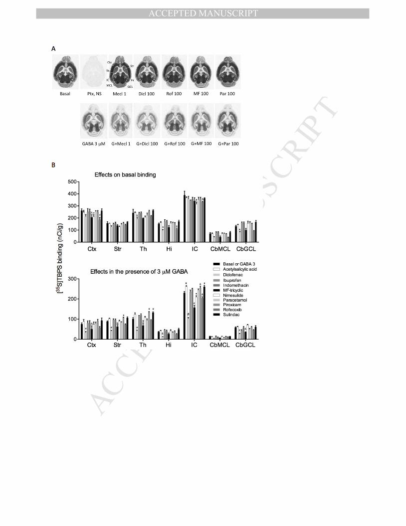
Fig. 4. The effects of nonsteroidal anti-inflammatory drugs on the basal and GABA-inhibited
[35
S]TBPS binding to GABAA receptors in various mouse brain regions. (A) Representative images,
demonstrating enhancement of the 3-µM GABA effect on binding by the positive control,
fenamate meclofenamic acid (Mecl, at 1 µM, see Fig. 2BC), and by 100 µM concentrations of
diclofenac (Dicl), rofecoxib (Rof) and MF-tricyclic (MF), but not by paracetamol (Par,
acetaminophen), which slightly increased the binding in some brain regions in the presence of
GABA. Ctx, cerebral cortex; (Cb)GCL, cerebellar granule cell layer; Hi, hippocampus; IC, inferior
colliculus; (Cb)ML, cerebellar molecular layer; Str, striatum; Th, thalamus. (B) Regional effects of
the studied NSAIDs on [35
S]TBPS binding in selected brain regions. Bars are means + S.E.M. (n=4).
* P < 0.05, &
P < 0.01, # P < 0.001 for the significance of the difference from the basal or GABA-
inhibited binding in the absence of drugs within the brain region (ANOVA, followed by Dunnett’s
test).
Fig. 5. (A) Representative current traces of niflumic acid and furosemide modulation on 10 µM and
20 µM GABA-induced currents in recombinant α1β1 and α1β3 GABAA receptors expressed in
Xenopus laevis oocytes. n = 7 and 5 for α1β1 and α1β3 combinations, respectively. F100,
furosemide 100 µM. (B) Niflumic acid effects represented as mean percentage ± S.E.M., the GABA
30 µM and GABA 20 µM control responses being set to 100 %. F, furosemide 100 µM + GABA 30
µM or GABA 20 µM; N, 1000 µM niflumic acid alone. *** P < 0.001 for the significance of the
difference from the corresponding control value (one-way ANOVA, followed by Dunnett’s test for
niflumic acid in the presence of GABA, Student’s t-test for furosemide or niflumic acid alone). The
dashed line represents the GABA control value. The EC50 values were 6.1 ± 1.3 µM and 10.6 ± 1.1
µM for α1β1 and α1β3, respectively.

MANUSCRIP
T
ACCEPTED
ACCEPTED MANUSCRIPT 25
Fig. 6. (A) Representative current traces of niflumic acid and furosemide modulation on 30 µM and
20 µM GABA-induced currents in recombinant α1β1γ2 and α1β3γ2 GABAA receptors, respectively,
expressed in Xenopus laevis oocytes. n = 7 and 5 for α1β1γ2 and α1β3γ2 combinations,
respectively. F, furosemide 100 µM. (B) Niflumic acid effects represented as mean percentage ±
S.E.M., the GABA 30 µM and GABA 20 µM control response being set to 100 %. F, furosemide 100
µM + GABA 30 µM or GABA 20 µM; N, 1000 µM niflumic acid alone. * P < 0.05, *** P < 0.001 for
the significance of the difference from the corresponding control value (one-way ANOVA, followed
by Dunnett’s test for niflumic acid in the presence of GABA, Student’s t-test for furosemide or
niflumic acid alone). The dashed line represents the GABA control value. The EC50 values were 14.3
± 3.8 µM and 33.0 ± 7.6 µM for α1β1γ2 and α1β3γ2, respectively.

MANUSCRIP
T
ACCEPTED
ACCEPTED MANUSCRIPT

MANUSCRIP
T
ACCEPTED
ACCEPTED MANUSCRIPT

MANUSCRIP
T
ACCEPTED
ACCEPTED MANUSCRIPT
MANUSCRIP
T
ACCEPTED
ACCEPTED MANUSCRIPT

MANUSCRIP
T
ACCEPTED
ACCEPTED MANUSCRIPT

MANUSCRIP
T
ACCEPTED
ACCEPTED MANUSCRIPT