mutagen and oncogen study of methylhydrazine final · pdf filemutagen and oncogen study of...
TRANSCRIPT

Michael G. MacNaughton, Lt Col, USAF, BSC
AMRL-TR-76-80 Deputy Director, Toxic Hazards Divisio
/1 p~5 47 7
MUTAGEN AND ONCOGEN STUDY OF METHYLHYDRAZINEFINAL REPORT
LITTON BIONETICS, INC.5516 NICHOLSON LANEKENSINGTON, MARYLAND 20795
DECEMBER 1976
Approved for public release; distribution unlimited
O13 o01
AEROSPACE MEDICAL RESEARCH LABORATORYAEROSPACE MEDICAL DIVISIONAIR FORCE SYSTEMS COMMANDWRIGHT-PATTERSON AIR FORCE BASE, OHIO 45433 STINFO CO"PY

NOTICES
When US Government drawings, specifications, or other data are used for any purpose other than a definitely relatedGovernment procurement operation, the Government thereby incurs no responsibility nor any obligation what-soever, and the fact that the Government may have formulated, furnished, or in any way supplied the said drawings,specifications, or other data, is not to be regarded by implication or otherwise, as in any manner licensing the holderor any other person or corporation, or conveying any rights or permission to manufacture, use, or sell any patentedinvention that may in any way be related thereto.
Please do not request copies of this report from Aerospace Medical Research Laboratory. Additional copies may bepurchased from:
National Technical Information Service5285 Port Royal RoadSpringfield, Virginia 22161
Federal Government agencies and their contractors registered with Defense Documentation Center should directrequests for copies of this report to:
Defense Documentation CenterCameron StationAlexandria, Virginia 22314
TECHNICAL REVIEW AND APPROVAL
AMRL-TR-76-80
This report has been reviewed by the Information Office (01) and is releasable to the National Technical InformationService (NTIS). At NTIS, it will be available to the general public, including foreign nations.
This technical report has been reviewed and is approved for publication.
FOR THE COMMANDER
VERNON L. CARTER, JR., COLONEL, USAF,.VCDeputy DirectorToxic Hazards DivisionAerospace Medical Research Laboratory
AIR FORCE - 17 JANUARAY 77 - 100

SECURITY CLASSIFICATION OF THIS PAGE (then Date Entered)
READ INSTRUCTIONSREPORT DOCUMENTATION PAGE BEFORE COMPLETING FORMI. REPORT NUMBER 2. GOVT ACCESSION NO. 3. RECIPIENT'S CATALOG NUMBER
AM•RL-TR-7 6-80 0
4. TITLE (and Subtitle) 5. TYPE OF REPORT & PERIOD COVERED
MUTAGEN AND ONCOGEN STUDY OF METHYLHYDRAZINE FINAL
6. PERFORMING ORG. REPORT NUMBER
7. AUTHOR(s) 8. CONTRACT OR GRANT NUMBER(s)
David Brusick, Ph.D., Dale W. Matheson, Ph.D. F33615-76-C-0515
9. PERFORMING ORGANIZATION NAME AND ADDRESS. 10. PROGRAM ELEMENT, PROJECT, TASKAREA & WORK UNIT NUMBERSLitton Bionetics, Incorporated 61102F, 2312, 2312V1,
5516 Nicholson Lane 2312V15
Kensington, MD 20795
11. CONTROLLING OFFICE NAME AND ADDRESS 12. REPORT DATEAerospace Medical Research Laboratory DECEKBER 1976Aerospace Medical Division, Air Force Sys Cmd 13. NUMBER OF PAGES
Wright-Patterson Air Force Base, Ohio 45433 3814. MONITORING AGENCY NAME & ADDRESS(If different from Controlling Office) IS. SECURITY CLASS. (of this report)
Unclassified
15a. DECL ASSI FICATION/,DOWN GRADINGSCHEDULE
16. DISTRIBUTION STATEMENT (of this Report)
Approved for public release; distribution unlimited.
17. DISTRIBUTION STATEMENT (of the abstract entered in Block 20, if different from Report)
18. SUPPLEMENTARY NOTES
19. KEY WORDS (Continue on reverse side if necessary and identify by block number)
Methylhydraz ineMutagenesisDNA RepairMicrosome Activation
20. ABSTRACT (Continue on reverse side If necessary and Identify by block number)
A three tier test was organized into a matrix of assays employing microbialcells, mammalian cells in culture, and in vivo experiments in rats and mice.Methylhydrazine (NH) was mutagenic for Salmonella typhimurium TA-1535 insuspension tests. Results from all other assays conducted as part of thisevaluation were considered to be negative.
DD JARM 1473 EDITION OF I NOV 65 IS OBSOLETE
SECURITY CLASSIFICATION OF THIS PAGE (When Data Entered)

PREFACE
This research was initiated by the Toxicology Branch, Toxic HazardsDivision, Aerospace Medical Research Laboratory. Experiments wereperformed under Contract F33615-76-C-0515 by Litton Bionetics, Inc.,5516 Nicholson Lane, Kensington, Maryland 20795.
The experiments were conducted by David Brusick, Ph.D., and Dale W.Matheson, Ph.D., of Litton Bionetics, Inc., Kensington, Maryland 20795.Kenneth C. Back, Ph.D., was contract monitor for the Aerospace MedicalResearch Laboratory.

1. INTRODUCTION
The detection and subsequent confirmation of mutagenic substancescapable of producing germ cell mutations requires a multifacetedtesting program. The components of such a program should be ableto detect both point mutations and chromosomal aberrations sincethese two classes of genetic alterations represent the types oftransmissible mutations that are of concern to man. The testsincluded in a mutagenicity evaluation program for chemicals shouldnot only be sensitive and reproducible, but also relevant to normalexposure and pharmacological conditions encountered in the environ-ment. These latter two conditions are often difficult to achievesince good human model systems are lacking. It may be argued thatif a single toxicologic end point, e.g., mutation, can be demon-strated in several different test species, then application of theresponse to a wide range of species, including man, can be made.Therefore, a mutagenicity evaluation program should contain aseries of assays covering many phylogenetic levels.
LBI feels that the program conducted in this study offered as many ofthe essential test criteria as possible for an accurate evaluationof methylhydrazine (MH) for genetic activity. Selected tests from TiersI, II, and III were organized into a matrix of assays employingmicrobial cells, mammalian cells in culture, and in vivo experimentsin rats and mice.
Tests utilizing these organisms measured point mutations (forwardand reverse), chromosomal aberrations, and mitotic recombinationalevents induced by acute and subchronic exposure to the test sub-stance.
Figure 1 illustrates the composition of the test program preparedfor the genetic evaluation of the test substance. A brief summaryof each of the assays is listed as follows:
A. In Vitro Microbial Assays
In these assays, the test substance was evaluated for muta-genic and recombinogenic activity in strains of Salmonella andSaccharomyces, respectively. Metabolic activation of thecompound was obtained by combining hepatic microsomes withthe test system. Nonactivation and activation semiquantita-tive plate tests were conducted.
B. In Vitro Mutation Assay in Mammalian Cells
In this assay, the mutations were measured in cultured mousecells (L5178Y). Both direct and in vitro activation assayswere performed. The specific event detected by these cellswas forward mutation at the thymidine kinase (TK+/- ÷ TK-/-)locus, which is an autosomal recessive trait. The combined invitro tests from A and B gave a very sensitive measurement ofthe test substance's ability to induce point mutations andmitotic recombination.
1

TEST SUBSTANCE
ITOXICITY TESTING
S~II .. . .. . ... .I .. .. . .. ..... I . ... .
IN VITRO FORWARD MUTATION INDUCTION DOMINANTPLATE"A-- YS TESTS USING OF UNSCHEDULED LETHAL
WITH SALMONELLA MOUSE LYMPHOMA DNA SYNTHESIS ASSAYS INTY P H -RUiRMR-- L5178Y CELLS IN WI-38 MICE AND
CELLS RATS
DETECTION OF POINT MUTATIONS DETECTION OF DETECTION OFINDUCED CHROMOSOME
DNA REPAIR ABERRATIONS
EVALUATION FOR GENERAL ACTIVITY
FIGURE 1
COMPOSITION OF THE GENETIC EVALUATION PROGRAM
2

C. Unscheduled DNA Synthesis
A second component of the in vitro mammalian cell assay systemutilized the human diploid WI-38 strain of cells. This cellstrain, obtained from human embryonic lung, was used to measuretest chemical-induced DNA repair in cells not undergoingscheduled (S phase) DNA synthesis.
Normal DNA synthesis occurs in the S phase of the cell cyclewith little or no synthesis occurring in any of the otherphases (GO, G1, G2 , or M). The detection of significant DNAsynthesis during these stages (UDS) is indicative of thestimulation of repair enzyme systems. Exposure of WI-38 cellsto various forms of radiation or to chemicals known to bemutagenic or carcinogenic has resulted in the stimulation ofUDS (1).
The detection of UDS in WI-38 cells involved exposure of thecells to the test chemical followed by the addition of tritiatedthymidine ( 3H-TdR) to the culture. If DNA damage has beeninduced, the 3H-TdR will be incorporated during the repair ofthe DNA. This incorporation can be detected by scintillationcounting.
D. Dominant Lethal Assay
This assay was designed to determine the ability of a compoundto induce genetic damage to the germ cells of treated male miceand rats leading to fetal wastage. Chromosome aberrations,including breaks, rearrangements, and deletions, are believedto produce the dominant lethality. Male mice and rats wereexposed to several dose levels of the test compound for fivedays and then sequentially mated to two virgin untreated femaleseach week over the period of spermatogenesis. At mid-pregnancythe females were killed and scored with respect to the numberof living and dead implants as well as to the level of fertility.These results were then compared to data from control animals.
E. Background
Hydrazines and MH react with pyrimidine bases, especially athigh pH, breaking the pyrimidine ring and causing the removalof the base from DNA (2). Based on this type of reaction withDNA, the potential for hydrazine and/or derivatives to exhibitmutagenic, teratogenic, and carcinogenic activity might beexpected.
Some information on the mutagenic and carcinogenic propertiesof hydrazine and some of its derivatives has been published.
3

Hydrazine in relatively high doses causes leukemia, reticulumcell sarcoma, and lung adenomas in mice (3). Hydrazine ismutagenic in T4 phage (4), S. typhimurium (5) and Drosophilamelanogaster (D. melanogaster) (6), but was not active in adominant lethal assay 7). Symetrical dimethylhydrazineinduced mitotic gene conversion (8).
Because the test agent is structurally related to chemicalsthat are established mutagens and carcinogens, and because ofthe excellent correlation between mutagenicity and carcino-genicity, mutagenesis studies might provide insight intopotential toxicologic problems associated with the testagent.
2. MATERIALS
A. , In Vitro Microbial Assays
The test chemical was examined in a series of microbialassays employing histidine-requiring mutants of S. typhimurium.The assays were conducted so that the compound was testeddirectly and in the presence of a mouse liver microsome activa-tion system.
The compound was evaluated at a minimum of four dose levelsunder both test conditions with the highest dose level showingsome evidence of toxicity. In addition to these tests, spottests (5) were conducted with the Salmonella mutants plusadditional strains of bacteria; S. typhimurium strain G-46 andE. coli strain WP2uvrA" (11).
1. Preparation of Tissue Homogenates and 9,000 x g Cell Fractions
Male mice (sufficient to provide the necessary quantitiesof tissues) were killed by cranial blow, decapitated, andbled. Organs were immediately dissected from the animalusing aseptic techniques and placed in ice-cold 0.25 Msucrose buffered with Tris buffer at a pH of 7.4. Uponcollection of the desired quantity of organs, they werewashed twice with fresh buffered sucrose and completelyhomogenized with a motor-drive homogenizing unit at 4C.The whole organ homogenate obtained from this step wascentrifuged for 20 minutes at 9,000 x 9 in a refrigeratedcentrifuge. The supernatant from the centrifuged samplewas retained and frozen at -80C. Samples from thesepreparations were used for the activation studies.
4
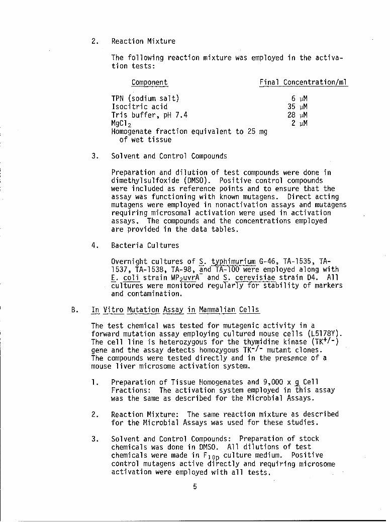
2. Reaction Mixture
The following reaction mixture was employed in the activa-tion tests:
Component Final Concentration/ml
TPN (sodium salt) 6 1MIsocitric acid 35 pMTris buffer, pH 7.4 28 ]MMgCI 2 2 pMHomogenate fraction equivalent to 25 mg
of wet tissue
3. Solvent and Control Compounds
Preparation and dilution of test compounds were done indimethylsulfoxide (DMSO). Positive control compoundswere included as reference points and to ensure that theassay was functioning with known mutagens. Direct actingmutagens were employed in nonactivation assays and mutagensrequiring microsomal activation were used in activationassays. The compounds and the concentrations employedare provided in the data tables.
4. Bacteria Cultures
Overnight cultures of S. typhimurium G-46, TA-1535, TA-1537, TA-1538, TA-98, and TA-1O0 were employed along withE. coli strain WP2uvrA and S. cerevisiae strain D4. Allcultures were monitored regularly for stability of markersand contamination.
B. In Vitro Mutation Assay in Mammalian Cells
The test chemical was tested for mutagenic activity in aforward mutation assay employing cultured mouse cells (L5178Y).The cell line is heterozygous for the thymidine kinase (TK+/-)gene and the assay detects homozygous TK-/- mutant clones.The compounds were tested directly and in the presence of amouse liver microsome activation system.
1. Preparation of Tissue Homogenates and 9,000 x R CellFractions: The activation system employed in this assaywas the same as described for the Microbial Assays.
2. Reaction Mixture: The same reaction mixture as describedfor the Microbial Assays was used for these studies.
3. Solvent and Control Compounds: Preparation of stockchemicals was done in DMSO. All dilutions of testchemicals were made in Flop culture medium. Positivecontrol mutagens active directly and requiring microsomeactivation were employed with all tests.
5

4. Cells and Media: TK+/- BUdR-sensitive L5178Y mouselymphoma cells were used in this assay. Growth medium(GM) for this line consists of Fischer's mouse leukemiamedium supplemented with 10% horse serum and sodiumpyruvate (Flo ). Cloning medium consists of Fischer'smedium plus 2B% horse serum and agar (0.37%). Selectivemedium for TK-/- cells was prepared by adding BUdR to thecloning medium.
C. Unscheduled DNA Synthesis (UDS) Assay
Nondividing WI-38 cells were exposed to three concentrationsof the test compound and 3H-thymidine. Treatment was directand under conditions of microsome activation. The amount of3H-thymidine incorporated into the DNA was measured by scin-tillation counting.
1. Preparation of Mouse Liver Microsomes
A 9,000 x g supernatant of mouse liver was prepared asdescribed in the Microbial Assays. This supernatant wasthen centrifuged at 105,000 x g for 60 minutes and thepelleted microsomes resuspended in 0.25 M sucrose. Thismicrosome preparation was added to the reaction mixturein place of the 9,000 x 9 cell fraction.
2. Reaction Mixture
The reaction mixture was the same as used in the Micro-bial Assays except purified microsomes replaced the 9,000x 9 supernatant.
3. Solvent and Control Compounds
Any stock solutions of chemicals were prepared and dilutedin DMSO. Positive control chemicals that act directlyand require microsome activation were employed.
4. Cells and Media
Human diploid embryonic lung cells (WI-38) were obtainedfrom Flow Laboratories and used in these assays. The GMemployed was Eagle's minimal essential medium (EMEM)supplemented with 10% fetal calf serum (FCS). Step-downmedium (SM) was amino acid depleted to reduce cell division,and hydroxyurea medium (HUM) was the medium used toinhibit S phase growth. All media were based on EMEM.
6

D. Dominant Lethal Assay (DLA)
The test chemical was tested in mouse and rat DLA. Allanimals were dosed by intraperitoneal (IP) injections overfive consecutive days, rested for two days, and mated.
1. Animals
a. Mice: Seven- to eight-week-old male random bredmice (ICR, Flow) were used for treatment. Femalemice of the same strain, age, and weight were usedfor the matings.
b. Rats: Ten- to twelve-week-old Sprague-Dawley malerats from a random bred closed colony (Flow) wereused for treatment. Females of the same strain,age, and weight were used for the matings.
2. Animal Husbandry
Each species was housed in separate rooms of our animalfacility.
Male mice were housed five to a cage while being dosedwith the compound, and then housed separately with twofemales for mating.
All animals were offered a 4% fat diet and water adlibitum. Water was acidified according to approvedlaboratory animal health standards.
Animals were identified by ear punch. Sanitary cages andbedding were used and changed two times per week at whichtimes water containers were cleaned, sanitized, andfilled. Cages were repositioned on racks once a week,and the racks repositioned within rooms monthly. Personnelhandling animals or working with animal facilities wearhead and face masks as well as suitable garments.Individuals with respiratory or other overt infectionsare excluded from the animal facility.
3. Positive and Negative Control Chemicals
Triethylenemelamine (TEM) was administered IP at a levelof 0.3 mg/kg in 0.85% saline as a positive control.Negative control animals received an IP injection of thecorn oil or water solvents.
E. Test Chemicals
The test sample was obtained from the United States AirForce. MH was a clear liquid (less than 100 ml) in an amberbottle.
7

3. METHODS
A. In Vitro Microbial Assays
Overnight cultures of S. typhimurium TA-1535, TA-1537, TA-1538,TA-98, TA-lO0, E. coli WP2 uvrA-, and S. cerevisiae D4 weregrown in complete broth. ApproximateTy 10- cells from aculture were added to test tubes containing 2.0 ml of moltenagar supplemented with biotin and a trace amount of histidine.
Four dose levels of the test chemical were added to the appro-priate tubes and the contents poured over selective medium.In activation tests 0.5 ml aliquots of the reaction mixturecontaining the microsomes were added to the tubes containingcells and chemical just prior to pouring onto the selectivemedium. After the overlays solidified, the plates were placedin a 37C incubator for 48 to 72 hours. The plates were thenscored for the number of colonies growing in the agar overlay.Positive and solvent controls using both direct-acting muta-gens and promutagens that required metabolic activation wererun with each assay. Supplementary spot tests were alsoconducted according to the methods described by Ames et al.(12).
The data are presented in Table 1. Concentrations of the testand positive control chemicals are given in the data tables.
B. In Vitro Mutation Assay in Mammalian Cells
1. Toxicity
The solubility, toxicity, and doses for the test chemicalwere determined prior to screening. The effect of thechemical on the survival of the indicator cells wasdetermined by exposing the cells to a wide range ofchemical concentrations in complete GM. Toxicity wasmeasured as loss in growth potential of the cells inducedby a five-hour exposure to the chemical. Four doses wereselected from the range of concentrations by using thehighest dose that showed no loss in growth potential asthe penultimate dose and by bracketing this with onehigher dose and two lower doses. Toxicity produced bychemical treatment was monitored during the experiment.
8

2. Test
a. Nonactivation assay
The procedure used was a modification of that reported byClive and Spector (13). Prior to each treatment, cellswere cleansed of spontaneous TK-/- by growing them in amedium containing thymidine, hypoxanthine, methotrexate,and glycine (THMG). This medium permits the survival ofonly those cells that produce the enzyme thymidine kinase,and can therefore utilize the exogenous thymidine from themedium. The test compound was added to the cleansed cellsin GM at the predetermined doses for five hours. Themutagenized cells were washed, fed, and allowed to expressin GM for three days. At the end of this expressionperiod, TK-/- mutants were detected by cloning the cells in theselection medium for ten days. Surviving cell popula-tions were determined by plating diluted aliquots innonselective GM.
b. Activation Assay
The activation assay differs from the nonactivationassay in the following manner only. Two and fivetenths ml of the reaction mixture was added to 10 mlof GM. The desired number of cleansed cells wasadded to this mixture, and the flask was incubatedon a rotary shaker for five hours. The incubationperiod was terminated by washing the cells twicewith GM. The washed mutagenized cells were thenallowed to express for three days and were cloned asindicated for the nonactivated cells.
c. Data Analysis
A mutation frequency for each test dose was deter-mined by dividing the number of mutants/ml by thenumber of surviving cells/ml (adjusted to 10-4) asindicated by plating efficiency. These data arepresented in Table 2. Concentrations of the testand positive control chemicals are given in the datatables.
C. Unscheduled DNA Synthesis
1. Cell Preparation
Normal human diploid WI-38 cells were seeded at 5 x 10⁵cells in a 100 mm tissue culture dish and grown to con-fluency in GM. Once reaching confluency, the cells wereswitched to SM for five days. The contact inhibitionimposed by confluency and the use of SM held the cells ina nonproliferating state.
9

2. Treatment
On the day of treatment, cells held in G, phase wereplaced in HUM. After 30 minutes this medium was replacedby 5 ml of HUM containing the control or test chemicaland 1.0 pCi of 3 HTdR. Each treatment was at three con-centrations. Exposure was terminated by washing thecells twice in cold BSS containing an excess of coldthymidine.
3. DNA Extraction and Measurement of 3 HTdR Incorporation
Treated plates were frozen at -20C until processed.After thawing, the cells on the 100 mm plate were coveredwith 2.5% SDS in 1 x SSC and scraped from the dish with arubber policeman. The cells were washed and precipitatedfrom the SDS by three changes of 95% ethanol and centri-fuged at 10,000 x g. Additional lipid components wereremoved by extraction in ethanol ether at 70C. Thispellet was washed in 70% ethanol, further incubated at70C in 0.3N NaOH, and the DNA extracted in 50 pl IN PCAat 70C. The DNA was separated into two 25 ul aliquots.One of these was dissolved in 10 ml of hydromix scintil-lation cocktail (Yorktown Co.) and counted in a Beckmanliquid scintillation spectrometer. The second aliquotwas added to 275 Pl of IN PCA and read at 260 nm in aGilford spectrophotometer. The values were corrected forlight scatter and converted to pg of DNA. Followingliquid scintillation counting, the data were combinedwith the DNA extraction values and expressed as disintegra-tion per minute per pg DNA (DPM/pg DNA).
4. Activation Assays
The activation tests were conducted according to themethods described above except that 0.62 ml of a purifiedmicrosome preparation (100,000 x g pellet) was added tothe test mixture.
5. Dosage Determinations
Doses were determined from preliminary toxicity tests inwhich cells were seeded in 16 mm wells (Linbro plate).A wide range of concentrations was tested in the wells,and toxicity was monitored visually by altered cell mor-phology and adhesion. The three doses used in theexperiments were selected.
The results of these tests are given in Table 3. Theconcentrations of test and control compounds are given inthe data tables.
10

D. Dominant Lethal Assay
The dominant lethal assay is designed to assess the ability ofthe test compound or its metabolic products to reach thetestes of treated male animals and induce genetic activity inthe developing gametes during spermatogenesis.MH was administered to male mice weighing 30 ± 2.5 gm. In
addition, MH was also dosed to male rats weighing 325 ± 25 gm.
1. Stock Solutions
The compound was prepared daily from stock solutions. MHwas dissolved in distilled water.
2. Compound Administration
Dosages were determined from LD50 data supplied by thecontract monitor with a high dose of 1/10 the LD50, anintermediate dose at 1/3 the high level, and a low doseof 1/10 the high level. Compound was injected IP intoeach animal daily for five days. All dosages and routesof administration were determined in consultation withDr. Kenneth Back of the United States Air Force.
Calculated dosages are as follows:
Methylhydrazine
Mice
LD50 26.0 mg/kgHigh 1/10 LD50 2.6 mg/kgInt. 1/30 LD50 0.86 mg/kgLow 1/100 LD50 0.26 mg/kg
Rats
LD50 21.5 mg/kgHigh 1/10 LD50 2.15 mg/kgInt. 1/30 LD50 0.72 mg/kgLow 1/100 LD50 0.215 mg/kg
3. Animal Husbandry
a. Mice: Ten male mice were housed five animals to acage during the five days of dosing. After two daysof rest, each male was caged with two virgin femalesfrom Monday through Friday. This sequence wasrepeated weekly with two new females each week foreight weeks. Fourteen days from the midweek in
11

which they were caged with the males, females werekilled, dissected, and the number of dead, living,and total embryos in the uterus recorded on standardforms. These data were statistically analyzed forindications of dominant lethality, and compared withcontrol data for significance.
b. Rats: The protocol for the experiment using ratsdiffered from that of the mice only in the sequenceof mating lasting seven weeks and in that corporalutea were counted and recorded. These scores wereused to determine evidence of compound-inducedpreimplantation losses.
4. Data
The results of the Dominant Lethal Assay are given inTables 4 to 17.
4. RESULTS
The results of the genetic studies are presented in the followingseries of tables:
12

CL
(V LM (Y) rý I M (Y) M k I m mc YS.-
4-)
oL o C +C$- -M-c ~-c
u I r-o ) c r ~ J CY)( 00 C' I (n0 C)c mt.0 -::-- ~ -:IJ -- an-J 0)0 .0 ko z- CY
-A -~ - I A
U
I-I S- D L \ICJC)Cj ) 0 DL
CD
C)~~C CL.. -+ -
4-3 co c+u-
LU 4-' IO N.0 (iiJ0 C'J C r(O-j C'JCo) CJ.- I N.- (M C\)L-
< U (Ii- A-(0 a
Ln CL c
<c -- + ++
<- 00) '- ) CI C---0CDC 0LnCj - 4-)aLUC\ C~ :d- m-. LCr-O\J' nt (n- ~
c > A A~ -P4-C- 4- ) 0
C) C.ULO- ++
IO I-- C-) 4-+mC C)- I. ýC oC ~ )C)(
F- -\ -o - e'jC o0-.- -m CoY)C I~d Y 4-)A A~ C
- 0-
i= 0- 0 S- C7S.- 4 -) S- 4-)s
(A -4- C :) 4-3O. O-- r-J 4-1
,-r- t
u, -) 5-LU 0 0 -0a)4-
4-) > 4-)4- 4 - - C a--C :-
CO C) to W40C D )43(S-4C> >- C C ) )C C, C) C, a)
4-) to .- 4-'> . . . 4- 4- >-
c,- 4-' 00 2DC) ) -C) CDC) LO t 00 CýC; .L,;0-D U V) 0> M: > (/)a-
C ~4-')C
13

TABLE 1A
Results From Suspension Tests of MH Using
S. typhimurium Strain TA-1535a
Compound Population Mutant MutationTest Concentration/ml Counts Counts Freq. (xlO- 8 )
Activation
Solvent Control (a) - 2698* 227 8.4(b) - 1369** 86 6.3
Positive Control DMN 100 vmoles 1264 4798 379.6MH 1 Pl 1043** 332 31.8MH 5 Ill 1958* 9855 503.3
"**Identifies treated group with appropriate solvent control.
aThe suspension assay was conducted using the same mouse liver activation
system described for the plate assays shown in Table I. The protocol waschanged such that rather than add all test components to semisolid overlayagar, they were suspended in saline and incubated 60 minutes at 370C on arotary shaker. After incubation samples were removed and assayed for thenumbers of surviving cells and numbers of revertants. Mutation frequencieswere calculated for each test. Dimethylnitrosamine (DMN) was used as thepositive control compound.
14

KEY
MOUSE LYMPHOMA ASSAY TABLE
COLUMN
A, B, C, D Day = Expression day cell counts (x 106)
E AGS = Represents cell population growth during expression.The value is obtained by subtracting the Day 1 countsfrom the terminal day counts.
F %GS = Percent suspension growth is obtained by expressingthe AGS values for treated cells as a percent of theAGS for the negative controls E treated x 100
E control
G MC = Mutant counts. The total number of colonies countedin the BUdR plates.
H VC = Viable counts. The total number of colonies countedin the VC plates.
VC counts in treated culturesI %CE = Cloning efficiency VC counts in control cultures x 100
J GF = Growth factor Percent suspension growth (column F) xPercent clonal growth (column I)
100
K MF(x 10-4) = Mutation frequency MC counts column G) X 10-4VC counts (column Hx
15

0-C C). 01O LCr r""0 )C) 0 00C:) Lt,r-.) CDC00 ) LOC 00 00
LL. 00 L L)-010 00 C)OC) O0C\J
LU r 0 0 m C C) C' 00 LoC\j0toC)i
C) L) r- 00 00 00J r- cO Lf *j d ,Lf f.01
0-2 0) r-co c 0N0 C C'~j- r--L0U~. ~ U C'J~C 1
ClV)C> (I))(j l-- ) Y Okocl lON. C) 0f Y L)DCY) Mf)'QC')O
00-ý m tm0 m- r~ 0) 001-
ýc C)
Ln
c2 1
CD( ' co 00 ,- O 0 CjC)C
r-/1 C) 0 0 0 U C~) N-t m
LUJ
0~~~ S-n 0fCcc S-U) acd5- -) -O 1 - S--. ~ 4-
C 0
0 L) 0)-
a) 0~- a) a)E4-3 >C LS 4-J >. .
4) C) C)C a. -L
>- C'.- 0-D CD>.- CDCC)C4- 004- (aL) 004' 0 .Iu V) 0-0 >r ,- 0 0
(0
16

TABLE 3
MEASUREMENT OF UDS IN WI-38 CELLS TREATED WITH MH
Concentration Activitya Percent
Test (Ial/ml) DNA(pg) DPM Index of Controlb
Nonactivation
Solvent Control - 9.02 76 8.4 -Positive Control MNNG (lOig/ml) 1.76 86 48.9 582MH 0.1 9.64 102 10.6 126
0.5 16.88 83 4.9 581.0 9.25 102 10.4 124
Activation
Solvent Control - 13.64 78 5.7 -Positive Control 2AAF (301ig/ml) 2.45 60 24.5 430MH 0.1 22.56 145 6.4 112
0.5 14.85 79 5.3 931.0 Sample Lost
aActivity Index = DPM/pg DNA (DPM = Disintegrations/minute)
bpercent of Control Activity Index Treated X 100Activity Index Control
17

-j u N N N- - r. .
l I In I-
N N
-c < <
.j~ N N '7 4.d
A'
- A
-A 18

~ti If !I '
Z6 . . a S
it ~ 0 ~ - ~ 0 -
41 - -Ij N! I II i
'A 0
.. jrA LA -L0 u0
Ln - - N N'40L- -
_j 1, II I T it4.1.f ~ ~ .
u) e
z Z
-Z Z - NA
.j z1J~~~~~ Z ZJ L
I- AIt0 d 0 . - S. - - -. ý ~ LA
2z :
0 A S.* 419

If It
ku 0
- K I ' It It It
C..
A- 4- L N
z be
Z. UA .. S 0
. -j I I If 0t It if It
WN N
le 4
~(A N 'A
LJ.I~~. 4d .wz 4
~~~Z -W A - .
'A'
-z z
- -3
20

A A~
'44
~~14%3
'.o
A~voSI
4 1§ 4
* 14 II21

0 ~~ 0 - 0
I- - r0
it z
0 - 0 o
~i 3 I I If It II It
LA
4 ~, z-. - z
0 0 OM w
Z Z
- ~ ~ ~ ~ I ;I * * * A -
0 ~~ Z 0C6 0 ' .
cc . o II I I I I .J ZU
Z -
220

-zI. F-
:Ic
- ff.f
-z . .? 0 0 0f0 * * S *LMell 0 0 ~
LON NL U.
?~z z
< 7.
13'
A~U -. 1S. S. ., S. S. .
- - .3
z~ ~~~~ z4 4 I 4 I
23

I*� � .t� � �'% 0 0
- r '7- = . . . S * -. .
-- - 0 0 0 0 0 0
-� I H I 'I I0 0 o 0 0
Z� N N N N N N N
0 J�' � � � '7- - - - -
N0 0 0 � -� 0
.� .t � �7 5 5 9 5 5 5 5
WO 0 0 0 0 0 0 0� 1% I II II H II II
- 0 0 0 0 0 0 0
W S N N N N N N N
.- -S -S - %
0 �' '7 .? C � *J.. �- - - - - -
-, - z- z
5. 0 0
..� . J9 - 0 � �. 0
� C 4 0 - -
� S * * S * * , A W
wO 0 0 0 0 0 0 0 .J�II I? II U 4
0 0 0 � �- 2
W W* N N N -� - - � �w
0 0 -. N. � '- .e� .�. z0 � - �
4 - - � - .� � - 0
0 - 0 4
0 z -
4�. 4
0 � *J� 0 t' 0 0
- � S * * S S S S �. -
- � 0 0 0 0 0 0 0 - =- N 0 C 0 0 0 - 0 � Z
- WS N N N N N N N 00
4 - . N N,' N N N, N. N, -
0 .0 f� � '0 N p s- U 0- - - - - - 4 �
-J -� -
- -..J
z eo <
Z �
o � C C 4 00
0 '7 4� .4 0 - 0 � -
S S C S * C S - - Zr -
_ �: 0 0 0 0 0 0 - - - 44 -�- - .- -
-� II Ii P I I I - - --
-- 0 0 0 0 0 0 0 Z �<� N N N N N N N � - -
- A� -
� N N, -. '. N N N A �= - 0 C '7 - ...J .j�.
- - - - N .�J .- � ..J..J 0
0�. �r020 .4-JO � --
44.-� w.�
=� -- -�
Z.J -� =2-- ..J 4�
)- o..� -.-
-Z Z�.) L. -0 --
=-� �.J Zz -�- �4 � -�
.A.-IJ -�'� -- ZZ7 - AA �i4
� -' N � C �- - Z�- $1 --
3 4 5-SA*.0 AA * �. --
�I�- .�- * -
5- .�J -. � �.;= .4A �
e* >0(.�h- A'- Z- 4 A
'I-
A
24

It It 4 1
Lw~
,. *L*
U. 6L*
- ' I ,
2~~L 661., .
-z ~ 3 r ~ ~ -616 eni I I I
66~ ' 3' ~ N '
-. Ji It - -
3mw
-. ~ ~~ z.. -- -
u- 0 11 11 4
3'~~~ z-3 ~ N ,
-3
25

- - -2
It 7 t- z
'..~ 4 -
6AJ a - -
4 II 4 4471
- .7 . .3 .~ 'O
L&. 0- - - - IV
A Nc
- - S ~ s .. -. S.LS
- - -~ N 9
- C 3 N 3' 3' .
-~1 N N -
.JJ ~7-1 S. z
C w 0m. - 4 3
- ~~~~ 3cIII
A ~ > N ~ ~ ~ 3 26

-u+
* 3 *
.-- ~ .~. -~ - 0 0
% -i
L. 4.
L
0A ~5; 9 9 .
> N N - - ~ 0 0fLu0
A~~~1 N<-- -
'S - N.NZ'. N
V).0w; I.7 .0 - 1n,
* * * * * * >N~~ -0x 0 ~
0~ z~..N~~L A I.5,jS ~ ~ - ~ - ~ . ~-
* - - - -27

It 11 'ý -tI
- =l it I It it It
2..
N t it it
.9 .A
oz Nn p- -. 9 .
it II 5 5
.9
a- -t I 0 9..63 -? N w
-1. 2. N m 11 - - aA-j
X- - - ' ' , - ~ ....
J~~~~~t "J9~
- . = N S s
'IL -28

Z 4j
0"' 0 l
> -~ N ~ 10 0 ~- 9 9 9 . , S
0 0 - -IV
-~~~L t 4 : 4?04 4
-w j
LL; m -4 it N
m -9 -a . 9 9
ýi a0 0 0 0 0I I i 4 if 11 It
- 0' 4 4 '0 ~ uýU.,V LA -- -
~ '66
z~ ~~ z~ .~ 'S - A--If - *j
~~6 LM 6 --
Z -~ 0 ~ 4 29

- z If 9t If *
vi 2 6mI I A I
-f it -
- ~s - - N N
-U A Ct3
.. iuS
LA-
LAfl -
6N
2 ~ ~~~ - z-N'
Z Z
'It ifJ,
A ~ ' - '- ~- U
.11 > 9 I I * * 30

- -
.� .� -�A �7 * � * �
-t 9 -' , ., - 0�
--. �- 0 � -� - -. - N '7 '7
N � - -� - - -'7'C - N N'7 N A A� N
N *i -�� N - - A *0 o o o - -�. 9 . . . -wo � � 0 0 0 o'7 N 0 N ,�W * � ! - 0AN - - N - - N0! N N N N N N N0 N N 0 � � 0 �
-S
'7 - -� -�-. '70 0. * . p pA ,..iO � 0 0 0 0 �
- - -Iz z -. a' '7 - -� -w � w . 'C '7 � '7 � 2- �.I �.'0
- - - - - -.,�J z - 'C N A
0 -A �
1=
I � -0 4 2 'A0 - N
- 0� Q A �
'C AN -Z 0 0 0 o o- . . p a * , *A WA - 0 0 0 0 0 0 A2 A �- 'C'7 - 0 r.. �'C 0 -
-- - N - - - WN N N N N N �
0 � .. '7 2 ,� Lf� � � U.
0
0 -
2
2 - A A =
00 Z� 0 0 0 0 0 � -p * * a , * -
--, 0 0 -. -- .� �
If I I� - 2
-� 0 - - z .� -- 2A .Z -- - - - N - A-� N N N N N N N A
AA- 0 0 - � -
-�0�. -- :.�. 2-xWO -� x
OX -.
- *W..J
-- -� <� 0�X 2 IA'A .. �LJ- 0.� � -- >.�2 2.-
z� � � 20 2- �� --
- -- AA -
*�J - N � '7 A 'C � 2- Zx
-- -- -- ijil -
-�
.� � j 2-A.� AA
-� .1 � . S -0 0>-2 )- 2
* � .Z- *
31

5. INTERPRETATIONS AND CONCLUSIONS
MH was evaluated for its genetic activity and ability to stimulateDNA repair using a battery of in vitro and in vivo assays.
A. Microbial Assays (Table 1)
There were no clear indications of mutagenic activity by MH inany of the microbial assays reported in Table 1. The toxicityof MH for bacteria and yeast was high and concentrations of 10pl/plate were consistently too toxic to use.
Because hydrazines have certain properties similar to nitrosamines,this compound was also examined for mutagenicity in a suspensionassay. Dimethylnitrosamine was found to be inactive in thestandard plate assay, but the chemical is highly mutagenicwhen tested in a suspension procedure. The results for MHunder the same test conditions were also found to be positive(Table ]A). Mutagenic activity was observed at MH concentrationsof I pl and 5 pl/ml after a 60-minute incubation with a mouseliver activation system.
B. Mouse Lymphoma Assays (Table 2)
The data from these tests were clearly negative.
C. UDS Assay in WI-38 Cells (Table 3)
The data from these tests were clearly negative. The samplecells at the high dose level (activation assay) were lost bybreakage in the centrifuge. However, there was no indicationof a trend or any activity in an equivalent dose in nonactiva-tion tests.
D. Dominant Lethal Assays
Dominant lethality, which is indicated by a high percentage ofdead implants to total implants, was not demonstrated by thedata from either mice or rats administered MH. In comparison,the positive control compound TEM demonstrated a clear domin-ant lethal effect in mice during weeks 1 through 3 and in ratsduring weeks 1 through 5.
1. Mice (Tables 4-9)
Implant data for week 5 were not statistically analyzeddue to the low number of pregnant females in the low andintermediate dosage groups. The reason for the lowfertility in these animals is unknown, but does notappear to be compound related since the negative controlgroup also only contained one pregnant animal. Implantdata from the other weeks did not reveal any of the dose-related trends indicative of compound-induced geneticactivity.
32

2. Rats (Tables 10-17)
Increased lethality compared to negative controls significantat p < 0.01 was indicated for week 7 dosages. The ratiosof dead to total implants for these animals fall within therange of variation encountered for all levels of tests through-out the testing period; arid, therefore, the statistical signi-ficance is associated with the week 7 negative control animalsnot having any dead implants. In addition, no dose-related trends were observed. In light of these observationsand since the ratios are considerably less than those forweeks 1 through 5 of the positive controls, we did notconsider the data as indicating biologically significantactivity for MH in rats.
E. Conclusions
MH was mutagenic in activation, microbial reversion tests ifthe tests were conducted as suspension tests and not if conductedas standard plate tests. This differential activity is similarto the type of results obtained with dimethyl- and diethylnitro-samines. Except for the mutagenicity of MH in tests with S.typhimurium TA-1535, there were no indications of genetic -activity for MH in any of the other tests conducted as part ofthis evaluation.
6. REFERENCES
1. Stich and Laishes, Pathobiology Annual, Vol. 3, p. 341, 1973.
2. Freese, Chemical Mutagens, Vol. 1, A. Hollaender (ed.), p. 1,Plenum Press, New York, 1971.
3. Juhasz, Potential Carcinogenic Hazards from Drugs, U.I.C.C.Monograph, Vol. 7, R. Truhart (ed.), p. 180, Springer-Verlag,Berlin, 1967.
4. Freese et al., Proc. Nat. Acad. Sci. (USA), 47:845, 1961.
5. Ames, Chemical Mutagens, Vol. 1, A. Hollaender (ed.), p. 267,Plenum Press, New York, 1971.
6. Auerbach, Science, 158:1141, 1967.
7. Epstein and Shafner, Nature, 219:385 1968.
8. Zimmermann and Schwaier, Naturwiss., 54:251, 1967.
9. Miller and Miller, Cancer Res., 25:1292, 1965.
33

10. Ames et al., Proc. Nat. Acad. Sci. (USA), 70:2281, 1973.
II. Green and Muriel, Mutation Res., 38:3, 1976.
12. Ames et al., Mutation Res., 31:347, 1975.
13. Clive and Spector, Mutation Res., 31:17, 1975.
34SU S. GOVERNMENT PRINTING OFrICE: 1976 - 757-001/153