mutagenesis of a specificity-determining residue in tyrosine hydroxylase establishes that the enzyme...
TRANSCRIPT
Mutagenesis of a Specificity-Determining Residue in TyrosineHydroxylase Establishes That the Enzyme Is a Robust PhenylalanineHydroxylase but a Fragile Tyrosine HydroxylaseS. Colette Daubner,*,† Audrey Avila,† Johnathan O. Bailey,‡ Dimitrios Barrera,§ Jaclyn Y. Bermudez,†
David H. Giles,∥ Crystal A. Khan,∥ Noel Shaheen,† Janie Womac Thompson,‡ Jessica Vasquez,‡
Susan P. Oxley,§ and Paul F. Fitzpatrick*,∥
†Department of Biological Sciences, St. Mary’s University, San Antonio, Texas 78228, United States‡Department of Biochemistry and Biophysics, Texas A&M University, College Station, Texas 77840, United States§Department of Chemistry and Biochemistry, St. Mary’s University, San Antonio, Texas 78228, United States∥Department of Biochemistry, The University of Texas Health Science Center at San Antonio, San Antonio, Texas 78229, UnitedStates
*S Supporting Information
ABSTRACT: The aromatic amino acid hydroxylases tyrosine hydrox-ylase (TyrH) and phenylalanine hydroxylase (PheH) have essentiallyidentical active sites; however, PheH is nearly incapable of hydroxylatingtyrosine, while TyrH can readily hydroxylate both tyrosine andphenylalanine. Previous studies have indicated that Asp425 of TyrH isimportant in determining the substrate specificity of that enzyme[Daubner, S. C., Melendez, J., and Fitzpatrick, P. F. (2000) Biochemistry39, 9652−9661]. Alanine-scanning mutagenesis of amino acids 423−427,a mobile loop containing Asp425, shows that only mutagenesis of Asp425alters the activity of the enzyme significantly. Saturation mutagenesis ofAsp425 results in large (up to 104) decreases in the Vmax and Vmax/Ktyrvalues for tyrosine hydroxylation, but only small decreases or evenincreases in the Vmax and Vmax/Kphe values for phenylalaninehydroxylation. The decrease in the tyrosine hydroxylation activity of the mutant proteins is due to an uncoupling oftetrahydropterin oxidation from amino acid hydroxylation with tyrosine as the amino acid substrate. In contrast, with theexception of the D425W mutant, the extent of coupling of tetrahydropterin oxidation and amino acid hydroxylation is unaffectedor increases with phenylalanine as the amino acid substrate. The decrease in the Vmax value with tyrosine as the substrate shows anegative correlation with the hydrophobicity of the amino acid residue at position 425. The results are consistent with a criticalrole of Asp425 being to prevent a hydrophobic interaction that results in a restricted active site in which hydroxylation of tyrosinedoes not occur.
The aromatic amino acid hydroxylases tyrosine hydroxylase(TyrH), phenylalanine hydroxylase (PheH), and trypto-
phan hydroxylase (TrpH) make up a small family of enzymeswith important roles in metabolism. They are all rate-limitingenzymes for particular pathways: PheH for phenylalaninecatabolism, TyrH for catecholamine synthesis, and TrpH forserotonin synthesis.1 The reactions they catalyze are shown inScheme 1. These enzymes all form tetramers of subunits thateach have two domains, with the larger (∼340 residues)carboxyl-terminal catalytic domains showing considerablesimilarity in sequence and three-dimensional structure.2,3 Thesmaller amino-terminal regulatory domains are of differentlengths and show low levels of identity among the threeenzymes. Gene analysis suggests that all three enzymes evolvedfrom an ancient hydroxylase. TyrH branched off first, 750million years ago, and TrpH and PheH diverged 600 million
years ago; the enzymes acquired different amino termini sometime after TyrH arose.4,5
The common mechanism proposed for the aromatic aminoacid hydroxylases is shown in Scheme 2.6 After thetetrahydropterin and the amino acid substrate bind, oxygenreacts to form a bridged peroxypterin; heterolytic cleavage ofthe proposed peroxy moiety would form the Fe(IV)Ohydroxylating intermediate and a 4a-hydroxypterin, the pterinproduct that is released by the enzyme. The Fe(IV)Ointermediate transfers an oxygen atom to the aromatic ring ofthe amino acid via electrophilic aromatic substitution.7,8
Received: January 8, 2013Revised: January 30, 2013Published: January 31, 2013
Article
pubs.acs.org/biochemistry
© 2013 American Chemical Society 1446 dx.doi.org/10.1021/bi400031n | Biochemistry 2013, 52, 1446−1455
The active sites of the aromatic amino acid hydroxylases arehighly conserved.9,10 Figure 1 shows an overlay of the activesites of TyrH and PheH to illustrate the similarity. The activesites of all three enzymes contain two histidines and a glutamatethat bind the single active site iron (His331, His336, andGlu376, respectively, in TyrH),11 a phenylalanine (Phe300 inTyrH)12,13 and a glutamate (Glu332 in TyrH)13,14 that bindthe pterin, an arginine and the associated aspartate (Arg316 andAsp328, respectively, in TyrH)14 that bind the carboxylate ofthe amino acid substrate, and several residues (Pro327, Phe377,and Trp372 in TyrH)15 that form a hydrophobic chamber forthe side chain of the amino acid substrate.The three enzymes differ in the extent of their specificity for
the amino acid substrate. TyrH can hydroxylate all threearomatic amino acids, with the following preference based onVmax/Km values: tyrosine > phenylalanine > tryptophan(∼5:1:0.2).15,16 PheH is quite specific for phenylalanine; itcan also hydroxylate tryptophan, but with a 6000-fold lowerVmax/Ktrp value,16,17 and the extent of formation of 3,4-
dihydroxyphenylalanine (DOPA) from tyrosine by PheH is solow that it is difficult to measure.16,18 TrpH can hydroxylateboth tryptophan and phenylalanine, with a preference for thephysiological substrate of ∼5-fold, but does not hydroxylatetyrosine.15,19 The substrate specificities are determined byresidues in the catalytic domains, in that mutant proteinslacking the regulatory domains and chimeric proteins thatcontain the regulatory domain of one hydroxylase attached tothe catalytic domain of the other display the same substratespecificities as the native enzymes.3
The structural basis for the amino acid substrate specificitiesof these enzymes has been studied by site-directed mutagenesisof residues in the active site cleft that differ among the threeenzymes. In general, the effects on substrate specificity havebeen modest, with changes in relative Vmax/Km values of anorder of magnitude or less.15,16,20,21 The exceptions have been
Scheme 1
Scheme 2
Figure 1. Comparison of the active sites of TyrH (blue carbons) andPheH (gray carbons). The residue numbers are for TyrH.Abbreviations: TA, thienylalanine; BH4, tetrahydrobiopterin. Thisfigure was drawn using PDB entries 1TOH, 1KW0, and 1DMW andChimera.60
Biochemistry Article
dx.doi.org/10.1021/bi400031n | Biochemistry 2013, 52, 1446−14551447
residues near the active site that do not interact directly withthe substrates. The most dramatic is at position 425 in TyrH,which is homologous to position 379 of PheH; this is aconserved aspartate in TyrH, a conserved valine in PheH, and aconserved isoleucine in TrpH. The (Vmax/Kphe)/(Vmax/Ktyr)value for TyrH D425V is ∼50000-fold higher than that of wild-type TyrH.16,18 Indeed, TyrH D425V is better than PheH atcatalyzing the formation of tyrosine from phenylalanine basedon the Vmax/Kphe values but has Vmax and Vmax/Ktyr values forcatalyzing the formation of DOPA from tyrosine that are lessthan 1% of the values for wild-type TyrH. Combining thereverse mutation in PheH, V379D, with a second mutation,H264Q, results in a PheH with significant activity at tyrosinehydroxylation and a 3000-fold decrease in its preference forphenylalanine over tyrosine as the substrate. Asp425 lies at thebend of a loop that reaches over the opening to the active sitecleft in the structures of TyrH and PheH (Figure 2).
Examination of the dynamics of TyrH using hydrogen−deuterium exchange has shown that this loop is quite mobile.22
In the work described here, we have conducted alaninescanning of residues in the loop and saturation mutagenesis ofAsp425 to understand the role of this residue in TyrH.
■ MATERIALS AND METHODSMaterials. Custom oligonucleotides were obtained from
Integrated DNA Technologies (Coralville, IA). Restrictionendonucleases were from New England Biolabs Inc. (Beverly,MA). Pfu DNA polymerase was obtained from Stratagene USA(La Jolla, CA). DNA sequencing was performed using the ABIBigDye V3.0 kit of Life Technologies (Grand Island, NY).Plasmids were purified using kits from Qiagen Inc. (Valencia,CA). 6-Methyltetrahydropterin (6-MePH4) was purchasedfrom B. Schircks Laboratories (Jona, Switzerland). Leupeptinand pepstatin were obtained from Peptides International(Louisville, KY). Escherichia coli strain BL21(DE3) fromEMD Millipore (Billerica, MA) was used for expression ofTyrH, and Invitrogen’s OmniMax strain from Life Technolo-
gies was used for DNA preparations and cloning. L-Tyrosine,DOPA, L-phenylalanine, glycerol, catalase, and dihydropteridinereductase were from Sigma-Aldrich Chemical Corp. (St. Louis,MO). Heparin-Sepharose CL-6B and phenyl-Sepharose FastFlow were purchased from GE Healthcare (Piscataway, NJ).Ampicillin and chloramphenicol were from Affymetrix (SantaClara, CA). HEPES and isopropyl β-thiogalactopyranoside(IPTG) were purchased from Research Products InternationalCorp. (Mount Prospect, IL). Dithiothreitol was from Inalco(San Luis Obispo, CA).
Expression and Purification of Recombinant Proteins.The plasmid for expression of wild-type rat TyrH (pETYH8)has been described previously.3,23 Mutagenesis was performedusing the QuikChange site-directed mutagenesis method(Stratagene). For each construct, the entire coding regionwas sequenced to ensure that no other mutations were present.E. coli strain BL21(DE3) pLysS was used for expression in allcases. Expression of TyrH D425Q, -E, and -N was conducted ina manner similar to that used for the wild-type enzyme, withgrowth at 37 °C for 3 h after induction with 0.25 mM IPTG; allthe other Asp425 variants were expressed by growing E. coli at18 °C for 15 h after induction with 0.25 mM IPTG. In all cases,enzyme purification was conducted as described for the wild-type enzyme,16 except that 1% streptomycin sulfate was used toprecipitate nucleic acids. Concentrations of wild-type TyrH andvariants were determined using an A280
1% value of 1.04 and amass of 56000 Da. Enzyme purity was assessed by denaturingpolyacrylamide gel electrophoresis.24
Enzyme Assays. The formation of DOPA from tyrosinewas measured using a colorimetric assay.25 Standard conditionswere 0.1 μM TyrH, 100 μM tyrosine, 400 μM 6-MePH4, 100μg/mL catalase, 10 μM ferrous ammonium sulfate, 1 μMdithiothreitol, 50 mM HEPES (pH 7.0), and 30 °C. Assayswere conducted for 2 min and then reactions were quenchedwith HCl. For Ktyr determinations, the concentration oftyrosine was 5−4000 μM, depending on the variant.For variants with a greatly weakened ability to hydroxylate
tyrosine, high-performance liquid chromatography (HPLC)was used to determine the Vmax for DOPA production. Thetyrosine hydroxylation reaction was performed as describedabove, but for 5 min and with 5−10 μM enzyme, inquadruplicate, at a tyrosine concentration ≥8 times the Ktyrvalue determined using the tetrahydropterin oxidation assay(see below). (For TyrH D425K, because of the limitedsolubility of tyrosine, the concentration of tyrosine was setequal to the Ktyr, 2.1 mM, and the number of moles of DOPAformed was multiplied by 2.) The quenched reaction mixtureswere centrifuged for 1 min, and 10 μL of the supernatants wasapplied to a Waters Atlantis C18 5 μm (4.6 mm × 250 mm)column. The column was eluted with a mobile phase of 30%methanol and 70% aqueous 0.5% trifluoroacetic acid at a flowrate of 0.7 mL/min; tyrosine eluted after DOPA. Tyrosine andDOPA were detected by fluorescence using a Waters 2475Multi λ fluorescence detector. The amount of DOPA producedwas determined by comparison with a standard curve.The formation of tyrosine from phenylalanine was measured
using a continuous assay, monitoring the absorbance change at275 nm due to the production of tyrosine.3 Standard assayscontained 0.1 μM enzyme, 5−3000 μM phenylalanine, 200 μM6-MePH4, 100 μg/mL catalase, 1 μM dithiothreitol, 10 μMferrous ammonium sulfate, and 80 mM Hepes (pH 7.0) at 30°C.
Figure 2. Ribbon drawing of TyrH (PDB entry 1TOH) with the ironligands and Asp425 shown.
Biochemistry Article
dx.doi.org/10.1021/bi400031n | Biochemistry 2013, 52, 1446−14551448
6-MePH4 oxidation rates were determined using a coupledassay with dihydropteridine reductase, monitoring the decreasein absorbance at 340 nm due to NADH oxidation.26 The assayscontained 5−2000 μM phenylalanine or 5−1300 μM tyrosinein addition to 200 μM 6-MePH4, 0.1 μM enzyme, 60 μg/mLcatalase, 200 μM NADH, 0.05 unit/mL sheep dihydropteridinereductase, and 80 mM Hepes (pH 7.0) at 30 °C.Steady-state kinetic data were fit directly to the Michaelis−
Menten equation using KaleidaGraph (Synergy) to obtain Vmax,Km, and Vmax/Km values. The ratio of moles of aromatic aminoacid hydroxylated per mole of 6-MePH4 oxidized was obtainedby dividing the Vmax value for amino acid hydroxylation by theVmax value for 6-MePH4 oxidation.
■ RESULTSAlanine-Scanning Mutagenesis of the Asp425 Loop.
The flexible loop that contains Asp425 includes residues 423−427. To learn whether the entire loop is important for tyrosinehydroxylation we carried out alanine scanning of the residues inthe loop. The steady-state kinetic parameters of TyrH variantsY423A, Q424A, D425A, Q426A, and T427A are listed in Table1. TyrH D425A exhibits a 500-fold decrease in tyrosine
hydroxylation activity, with the Vmax and Vmax/Ktyr valuesexhibiting identical decreases. In contrast, replacing any of theother residues in the loop with alanine has only modest effectson these steady-state kinetic parameters, suggesting that theside chains of Tyr423, Gln424, Gln426, and Thr427 are notcritical for the ability of the enzyme to hydroxylate tyrosine. Ofthe residues in this loop, only Asp425 is critical for DOPAformation.Effects of Saturation Mutagenesis of Asp425 on
Steady-State Kinetics. To gain further insight into the roleof Asp425, we substituted Asp425 of TyrH with every otheramino acid but proline. (We did not make the proline variant,because we thought it would cause drastic structural changes ina hairpin loop.) Most of the variants did not express as readilyas the wild-type enzyme. For many, the bacterial cells had to begrown at 18 °C to minimize formation of inclusion bodies, andwe obtained only ∼2 mg of protein per liter of bacterial culture.Because the previous analysis of the D425V enzyme showed alarge change in the preference for phenylalanine versus tyrosineas the amino acid substrate,16 the kinetics of each variant werecharacterized with both of these amino acids. This was doneboth by measuring directly the amount of hydroxylated aminoacid product formed and by measuring the rate oftetrahydropterin oxidation in the presence of phenylalanineor tyrosine.
The steady-state kinetic parameters for catalysis ofhydroxylation of tyrosine to DOPA by the Asp425 variantsare listed in Table 2. Only the D425E and D425Q enzymes
produce sufficient DOPA for measurement of the Vmax and Ktyrvalues using the standard colorimetric assay. For the othervariants, we used HPLC separation of the products detected byfluorescence to determine the amount of DOPA produced fromtyrosine. For these variants, the Ktyr value was determined bymeasuring the rate of 6-MePH4 oxidation as a function oftyrosine concentration; this was combined with the Vmax valuefrom HPLC analyses to obtain the Vmax/Ktyr value. For thewild-type enzyme and the D425E and D425Q enzymes, the Ktyrvalues determined from the two assays are identical (results notshown). In the case of TyrH D425W, no DOPA could bedetected even with the more sensitive HPLC assay; therefore,we are able to set only an upper limit for the Vmax value for thisvariant.For most of the variants, replacing Asp425 with another
amino acid has modest effects on the Ktyr value. With theexception of the D425K and D425T enzymes, the change is nomore than 4-fold, with both increases and decreases seen. Incontrast, the changes in the Vmax and Vmax/Ktyr values are muchgreater. In addition, the relative changes in these twoparameters are comparable for each variant. This is readilyillustrated by plotting Vmax versus Vmax/Ktyr, as is done in Figure3A. Here, the data are plotted in logarithmic form toaccommodate the large range of the changes, which span 4orders of magnitude. As might be expected, the most activevariant for tyrosine hydroxylation is TyrH D425E, with lessthan 10% of the activity of the wild-type enzyme. The variantscontaining amide residues, TyrH D425Q and D425N, havecomparable but lower activity, with the Vmax value beingaffected less than the Vmax/Ktyr value, while the variantscontaining alcohol residues, TyrH D425S and D425T, exhibitthe opposite pattern, in that the Vmax value is affected more
Table 1. Steady-State Kinetic Parameters for Alanine-Scanning Mutants of TyrH Residues 423−427a
enzyme Ktyr (μM) Vmax (min−1) Vmax/Ktyr (μM
−1 min−1)
wild-type TyrH 40 ± 4 200 ± 12 5.00 ± 0.25Y423A 30 ± 6 108 ± 6 3.6 ± 0.7Q424A 44 ± 5 125 ± 5 2.8 ± 0.3D425A 56 ± 19 0.54 ± 0.01 0.0097 ± 0.0033Q426A 66 ± 12 144 ± 12 2.2 ± 0.4T427A 51 ± 10 174 ± 10 3.4 ± 0.7
aConditions: 0.1 μM enzyme, 10−400 μM tyrosine, 400 μM 6-methyltetrahydropterin, 100 μg/mL catalase, 10 μM ferrousammonium sulfate, 1 μM dithiothreitol, 50 mM HEPES (pH 7.0),and 30 °C.
Table 2. Steady-State Kinetic Parameters for TyrosineHydroxylation by TyrH Asp425 Variantsa
enzyme Ktyr (μM) Vmax (min−1) Vmax/Ktyr (mM−1 min−1)
wild-type 40 ± 4 200 ± 12 5000 ± 250D425A 56 ± 19 0.54 ± 0.01 9.7 ± 3.3D425C 15 ± 4.3 0.33 ± 0.02 22 ± 6D425E 97 ± 10 13.4 ± 0.4 140 ± 10D425F 17.2 ± 4.2 0.12 ± 0.03 7.2 ± 2.7D425G 10.6 ± 2.8 0.4 ± 0.1 37 ± 13D425H 18.7 ± 5.7 0.067 ± 0.022 3.6 ± 1.6D425I 41 ± 8 0.08 ± 0.01 1.9 ± 0.5D425K 2390 ± 910 0.38 ± 0.07 0.16 ± 0.07D425L 6.0 ± 1.9 0.042 ± 0.005 7.0 ± 2.4D425M 6.9 ± 1.6 0.27 ± 0.06 39 ± 13D425N 172 ± 27 4.6 ± 0.2 26.7 ± 4.4D425Q 162 ± 32 7.3 ± 0.4 45 ± 7D425R 88 ± 28 0.018 ± 0.006 0.204 ± 0.093D425S 8.9 ± 1.6 1.01 ± 0.007 113 ± 20D425T 1.05 ± 0.22 0.44 ± 0.02 423 ± 91D425V 40 ± 17 0.038 ± 0.003 0.94 ± 0.40D425W ndb <0.008 ndb
D425Y 30.8 ± 4.4 0.15 ± 0.02 4.84 ± 0.95WT PheHc <0.1 0.036 ± 0.003
aConditions as for Table 1. bNo detectable DOPA formation. cFromref 16.
Biochemistry Article
dx.doi.org/10.1021/bi400031n | Biochemistry 2013, 52, 1446−14551449
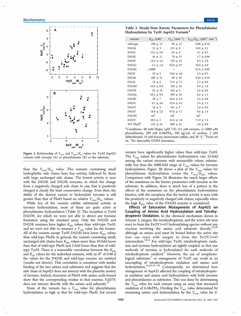
than the Vmax/Ktyr value. The variants containing smallhydrophobic side chains have less activity, followed by thosewith large uncharged side chains. The lowest activity is seenwith the D425K and D425R enzymes, in which the changefrom a negatively charged side chain to one that is positivelycharged is clearly the least conservative change. Even then, theability of the slowest variant to hydroxylate tyrosine is stillgreater than that of PheH based on relative Vmax/Ktyr values.While few of the variants exhibit substantial activity at
tyrosine hydroxylation, most of them are quite active atphenylalanine hydroxylation (Table 3). The exception is TyrHD425W, for which we were not able to detect any tyrosineformation using the standard assay. Only the D425H andD425K enzymes have higher Kphe values than wild-type TyrH,and we were not able to measure a Vmax value for the former.All of the variants except TyrH D425H have lower Kphe valuesthan wild-type PheH; in general, the variants containing small,uncharged side chains have Kphe values more than 10-fold lowerthan that of wild-type PheH and 5-fold lower than that of wild-type TyrH. There is a reasonable correlation between the Kpheand Ktyr values for the individual mutants, with an R2 of 0.90 ifthe values for the D425K and wild-type enzyme are omitted(results not shown). This correlation is consistent with similarbinding of the two amino acid substrates and suggests that theside chain of Asp425 does not interact with the phenolic moietyof tyrosine. Indeed, structures of PheH with amino acids boundshow that the corresponding residue in that enzyme, Val379,does not interact directly with the amino acid substrate.27
None of the variants has a Vmax value for phenylalaninehydroxylation as high as that for wild-type PheH, but several
variants have significantly higher values than wild-type TyrH.The Vmax values for phenylalanine hydroxylation vary 25-foldamong the variant enzymes with measurable values, substan-tially less than the 5000-fold range of Vmax values for tyrosinehydroxylation. Figure 3B shows a plot of the Vmax values forphenylalanine hydroxylation versus the Vmax/Kphe values.Comparison with Figure 3A illustrates the much larger effectsof the mutations on the kinetic parameters with tyrosine as thesubstrate. In addition, there is much less of a pattern in theeffects of the mutations on the phenylalanine hydroxylationkinetics, with the exception that the lowest activity is seen withthe positively or negatively charged side chains, especially whenthe high Kphe value of the D425H enzyme is considered.
Effects of Saturation Mutagenesis of Asp425 onCoupling of Amino Acid Hydroxylation and Tetrahy-dropterin Oxidation. In the chemical mechanism shown inScheme 2, oxygen, the tetrahydropterin, and the active site ironreact to form the Fe(IV)O hydroxylating species without anyreaction involving the amino acid substrate directly,28,29
although an amino acid must be bound before the active siteiron can react with oxygen to form the Fe(IV)Ointermediate.30,31 For wild-type TyrH, tetrahydropterin oxida-tion and tyrosine hydroxylation are tightly coupled, so that onemolecule of tyrosine is hydroxylated for each molecule oftetrahydropterin oxidized.2 However, the use of nonphysio-logical substrates7 or mutagenesis of TyrH can result in anuncoupling of tetrahydropterin oxidation and amino acidhydroxylation.14,18,32−34 Consequently, we determined howmutagenesis of Asp425 affected the coupling of tetrahydropter-in oxidation and amino acid hydroxylation with both tyrosineand phenylalanine as substrates. This was done by determiningthe Vmax value for each mutant using an assay that measuredoxidation of 6-MePH4. Dividing the Vmax value determined bymeasuring amino acid hydroxylation by the Vmax value for 6-
Figure 3. Relationship of Vmax and Vmax/Km values for TyrH Asp425variants with tyrosine (A) or phenylalanine (B) as the substrate.
Table 3. Steady-State Kinetic Parameters for PhenylalanineHydroxylation by TyrH Asp425 Variantsa
enzyme Kphe (μM) Vmax (min−1) Vmax/Kphe (μM−1 min−1)
wild-type 100 ± 15 96 ± 12 0.96 ± 0.19D425A 21 ± 3 313 ± 8 14.8 ± 2.1D425C 15.2 ± 2.0 63 ± 6 4.1 ± 0.2D425E 45 ± 11 76 ± 13 1.7 ± 0.06D425F 14.4 ± 4.2 119 ± 22 8.3 ± 2.9D425G 5.3 ± 1.4 82.6 ± 0.7 18.4 ± 4.9D425H >5000 − 0.14 ± 0.02D425I 22 ± 4 54.6 ± 4.6 2.5 ± 0.5D425K 200 ± 15 69 ± 10 0.34 ± 0.10D425L 19 ± 6 71.6 ± 7.3 2.7 ± 0.6D425M 16.4 ± 0.4 150 ± 16 9.4 ± 1.8D425N 62 ± 14 162 ± 5 2.6 ± 0.6D425Q 56.5 ± 9.4 349 ± 16 6.2 ± 1.1D425R 28 ± 7 64.4 ± 6.4 2.2 ± 0.8D425S 9.2 ± 4.6 31.6 ± 4.5 3.4 ± 1.3D425T 16 ± 5 141 ± 2 1.8 ± 0.6D425V 14.8 ± 2.2 87.8 ± 1.7 8.6 ± 1.4D425W ndc ndc ndc
D425Y 10.5 ± 2 41.6 ± 1.6 11.9 ± 1.4WT PheHb 252 ± 54 960 ± 41 3.8 ± 0.8
aConditions: 80 mM Hepes (pH 7.0), 0.1 μM enzyme, 5−2000 μMphenylalanine, 200 μM 6-MePH4, 100 μg/mL of catalase, 1 μMdithiothreitol, 10 μM ferrous ammonium sulfate, and 30 °C. bFrom ref16. cNo detectable DOPA formation.
Biochemistry Article
dx.doi.org/10.1021/bi400031n | Biochemistry 2013, 52, 1446−14551450
MePH4 oxidation yielded the extent of coupling. These data arelisted in Table 4.
All of the variants oxidize 6-MePH4 in the presence oftyrosine, including TyrH D425W, for which we were unable todetect any DOPA formation, but none are as active at tyrosine-dependent 6-MePH4 oxidation as the wild-type enzyme. Inaddition, all of the mutations significantly uncouple 6-MePH4oxidation and tyrosine hydroxylation. Even for the most activevariant, TyrH D425E, only 11% of the oxidizing equivalentsfrom the tetrahydropterin are consumed productively. For anumber of the variants, the level of uncoupling is greater than99%, with TyrH D425V being the most uncoupled.All of the variants also oxidize 6-MePH4 in the presence of
phenylalanine. Again, TyrH D425W has detectable but verylow activity as a phenylalanine-dependent tetrahydropterinoxidase. For all of the variants except TyrH D425Y and even forwild-type TyrH, the Vmax value for 6-MePH4 oxidation withphenylalanine is equal to or greater than that with tyrosine asthe amino acid substrate. The phenylalanine-dependent 6-MePH4 oxidation activity with TyrH D425A is twice that of thewild-type enzyme and approaches the Vmax value for phenyl-alanine hydroxylation by liver PheH (Table 3). The extent ofuncoupling of 6-MePH4 oxidation and phenylalanine hydrox-ylation for the variants is much lower than for tyrosinehydroxylation. Indeed, in no case does mutagenesis of Asp425decrease the level of coupling of phenylalanine hydroxylationand 6-MePH4 oxidation below what is seen with wild-typeTyrH, and for the D425F, D425Q, and D425Y enzymes, thereactions are completely coupled, unlike that of the wild-typeenzyme.The magnitude (up to 104) of the changes in the extent of
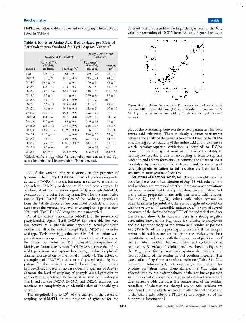
coupling of 6-MePH4 in the presence of tyrosine for the
different variants resembles the large changes seen in the Vmaxvalue for formation of DOPA from tyrosine. Figure 4 shows a
plot of the relationship between these two parameters for bothamino acid substrates. There is clearly a direct relationshipbetween the ability of the variants to convert tyrosine to DOPAat saturating concentrations of the amino acid and the extent towhich tetrahydropterin oxidation is coupled to DOPAformation, establishing that most of the loss of the ability tohydroxylate tyrosine is due to uncoupling of tetrahydropterinoxidation and DOPA formation. In contrast, the ability of TyrHto catalyze hydroxylation of phenylalanine and the coupling oftetrahydropterin oxidation to this reaction are both far lesssensitive to mutagenesis of Asp425.
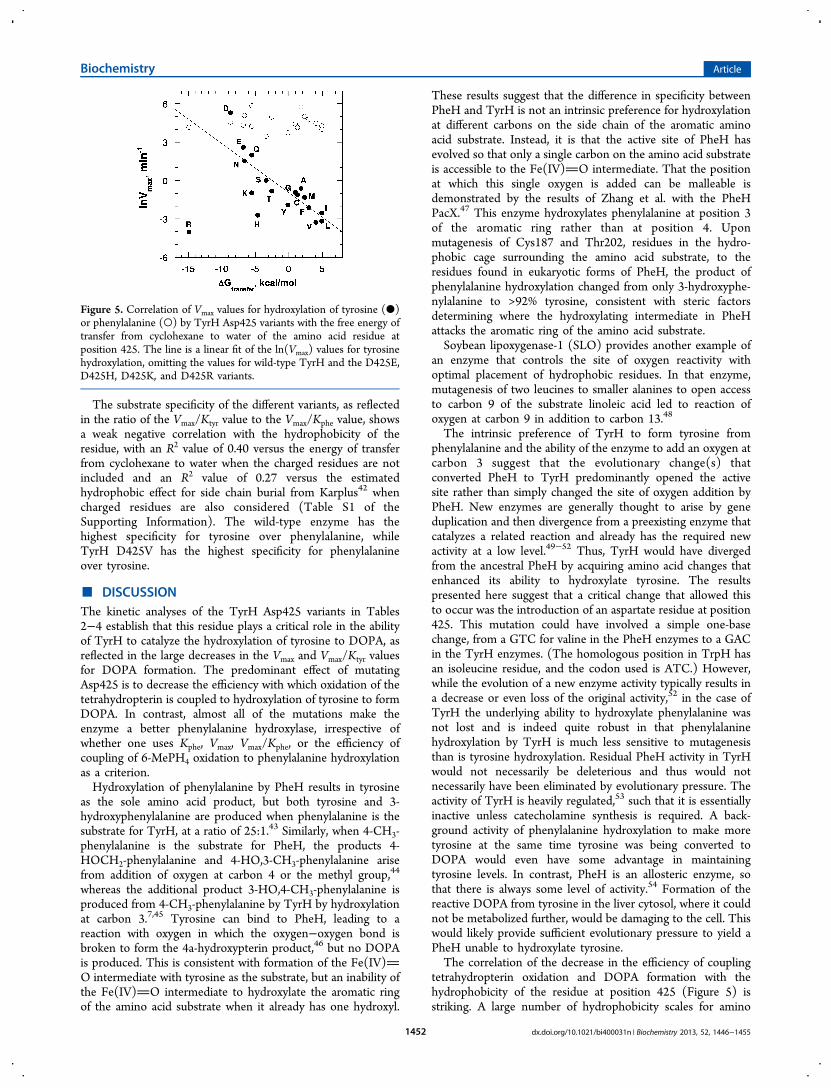
Structure−Function Analyses. To gain insight into thebasis for the effects of substitution of Asp425 with other aminoacid residues, we examined whether there are any correlationsbetween the individual kinetic parameters given in Tables 2−4and physical properties of the individual amino acid residues.For the Km and Vmax/Km values with either tyrosine orphenylalanine as the substrate, there is no significant correlationwith the volume,35,36 accessible surface area,37 or any of severalmeasures of the hydrophobicity38−42 of the individual residues(results not shown). In contrast, there is a strong negativecorrelation between the Vmax value for tyrosine hydroxylationand the hydrophobicity of the amino acid residue at position425 (Table S1 of the Supporting Information). If the chargedamino acid residues are omitted from the analysis, the bestquantitative correlation is with the free energy of partitioning ofthe individual residues between water and cyclohexane asreported by Radzicka and Wolfenden.41 As shown in Figure 5,the Vmax value for tyrosine hydroxylation decreases as thehydrophobicity of the residue at that position increases. Theextent of coupling shows a similar correlation (Table S1 of theSupporting Information), not surprisingly. In contrast, fortyrosine formation from phenylalanine, the Vmax value isaffected little by the hydrophobicity of the residue at position425. The extent of coupling with phenylalanine as the substratedoes correlate with the accessible surface area of the residue,regardless of whether the charged amino acid residues areconsidered, but the effects are much smaller than when tyrosineis the amino acid substrate (Table S1 and Figure S1 of theSupporting Information).
Table 4. Moles of Amino Acid Hydroxylated per Mole ofTetrahydropterin Oxidized for TyrH Asp425 Variantsa
tyrosine as the substratephenylalanine as the
substrate
enzyme
Vmax (min−1),
6-MePH4oxidation coupling (%)
Vmax (min−1),
6-MePH4oxidation
coupling(%)
TyrH 239 ± 17 84 ± 9 349 ± 22 28 ± 4D425A 71 ± 9 0.76 ± 0.22 715 ± 20 44 ± 1D425C 30.2 ± 1.6 1.1 ± 0.1 100 ± 3 63 ± 7D425E 119 ± 12 11.0 ± 0.6 125 ± 5 61 ± 12D425F 48.2 ± 2.8 0.26 ± 0.09 118 ± 9 101 ± 27D425G 37 ± 2 1.1 ± 0.3 210 ± 8.4 39 ± 2D425H 48 ± 7 0.14 ± 0.05 107 ± 2 ndb
D425I 52 ± 12 0.15 ± 0.03 111 ± 8 49 ± 5D425K 42 ± 9 0.46 ± 0.18 115 ± 5 60 ± 10D425L 31.5 ± 1.4 0.13 ± 0.05 192 ± 11 37 ± 4D425M 159 ± 6 0.17 ± 0.04 279 ± 11 54 ± 6D425N 117 ± 6 3.9 ± 0.4 266 ± 10 61 ± 2D425Q 214 ± 13 3.40 ± 0.03 358 ± 17 98 ± 6D425R 19.6 ± 1.3 0.092 ± 0.058 96 ± 71 67 ± 8D425S 91.7 ± 2.5 1.1 ± 0.04 89.4 ± 3.5 35 ± 5D425T 44 ± 1 1.00 ± 0.07 221 ± 12 64 ± 4D425V 46.6 ± 7.1 0.081 ± 0.007 214 ± 1 41 ± 1D425W 3.2 ± 0.5 ndb 1.6 ± 0.3 ndb
D425Y 105 ± 4 0.14 ± 0.02 31.5 ± 1.8 132 ± 9aCalculated from Vmax values for tetrahydropterin oxidation and Vmaxvalues for amino acid hydroxylation. bNone detected.
Figure 4. Correlation between the Vmax values for hydroxylation oftyrosine (●) or phenylalanine (○) and the extent of coupling of 6-MePH4 oxidation and amino acid hydroxylation for TyrH Asp425variants.
Biochemistry Article
dx.doi.org/10.1021/bi400031n | Biochemistry 2013, 52, 1446−14551451
The substrate specificity of the different variants, as reflectedin the ratio of the Vmax/Ktyr value to the Vmax/Kphe value, showsa weak negative correlation with the hydrophobicity of theresidue, with an R2 value of 0.40 versus the energy of transferfrom cyclohexane to water when the charged residues are notincluded and an R2 value of 0.27 versus the estimatedhydrophobic effect for side chain burial from Karplus42 whencharged residues are also considered (Table S1 of theSupporting Information). The wild-type enzyme has thehighest specificity for tyrosine over phenylalanine, whileTyrH D425V has the highest specificity for phenylalanineover tyrosine.
■ DISCUSSIONThe kinetic analyses of the TyrH Asp425 variants in Tables2−4 establish that this residue plays a critical role in the abilityof TyrH to catalyze the hydroxylation of tyrosine to DOPA, asreflected in the large decreases in the Vmax and Vmax/Ktyr valuesfor DOPA formation. The predominant effect of mutatingAsp425 is to decrease the efficiency with which oxidation of thetetrahydropterin is coupled to hydroxylation of tyrosine to formDOPA. In contrast, almost all of the mutations make theenzyme a better phenylalanine hydroxylase, irrespective ofwhether one uses Kphe, Vmax, Vmax/Kphe, or the efficiency ofcoupling of 6-MePH4 oxidation to phenylalanine hydroxylationas a criterion.Hydroxylation of phenylalanine by PheH results in tyrosine
as the sole amino acid product, but both tyrosine and 3-hydroxyphenylalanine are produced when phenylalanine is thesubstrate for TyrH, at a ratio of 25:1.43 Similarly, when 4-CH3-phenylalanine is the substrate for PheH, the products 4-HOCH2-phenylalanine and 4-HO,3-CH3-phenylalanine arisefrom addition of oxygen at carbon 4 or the methyl group,44
whereas the additional product 3-HO,4-CH3-phenylalanine isproduced from 4-CH3-phenylalanine by TyrH by hydroxylationat carbon 3.7,45 Tyrosine can bind to PheH, leading to areaction with oxygen in which the oxygen−oxygen bond isbroken to form the 4a-hydroxypterin product,46 but no DOPAis produced. This is consistent with formation of the Fe(IV)O intermediate with tyrosine as the substrate, but an inability ofthe Fe(IV)O intermediate to hydroxylate the aromatic ringof the amino acid substrate when it already has one hydroxyl.
These results suggest that the difference in specificity betweenPheH and TyrH is not an intrinsic preference for hydroxylationat different carbons on the side chain of the aromatic aminoacid substrate. Instead, it is that the active site of PheH hasevolved so that only a single carbon on the amino acid substrateis accessible to the Fe(IV)O intermediate. That the positionat which this single oxygen is added can be malleable isdemonstrated by the results of Zhang et al. with the PheHPacX.47 This enzyme hydroxylates phenylalanine at position 3of the aromatic ring rather than at position 4. Uponmutagenesis of Cys187 and Thr202, residues in the hydro-phobic cage surrounding the amino acid substrate, to theresidues found in eukaryotic forms of PheH, the product ofphenylalanine hydroxylation changed from only 3-hydroxyphe-nylalanine to >92% tyrosine, consistent with steric factorsdetermining where the hydroxylating intermediate in PheHattacks the aromatic ring of the amino acid substrate.Soybean lipoxygenase-1 (SLO) provides another example of
an enzyme that controls the site of oxygen reactivity withoptimal placement of hydrophobic residues. In that enzyme,mutagenesis of two leucines to smaller alanines to open accessto carbon 9 of the substrate linoleic acid led to reaction ofoxygen at carbon 9 in addition to carbon 13.48
The intrinsic preference of TyrH to form tyrosine fromphenylalanine and the ability of the enzyme to add an oxygen atcarbon 3 suggest that the evolutionary change(s) thatconverted PheH to TyrH predominantly opened the activesite rather than simply changed the site of oxygen addition byPheH. New enzymes are generally thought to arise by geneduplication and then divergence from a preexisting enzyme thatcatalyzes a related reaction and already has the required newactivity at a low level.49−52 Thus, TyrH would have divergedfrom the ancestral PheH by acquiring amino acid changes thatenhanced its ability to hydroxylate tyrosine. The resultspresented here suggest that a critical change that allowed thisto occur was the introduction of an aspartate residue at position425. This mutation could have involved a simple one-basechange, from a GTC for valine in the PheH enzymes to a GACin the TyrH enzymes. (The homologous position in TrpH hasan isoleucine residue, and the codon used is ATC.) However,while the evolution of a new enzyme activity typically results ina decrease or even loss of the original activity,52 in the case ofTyrH the underlying ability to hydroxylate phenylalanine wasnot lost and is indeed quite robust in that phenylalaninehydroxylation by TyrH is much less sensitive to mutagenesisthan is tyrosine hydroxylation. Residual PheH activity in TyrHwould not necessarily be deleterious and thus would notnecessarily have been eliminated by evolutionary pressure. Theactivity of TyrH is heavily regulated,53 such that it is essentiallyinactive unless catecholamine synthesis is required. A back-ground activity of phenylalanine hydroxylation to make moretyrosine at the same time tyrosine was being converted toDOPA would even have some advantage in maintainingtyrosine levels. In contrast, PheH is an allosteric enzyme, sothat there is always some level of activity.54 Formation of thereactive DOPA from tyrosine in the liver cytosol, where it couldnot be metabolized further, would be damaging to the cell. Thiswould likely provide sufficient evolutionary pressure to yield aPheH unable to hydroxylate tyrosine.The correlation of the decrease in the efficiency of coupling
tetrahydropterin oxidation and DOPA formation with thehydrophobicity of the residue at position 425 (Figure 5) isstriking. A large number of hydrophobicity scales for amino
Figure 5. Correlation of Vmax values for hydroxylation of tyrosine (●)or phenylalanine (○) by TyrH Asp425 variants with the free energy oftransfer from cyclohexane to water of the amino acid residue atposition 425. The line is a linear fit of the ln(Vmax) values for tyrosinehydroxylation, omitting the values for wild-type TyrH and the D425E,D425H, D425K, and D425R variants.
Biochemistry Article
dx.doi.org/10.1021/bi400031n | Biochemistry 2013, 52, 1446−14551452
acid residues have been developed over the years, primarily togain insight into the contribution of hydrophobic interactionsto protein folding.55,56 The accuracy with which any specifichydrophobicity scale predicts the effects of site-directedmutagenesis on protein structure is clearly context-dependent,and charged and polar amino acid side chains are capable ofinteractions other than the hydrophobic kind.42 In light of thesecomplications, we did not attempt to determine the hydro-phobicity scale that gave the best fit to the data from the manyavailable scales but instead focused on a representative selectionof hydrophobicity scales and other properties of amino acidside chains. The negative correlation of the coupling with thefree energy of transfer from water to cyclohexane suggests thata hydrophobic interaction involving the residue at position 425results in a restricted active site in which hydroxylation oftyrosine is not possible. This is consistent with the finding thatTyrH D425V, the least coupled variant other than TyrHD425W, contains the residue found at this position in PheH, anenzyme that is essentially unable to catalyze tyrosinehydroxylation.The hydrophobic interaction is likely with Phe184, which is
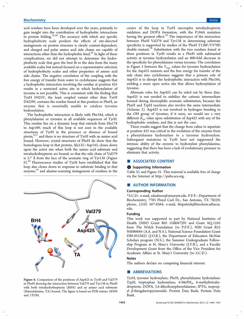
phenylalanine or tyrosine in all available sequences of TyrH.This residue lies on a dynamic loop that extends from His178to Asp189; much of this loop is not seen in the availablestructures of TyrH in the presence or absence of boundpterin,9,57 and there is no structure of TyrH with an amino acidbound. However, crystal structures of PheH do show that thehomologous loop in that protein, Ala132−Asp143, closes downupon the active site when both the amino acid substrate andtetrahydrobiopterin are bound, so that the side chain of Val379is 3.7 Å from the face of the aromatic ring of Tyr138 (Figure6).58 Fluorescence studies of TyrH have established that thisloop also closes down in response to substrate binding to thatenzyme,59 and alanine-scanning mutagenesis of residues in the
center of the loop in TyrH uncouples tetrahydropterinoxidation and DOPA formation, with the F184A mutationhaving the greatest effect.18 The importance of the interactionbetween PheH Val379 and Tyr138 in determining substratespecificity is supported by studies of the PheH Y138F/V379Ddouble mutant.18 Substitution with the two residues found atthese positions in TyrH results in a PheH with substantialactivity at tyrosine hydroxylation and an 800-fold decrease inthe specificity for phenylalanine versus tyrosine. The correlationin Figure 5 between the Vmax values for tyrosine hydroxylationfor the Asp425 mutants and the free energy for transfer of theside chain into cyclohexane suggests that a primary role ofAsp425 is to disrupt the hydrophobic interaction with Phe184,yielding a more open active site that allows hydroxylation oftyrosine.Alternate roles for Asp425 can be ruled out by these data.
Asp425 is not needed to stabilize the cationic intermediateformed during electrophilic aromatic substitution, because thePheH and TrpH reactions also involve the same intermediate(Scheme 2). Asp425 is not involved in hydrogen bonding tothe OH group of tyrosine; if it were, we would see a verydifferent Ktyr value upon substitution of Asp425 with any of thehydrophobic residues, and this is not the case.These results suggest that the change from valine to aspartate
at position 425 was critical in the evolution of the enzyme froma phenylalanine hydroxylase to a tyrosine hydroxylase.Subsequent mutations in TyrH have not suppressed theintrinsic ability of the enzyme to hydroxylate phenylalanine,suggesting that there has been a lack of evolutionary pressure toeliminate that activity.
■ ASSOCIATED CONTENT
*S Supporting InformationTable S1 and Figure S1. This material is available free of chargevia the Internet at http://pubs.acs.org.
■ AUTHOR INFORMATION
Corresponding Author*S.C.D.: e-mail, [email protected]. P.F.F.: Department ofBiochemistry, 7703 Floyd Curl Dr., San Antonio, TX 78229;phone, (210) 567-8264; e-mail, [email protected].
FundingThis work was supported in part by National Institutes ofHealth (NIH) Grant R01 GM047291 and Grant AQ-1245from The Welch Foundation (to P.F.F.), NIH Grant R25NS080684 (A.A. and N.S.), National Science Foundation GrantDBI-0552822 (J.O.B.), the Department of Education McNairScholars program (N.S.), the Summer Undergraduate Fellow-ship Program at St. Mary’s University (J.Y.B.), and a FacultyDevelopment Grant from the Office of the Vice President forAcademic Affairs at St. Mary’s University (to S.C.D.).
NotesThe authors declare no competing financial interest.
■ ABBREVIATIONS
TyrH, tyrosine hydroxylase; PheH, phenylalanine hydroxylase;TrpH, tryptophan hydroxylase; 6-MePH4, 6-methyltetrahy-dropterin; DOPA, 3,4-dihydroxyphenylalanine; IPTG, isoprop-yl β-thiogalactopyranoside; Protein Data Bank, Protein DataBank.
Figure 6. Comparison of the positions of Asp425 in TyrH and Val379in PheH showing the interaction between Val379 and Tyr138 in PheHwith both tetrahydrobiopterin (BH4) and an amino acid substrate(thienylalanine, TA) bound. The figure is based on PDB entries 1KW0and 1TOH.
Biochemistry Article
dx.doi.org/10.1021/bi400031n | Biochemistry 2013, 52, 1446−14551453
■ REFERENCES(1) Fitzpatrick, P. F. (2000) The aromatic amino acid hydroxylases.In Advances in Enzymology and Related Areas of Molecular Biology(Purich, D. L., Ed.) pp 235−294, John Wiley & Sons, Inc., New York.(2) Fitzpatrick, P. F. (1999) The tetrahydropterin-dependent aminoacid hydroxylases. Annu. Rev. Biochem. 68, 355−381.(3) Daubner, S. C., Hillas, P. J., and Fitzpatrick, P. F. (1997)Characterization of chimeric pterin-dependent hydroxylases: Con-tributions of the regulatory domains of tyrosine and phenylalaninehydroxylase to substrate specificity. Biochemistry 36, 11574−11582.(4) Ledley, F. D., DiLella, A. G., Kwok, S. C. M., and Woo, S. L. C.(1985) Homology between phenylalanine and tyrosine hydroxylasesreveals common structural and functional domains. Biochemistry 24,3389−3394.(5) Grenett, H. E., Ledley, F. D., Reed, L. L., and Woo, S. L. C.(1987) Full-length cDNA for rabbit tryptophan hydroxylase: Func-tional domains and evolution of aromatic amino acid hydroxylases.Proc. Natl. Acad. Sci. U.S.A. 84, 5530−5534.(6) Fitzpatrick, P. F. (2003) Mechanism of aromatic amino acidhydroxylation. Biochemistry 42, 14083−14091.(7) Hillas, P. J., and Fitzpatrick, P. F. (1996) A mechanism forhydroxylation by tyrosine hydroxylase based on partitioning ofsubstituted phenylalanines. Biochemistry 35, 6969−6975.(8) Pavon, J. A., and Fitzpatrick, P. F. (2006) Insights into thecatalytic mechanisms of phenylalanine and tryptophan hydroxylasefrom kinetic isotope effects on aromatic hydroxylation. Biochemistry 45,11030−11037.(9) Goodwill, K. E., Sabatier, C., Marks, C., Raag, R., Fitzpatrick, P.F., and Stevens, R. C. (1997) Crystal structure of tyrosine hydroxylaseat 2.3 Å and its implications for inherited neurodegenerative diseases.Nat. Struct. Biol. 4, 578−585.(10) Kobe, B., Jennings, I. G., House, C. M., Michell, B. J., Goodwill,K. E., Santarsiero, B. D., Stevens, R. C., Cotton, R. G. H., and Kemp, B.E. (1999) Structural basis of autoregulation of phenylalaninehydroxylase. Nat. Struct. Biol. 6, 442−448.(11) Ramsey, A. J., Daubner, S. C., Ehrlich, J. I., and Fitzpatrick, P. F.(1995) Identification of Iron Ligands in Tyrosine-Hydroxylase byMutagenesis of Conserved Histidinyl Residues. Protein Sci. 4, 2082−2086.(12) Ellis, H. R., Daubner, S. C., McCulloch, R. I., and Fitzpatrick, P.F. (1999) Phenylalanine residues in the active site of tyrosinehydroxylase: Mutagenesis of Phe300 and Phe309 to alanine and metalion-catalyzed hydroxylation of Phe300. Biochemistry 38, 10909−10914.(13) Erlandsen, H., Bjorgo, E., Flatmark, T., and Stevens, R. C.(2000) Crystal structure and site-specific mutagenesis of pterin-boundhuman phenylalanine hydroxylase. Biochemistry 39, 2208−2217.(14) Daubner, S. C., and Fitzpatrick, P. F. (1999) Site-directedmutants of charged residues in the active site of tyrosine hydroxylase.Biochemistry 38, 4448−4454.(15) Daubner, S. C., Moran, G. R., and Fitzpatrick, P. F. (2002) Roleof tryptophan hydroxylase Phe313 in determining substrate specificity.Biochem. Biophys. Res. Commun. 292, 639−641.(16) Daubner, S. C., Melendez, J., and Fitzpatrick, P. F. (2000)Reversing the substrate specificities of phenylalanine and tyrosinehydroxylase: Aspartate 425 of tyrosine hydroxylase is essential for L-DOPA formation. Biochemistry 39, 9652−9661.(17) Kaufman, S., and Mason, K. (1982) Specificity of amino acids asactivators and substrates for phenylalanine hydroxylase. J. Biol. Chem.257, 14667−14678.(18) Daubner, S. C., McGinnis, J. T., Gardner, M., Kroboth, S. L.,Morris, A. R., and Fitzpatrick, P. F. (2006) A flexible loop in tyrosinehydroxylase controls coupling of amino acid hydroxylation totetrahydropterin oxidation. J. Mol. Biol. 359, 299−307.(19) Moran, G. R., Derecskei-Kovacs, A., Hillas, P. J., and Fitzpatrick,P. F. (2000) On the catalytic mechanism of tryptophan hydroxylase. J.Am. Chem. Soc. 122, 4535−4541.(20) Daubner, S. C., and Fitzpatrick, P. F. (1998) Mutation tophenylalanine of tyrosine 371 in tyrosine hydroxylase increases theaffinity for phenylalanine. Biochemistry 37, 16440−16444.
(21) McKinney, J., Teigen, K., Frøystein, N. A., Salaun, C.,Knappskog, P. M., Haavik, J., and Martínez, A. (2001) Conformationof the substrate and pterin cofactor bound to human tryptophanhydroxylase. Important role of Phe313 in substrate specificity.Biochemistry 40, 15591−15601.(22) Wang, S., Sura, G. R., Dangott, L. J., and Fitzpatrick, P. F.(2009) Identification by hydrogen/deuterium exchange of structuralchanges in tyrosine hydroxylase associated with regulation. Bio-chemistry 48, 4972−4979.(23) Daubner, S., Lauriano, C., Haycock, J., and Fitzpatrick, P.(1992) Site-directed mutagenesis of serine 40 of rat tyrosinehydroxylase. Effects of dopamine and cAMP-dependent phosphor-ylation on enzyme activity. J. Biol. Chem. 267, 12639−12646.(24) Laemmli, U. K. (1970) Cleavage of structural proteins duringthe assembly of the head of bacteriophage T4. Nature 227, 680−685.(25) Fitzpatrick, P. F. (1991) The steady state kinetic mechanism ofrat tyrosine hydroxylase. Biochemistry 30, 3658−3662.(26) Daubner, S. C., Hillas, P. J., and Fitzpatrick, P. F. (1997)Expression and characterization of the catalytic domain of humanphenylalanine hydroxylase. Arch. Biochem. Biophys. 348, 295−302.(27) Andersen, O. A., Stokka, A. J., Flatmark, T., and Hough, E.(2003) 2.0 Å Resolution crystal structures of the ternary complexes ofhuman phenylalanine hydroxylase catalytic domain with tetrahydro-biopterin and 3-(2-thienyl)-L-alanine or L-norleucine: Substratespecificity and molecular motions related to substrate binding. J.Mol. Biol. 333, 747−757.(28) Panay, A. J., Lee, M., Krebs, C., Bollinger, J. M., Jr., andFitzpatrick, P. F. (2011) Evidence for a high spin Fe(IV) species in thecatalytic cycle of a bacterial phenylalanine hydroxylase. Biochemistry 50,1928−1933.(29) Eser, B. E., Barr, E. W., Frantom, P. A., Saleh, L., Bollinger, J. M.,Jr., Krebs, C., and Fitzpatrick, P. F. (2007) Direct spectroscopicevidence for a high-spin Fe(IV) intermediate in tyrosine hydroxylase. J.Am. Chem. Soc. 129, 11334−11335.(30) Fitzpatrick, P. F. (1991) Studies of the rate-limiting step in thetyrosine hydroxylase reaction: Alternate substrates, solvent isotopeeffects, and transition state analogs. Biochemistry 30, 6386−6391.(31) Chow, M. S., Eser, B. E., Wilson, S. A., Hodgson, K. O.,Hedman, B., Fitzpatrick, P. F., and Solomon, E. I. (2009) Spectroscopyand kinetics of wild-type and mutant tyrosine hydroxylase:Mechanistic insight into O2 activation. J. Am. Chem. Soc. 131, 7685−7698.(32) Ellis, H. R., Daubner, S. C., and Fitzpatrick, P. F. (2000)Mutation of serine 395 of tyrosine hydroxylase decouples oxygen-oxygen bond cleavage and tyrosine hydroxylation. Biochemistry 39,4174−4181.(33) Fitzpatrick, P. F., Ralph, E. C., Ellis, H. R., Willmon, O. J., andDaubner, S. C. (2003) Characterization of metal ligand mutants oftyrosine hydroxylase: insights into the plasticity of a 2-histidine-1-carboxylate triad. Biochemistry 42, 2081−2088.(34) Frantom, P. A., and Fitzpatrick, P. F. (2003) Uncoupled formsof tyrosine hydroxylase unmask kinetic isotope effects on chemicalsteps. J. Am. Chem. Soc. 125, 16190−16191.(35) Richards, F. M. (1974) The interpretation of protein structures:Total volume, group volume distributions and packing density. J. Mol.Biol. 82, 1−14.(36) Creighton, T. E. (1984) Proteins: Structures and MolecularProperties, 2nd ed., W. H. Freeman and Co., New York.(37) Chothia, C. (1976) The nature of the accessible and buriedsurfaces in proteins. J. Mol. Biol. 105, 1−12.(38) Fauchere, J.-L., and Pliska, V. (1983) Hydrophobic parameters πof amino-acid side chains from the partitioning of N-acetyl-amino-acidamides. Eur. J. Med. Chem. 18, 369−375.(39) Guy, H. R. (1985) Amino acid side-chain partition energies anddistribution of residues in soluble proteins. Biophys. J. 47, 61−70.(40) Damodaran, S., and Song, K. B. (1986) The role of solventpolarity in the free energy of transfer of amino acid side chains fromwater to organic solvents. J. Biol. Chem. 261, 7220−7222.
Biochemistry Article
dx.doi.org/10.1021/bi400031n | Biochemistry 2013, 52, 1446−14551454
(41) Radzicka, A., and Wolfenden, R. (1988) Comparing thepolarities of the amino acids: Side-chain distribution coefficientsbetween the vapor phase, cyclohexane, 1-octanol, and neutral aqueoussolution. Biochemistry 27, 1664−1670.(42) Karplus, P. A. (1997) Hydrophobicity regained. Protein Sci. 6,1302−1307.(43) Fitzpatrick, P. F. (1994) Kinetic isotope effects on hydroxylationof ring-deuterated phenylalanines by tyrosine hydroxylase provideevidence against partitioning of an arene oxide intermediate. J. Am.Chem. Soc. 116, 1133−1134.(44) Siegmund, H.-U., and Kaufman, S. (1991) Hydroxylation of 4-methylphenylalanine by rat liver phenylalanine hydroxylase. J. Biol.Chem. 266, 2903−2910.(45) Pavon, J. A., and Fitzpatrick, P. F. (2005) Intrinsic isotopeeffects on benzylic hydroxylation by the aromatic amino acidhydroxylases: Evidence for hydrogen tunneling, coupled motion, andsimilar reactivities. J. Am. Chem. Soc. 127, 16414−16415.(46) Davis, M. D., and Kaufman, S. (1993) Products of the tyrosine-dependent oxidation of tetrahydrobiopterin by rat liver phenylalaninehydroxylase. Arch. Biochem. Biophys. 304, 9−16.(47) Zhang, W., Ames, B. D., and Walsh, C. T. (2011) Identificationof Phenylalanine 3-Hydroxylase for meta-Tyrosine Biosynthesis.Biochemistry 50, 5401−5403.(48) Knapp, M. J., Seebeck, F. P., and Klinman, J. P. (2001) StericControl of Oxygenation Regiochemistry in Soybean Lipoxygenase-1. J.Am. Chem. Soc. 123, 2931−2932.(49) Jensen, R. A. (1976) Enzyme Recruitment in Evolution of NewFunction. Annu. Rev. Microbiol. 30, 409−425.(50) O’Brien, P. J., and Herschlag, D. (1999) Catalytic promiscuityand the evolution of new enzymatic activities. Chem. Biol. 6, R91−R105.(51) Tawfik, D. S., and Khersonsky, O. (2010) Enzyme Promiscuity:A Mechanistic and Evolutionary Perspective. Annu. Rev. Biochem. 79,471−505.(52) Copley, S. D. (2012) Toward a systems biology perspective onenzyme evolution. J. Biol. Chem. 287, 3−10.(53) Daubner, S. C., Le, T., and Wang, S. (2011) Tyrosinehydroxylase and regulation of dopamine synthesis. Arch. Biochem.Biophys. 508, 1−12.(54) Fitzpatrick, P. F. (2012) Allosteric regulation of phenylalaninehydroxylase. Arch. Biochem. Biophys. 519, 194−201.(55) Biswas, K. M., DeVido, D. R., and Dorsey, J. G. (2003)Evaluation of methods for measuring amino acid hydrophobicities andinteractions. J. Chromatogr., A 1000, 637−655.(56) Mant, C. T., Kovacs, J. M., Kim, H.-M., Pollock, D. D., andHodges, R. S. (2009) Intrinsic amino acid side-chain hydrophilicity/hydrophobicity coefficients determined by reversed-phase high-performance liquid chromatography of model peptides: Comparisonwith other hydrophilicity/hydrophobicity scales. Pept. Sci. 92, 573−595.(57) Goodwill, K. E., Sabatier, C., and Stevens, R. C. (1998) Crystalstructure of tyrosine hydroxylase with bound cofactor analogue andiron at 2.3 Å resolution: Self-hydroxylation of Phe300 and the pterin-binding site. Biochemistry 37, 13437−13445.(58) Andersen, O. A., Flatmark, T., and Hough, E. (2002) Crystalstructure of the ternary complex of the catalytic domain of humanphenylalanine hydroxylase with tetrahydrobiopterin and 3-(2-thienyl)-L-alanine, and its implications for the mechanism of catalysis andsubstrate activation. J. Mol. Biol. 320, 1095−1108.(59) Sura, G. R., Lasagna, M., Gawandi, V., Reinhart, G. D., andFitzpatrick, P. F. (2006) Effects of ligands on the mobility of an active-site loop in tyrosine hydroxylase as monitored by fluorescenceanisotropy. Biochemistry 45, 9632−9638.(60) Pettersen, E. F., Goddard, T. D., Huang, C. C., Couch, G. S.,Greenblatt, D. M., Meng, E. C., and Ferrin, T. E. (2004) UCSFChimera: A visualization system for exploratory research and analysis.J. Comput. Chem. 25, 1605−1612.
Biochemistry Article
dx.doi.org/10.1021/bi400031n | Biochemistry 2013, 52, 1446−14551455