mutation detection by single strand conformation ... · pdf filechapter | 4. mutation...
TRANSCRIPT
Mutation Detection by Single Strand Conformation Polymorphism and Heteroduplex Analysis
Panayiotis G. Menounos1 and George P. Patrinos2,3
1Nursing Military Academy, Laboratory of Research, Athens, Greece;2Department of Pharmacy, School of Health Sciences, University of Patras, Patras, Greece;3Erasmus University Medical Center, Faculty of Medicine, and Health Sciences, Department of Bioinformatics,
Rotterdam, The Netherlands
Chapter 4
Molecular DiagnosticsCopyright © 2010 Elsevier Ltd. All rights reserved.
4.1 IntroductIon
Single strand conformation polymorphism (SSCP) and heteroduplex analysis (HDA) are two of the most popular electrophoresis-based mutation detection methods. Coupled to DNA amplification of the sequence to be analyzed, these techniques have become the methods of choice for a number of molecular diagnostic laboratories. This can be explained mainly by the numerous advantages, namely their technical simplicity and relatively high specificity for the detection of sequence variations, the low operation costs, and the poten-tial for automation for high-throughput mutation analysis. If fluorescently labeled primers are employed during DNA amplification, SSCP analysis can be also performed in gel- or capillary electrophoresis-based automated sequencers (F-SSCP, CE-SSCP), hence allowing for precise, reproduc-ible and high-throughput analysis of the genomic varia-tion. There are several factors that influence sensitivity, and therefore need to be taken into account in order to obtain reproducible results as well as to maximize the sensitivity of mutation detection. In the following pages, the theory and practice of both SSCP and HDA will be discussed. In par-ticular, emphasis will be given to the principle, the param-eters influencing the sensitivity and reproducibility of the results, the available detection schemes, and the limitations of both techniques. Finally, a number of applications for screening genomic loci in order to investigate the underly-ing molecular heterogeneity are also discussed.
45
4.2 PrIncIPles of sIngle strand conformatIon PolymorPhIsm analysIs
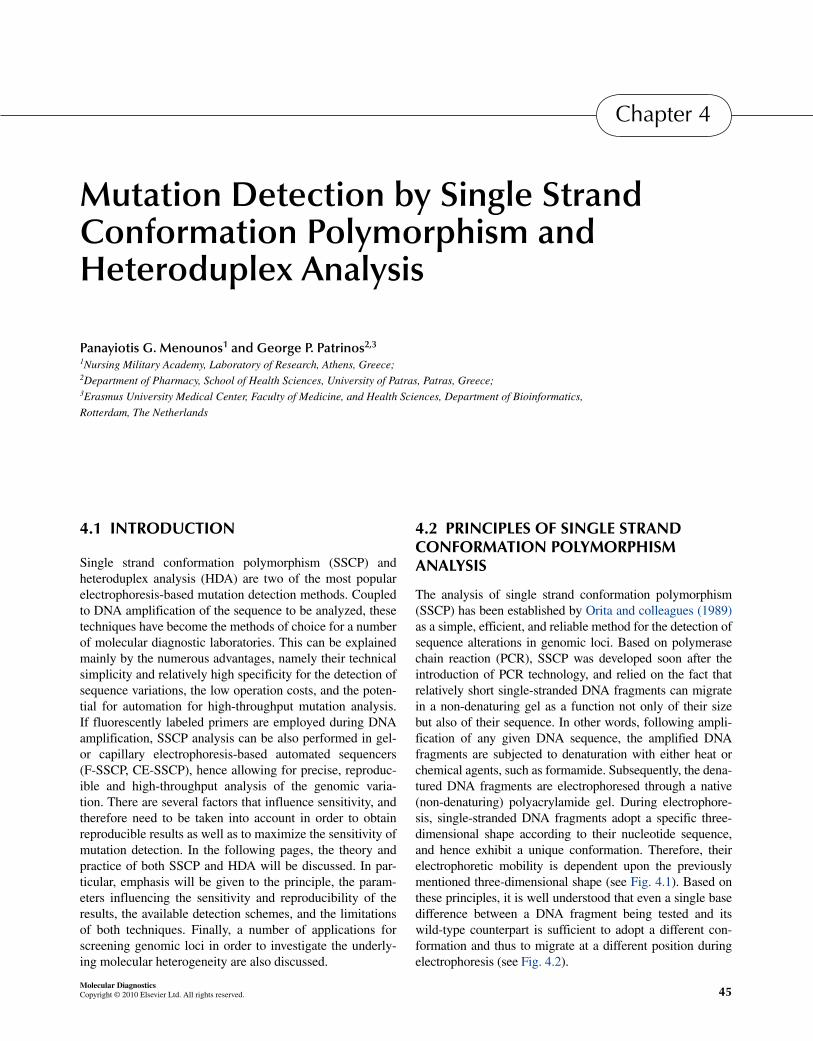
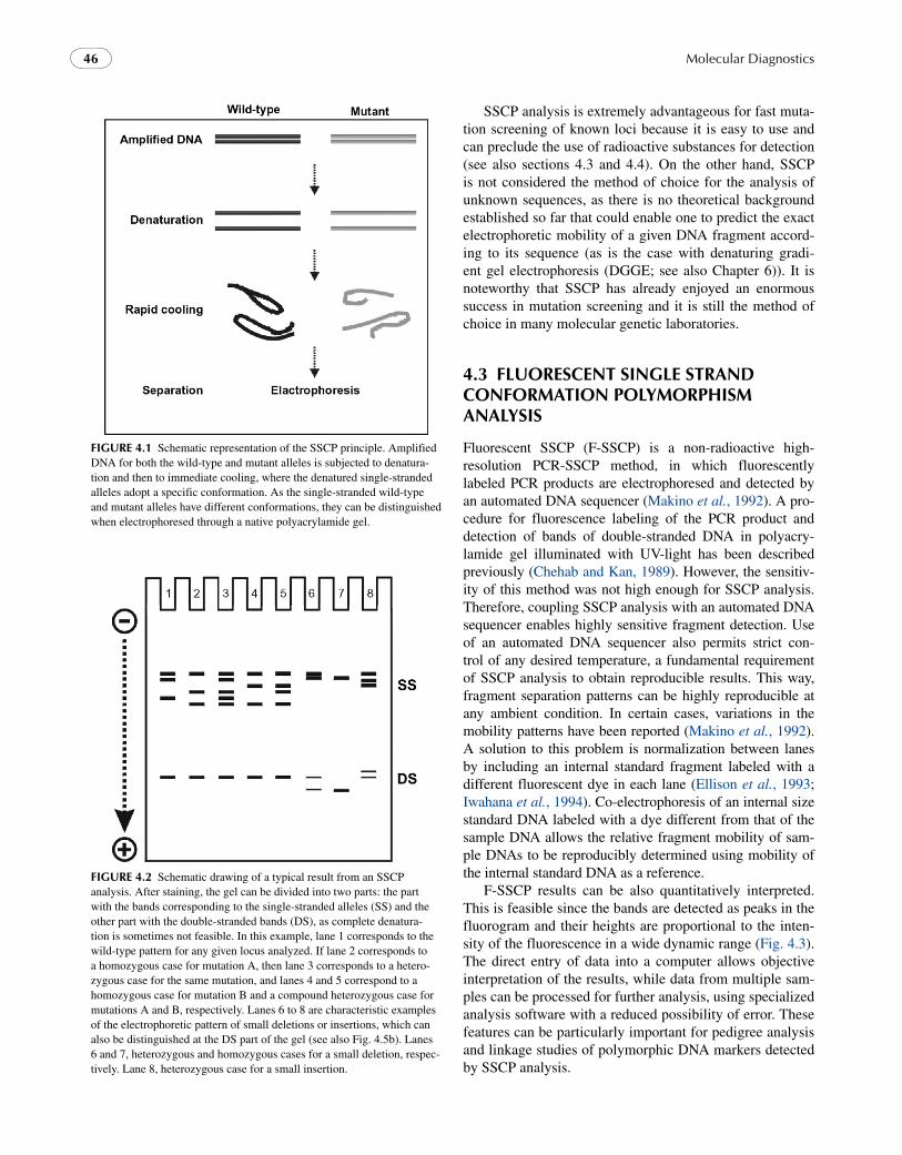
The analysis of single strand conformation polymorphism (SSCP) has been established by Orita and colleagues (1989) as a simple, efficient, and reliable method for the detection of sequence alterations in genomic loci. Based on polymerase chain reaction (PCR), SSCP was developed soon after the introduction of PCR technology, and relied on the fact that relatively short single-stranded DNA fragments can migrate in a non-denaturing gel as a function not only of their size but also of their sequence. In other words, following ampli-fication of any given DNA sequence, the amplified DNA fragments are subjected to denaturation with either heat or chemical agents, such as formamide. Subsequently, the dena-tured DNA fragments are electrophoresed through a native (non-denaturing) polyacrylamide gel. During electrophore-sis, single-stranded DNA fragments adopt a specific three-dimensional shape according to their nucleotide sequence, and hence exhibit a unique conformation. Therefore, their electrophoretic mobility is dependent upon the previously mentioned three-dimensional shape (see Fig. 4.1). Based on these principles, it is well understood that even a single base difference between a DNA fragment being tested and its wild-type counterpart is sufficient to adopt a different con-formation and thus to migrate at a different position during electrophoresis (see Fig. 4.2).
Molecular Diagnostics46
fIgure 4.1 Schematic representation of the SSCP principle. Amplified DNA for both the wild-type and mutant alleles is subjected to denatura-tion and then to immediate cooling, where the denatured single-stranded alleles adopt a specific conformation. As the single-stranded wild-type and mutant alleles have different conformations, they can be distinguished when electrophoresed through a native polyacrylamide gel.
fIgure 4.2 Schematic drawing of a typical result from an SSCP analysis. After staining, the gel can be divided into two parts: the part with the bands corresponding to the single-stranded alleles (SS) and the other part with the double-stranded bands (DS), as complete denatura-tion is sometimes not feasible. In this example, lane 1 corresponds to the wild-type pattern for any given locus analyzed. If lane 2 corresponds to a homozygous case for mutation A, then lane 3 corresponds to a hetero-zygous case for the same mutation, and lanes 4 and 5 correspond to a homozygous case for mutation B and a compound heterozygous case for mutations A and B, respectively. Lanes 6 to 8 are characteristic examples of the electrophoretic pattern of small deletions or insertions, which can also be distinguished at the DS part of the gel (see also Fig. 4.5b). Lanes 6 and 7, heterozygous and homozygous cases for a small deletion, respec-tively. Lane 8, heterozygous case for a small insertion.
SSCP analysis is extremely advantageous for fast muta-tion screening of known loci because it is easy to use and can preclude the use of radioactive substances for detection (see also sections 4.3 and 4.4). On the other hand, SSCP is not considered the method of choice for the analysis of unknown sequences, as there is no theoretical background established so far that could enable one to predict the exact electrophoretic mobility of a given DNA fragment accord-ing to its sequence (as is the case with denaturing gradi-ent gel electrophoresis (DGGE; see also Chapter 6)). It is noteworthy that SSCP has already enjoyed an enormous success in mutation screening and it is still the method of choice in many molecular genetic laboratories.
4.3 fluorescent sIngle strand conformatIon PolymorPhIsm analysIs
Fluorescent SSCP (F-SSCP) is a non-radioactive high- resolution PCR-SSCP method, in which fluorescently labeled PCR products are electrophoresed and detected by an automated DNA sequencer (Makino et al., 1992). A pro-cedure for fluorescence labeling of the PCR product and detection of bands of double-stranded DNA in polyacry-lamide gel illuminated with UV-light has been described previously (Chehab and Kan, 1989). However, the sensitiv-ity of this method was not high enough for SSCP analysis. Therefore, coupling SSCP analysis with an automated DNA sequencer enables highly sensitive fragment detection. Use of an automated DNA sequencer also permits strict con-trol of any desired temperature, a fundamental requirement of SSCP analysis to obtain reproducible results. This way, fragment separation patterns can be highly reproducible at any ambient condition. In certain cases, variations in the mobility patterns have been reported (Makino et al., 1992). A solution to this problem is normalization between lanes by including an internal standard fragment labeled with a different fluorescent dye in each lane (Ellison et al., 1993; Iwahana et al., 1994). Co-electrophoresis of an internal size standard DNA labeled with a dye different from that of the sample DNA allows the relative fragment mobility of sam-ple DNAs to be reproducibly determined using mobility of the internal standard DNA as a reference.
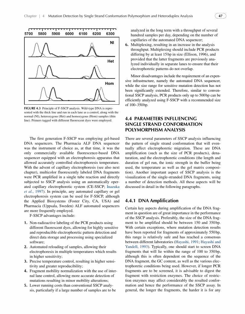
F-SSCP results can be also quantitatively interpreted. This is feasible since the bands are detected as peaks in the fluorogram and their heights are proportional to the inten-sity of the fluorescence in a wide dynamic range (Fig. 4.3). The direct entry of data into a computer allows objective interpretation of the results, while data from multiple sam-ples can be processed for further analysis, using specialized analysis software with a reduced possibility of error. These features can be particularly important for pedigree analysis and linkage studies of polymorphic DNA markers detected by SSCP analysis.
chapter | 4 Mutation Detection by Single Strand Conformation Polymorphism and Heteroduplex Analysis 47
The first generation F-SSCP was employing gel-based DNA sequencers. The Pharmacia ALF DNA sequencer was the instrument of choice as, at that time, it was the only commercially available fluorescence-based DNA sequencer equipped with an electrophoresis apparatus that allowed accurately controlled electrophoresis temperature. With the advent of capillary electrophoresis (see also next chapter), multicolor fluorescently labeled DNA fragments were PCR amplified in a single tube reaction and directly subjected to SSCP analysis using an automatically oper-ated capillary electrophoretic system (CE-SSCP; Inazuka et al., 1997). In principle, any automated capillary or gel electrophoresis system can be used for F-SSCP, although the Applied Biosystems (Foster City, CA, USA) and Pharmacia (Uppsala, Sweden) ALF automated sequencers are more frequently employed.
F-SSCP advantages include:
1. Non-radioactive labeling of the PCR products using different fluorescent dyes, allowing for highly sensitive and reproducible electrophoretic pattern detection and direct data storage and processing using specialized software;
2. Automated reloading of samples, allowing their electrophoresis in multiple temperatures which results in higher sensitivity;
3. Precise temperature control, resulting in higher sensi-tivity and greater reproducibility;
4. Fragment mobility normalization with the use of inter-nal lane control, allowing more accurate detection of mutations resulting in minor mobility alterations;
5. Lower running costs than conventional SSCP analy-sis, particularly if a large number of samples are to be
fIgure 4.3 Principle of F-SSCP analysis. Wild-type DNA is repre-sented with the thick line and run in each lane as a control, along with the normal (Nl), heterozygous (Het) and homozygous (Hom) samples (thin line). Primers tagged with different fluorescent dyes were employed.
analyzed in the long term with a throughput of several hundred samples per day, depending on the number of capillaries of the automated DNA sequencer;
6. Multiplexing, resulting in an increase in the analysis throughput. Multiplexing should include PCR products differing by at least 15 bp in size (Ellison, 1996), and provided that the latter fragments are previously ana-lyzed individually in separate lanes to ensure that their electrophoretic patterns do not overlap.
Minor disadvantages include the requirement of an expen-sive infrastructure, namely the automated DNA sequencer, while the size range for sensitive mutation detection has not been significantly extended. Therefore, similar to conven-tional SSCP analysis, PCR products only up to 500 bp can be efficiently analyzed using F-SSCP with a recommended size of 100–350 bp.
4.4 Parameters InfluencIng sIngle strand conformatIon PolymorPhIsm analysIs
There are several parameters of SSCP analysis influencing the pattern of single strand conformation that will even-tually affect electrophoretic migration. These are DNA amplification (such as the size of PCR products), dena-turation, and the electrophoretic conditions (the length and duration of gel run, the ionic strength in the buffer being used, the temperature as well as the gel matrix composi-tion). Another important aspect of SSCP analysis is the visualization of the single-stranded DNA fragments, using a number of detection methods. All these aspects will be discussed in detail in the following paragraphs.
4.4.1 dna amplification
Certain key aspects during amplification of the DNA frag-ment in question are of great importance in the performance of the SSCP analysis. Preferably, the size of the DNA frag-ment to be amplified should be between 150 and 350 bp. With certain exceptions, where mutation detection results have been reported for fragments of approximately 550 bp, this range is relatively safe and has reached a consensus between different laboratories (Hayashi, 1991; Hayashi and Yandell, 1993). Typically, one should start to screen DNA fragments that will lie within the range of 100 to 350 bp, although this is often dependent on the sequence of the DNA fragment, the GC content, as well as the various elec-trophoretic conditions being used. However, if longer PCR fragments are to be screened, it is advisable to digest the fragment with restriction enzymes. The choice of restric-tion enzymes may affect considerably the resulted confor-mation and hence the performance of the SSCP assay. In general, the longer the fragments, the harder it is for any
Molecular Diagnostics48
given single nucleotide change to have an effect in the conformation of the fragment, although under certain cir-cumstances detection efficiency is not uniformly decreased with increasing DNA fragment length. It is noteworthy that sensitivity of SSCP can be greatly improved even for frag-ments as big as 800 bp, by running the electrophoresis in low pH buffer systems and at a fixed temperature (Kukita et al., 1997).
After amplification, all PCR products should be screened on an agarose gel for the desired product length. If, despite considerable efforts, undesired side products are difficult to eliminate, a PCR product purification protocol should be applied. In addition, a negative and, where possible, a positive control of each PCR product should be included during each amplification step, since this will be the only indirect evidence that the screening assay is performed on the desired PCR products. PCR products can be stored on DNase-free tubes at 4°C. However, prolonged storage should be avoided and the subsequent steps of SSCP protocol should be performed as soon as possible.
4.4.2 denaturation
In SSCP analysis, it is important to achieve complete and as much irreversible denaturation of the DNA strands as possible. Incomplete denaturation, partial folding, and reannealing to double-stranded DNA will greatly reduce the amount of single-stranded DNA in the assay and will subsequently affect detection of the single-stranded mol-ecules. Usually, denaturation of the PCR products is car-ried out by incubation to high temperature, that is, 95°C for 5 to 7 min, and immediately after are chilled on ice for approximately 10 min. Alternatively, there are numerous denaturing agents, such as formamide, methyl-mercuric hydroxide, sodium hydroxide, and urea, which seem to per-form well (Humphries et al., 1997). Formamide is the most commonly used denaturing agent. In certain conditions, it is advisable to use 5–10% glycerol prior to loading the samples on the gel. This strategy was previously shown to produce sharper DNA bands, which greatly facilitate sub-sequent interpretation of results. Notably, an alternative method to increase the concentration of the single-stranded DNA molecules is asymmetric PCR. Following the first amplification step, an aliquot of the PCR product is used as template for nested PCR using only one of the primers employed in the previous amplification round (Lázaro and Estivill, 1992). This approach overcomes the incomplete DNA denaturation and reannealing.
4.4.3 electrophoretic Parameters
Prior to reviewing the various electrophoretic parameters that may influence the SSCP analysis, it is worthwhile mentioning that currently there is neither adequate theory
nor any physicochemical model available that could allow one to predict the three-dimensional structure of any given single-stranded DNA fragment, and as a result, its electro-phoretic mobility. Apart from the size of the DNA frag-ment and its GC content, the following parameters have been empirically found to affect the sensitivity of SSCP analysis: the gel matrix composition, the buffer composi-tion (ionic strength, the pH and buffer supplements, such as glycerol), the duration of gel run, the gel length, the DNA concentration, and the electrophoresis temperature. The effect of these factors on SSCP resolution and sensitivity is outlined next.
1. Gel matrix composition. For conventional SSCP analy-sis, the most common and widely accepted matrix is a cross-linked acrylamide polymer (8–12%). The small pore size of acrylamide-derived matrices makes it ideal for enhanced resolution and discrimination even at the nucleotide level. Higher resolution can be achieved upon addition of 10–15% of sucrose or glycerol. It has been previously shown that the mutation detection enhancement (MDE) gel (FMC Bioproducts) had a mutation detection rate of approximately 95% (Ravnik-Glavac et al., 1994). Although there is a considerable variability in the percentage of mutations detected with this commercially available gel matrix, it should be noted that in a considerable and rather growing number of studies for which SSCP has been implemented, MDE gels were used instead of the standard acryla-mide. For CE-SSCP analysis, data suggest that a sen-sitivity of 98–99% can be obtained using a 10% long chain poly-N,N-dimethylacrylamide polymer (LPDMA; Jespersgaard et al., 2006).
2. Buffer composition. So far, the Tris-borate buffer is the buffer of choice for most investigators in SSCP analysis. However, in certain instances HEPES buffer has been demonstrated to offer an alternative solu-tion that may increase the sensitivity of the SSCP. The addition of 5% glycerol has been shown to lower the pH and to decrease the electrostatic repulsion between the negatively charged phosphates in the nucleic acid backbone, resulting in a higher resolution between the mutant and wild-type DNA fragments. Conformational structures can also be more compacted by increasing the salt concentration. Finally, buffer systems with low pH have been shown to increase the sensitivity for mutation detection in larger DNA fragments (Kukita et al., 1997).
3. Gel length and duration of gel run. There is a consid-erable variation in the duration of the gel run that has been adopted by the various diagnostic laboratories. It is inevitable that the time of electrophoresis is depend-ent on both the length of the gel as well as the applied voltage. It is preferable to start the electrophoresis with a relatively moderate voltage and increase it as soon as the PCR fragments have been migrated into the gel.
chapter | 4 Mutation Detection by Single Strand Conformation Polymorphism and Heteroduplex Analysis 49
The length could vary between 10 and 40 cm. In gen-eral the bigger the gel length, the better the resolution, since at several occasions the conformational changes of the wild-type and mutant single-stranded alleles are so minor that they may migrate at very close proximity to each other.
4. Temperature. Several laboratories have demonstrated in the past the importance of temperature on the conformational changes of the DNA fragments. It is conceivable that the known temperature effect on the stabilization of the secondary structure of single-stranded DNA fragments may affect to a varying degree (depending on the primary sequence) the SSCP results. In addition to a lower pH, decreasing the temperature to 4°C has been shown to enhance the stability of the conformation for any given single-stranded DNA frag-ment. It is advisable that one should try a gradual tem-perature decrease, starting from 15°C and descending with an increment of 2–4°C at a time. For CE-SSCP, Jespersgaard and coworkers (2006) concluded that a sensitivity of 98–99% could be obtained when using the LPDMA polymers and a single electrophoresis temperature of 27°C, by analyzing a temperature range between 18 and 35°C.
5. DNA concentration. In the past, several investiga-tors have used high DNA concentrations in order to enhance detection. Unfortunately, this has often led to a decrease in the specific concentration of the single-stranded DNA. Even after the addition of formamide, it has been empirically shown that at high concentra-tions, the two single-stranded molecules tend to rean-neal and form a double-stranded DNA. It is preferable, therefore, to keep the DNA concentration relatively low in the loading buffer. Also, gel overloading can sometimes result in abnormal migration of the bands, leading to decreased resolution. Similarly, in F-SSCP a critical step includes dilution of the PCR products. If the sample is too concentrated, then peaks will not be sharp enough (also known as peak broadening) to allow unambiguous detection, leading to false positive results. On the other hand, if the sample is overdiluted, the poor signal-to-noise ratio will also result in false negative results. Therefore, the appropriate dilution factor should be estimated for each PCR, ranging from 1/10 to 1/80.
4.4.4 detection
Most molecular diagnostic laboratories are well adapted in the use of radioactivity and have the equipment and exper-tise required for this type of protocol. Typically, autoradi-ography requires immobilization of the gel, a drying step, and finally film exposure. However, this approach is more time consuming than some of the more recently described protocols that utilize silver staining or detection with flu-orescent dyes. Alternatively, in F-SSCP, analysis can be
done on automated sequencers with fluorescent dye-labeled DNA fragments. In relative terms, and although ethidium bromide staining can be considered an attractive alterna-tive to the use of fluorescent substances (Yap and McGee, 1992a; Lázaro and Estivill, 1992), the silver staining approach comprises one of the most straightforward, fast, as well as sensitive methods for the visualization of bands on conventional SSCP gels. In brief, after electrophoresis, the polyacrylamide gels are first fixed with 10% acetic acid for approximately 30 min at room temperature and sub-sequently washed with water. Depending on the concen-tration of silver nitrate, incubation with the silver nitrate solution can last for approximately 60 min (in a 0.001% AgNO3, 0.036% formaldehyde solution). This incubation step is performed in the dark, while avoiding any con-tamination with protein-containing solution (proteins are stained extensively with silver nitrate leading to immense background). Subsequently, the polyacrylamide gels are washed with water and color development is performed by incubating the gel for 5 to 10 min with a color development solution (containing 2.5% Na2CO3, 0.036% formaldehyde, and 0.002% sodium thiosulfate). Color development can be stopped with a solution containing a chelating agent (such as 1.5% EDTA). Gels can be subsequently fixed with 30% ethanol and 4% glycerol. The stained gels are transferred to a vacuum dryer and are immobilized to a porous paper. Results are interpreted visually or can be analyzed by means of an image analysis system.
4.5 heteroduPlex analysIs for mutatIon detectIon
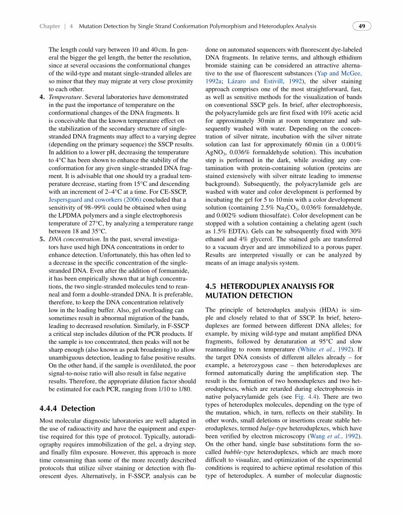
The principle of heteroduplex analysis (HDA) is sim-ple and closely related to that of SSCP. In brief, hetero-duplexes are formed between different DNA alleles; for example, by mixing wild-type and mutant amplified DNA fragments, followed by denaturation at 95°C and slow reannealing to room temperature (White et al., 1992). If the target DNA consists of different alleles already – for example, a heterozygous case – then heteroduplexes are formed automatically during the amplification step. The result is the formation of two homoduplexes and two het-eroduplexes, which are retarded during electrophoresis in native polyacrylamide gels (see Fig. 4.4). There are two types of heteroduplex molecules, depending on the type of the mutation, which, in turn, reflects on their stability. In other words, small deletions or insertions create stable het-eroduplexes, termed bulge-type heteroduplexes, which have been verified by electron microscopy (Wang et al., 1992). On the other hand, single base substitutions form the so-called bubble-type heteroduplexes, which are much more difficult to visualize, and optimization of the experimental conditions is required to achieve optimal resolution of this type of heteroduplex. A number of molecular diagnostic
50
techniques, based on heteroduplex formation, are reported in the literature, such as enzymatic or chemical cleavage (see Chapter 3) and denaturing gradient gel electrophoresis (DGGE; see Chapter 6), but HDA appears to be the most attractive one, as it can be performed rapidly on short gels without the need of specialized equipment and the use of radioactivity. The most typical example of the use of HDA for mutation screening is the rapid detection of the 3 bp p.F508del deletion in the CFTR gene, leading to cystic fibrosis (Wang et al., 1992).
HDA can be also adapted to a fluorescent capillary system, such as the currently available automated DNA sequencers. This is possible by labeling the primers with fluorescent dyes, such as 6-carboxyfluorescein (FAM), hexachloro-6-carboxyfluorescein (HEX), etc. Like F-SSCP and CE-SSCP, capillary-based HDA (CE-HDA) also has the advantage of multiplexing, which together with the use of different fluorescent dyes can significantly increase the analysis throughput (Kozlowski and Krzyzosiak, 2001). Use of multi-capillary systems, e.g. 96-capillary platforms, can also reduce the time needed for processing each sam-ple. For example, up to 10 PCR products can be analyzed in one capillary per run, indicating that the entire analysis of a patient’s BRCA1 and BRCA2 coding regions can be completed in a single run within 1.5 h (Esteban-Cardeñosa
fIgure 4.4 The HDA principle. Unlike SSCP analysis, amplified DNA is now subjected to denaturation followed by slow reannealing of the denatured alleles, leading to both homoduplexes (HmD) and heterodu-plexes (HtD). The latter migrate more slowly during gel electrophoresis, due to their sequence mismatch(es) and therefore heterozygous (Het) and homozygous (Hom) cases can be easily distinguished from the wild type (Wt) based on their electrophoretic pattern. M: size marker.
Molecular Diagnostics
et al., 2004). In brief, CE-HDA is a high-throughput, suf-ficiently sensitive and cost-effective methodology and can be easily implemented to offer reliable genetic analy-sis in molecular diagnostic laboratories with large sample volumes.
4.6 sensItIvIty and lImItatIons
Several factors can affect the sensitivity of both SSCP and HDA analysis and their optimization is highly empirical, as there is no adequate theoretical basis or type of algo-rithm (as in DGGE; Myers et al., 1987) that would enable researchers to predict the three-dimensional conforma-tion of the single-stranded DNA fragments under specific experimental procedures. Those elements that frequently affect the sensitivity of both methods will be discussed in this section.
4.6.1 optimizing sscP sensitivity
In general, most of the single-stranded DNA fragments will have a more compacted conformation at lower temperatures or in the presence of higher salt concentrations. It follows, however, that by increasing the salt concentration, the con-ductivity is also increased, thus having a rather significant effect on temperature. Depending on these parameters, which largely have been determined empirically, it is sug-gested that electrophoresis be performed at room tempera-ture with 5–10% glycerol or at 4°C without addition of glycerol or salt; this is considered a good starting point. In addition, it is highly suggestive that an empirical estimate of SSCP sensitivity will be based on the likelihood of detect-ing known sequence variations under controlled conditions. This is expected to provide a valuable means by which the effect on electrophoretic mobility of known sequence varia-tions will be determined. Under no circumstances should a perfect overall detection efficiency be anticipated. Hayashi and Yandell (1993) have conducted a valuable study to determine which are the most effective sets of parameters influencing the detection of a single mutation. Given that the latter is largely dependent on DNA fragment’s size, it is expected that for most fragments shorter than 200 nucle-otides, more than 90% of the sequence variations will be detected. Gradually, as the size of the PCR fragment increases to 300–350 nucleotides, a safe prediction is that more than 80% of mutations will be detected. It should be noted, however, that within this range, Sheffield and col-leagues (1993) have shown that the overall sensitivity of the SSCP is not affected by the position of a given mutation. On the contrary, Ellis and coworkers (2000) have shown that the increase in fragment size did not have a substantial effect on F-SSCP sensitivity, by analyzing ABCC7 exon 11 amplicons of 190 bp and 490 bp, respectively. Alternatively, a more quantitative method for estimating the sensitivity of
chapter | 4 Mutation Detection by Single Strand Conformation Polymorphism and Heteroduplex Analysis 51
the SSCP detection method based on statistical arguments has been developed. This method is based on the fact that the chance of any given strand to exhibit a mobility shift is independent from the other strand. On this assumption, the probability of observing shifts in both strands (P2), in one strand (P1), or in none of the strands (P0), is equal to x2, 2x(1 x) and (1 x)2, respectively, where x is the sensitiv-ity of the technique when only one of the strands is labeled. The ratio of P2 to P1 is equal to the observed number of the mutations on both strands over the number of the observed mutations on the single strand [r (x/2(1 x)]. The useful sensitivity of the technique can be calculated as being equal to 1 P0. Based on this probabilistic theory, the estimates from previous studies are that the sensitivity for 100–200 bp fragments is approximately 96%, regardless of the pres-ence or absence of glycerol (Hayashi and Yandell, 1993). However, by adding glycerol, the sensitivity is still high for fragments ranging from 200 to 300 bp, but is decreas-ing when glycerol is not used. The latter may indicate the inability of the former calculation to depict the actual elec-trophoretic mobility, which to some extent is expected. In addition, it may also explain the fact that substitutions, which induce significant conformational changes, are most likely to have an effect, to a variable extent, on the other strand. Despite this discrepancy, it can be safely concluded that decreasing the fragment size will greatly enhance the sensitivity. It seems rational that the overall effect of a given mutation is displayed more efficiently when the total number of nucleotides surrounding that particular mutation is less. It is also profound that glycerol greatly increases the overall sensitivity when electrophoresis is performed at room temperature.
Practical observations suggest that any fragment that exhibits a differential mobility will often migrate very close to the reference fragment. However, the overall frag-ment number is not always predictable in advance. Any given number of conformations may be supported to a variable extent by the applied electrophoretic parameters. Furthermore, the band intensity is irrelevant to allelic dif-ferences, as it is strictly dependent on the different confor-mations. Therefore, SSCP is not a safe method to predict gene-dosage effects. In addition, the simultaneous detec-tion of more than one mutation in a single DNA sample is not easily predictable, as previous data have shown that the electrophoretic pattern may vary considerably within differ-ent experiments. In general, although highly reproducible, the mobility of single-stranded DNA conformers cannot be predicted in advance from sequence information. Such attempts to predict SSCP mobility changes by modeling single-stranded DNA conformations using the structure prediction program Mfold (http://frontend.bioinfo.rpi.edu/applications/mfold/) have not provided consistent results. Improvements in modeling the structures of single-stranded DNA would make it possible to more accurately predicting SSCP mobility (Nakabayashi and Nishigaki, 1996), the
idiosyncratic nature of SSCP remains its main weakness as a diagnostic tool (Liu et al., 1999).
Attempts to further improve the sensitivity of the SSCP have led to the development of the RNA-SSCP approach (Sarkar et al., 1992). Here, although the method is essen-tially the same, the double-stranded DNA is converted to the corresponding single-stranded RNA by means of one of the two primers that has phage promoter sequences on its 5 end. This method has shown a higher sensitivity com-pared to the previously described SSCP methodology. Its sensitivity may rely on the fact that the in vitro transcribed strand has no complementary strand to reanneal with. Therefore, sufficient amounts of the in vitro transcribed product can be electrophoresed and easily analyzed even by ethidium bromide staining (Sarkar et al., 1992).
So far, there has been much discussion concerning the false negative results of this assay and possible ways to minimize them. However, false positive results also affect the net outcome of the SSCP analysis. In order to mini-mize the frequency of reporting false positive results, it is advisable to perform repeated SSCP electrophoretic runs (particularly when SSCP data are of clinical interest). An additional way is to determine the minimum mobility vari-ation, which is detectable within the context of laboratory SSCP conditions. In practical terms, mobility differences of 3 mm are generally clear, but a detection difference of only 2 mm requires excellent gel running conditions and is often subjective. Therefore, any difference smaller than or equal to 2 mm can be considered only with reservation.
Reproducibility is the last and perhaps most essential parameter applicable to most techniques in molecular diag-nostics. In general, if conditions are kept constant then the resulting reproducibility is usually high. Nevertheless, Hayashi (1991) has previously suggested that DNA sequences may have different stable conformations. The latter is thus interpreted as a variable that may compromise SSCP repro-ducibility. It relies on the possibility that when the free energy difference between different conformations is small, an oscil-lation between different structures of comparable energies may be observed.
4.6.2 sensitivity of hda
So far, the sensitivity of the HDA methodology has not been determined to the extent of the SSCP analysis. Rossetti and colleagues (1995) compared directly both SSCP and HDA assays for the detection of known mutations in a panel of four genes. Despite the fact that none of the assays was per-formed with 100% efficiency, HDA detected slightly more mutations than SSCP in the same samples. Interestingly, these authors suggested that both techniques could be used in concert to detect all mutations.
Another aspect to improve the performance of HDA is the gel matrix. As in SSCP, the use of MDE (derived from HydroLink D5000™; Keen et al., 1991) has basically made
Molecular Diagnostics52
HDA a valuable mutation detection technique. Today, the majority of the diagnostic laboratories, in which HDA is the method of choice to detect genomic variation, are employing MDE gels.
Finally, as already mentioned in section 4.5, the bubble-type heteroduplexes, which are formed due to the presence of single base substitutions, are much more difficult to visu-alize compared to the bulge-type heteroduplexes. In order to overcome this bottleneck, and based on the observa-tion that heteroduplexes are much easier to visualize when a deletion or insertion mutation is involved, the Universal Heteroduplex Generator (UHG) was conceived (Wood et al., 1993a, b). In brief, the UHG consists of a synthetic DNA fragment, which bears a small (that is, 2 to 5 bp) dele-tion. This synthetic fragment is amplified by the use of the same oligonucleotide primers as the DNA under study. After amplification, the test amplicon is mixed with the amplified UHG, denatured, and then slowly reannealed, followed by electrophoresis. If no mutation is present in the test DNA, then only a bulge-type heteroduplex will be present, slightly retarded compared to the homoduplex. If, however, a single base substitution is also present, then the resulting heterodu-plex will have two mismatches: a bulge and a bubble type, which will result in the heteroduplex migrating significantly lower, compared to the simple bulge-type heteroduplex. The use of UHG has been reported for the detection of known mutations within a number of loci, such as von Willebrand disease (Wood et al., 1995), phenylketonurea (Wood et al., 1993a), and for prenatal determination of blood group alle-les (Stoerker et al., 1996).
4.7 detectIon of the underlyIng genomIc varIatIon usIng sscP and hda
Due to their numerous advantages, SSCP and HDA anal-yses are nowadays the methods of choice in a growing number of private or public molecular diagnostic labo-ratories to either interrogate known mutations or scan for known or unknown mutations in short stretches of DNA and in relatively short time. Similarly, SSCP and HDA have also been proven to be invaluable tools for basic sci-ence, enabling both the identification of causative genes for human hereditary diseases and mapping of genomic loci.
A short summary of the existing applications of SSCP and HDA follows in the next paragraphs, which is only indicative for the applicability of these techniques in almost every genomic locus.
4.7.1 applications in Basic science
The utilization of SSCP in basic science as a tool for genomic DNA analysis is well established. Mutations in sev-eral key candidate genes implicated in various cell processes
have now been identified, and the extent of those mutations as well as their frequency has given an insight into the role of these molecules in the relevant processes.
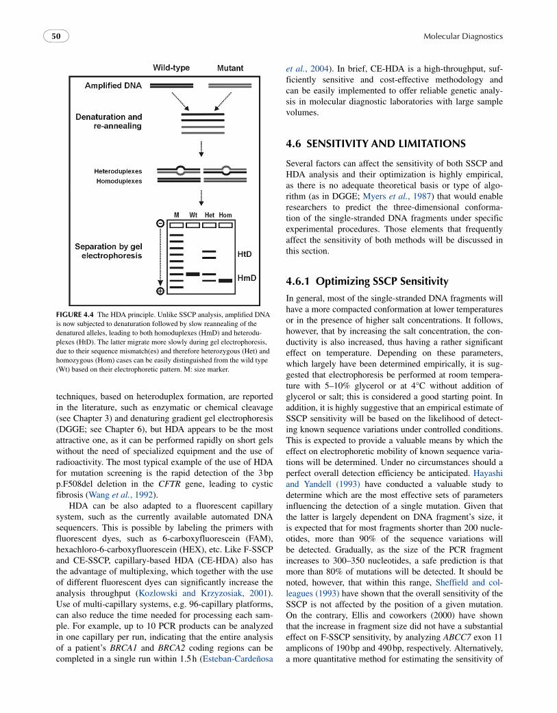
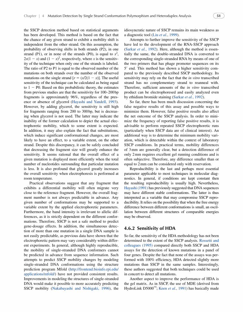
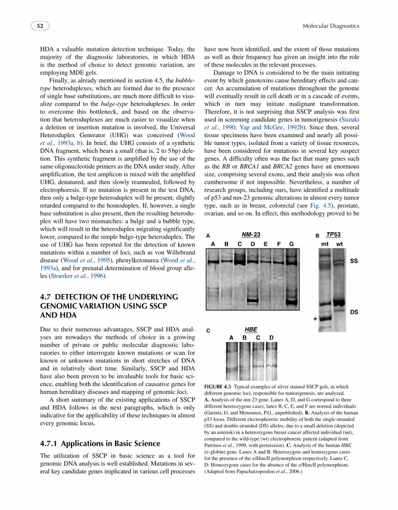
Damage to DNA is considered to be the main initiating event by which genotoxins cause hereditary effects and can-cer. An accumulation of mutations throughout the genome will eventually result in cell death or in a cascade of events, which in turn may initiate malignant transformation. Therefore, it is not surprising that SSCP analysis was first used in screening candidate genes in tumorigenesis (Suzuki et al., 1990; Yap and McGee, 1992b). Since then, several tissue specimens have been examined and nearly all possi-ble tumor types, isolated from a variety of tissue resources, have been considered for mutations in several key suspect genes. A difficulty often was the fact that many genes such as the RB or BRCA1 and BRCA2 genes have an enormous size, comprising several exons, and their analysis was often cumbersome if not impossible. Nevertheless, a number of research groups, including ours, have identified a multitude of p53 and nm-23 genomic alterations in almost every tumor type, such as in breast, colorectal (see Fig. 4.5), prostate, ovarian, and so on. In effect, this methodology proved to be
fIgure 4.5 Typical examples of silver stained SSCP gels, in which different genomic loci, responsible for tumorigenesis, are analyzed. A. Analysis of the nm-23 gene. Lanes A, D, and G correspond to three different heterozygous cases; lanes B, C, E, and F are normal individuals (Garinis, G. and Menounos, P.G., unpublished). B. Analysis of the human p53 locus. Different electrophoretic mobility of both the single-stranded (SS) and double-stranded (DS) alleles, due to a small deletion (depicted by an asterisk) in a heterozygous breast cancer affected individual (mt), compared to the wild-type (wt) electrophoretic pattern (adapted from Patrinos et al., 1999, with permission). C. Analysis of the human HBE (-globin) gene. Lanes A and B: Heterozygote and homozygous cases for the presence of the /HincII polymorphism respectively. Lanes C, D: Homozygous cases for the absence of the /HincII polymorphism. (Adapted from Papachatzopoulou et al., 2006.)
chapter | 4 Mutation Detection by Single Strand Conformation Polymorphism and Heteroduplex Analysis 53
particularly useful in revealing, in a stepwise approach, that the altered expression of several cell cycle regulatory mol-ecules either at the genomic or transcriptional and protein levels may exert a synergetic effect on tumor growth and chromosomal instability on breast cancer and non-small cell lung and colorectal carcinomas.
In addition to the utilization of SSCP methodology as a screening tool, several investigators have previously employed this technique for gene mapping in mouse genes (Beier, 1993 and references therein). The methodology is based on the fact that a given polymorphism readily can be found in non-coding regions of genes such as the 3 untrans-lated regions or introns, between alleles of mouse species, and in several occasions between inbred strains as well. The segregations of these polymorphisms can be analyzed with recombinant inbred or interspecific crosses and the strain distribution pattern obtained can be compared with that for other markers and analyzed by standard linkage analy-sis algorithms. For instance, SSCP has previously been employed to localize 39 mouse-specific sequence-tagged
sites (STSs), generated from mouse–hamster somatic cell hybrids. These were subsequently integrated with other markers to generate a high-density map of mouse chro-mosome 1 containing over 100 markers typed on a single interspecific backcross (Watson et al., 1992). SSCP analy-sis also has been applied in relatively fewer cases for the linkage analysis of human genes (Nishimura et al., 1993; Avramopoulos et al., 1993).
4.7.2 molecular diagnostic applications
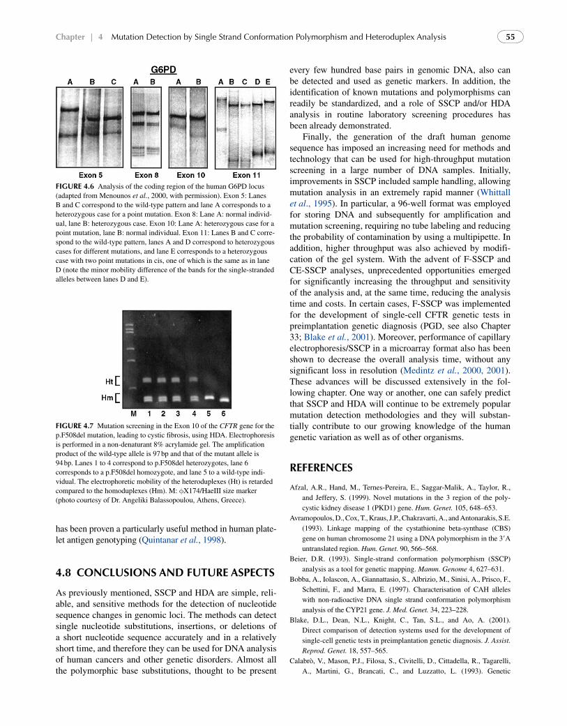
Both conventional and fluorescent or capillary-based SSCP and HDA can be successfully used for the detection of known mutations in any genomic locus. A brief summary of the numerous applications of SSCP analysis for various human genes is given in Table 4.1. When SSCP analysis is coupled to non-radioactive detection schemes, then it most certainly becomes the method of choice for routine molec-ular diagnostic analysis (see Fig. 4.6 for representative examples from G6PD mutation screening (Menounos et al.,
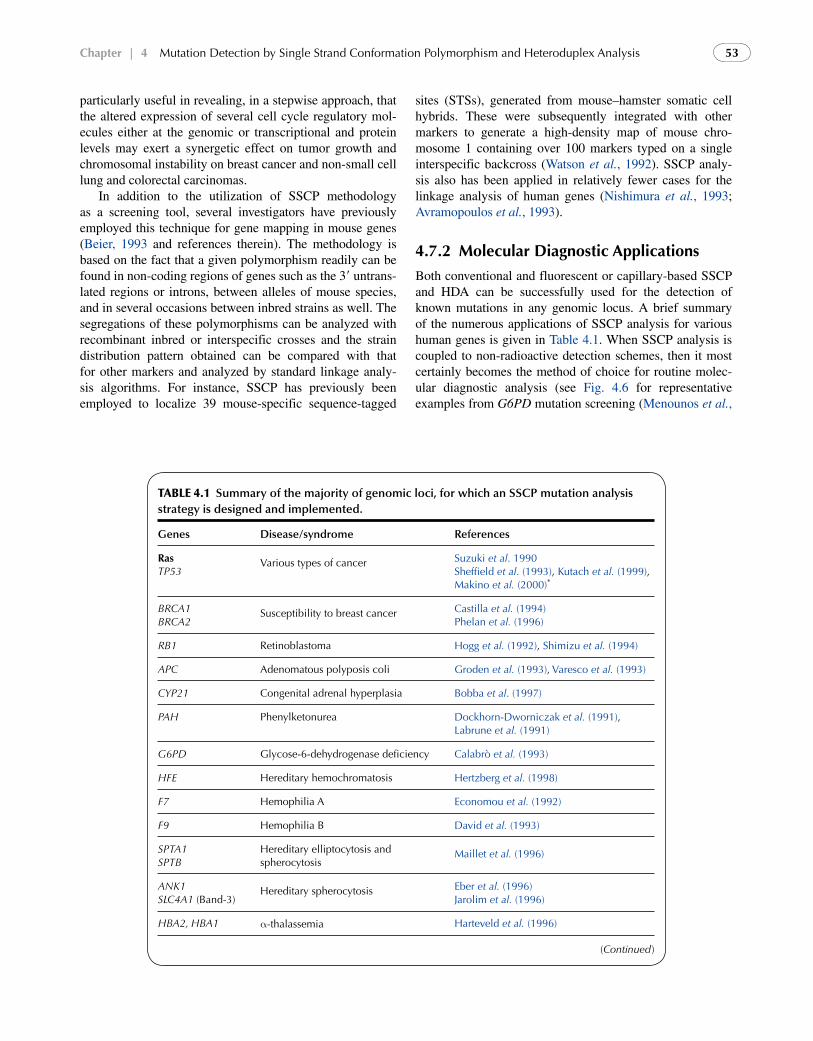
Table 4.1 Summary of the majority of genomic loci, for which an SSCP mutation analysis strategy is designed and implemented.
Genes Disease/syndrome References
rasTP53
Various types of cancer Suzuki et al. 1990Sheffield et al. (1993), Kutach et al. (1999), Makino et al. (2000)*
BRCA1BRCA2
Susceptibility to breast cancer Castilla et al. (1994)Phelan et al. (1996)
RB1 Retinoblastoma Hogg et al. (1992), Shimizu et al. (1994)
APC Adenomatous polyposis coli Groden et al. (1993), Varesco et al. (1993)
CYP21 Congenital adrenal hyperplasia Bobba et al. (1997)
PAH Phenylketonurea Dockhorn-Dworniczak et al. (1991), Labrune et al. (1991)
G6PD Glycose-6-dehydrogenase deficiency Calabrò et al. (1993)
HFE Hereditary hemochromatosis Hertzberg et al. (1998)
F7 Hemophilia A Economou et al. (1992)
F9 Hemophilia B David et al. (1993)
SPTA1SPTB
Hereditary elliptocytosis and spherocytosis
Maillet et al. (1996)
ANK1SLC4A1 (Band-3)
Hereditary spherocytosis Eber et al. (1996)Jarolim et al. (1996)
HBA2, HBA1 -thalassemia Harteveld et al. (1996)
(Continued)
Molecular Diagnostics54
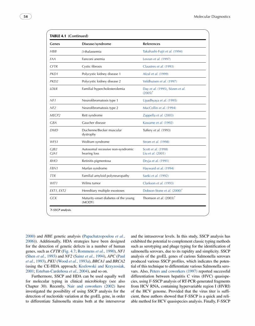
Table 4.1 (Continued)
Genes Disease/syndrome References
HBB -thalassemia Takahashi-Fujii et al. (1994)
FAA Fanconi anemia Levran et al. (1997)
CFTR Cystic fibrosis Claustres et al. (1993)
PKD1 Polycystic kidney disease 1 Afzal et al. (1999)
PKD2 Polycystic kidney disease 2 Veldhuisen et al. (1997)
LDLR Familial hypercholesterolemia Day et al. (1995), Sözen et al. (2005)*
NF1 Neurofibromatosis type 1 Upadhyaya et al. (1995)
NF2 Neurofibromatosis type 2 MacCollin et al. (1994)
MECP2 Rett syndrome Zappella et al. (2003)
GBA Gaucher disease Kawame et al. (1992)
DMD Duchenne/Becker muscular dystrophy
Tuffery et al. (1993)
WFS1 Wolfram syndrome Strom et al. (1998)
GJB2GJA1
Autosomal recessive non-syndromic hearing loss
Scott et al. (1998)Liu et al. (2001)
RHO Retinitis pigmentosa Dryja et al. (1991)
FBN1 Marfan syndrome Hayward et al. (1994)
TTR Familial amyloid polyneuropathy Saeki et al. (1992)
WIT1 Wilms tumor Clarkson et al. (1993)
EXT1, EXT2 Hereditary multiple exostoses Dobson-Stone et al. (2000)*
GCK Maturity-onset diabetes of the young (MODY)
Thomson et al. (2003)*
*F-SSCP analysis.
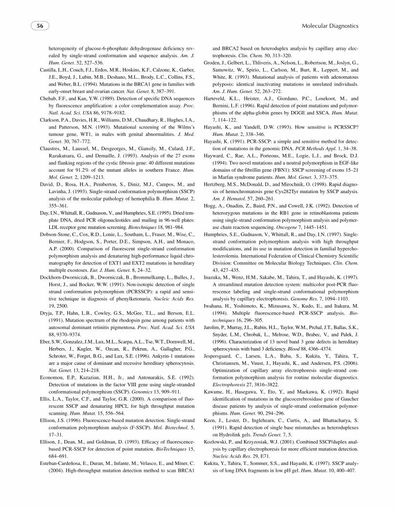
2000) and HBE genetic analysis (Papachatzopoulou et al., 2006)). Additionally, HDA strategies have been designed for the detection of genetic defects in a number of human genes, such as CFTR (Fig. 4.7; Rommens et al., 1990), NF1 (Shen et al., 1993) and NF2 (Sainz et al., 1994), APC (Paul et al., 1993), PKU (Wood et al., 1993a), BRCA1 and BRCA2 (using the CE-HDA approach; Kozlowski and Krzyzosiak, 2001; Esteban-Cardeñosa et al., 2004), and so on.
Furthermore, SSCP and HDA can be used equally well for molecular typing in clinical microbiology (see also Chapter 30). Recently, Nair and coworkers (2002) have investigated the possibility of using SSCP analysis for the detection of nucleotide variation at the groEL gene, in order to differentiate Salmonella strains both at the interserovar
and the intraserovar levels. In this study, SSCP analysis has exhibited the potential to complement classic typing methods such as serotyping and phage typing for the identification of salmonella serovars, due to its rapidity and simplicity. SSCP analysis of the groEL genes of carious Salmonella serovars produced various SSCP profiles, which indicates the poten-tial of this technique to differentiate various Salmonella sero-vars. Also, Peters and coworkers (1997) reported successful differentiation between hepatitis C virus (HVC) quasispe-cies, using F-SSCP analysis of RT-PCR-generated fragments from HCV RNA, containing hypervariable region I (HVRI) of the HCV genome. Provided that the virus titer is suffi-cient, these authors showed that F-SSCP is a quick and reli-able method for HCV quasispecies analysis. Finally, F-SSCP
chapter | 4 Mutation Detection by Single Strand Conformation Polymorphism and Heteroduplex Analysis 55
has been proven a particularly useful method in human plate-let antigen genotyping (Quintanar et al., 1998).
4.8 conclusIons and future asPects
As previously mentioned, SSCP and HDA are simple, reli-able, and sensitive methods for the detection of nucleotide sequence changes in genomic loci. The methods can detect single nucleotide substitutions, insertions, or deletions of a short nucleotide sequence accurately and in a relatively short time, and therefore they can be used for DNA analysis of human cancers and other genetic disorders. Almost all the polymorphic base substitutions, thought to be present
fIgure 4.6 Analysis of the coding region of the human G6PD locus (adapted from Menounos et al., 2000, with permission). Exon 5: Lanes B and C correspond to the wild-type pattern and lane A corresponds to a heterozygous case for a point mutation. Exon 8: Lane A: normal individ-ual, lane B: heterozygous case. Exon 10: Lane A: heterozygous case for a point mutation, lane B: normal individual. Exon 11: Lanes B and C corre-spond to the wild-type pattern, lanes A and D correspond to heterozygous cases for different mutations, and lane E corresponds to a heterozygous case with two point mutations in cis, one of which is the same as in lane D (note the minor mobility difference of the bands for the single-stranded alleles between lanes D and E).
fIgure 4.7 Mutation screening in the Exon 10 of the CFTR gene for the p.F508del mutation, leading to cystic fibrosis, using HDA. Electrophoresis is performed in a non-denaturant 8% acrylamide gel. The amplification product of the wild-type allele is 97 bp and that of the mutant allele is 94 bp. Lanes 1 to 4 correspond to p.F508del heterozygotes, lane 6 corresponds to a p.F508del homozygote, and lane 5 to a wild-type indi-vidual. The electrophoretic mobility of the heteroduplexes (Ht) is retarded compared to the homoduplexes (Hm). M: X174/HaeIII size marker (photo courtesy of Dr. Angeliki Balassopoulou, Athens, Greece).
every few hundred base pairs in genomic DNA, also can be detected and used as genetic markers. In addition, the identification of known mutations and polymorphisms can readily be standardized, and a role of SSCP and/or HDA analysis in routine laboratory screening procedures has been already demonstrated.
Finally, the generation of the draft human genome sequence has imposed an increasing need for methods and technology that can be used for high-throughput mutation screening in a large number of DNA samples. Initially, improvements in SSCP included sample handling, allowing mutation analysis in an extremely rapid manner (Whittall et al., 1995). In particular, a 96-well format was employed for storing DNA and subsequently for amplification and mutation screening, requiring no tube labeling and reducing the probability of contamination by using a multipipette. In addition, higher throughput was also achieved by modifi-cation of the gel system. With the advent of F-SSCP and CE-SSCP analyses, unprecedented opportunities emerged for significantly increasing the throughput and sensitivity of the analysis and, at the same time, reducing the analysis time and costs. In certain cases, F-SSCP was implemented for the development of single-cell CFTR genetic tests in preimplantation genetic diagnosis (PGD, see also Chapter 33; Blake et al., 2001). Moreover, performance of capillary electrophoresis/SSCP in a microarray format also has been shown to decrease the overall analysis time, without any significant loss in resolution (Medintz et al., 2000, 2001). These advances will be discussed extensively in the fol-lowing chapter. One way or another, one can safely predict that SSCP and HDA will continue to be extremely popular mutation detection methodologies and they will substan-tially contribute to our growing knowledge of the human genetic variation as well as of other organisms.
references
Afzal, A.R., Hand, M., Ternes-Pereira, E., Saggar-Malik, A., Taylor, R., and Jeffery, S. (1999). Novel mutations in the 3 region of the poly-cystic kidney disease 1 (PKD1) gene. Hum. Genet. 105, 648–653.
Avramopoulos, D., Cox, T., Kraus, J.P., Chakravarti, A., and Antonarakis, S.E. (1993). Linkage mapping of the cystathionine beta-synthase (CBS) gene on human chromosome 21 using a DNA polymorphism in the 3A untranslated region. Hum. Genet. 90, 566–568.
Beier, D.R. (1993). Single-strand conformation polymorphism (SSCP) analysis as a tool for genetic mapping. Mamm. Genome 4, 627–631.
Bobba, A., Iolascon, A., Giannattasio, S., Albrizio, M., Sinisi, A., Prisco, F., Schettini, F., and Marra, E. (1997). Characterisation of CAH alleles with non-radioactive DNA single strand conformation polymorphism analysis of the CYP21 gene. J. Med. Genet. 34, 223–228.
Blake, D.L., Dean, N.L., Knight, C., Tan, S.L., and Ao, A. (2001). Direct comparison of detection systems used for the development of single-cell genetic tests in preimplantation genetic diagnosis. J. Assist. Reprod. Genet. 18, 557–565.
Calabrò, V., Mason, P.J., Filosa, S., Civitelli, D., Cittadella, R., Tagarelli, A., Martini, G., Brancati, C., and Luzzatto, L. (1993). Genetic
Molecular Diagnostics56
heterogeneity of glucose-6-phosphate dehydrogenase deficiency rev-ealed by single-strand conformation and sequence analysis. Am. J. Hum. Genet. 52, 527–536.
Castilla, L.H., Couch, F.J., Erdos, M.R., Hoskins, K.F., Calzone, K., Garber, J.E., Boyd, J., Lubin, M.B., Deshano, M.L., Brody, L.C., Collins, F.S., and Weber, B.L. (1994). Mutations in the BRCA1 gene in families with early-onset breast and ovarian cancer. Nat. Genet. 8, 387–391.
Chehab, F.F., and Kan, Y.W. (1989). Detection of specific DNA sequences by fluorescence amplification: a color complementation assay. Proc. Natl. Acad. Sci. USA 86, 9178–9182.
Clarkson, P.A., Davies, H.R., Williams, D.M., Chaudhary, R., Hughes, I.A., and Patterson, M.N. (1993). Mutational screening of the Wilms’s tumour gene, WT1, in males with genital abnormalities. J. Med. Genet. 30, 767–772.
Claustres, M., Laussel, M., Desgeorges, M., Giansily, M., Culard, J.F., Razakatsara, G., and Demaille, J. (1993). Analysis of the 27 exons and flanking regions of the cystic fibrosis gene: 40 different mutations account for 91.2% of the mutant alleles in southern France. Hum. Mol. Genet. 2, 1209–1213.
David, D., Rosa, H.A., Pemberton, S., Diniz, M.J., Campos, M., and Lavinha, J. (1993). Single-strand conformation polymorphism (SSCP) analysis of the molecular pathology of hemophilia B. Hum. Mutat. 2, 355–361.
Day, I.N., Whittall, R., Gudnason, V., and Humphries, S.E. (1995). Dried tem-plate DNA, dried PCR oligonucleotides and mailing in 96-well plates: LDL receptor gene mutation screening. Biotechniques 18, 981–984.
Dobson-Stone, C., Cox, R.D., Lonie, L., Southam, L., Fraser, M., Wise, C., Bernier, F., Hodgson, S., Porter, D.E., Simpson, A.H., and Monaco, A.P. (2000). Comparison of fluorescent single-strand conformation polymorphism analysis and denaturing high-performance liquid chro-matography for detection of EXT1 and EXT2 mutations in hereditary multiple exostoses. Eur. J. Hum. Genet. 8, 24–32.
Dockhorn-Dworniczak, B., Dworniczak, B., Brommelkamp, L., Bulles, J., Horst, J., and Bocker, W.W. (1991). Non-isotopic detection of single strand conformation polymorphism (PCRSSCP): a rapid and sensi-tive technique in diagnosis of phenylketonuria. Nucleic Acids Res. 19, 2500.
Dryja, T.P., Hahn, L.B., Cowley, G.S., McGee, T.L., and Berson, E.L. (1991). Mutation spectrum of the rhodopsin gene among patients with autosomal dominant retinitis pigmentosa. Proc. Natl. Acad. Sci. USA 88, 9370–9374.
Eber, S.W., Gonzalez, J.M., Lux, M.L., Scarpa, A.L., Tse, W.T., Dornwell, M., Herbers, J., Kugler, W., Ozcan, R., Pekrun, A., Gallagher, P.G., Schroter, W., Forget, B.G., and Lux, S.E. (1996). Ankyrin-1 mutations are a major cause of dominant and recessive hereditary spherocytosis. Nat. Genet. 13, 214–218.
Economou, E.P., Kazazian, H.H., Jr., and Antonarakis, S.E. (1992). Detection of mutations in the factor VIII gene using single-stranded conformational polymorphism (SSCP). Genomics 13, 909–911.
Ellis, L.A., Taylor, C.F., and Taylor, G.R. (2000). A comparison of fluo-rescent SSCP and denaturing HPCL for high throughput mutation scanning. Hum. Mutat. 15, 556–564.
Ellison, J.S. (1996). Fluorescence-based mutation detection. Single-strand conformation polymorphism analysis (F-SSCP). Mol. Biotechnol. 5, 17–31.
Ellison, J., Dean, M., and Goldman, D. (1993). Efficacy of fluorescence-based PCR-SSCP for detection of point mutation. BioTechniques 15, 684–691.
Esteban-Cardeñosa, E., Duran, M., Infante, M., Velasco, E., and Miner, C. (2004). High-throughput mutation detection method to scan BRCA1
and BRCA2 based on heteroduplex analysis by capillary array elec-trophoresis. Clin. Chem. 50, 313–320.
Groden, J., Gelbert, L., Thliveris, A., Nelson, L., Robertson, M., Joslyn, G., Samowitz, W., Spirio, L., Carlson, M., Burt, R., Leppert, M., and White, R. (1993). Mutational analysis of patients with adenomatous polyposis: identical inactivating mutations in unrelated individuals. Am. J. Hum. Genet. 52, 263–272.
Harteveld, K.L., Heister, A.J., Giordano, P.C., Losekoot, M., and Bernini, L.F. (1996). Rapid detection of point mutations and polymor-phisms of the alpha-globin genes by DGGE and SSCA. Hum. Mutat. 7, 114–122.
Hayashi, K., and Yandell, D.W. (1993). How sensitive is PCRSSCP? Hum. Mutat. 2, 338–346.
Hayashi, K. (1991). PCR-SSCP: a simple and sensitive method for detec-tion of mutations in the genomic DNA. PCR Methods Appl. 1, 34–38.
Hayward, C., Rae, A.L., Porteous, M.E., Logie, L.J., and Brock, D.J. (1994). Two novel mutations and a neutral polymorphism in EGF-like domains of the fibrillin gene (FBN1): SSCP screening of exons 15–21 in Marfan syndrome patients. Hum. Mol. Genet. 3, 373–375.
Hertzberg, M.S., McDonald, D., and Mirochnik, O. (1998). Rapid diagno-sis of hemochromatosis gene Cys282Tyr mutation by SSCP analysis. Am. J. Hematol. 57, 260–261.
Hogg, A., Onadim, Z., Baird, P.N., and Cowell, J.K. (1992). Detection of heterozygous mutations in the RB1 gene in retinoblastoma patients using single-strand conformation polymorphism analysis and polymer-ase chain reaction sequencing. Oncogene 7, 1445–1451.
Humphries, S.E., Gudnason, V., Whittall, R., and Day, I.N. (1997). Single-strand conformation polymorphism analysis with high throughput modifications, and its use in mutation detection in familial hypercho-lesterolemia. International Federation of Clinical Chemistry Scientific Division: Committee on Molecular Biology Techniques. Clin. Chem. 43, 427–435.
Inazuka, M., Wenz, H.M., Sakabe, M., Tahira, T., and Hayashi, K. (1997). A streamlined mutation detection system: multicolor post-PCR fluo-rescence labeling and single-strand conformational polymorphism analysis by capillary electrophoresis. Genome Res. 7, 1094–1103.
Iwahana, H., Yoshimoto, K., Mizusawa, N., Kudo, E., and Itakura, M. (1994). Multiple fluorescence-based PCR-SSCP analysis. Bio- techniques 16, 296–305.
Jarolim, P., Murray, J.L., Rubin, H.L., Taylor, W.M., Prchal, J.T., Ballas, S.K., Snyder, L.M., Chrobak, L., Melrose, W.D., Brabec, V., and Palek, J. (1996). Characterization of 13 novel band 3 gene defects in hereditary spherocytosis with band 3 deficiency. Blood 88, 4366–4374.
Jespersgaard, C., Larsen, L.A., Baba, S., Kukita, Y., Tahira, T., Christiansen, M., Vuust, J., Hayashi, K., and Andersen, P.S. (2006). Optimization of capillary array electrophoresis single-strand con-formation polymorphism analysis for routine molecular diagnostics. Electrophoresis 27, 3816–3822.
Kawame, H., Hasegawa, Y., Eto, Y., and Maekawa, K. (1992). Rapid identification of mutations in the glucocerebrosidase gene of Gaucher disease patients by analysis of single-strand conformation polymor-phisms. Hum. Genet. 90, 294–296.
Keen, J., Lester, D., Inglehearn, C., Curtis, A., and Bhattacharya, S. (1991). Rapid detection of single base mismatches as heteroduplexes on Hydrolink gels. Trends Genet. 7, 5.
Kozlowski, P., and Krzyzosiak, W.J. (2001). Combined SSCP/duplex anal-ysis by capillary electrophoresis for more efficient mutation detection. Nucleic Acids Res. 29, E71.
Kukita, Y., Tahira, T., Sommer, S.S., and Hayashi, K. (1997). SSCP analy-sis of long DNA fragments in low pH gel. Hum. Mutat. 10, 400–407.
chapter | 4 Mutation Detection by Single Strand Conformation Polymorphism and Heteroduplex Analysis 57
Kutach, L.S., Bolshakov, S., and Ananthaswamy, H.N. (1999). Detection of mutations and polymorphisms in the p53 tumor suppressor gene by single-strand conformation polymorphism analysis. Electrophoresis 20, 1204–1210.
Labrune, P., Melle, D., Rey, F., Berthelon, M., Caillaud, C., Rey, J., Munnich, A., and Lyonnet, S. (1991). Single-strand conformation polymorphism for detection of mutations and base substitutions in phenylketonuria. Am. J. Hum. Genet. 48, 1115–1120.
Lázaro, C., and Estivill, X. (1992). Mutation analysis of genetic dis-eases by asymmetric-PCR SSCP and ethidium bromide staining: application to neurofibromatosis and cystic fibrosis. Mol. Cell. Probes 6, 357–359.
Levran, O., Erlich, T., Magdalena, N., Gregory, J.J., Batish, S.D., Verlander, P.C., and Auerbach, A.D. (1997). Sequence variation in the Fanconi anemia gene FAA. Proc. Natl. Acad. Sci. USA 94, 13051–13056.
Liu, Q., Feng, J., Buzin, C., Wen, C., Nozari, G., Mengos, A., Nguyen, V., Liu, J., Crawford, L., Fujimura, F.K., and Sommer, S.S. (1999). Detection of virtually all mutations-SSCP (DOVAM-S): a rapid method for mutation scanning with virtually 100% sensitivity. Biotechniques 26, 932.
Liu, X.Z., Xia, X.J., Adams, J., Chen, Z.Y., Welch, K.O., Tekin, M., Ouyang, X.M., Kristiansen, A., Pandya, A., Balkany, T., Arnos, K.S., and Nance, W.E. (2001). Mutations in GJA1 (connexin 43) are asso-ciated with non-syndromic autosomal recessive deafness. Hum. Mol. Genet. 10, 2945–2951.
MacCollin, M., Ramesh, V., Jacoby, L.B., Louis, D.N., Rubio, M.P., Pulaski, K., Trofatter, J.A., Short, M.P., Bove, C., Eldridge, R., Parry, D.M., and Gusella, J.F. (1994). Mutational analysis of patients with neurofibromatosis 2. Am. J. Hum. Genet. 55, 314–320.
Maillet, P., Alloisio, N., Morle, L., and Delaunay, J. (1996). Spectrin mutations in hereditary elliptocytosis and hereditary spherocytosis. Hum. Mutat. 8, 97–107.
Makino, R., Yazyu, H., Kishimoto, Y., Sekiya, T., and Hayashi, K. (1992). F-SSCP: fluorescence-based polymerase chain reaction-single-strand conformation polymorphism (PCR-SSCP) analysis. PCR Methods Applic. 2, 10–13.
Makino, R., Kaneko, K., Kurahashi, T., Matsumura, T., and Mitamura, K. (2000). Detection of mutation of the p53 gene with high sensitivity by fluorescence-based PCR-SSCP analysis using low-pH buffer and an automated DNA sequencer in a large number of DNA samples. Mutat. Res. 452, 83–90.
Medintz, I., Wong, W.W., Sensabaugh, G., and Mathies, R.A. (2000). High speed single nucleotide polymorphism typing of a hereditary haemochromatosis mutation with capillary array electrophoresis microplates. Electrophoresis 21, 2352–2358.
Medintz, I.L., Paegel, B.M., Blazej, R.G., Emrich, C.A., Berti, L., Scherer, J.R., and Mathies, R.A. (2001). High-performance genetic analysis using micro-fabricated capillary array electrophoresis microplates. Electrophoresis 22, 3845–3856.
Menounos, P., Zervas, C., Garinis, G., Doukas, C., Kolokithopoulos, D., Tegos, C., and Patrinos, G.P. (2000). Molecular heterogeneity of the glucose-6-phosphate dehydrogenase deficiency in the Hellenic popu-lation. Hum. Hered. 50, 237–241.
Myers, R.M., Maniatis, T., and Lerman, L.S. (1987). Detection and locali-zation of single base changes by denaturing gradient gel electrophore-sis. Methods Enzymol. 155, 501–527.
Nair, S., Lin, T.K., Pang, T., and Altwegg, M. (2002). Characterization of Salmonella serovars by PCR-single-strand conformation polymor-phism analysis. J. Clin. Microbiol. 40, 2346–2351.
Nakabayashi, Y., and Nishigaki, K. (1996). Single-strand conformation polymorphism (SSCP) can be explained by semistable conformation dynamics of single-stranded DNA. J. Biochem. (Tokyo) 120, 320–325.
Nishimura, D.Y., Purchio, A.F., and Murray, J.C. (1993). Linkage locali-zation of TGFB2 and the human homeobox gene HLX1 to chromo-some 1q. Genomics 15, 357–364.
Orita, M., Iwahana, H., Kanazawa, H., Hayashi, K., and Sekiya, T. (1989). Detection of polymorphisms of human DNA by gel electrophoresis as single-strand conformation polymorphisms. Proc. Natl. Acad. Sci. USA 86, 2766–2770.
Papachatzopoulou, A., Menounos, P.G., Kolonelou, C., and Patrinos, G.P. (2006). Mutation screening in the human epsilon-globin gene using single-strand conformation polymorphism analysis. Am. J. Hematol. 81, 136–138.
Patrinos, G.P., Garinis, G., Kounelis, S., Kouri, E., and Menounos, P. (1999). A novel 23-bp deletion in exon 5 of the p53 tumor suppressor gene. J. Mol. Med. 77, 686–689.
Paul, P., Letteboer, T., Gelbert, L., Groden, J., White, R., and Coppes, M.J. (1993). Identical APC exon 15 mutations result in a variable phenotype in familial adenomatous polyposis. Hum. Mol. Genet. 2, 925–931.
Peters, T., Schlayer, H.J., Hiller, B., Rösler, B., Blum, H., and Rasenack, J. (1997). Quasispecies analysis in hepatitis C virus infection by fluores-cent single strand conformation polymorphism. J. Virol. Methods. 64, 95–102.
Phelan, C.M., Lancaster, J.M., Tonin, P., Gumbs, C., Cochran, C., Carter, R., Ghadirian, P., Perret, C., Moslehi, R., Dion, F., Faucher, M.C., Dole, K., Karimi, S., Foulkes, W., Lounis, H., Warner, E., Goss, P., Anderson, D., Larsson, C., Narod, S.A., and Futreal, P.A. (1996). Mutation analysis of the BRCA2 gene in 49 site-specific breast cancer families. Nat. Genet. 13, 120–122.
Quintanar, A., Jallu, V., Legros, Y., and Kaplan, C. (1998). Human platelet antigen genotyping using a fluorescent SSCP technique with an auto-matic sequencer. Br. J. Haematol. 103, 437–444.
Ravnik-Glavac, M., Glavac, D., and Dean, M. (1994). Sensitivity of single-strand conformation polymorphism and heteroduplex method for mutation detection in the cystic fibrosis gene. Hum. Mol. Genet. 3, 801–807.
Rommens, J., Kerem, B.S., Greer, W., Chang, P., Tsui, L.C., and Ray, P. (1990). Rapid nonradioactive detection of the major cystic fibrosis mutation. Am. J. Hum. Genet. 46, 395–396.
Rossetti, S., Corra, S., Biasi, M.O., Turco, A.E., and Pignatti, P.F. (1995). Comparison of heteroduplex and single-strand conformation analyses, followed by ethidium fluorescence visualization, for the detection of mutations in four human genes. Mol. Cell. Probes 9, 195–200.
Saeki, Y., Ueno, S., Takahashi, N., Soga, F., and Yanagihara, T. (1992). A novel mutant (transthyretin Ile-50) related to amyloid polyneuropa-thy. Single-strand conformation polymorphism as a new genetic marker. FEBS Lett. 308, 35–37.
Sainz, J., Huynh, D.P., Figueroa, K., Ragge, N.K., Baser, M.E., and Pulst, S.M. (1994). Mutations of the neurofibromatosis gene and lack of the gene product in vestibular schwannomas. Hum. Mol. Genet. 3, 885–891.
Sarkar, G., Yoon, H.S., and Sommer, S.S. (1992). Screening for mutations by RNA single-strand conformation polymorphism (rSSCP): compar-ison with DNA-SSCP. Nucleic Acids Res. 20, 871–878.
Scott, D.A., Kraft, M.L., Carmi, R., Ramesh, A., Elbedour, K., Yairi, Y., Srisailapathy, C.R., Rosengren, S.S., Markham, A.F., Mueller, R.F., Lench, N.J., Van Camp, G., Smith, R.J., and Sheffield, V.C. (1998). Identification of mutations in the connexin 26 gene that cause auto-somal recessive nonsyndromic hearing loss. Hum. Mutat. 11, 387–394.
Molecular Diagnostics58
Sheffield, V.C., Beck, J.S., Kwitek, A.E., Sandstrom, D.W., and Stone, E.M. (1993). The sensitivity of single-strand conformation polymorphism anal-ysis for the detection of single base substitutions. Genomics 16, 325–332.
Shen, M.H., Harper, P.S., and Upadhyaya, M. (1993). Neurofibromatosis type 1 (NF1): the search for mutations by PCR heteroduplex analysis on Hydrolink gels. Hum. Mol. Genet. 2, 1861–1864.
Shimizu, T., Toguchida, J., Kato, M.V., Kaneko, A., Ishizaki, K., and Sasaki, M.S. (1994). Detection of mutations of the RB1 gene in retin-oblastoma patients by using exon-by-exon PCRSSCP analysis. Am. J. Hum. Genet. 54, 793–800.
Sözen, M.M., Whittall, R., Oner, C., Tokatli, A., Kalkanoglu, H.S., Dursun, A., Coskun, T., Oner, R., and Humphries, S.E. (2005). The molecular basis of familial hypercholesterolaemia in Turkish patients. Atherosclerosis 180, 63–71.
Stoerker, J., Hurwitz, C., Rose, N.C., Silberstein, L.E., and Highsmith, W.E. (1996). Heteroduplex generator in analysis of Rh blood group alleles. Clin. Chem. 42, 356–360.
Strom, T.M., Hortnagel, K., Hofmann, S., Gekeler, F., Scharfe, C., Rabl, W., Gerbitz, K.D., and Meitinger, T. (1998). Diabetes insipidus, diabetes mellitus, optic atrophy and deafness (DIDMOAD) caused by mutations in a novel gene (wolframin) coding for a predicted transmembrane pro-tein. Hum. Mol. Genet. 7, 2021–2028.
Suzuki, Y., Orita, M., Shiraishi, M., Hayashi, K., and Sekiya, T. (1990). Detection of ras gene mutations in human lung cancers by single-strand conformation polymorphism analysis of polymerase chain reaction products. Oncogene 5, 1037–1043.
Takahashi-Fujii, A., Ishino, Y., Kato, I., and Fukumaki, Y. (1994). Rapid and practical detection of beta-globin mutations causing beta-tha-lassemia by fluorescence-based PCR-single-stranded conformation polymorphism analysis. Mol. Cell. Probes 8, 385–393.
Thomson, K.L., Gloyn, A.L., Colclough, K., Batten, M., Allen, L.I. Beards, F., Hattersley, A.T., and Ellard, S. (2003). Identification of 21 novel glucokinase (GCK) mutations in UK and European Caucasians with maturity-onset diabetes of the young (MODY). Hum. Mutat. 22, 417.
Tuffery, S., Moine, P., Demaille, J., and Claustres, M. (1993). Base substi-tutions in the human dystrophin gene: detection by using the single-strand conformation polymorphism (SSCP) technique. Hum. Mutat. 2, 368–374.
Upadhyaya, M., Maynard, J., Osborn, M., Huson, S.M., Ponder, M., Ponder, B.A., and Harper, P.S. (1995). Characterisation of germline mutations in the neurofibromatosis type 1 (NF1) gene. J. Med. Genet. 32, 706–710.
Varesco, L., Gismondi, V., James, R., Robertson, M., Grammatico, P., Groden, J., Casarino, L., De Benedetti, L., Bafico, A., Bertario, L., Sala, P., Sassatelli, R., Ponz de Leon, M., Biasco, G., Allergetti,
A., Aste, H., De Sanctis, S., Rossetti, C., Illeni, M.T., Sciarra, A., Del Porto, G., White, R., and Ferrara, G.B. (1993). Identification of APC gene mutations in Italian adenomatous polyposis coli patients by PCR-SSCP analysis. Am. J. Hum. Genet. 52, 280–285.
Veldhuisen, B., Saris, J.J., de Haij, S., Hayashi, T., Reynolds, D.M., Mochizuki, T., Elles, R., Fossdal, R., Bogdanova, N., van Dijk, M.A., Coto, E., Ravine, D., Norby, S., Verellen-Dumoulin, C., Breuning, M.H., Somlo, S., and Peters, D.J. (1997). A spectrum of mutations in the sec-ond gene for autosomal dominant polycystic kidney disease (PKD2). Am. J. Hum. Genet. 61, 547–555.
Wang, Y.H., Barker, P., and Griffith, J. (1992). Visualization of diagnos-tic heteroduplex DNAs from cystic fibrosis deletion heterozygotes provides an estimate of the kinking of DNA by bulged bases. J. Biol. Chem. 267, 4911–4915.
Watson, M.L., D’Eustachio, P., Mock, B.A., Steinberg, A.D., Morse, H.C., 3rd, Oakey, R.J., Howard, T.A., Rochelle, J.M., and Seldin, M.F. (1992). A linkage map of mouse chromosome 1 using an interspecific cross segregating for the gld autoimmunity mutation. Mamm. Genome 2, 158–171.
White, M.B., Carvalho, M., Derse, D., O’Brien, S.J., and Dean, M. (1992). Detecting single base substitutions as heteroduplex polymorphisms. Genomics 12, 301–306.
Whittall, R., Gudnason, V., Weavind, G.P., Day, L.B., Humphries, S.E., and Day, I.N. (1995). Utilities for high throughput use of the single strand conformational polymorphism method: screening of 791 patients with familial hypercholesterolaemia for mutations in exon 3 of the low den-sity lipoprotein receptor gene. J. Med. Genet. 32, 509–515.
Wood, N., Tyfield, L., and Bidwell, J. (1993a). Rapid classification of phenylketonuria genotypes by analysis of heteroduplexes generated by PCR-amplifiable synthetic DNA. Hum. Mutat. 2, 131–137.
Wood, N., Standen, G., Hows, J., Bradley, B., and Bidwell, J. (1993b). Diagnosis of sickle-cell disease with a universal heteroduplex genera-tor. Lancet 342, 1519–1520.
Wood, N., Standen, G.R., Murray, E.W., Lillicrap, D., Holmberg, L., Peake, I.R., and Bidwell, J. (1995). Rapid genotype analysis in type 2B von Willebrand’s disease using a universal heteroduplex generator. Br. J. Haematol. 89, 152–156.
Yap, E.P., and McGee, J.O. (1992a). Nonisotopic SSCP detection in PCR products by ethidium bromide staining. Trends Genet. 8, 49.
Yap, E.P., and McGee, J.O. (1992b). Nonisotopic SSCP and competitive PCR for DNA quantification: p53 in breast cancer cells. Nucleic Acids Res. 20, 145.
Zappella, M., Meloni, I., Longo, I., Canitano, R., Hayek, G., Rosaia, L., Mari, F., and Renieri, A. (2003). Study of MECP2 gene in Rett syn-drome variants and autistic girls. Am. J. Med. Genet. 119B, 102–107.