mutation research/fundamental and molecular mechanisms of
TRANSCRIPT

Mutation Research 647 (2008) 3–12
Contents lists available at ScienceDirect
Mutation Research/Fundamental and MolecularMechanisms of Mutagenesis
journa l homepage: www.e lsev ier .com/ locate /molmutCommuni ty address : www.e lsev ier .com/ locate /mutres
Review
PHD fingers in human diseases: Disorders arising from misinterpretingepigenetic marks
Lindsey A. Baker, C. David Allis ∗, Gang G. Wang ∗
The Rockefeller University, Laboratory of Chromatin Biology & Epigenetics, 1230 York Avenue, Box 78, New York, NY 10065, United States
a r t i c l e i n f o
Article history:Received 20 May 2008Accepted 4 July 2008Available online 17 July 2008
Keywords:Plant Homeodomain (PHD) fingerH3K4 methylationRAG2INGNSD1Epigenetics
a b s t r a c t
Histone covalent modifications regulate many, if not all, DNA-templated processes, including geneexpression and DNA damage response. The biological consequences of histone modifications are medi-ated partially by evolutionarily conserved “reader/effector” modules that bind to histone marks in amodification- and context-specific fashion and subsequently enact chromatin changes or recruit otherproteins to do so. Recently, the Plant Homeodomain (PHD) finger has emerged as a class of specialized“reader” modules that, in some instances, recognize the methylation status of histone lysine residues,such as histone H3 lysine 4 (H3K4). While mutations in catalytic enzymes that mediate the addition orremoval of histone modifications (i.e., “writers” and “erasers”) are already known to be involved in var-ious human diseases, mutations in the modification-specific “reader” proteins are only beginning to berecognized as contributing to human diseases. For instance, point mutations, deletions or chromosomaltranslocations that target PHD fingers encoded by many genes (such as recombination activating gene 2(RAG2), Inhibitor of Growth (ING), nuclear receptor-binding SET domain-containing 1 (NSD1) and AlphaThalassaemia and Mental Retardation Syndrome, X-linked (ATRX)) have been associated with a widerange of human pathologies including immunological disorders, cancers, and neurological diseases. Inthis review, we will discuss the structural features of PHD fingers as well as the diseases for which directmutation or dysregulation of the PHD finger has been reported. We propose that misinterpretation ofthe epigenetic marks may serve as a general mechanism for human diseases of this category. Determin-
ing the regulatory roles of histone covalent modifications in the context of human disease will allow fora more thorough understanding of normal and pathological development, and may provide innovativeC
0d
therapeutic strategies wherein “chromatin readers” stand as potential drug targets.Published by Elsevier B.V.
ontents
1. Introduction . . . . . . . . . . . . . . . . . . . . . . . . . . . . . . . . . . . . . . . . . . . . . . . . . . . . . . . . . . . . . . . . . . . . . . . . . . . . . . . . . . . . . . . . . . . . . . . . . . . . . . . . . . . . . . . . . . . . . . . . . . . . . . . . . . . . . . . . . . . . 42. The structure of PHD fingers. . . . . . . . . . . . . . . . . . . . . . . . . . . . . . . . . . . . . . . . . . . . . . . . . . . . . . . . . . . . . . . . . . . . . . . . . . . . . . . . . . . . . . . . . . . . . . . . . . . . . . . . . . . . . . . . . . . . . . . . . . . 43. PHD finger dysregulation in immunodeficiency syndromes . . . . . . . . . . . . . . . . . . . . . . . . . . . . . . . . . . . . . . . . . . . . . . . . . . . . . . . . . . . . . . . . . . . . . . . . . . . . . . . . . . . . . . . . . . 5
3.1. Recombination activating gene 2. . . . . . . . . . . . . . . . . . . . . . . . . . . . . . . . . . . . . . . . . . . . . . . . . . . . . . . . . . . . . . . . . . . . . . . . . . . . . . . . . . . . . . . . . . . . . . . . . . . . . . . . . . . . . . . 53.2. The autoimmune regulator protein . . . . . . . . . . . . . . . . . . . . . . . . . . . . . . . . . . . . . . . . . . . . . . . . . . . . . . . . . . . . . . . . . . . . . . . . . . . . . . . . . . . . . . . . . . . . . . . . . . . . . . . . . . . . 6
4. PHD finger dysregulation in cancers . . . . . . . . . . . . . . . . . . . . . . . . . . . . . . . . . . . . . . . . . . . . . . . . . . . . . . . . . . . . . . . . . . . . . . . . . . . . . . . . . . . . . . . . . . . . . . . . . . . . . . . . . . . . . . . . . . 64.1. INhibitor of Growth 1. . . . . . . . . . . . . . . . . . . . . . . . . . . . . . . . . . . . . . . . . . . . . . . . . . . . . . . . . . . . . . . . . . . . . . . . . . . . . . . . . . . . . . . . . . . . . . . . . . . . . . . . . . . . . . . . . . . . . . . . . . . 64.2. PHD fingers fused to NUP98 in blood cancers . . . . . . . . . . . . . . . . . . . . . . . . . . . . . . . . . . . . . . . . . . . . . . . . . . . . . . . . . . . . . . . . . . . . . . . . . . . . . . . . . . . . . . . . . . . . . . . . . 84.3. Mixed lineage leukaemia (MLL) gene . . . . . . . . . . . . . . . . . . . . . . . . . . . . . . . . . . . . . . . . . . . . . . . . . . . . . . . . . . . . . . . . . . . . . . . . . . . . . . . . . . . . . . . . . . . . . . . . . . . . . . . . . . 9
4.4. Other P5. PHD finger dys5.1. NSD1 . .5.2. Alpha T
∗ Corresponding authE-mail addresses: alli
027-5107/$ – see frontoi:10.1016/j.mrfmmm.
HD proteins implicated in cancer . . . . . . . . . . . . . . . . . . . . . . . . . . . . . . . . . . . . . . . . . . . . . . . . . . . . . . . . . . . . . . . . . . . . . . . . . . . . . . . . . . . . . . . . . . . . . . . . . . . . . . 9regulation in neurological disorders . . . . . . . . . . . . . . . . . . . . . . . . . . . . . . . . . . . . . . . . . . . . . . . . . . . . . . . . . . . . . . . . . . . . . . . . . . . . . . . . . . . . . . . . . . . . . . . . . . 9. . . . . . . . . . . . . . . . . . . . . . . . . . . . . . . . . . . . . . . . . . . . . . . . . . . . . . . . . . . . . . . . . . . . . . . . . . . . . . . . . . . . . . . . . . . . . . . . . . . . . . . . . . . . . . . . . . . . . . . . . . . . . . . . . . . . . . . . . . 9halassaemia and Mental Retardation Syndrome, X-linked (ATRX) . . . . . . . . . . . . . . . . . . . . . . . . . . . . . . . . . . . . . . . . . . . . . . . . . . . . . . . . . . . . . . . . . . 10
ors. Tel.: +1 212 327 7872; fax: +1 212 327 [email protected] (C.D. Allis), [email protected] (G.G. Wang).
matter. Published by Elsevier B.V.2008.07.004

4 L.A. Baker et al. / Mutation Research 647 (2008) 3–12
5.3. CREB Binding Protein (CBP/CREBBP) and PHD finger protein 6 (PHF6) . . . . . . . . . . . . . . . . . . . . . . . . . . . . . . . . . . . . . . . . . . . . . . . . . . . . . . . . . . . . . . . . . . . . . . 106. Conclusions . . . . . . . . . . . . . . . . . . . . . . . . . . . . . . . . . . . . . . . . . . . . . . . . . . . . . . . . . . . . . . . . . . . . . . . . . . . . . . . . . . . . . . . . . . . . . . . . . . . . . . . . . . . . . . . . . . . . . . . . . . . . . . . . . . . . . . . . . . . 10
Conflict of interest . . . . . . . . . . . . . . . . . . . . . . . . . . . . . . . . . . . . . . . . . . . . . . . . . . . . . . . . . . . . . . . . . . . . . . . . . . . . . . . . . . . . . . . . . . . . . . . . . . . . . . . . . . . . . . . . . . . . . . . . . . . . . . . . . . . . 10Acknowledgements . . . . . . . . . . . . . . . . . . . . . . . . . . . . . . . . . . . . . . . . . . . . . . . . . . . . . . . . . . . . . . . . . . . . . . . . . . . . . . . . . . . . . . . . . . . . . . . . . . . . . . . . . . . . . . . . . . . . . . . . . . . . . . . . . . 10
. . . . . .
1
cahtcoambaaeHtHwHtuattslomaPioNaaml
alidgtt“irtncdcelU
iaaat
2
tpffii(rCPlamawras
Baifbpsf(YrhPtb
astlefiaPb
References . . . . . . . . . . . . . . . . . . . . . . . . . . . . . . . . . . . . . . . . . . . . . . . . . . . . . . . . . . . .
. Introduction
The fundamental repeating unit of chromatin, the nucleosomeore particle, consists of ∼146 base pairs of DNA wrapped around
histone octamer consisting of two copies each of the coreistones—H2A, H2B, H3 and H4 [1]. Covalent modification of his-ones and DNA methylation may serve as the potential moleculararriers of epigenetic inheritance, ensuring the correct storage,rganization, and interpretation of genetic information spatiallynd temporally during development [2]. Histone post-translationalodification is often a dynamic and reversible process mediated
y two antagonizing sets of enzymatic complexes: the “writer”nd “eraser” proteins and associated factors that site-specificallyttach and remove the modifications, respectively (Fig. 1A). Forxample, methylation of histone H3, lysines 4 and 36 (H3K4 and3K36) is generally associated with “open” euchromatin struc-
ure and transcriptional activation, whereas methylation of histone3, lysines 9 and 27 (H3K9 and H3K27) is generally associatedith “closed” heterochromatin structure and gene silencing [2,3].owever, mechanisms by which histone modification marks con-
ribute to their specific functional consequences are not fullynderstood. While some histone modifications, such as lysinecetylation, alter chromatin structure directly via charge abla-ion [4], other modifications serve as binding sites, recruitinghe so-called “reader/effector” proteins that specifically recognizeuch marks and translate them into subsequent meaningful bio-ogical consequences via either their intrinsic activities or thosef their interacting partners (Fig. 1A) [5,6]. For example, bro-odomains and chromodomains interact with specific histone
cetylation and methylation marks respectively [7,8]. Recently, thelant Homeodomain (PHD) finger has emerged as a motif that,n some cases, differentially recognizes either methylated [9–13]r unmodified [14,15] lysine residues in histone tails (Fig. 1B–C).otably, many “reader” module-containing factors or complexeslso harbor “writer” or “eraser” activities, and these combinedctivities coordinate “read–write” or “read–erase” processes thatight underlie the spreading or erasing of epigenetic marks over a
arge domain in the genome [16].A prediction of the “histone code hypothesis” [17] is that alter-
tions in the “balance” between “on” versus “off” chromatin statesead to inappropriate expression or silencing of gene programs that,n turn, alter states of cellular identity and may lead to humanisease. Cancer development has long been recognized as a min-led process of genetic and epigenetic alterations that contributeo its initiation and progression [18], and cancer-associated muta-ion or dysregulation has been identified in various “writer” anderaser” enzymes [19,20]. Recently, a variety of diseases includingmmunodeficiency syndrome, solid and blood cancers, and neu-ological disorders, have been linked to dysregulation of factorshat harbor the chromatin-recognizing “reader/effector” modules,otably PHD fingers in many cases (Table 1). In this review, we dis-uss the structural features of PHD fingers and elaborate on those
iseases associated with PHD finger dysregulation. We propose aategory of human diseases that stems from misinterpreting thepigenetic marks, including histone modifications and DNA methy-ation (exemplified by MeCP2 mutations in Rett’s Syndrome [21]).nderstanding the regulatory signals provided by epigenetic marksin
sd
. . . . . . . . . . . . . . . . . . . . . . . . . . . . . . . . . . . . . . . . . . . . . . . . . . . . . . . . . . . . . . . . . . . . . . . . . . 10
n the context of human disease will not only broaden the mech-nistic appreciation of normal and pathological development, butlso pinpoint the significance of “epigenetic codes” in our genomes an additional indexing system that operates beyond the DNAemplate itself.
. The structure of PHD fingers
Since its initial identification in two plant homeodomain pro-eins that gave the domain its name, 14 PHD finger-containingroteins have been found in the budding yeast genome, 50 in theruit fly, and up to several hundred in humans [22]. The typical PHDnger consists of two interleaved atypical zinc fingers, character-
zed by a Cys4-His-Cys3 architecture that coordinates two Zn2+ ionsFig. 1B–C) [22], although there are noted exceptions, such as theecombination activating gene 2 (RAG2) PHD finger, containing ays3-His2-Cys2-His architecture (Fig. 2B) [23,24]. Structurally, theHD finger resembles the RING finger which functions as an E3igase in the ubiquitylation pathway, but the PHD domain gener-lly lacks the E2 ligase-interacting surface that is characteristic ofany RING domains [22]. Because many PHD-containing proteins
ssociate with chromatin and regulate its activities, the PHD fingeras initially suggested to interact with chromatin [22,25]. Indeed,
ecent studies have revealed that tri-methylated H3K4 (H3K4me3)nd unmodified H3K4 (H3K4me0) serve as ligands for two distinctubclasses of PHD fingers [5,7–11,13].
As founding members of the first subclass, the PHD fingers ofPTF and ING2 engage H3K4me3 and H3R2 simultaneously in twodjacent channels that are separated by a conserved tryptophann the PHD [9–11,13]. The aromatic or hydrophobic residues thatorm a channel or cage around H3K4me3 stabilize the interactionetween the PHD finger and the H3K4me3 side chain via a com-osite of cation-� and hydrophobic interactions (Fig. 1B). Withlight variations, a similar H3K4me3-engaging aromatic cage is alsoound in other PHD fingers including those of other ING membersINhibitor of Growth 1 (ING1), ING3-5, and their yeast homologueng1) and RAG2, indicating a common mechanism for H3K4me3ecognition conserved in evolution [5,7,8,23,24,26,27]. On the otherand, the H3R2-engaging channel or pocket differs among theseHD domains. H3R2 methylation inhibits the H3K4me-binding byhe ING2, BPTF and Spp1 PHD fingers [24,28,29], whereas H3K4me-inding by the RAG2 PHD finger tolerates H3R2 methylation [24].
A second subclass of PHD fingers, including those of DNMT3Lnd BHC80/PHF21A interact with unmodified H3K4 (H3K4me0)pecifically [14,15]. Instead of utilizing an aromatic cage/channel,he specificity for the H3K4me0-PHD finger association is estab-ished through an electrostatic bridge between the unmodifiedpsilon amino group of H3K4me0 and an acidic residue in PHDnger (Asp90 in DNMT3L or Asp489 in BHC80), and methylationt H3K4 sterically excludes such interaction (Fig. 1C). The firstHD finger of the autoimmune regulator (AIRE) protein has alsoeen reported as an H3K4me0 binder [30], suggesting that these
nstances may portend a more generalized mechanism in recog-izing unmodified histone tails.
Many other PHD fingers do not seem to fit into the two knownubclasses above as they lack those critical engaging residuesescribed. Indeed, emerging evidence shows that some of them

L.A. Baker et al. / Mutation Research 647 (2008) 3–12 5
Fig. 1. PHD fingers are “reader/effector” modules that recognize histone lysine methylation status. (A) A schematic model that illustrates the “writing”, “erasing” and “reading”of covalent modification marks on histone tails. (B and C) Two specialized subsets of PHD fingers specifically recognize and bind to the highly methylated-H3K4 (B) andu 11] ani e. Form
aytnf
3s
3
PmcTcrrrsspridfhr(tsmf
a[tp
achmtaeostabr[oR
oddswoa
nmodified H3K4 (C) respectively. The structures shown are PHD fingers of BPTF [on in cyan sphere. Side chains of the critical engaging residues are shown in purpl
ultivalent engaging modules have been described elsewhere [7].
ssociate with different methyl marks, with some PHD fingers ineast binding to H3K36me and PHD fingers in SMCX and ICBP90o H3K9me [31–33]. In addition, many PHD fingers may recog-ize modifications other than methyl-lysines or have unknown
unctions.
. PHD finger dysregulation in immunodeficiencyyndromes
.1. Recombination activating gene 2
The immunodeficiency syndromes caused by mutations in theHD finger of RAG2 provide a paradigmatic example of how PHDutations contribute to human disease. RAG2 recombinase is the
atalytic engine of V(D)J recombination, whereby developing B andcells fuse different combinations of receptor gene segments to
reate B and T cell receptor diversity [34,35]. These somatic cellecombination events are the centerpiece of the adaptive immuneesponse. During V(D)J recombination, RAG2 and its associatedecombinase RAG1 work together to recognize and create double-trand breaks at recombination signal sequences (RSSs) withinpecific V(D)J gene segments [36]. Once the breaks are made, repairroteins ligate the broken ends together to generate a functionaleceptor gene (Fig. 2D). Deleting the RAG2 gene in mice resultsn the disruption of V(D)J recombination, the failure of B/T cellifferentiation and a compromised immune system [37]. Loss-of-unction point mutations in RAG2 cause similar phenotypes inumans (Fig. 2A) [38]. Severe RAG2 mutations completely dis-upt V(D)J recombination, causing a condition known as T-B-SCID
“Severe Combined Immunodeficiency”) where patients lack func-ional B or T cells and are susceptible to infections [38]. In a lessevere disorder called “Omenn Syndrome”, hypomorphic RAG2utations partially impair V(D)J recombination, causing a lack ofunctional B cells with normal or elevated levels of T cells, which
artwt
d BHC80 [15], with PHD finger depicted in silver, H3K4 peptide in green and Zn2+
simplicity, only modules engaging single marks are shown, and aspects involving
re often activated and only express a limited set of receptors39,40]. Patients with Omenn Syndrome suffer from chronic infec-ions, alopecia, lymphopenia, diarrhea, and autoimmune problemsresumably caused by the inappropriately activated T cells [39,41].
Although the mechanism by which the RAG2–RAG1 complexesre targeted to the correct receptor gene segments remains to belarified, evidence has linked it to the status of transcription andistone modifications at appropriate recombining loci. Gene seg-ents poised to undergo V(D)J recombination are usually actively
ranscribed prior to recombination, and are often marked by H3/H4cetylation and H3K4 methylation [35,42–45]. In addition, recentvidence demonstrated that the RAG2 PHD finger specifically rec-gnizes and binds to the H3K4me3 marks enriched in the V(D)Jegments poised to undergo recombination [23,24,46]. Althoughhe RAG2 PHD finger was dispensable for in vitro recombinationssays, it was essential for efficient V(D)J recombination in vivoecause deletion of the PHD finger resulted in a reduced V(D)Jecombination frequency (∼20–40% of that for wildtype RAG2)23,47]. The reduction in H3K4me levels by knocking down WDR5r over-expressing SMCX in human HT1080 fibroblasts also reducedAG2 recombination activity [23].
Strikingly, out of the 24 known RAG2 mutations linked to SCIDr Omenn Syndrome, 6 are located within the RAG2 PHD fingeromain (Fig. 2A), and the severity with which these mutationsisrupt the RAG2–H3K4me3 interaction often correlates with theeverity of the disease [38,40,48–50]. Mutation W453R (Fig. 2B–C),hich is found in patients with the less severe form of immun-
deficiency, Omenn Syndrome [48,49], targets a highly conservedromatic residue that participates in the H3K4me3–RAG2 inter-
ction. This mutation destabilizes the H3K4me3 interaction andeduces RAG2 recombination activity in vivo without the perturba-ion of RAG2 PHD folding [23,24]. Mutations K440N and W416L,hich are also linked to Omenn Syndrome [40], have not beenested for recombination activity. These mutations may interfere
6 L.A. Baker et al. / Mutation Research 647 (2008) 3–12
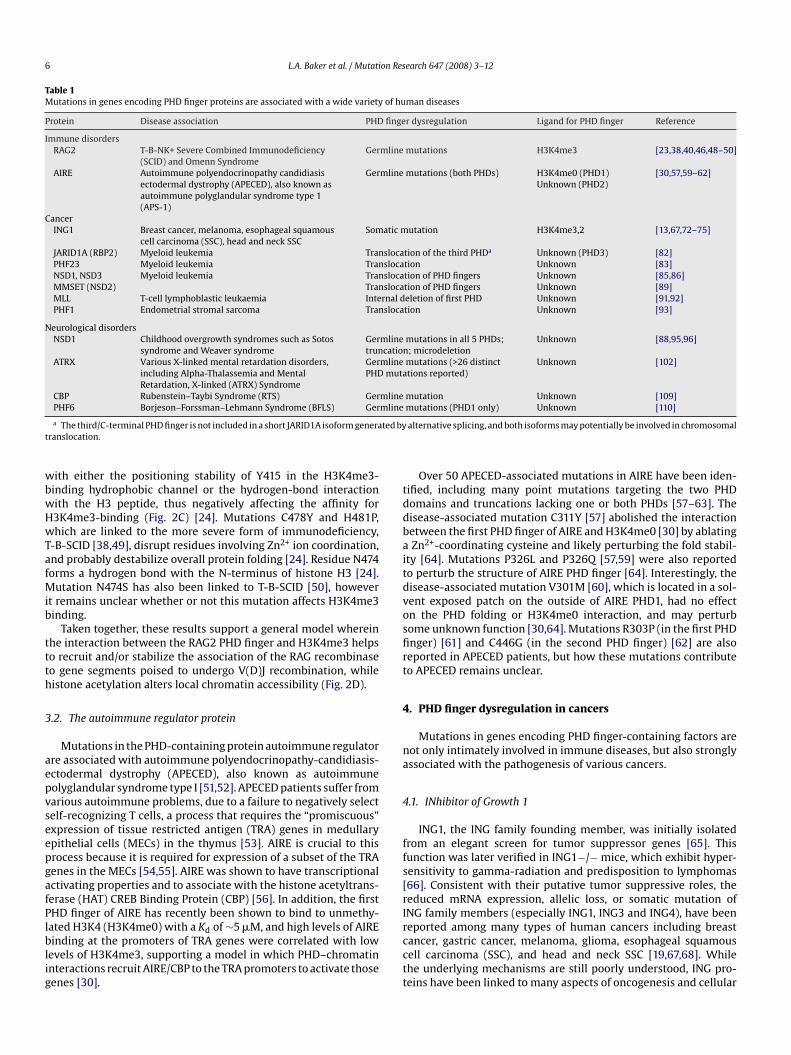
Table 1Mutations in genes encoding PHD finger proteins are associated with a wide variety of human diseases
Protein Disease association PHD finger dysregulation Ligand for PHD finger Reference
Immune disordersRAG2 T-B-NK+ Severe Combined Immunodeficiency
(SCID) and Omenn SyndromeGermline mutations H3K4me3 [23,38,40,46,48–50]
AIRE Autoimmune polyendocrinopathy candidiasisectodermal dystrophy (APECED), also known asautoimmune polyglandular syndrome type 1(APS-1)
Germline mutations (both PHDs) H3K4me0 (PHD1)Unknown (PHD2)
[30,57,59–62]
CancerING1 Breast cancer, melanoma, esophageal squamous
cell carcinoma (SSC), head and neck SSCSomatic mutation H3K4me3,2 [13,67,72–75]
JARID1A (RBP2) Myeloid leukemia Translocation of the third PHDa Unknown (PHD3) [82]PHF23 Myeloid leukemia Translocation Unknown [83]NSD1, NSD3 Myeloid leukemia Translocation of PHD fingers Unknown [85,86]MMSET (NSD2) Translocation of PHD fingers Unknown [89]MLL T-cell lymphoblastic leukaemia Internal deletion of first PHD Unknown [91,92]PHF1 Endometrial stromal sarcoma Translocation Unknown [93]
Neurological disordersNSD1 Childhood overgrowth syndromes such as Sotos
syndrome and Weaver syndromeGermline mutations in all 5 PHDs;truncation; microdeletion
Unknown [88,95,96]
ATRX Various X-linked mental retardation disorders,including Alpha-Thalassemia and MentalRetardation, X-linked (ATRX) Syndrome
Germline mutations (>26 distinctPHD mutations reported)
Unknown [102]
CBP Rubenstein–Taybi Syndrome (RTS) Germline mutation Unknown [109]mline
ted byt
wbwHwTafMib
ttth
3
aepvseepgafPlblig
tddbaitdvosfirt
4
na
4
ffs[rI
PHF6 Borjeson–Forssman–Lehmann Syndrome (BFLS) Ger
a The third/C-terminal PHD finger is not included in a short JARID1A isoform generaranslocation.
ith either the positioning stability of Y415 in the H3K4me3-inding hydrophobic channel or the hydrogen-bond interactionith the H3 peptide, thus negatively affecting the affinity for3K4me3-binding (Fig. 2C) [24]. Mutations C478Y and H481P,hich are linked to the more severe form of immunodeficiency,
-B-SCID [38,49], disrupt residues involving Zn2+ ion coordination,nd probably destabilize overall protein folding [24]. Residue N474orms a hydrogen bond with the N-terminus of histone H3 [24].
utation N474S has also been linked to T-B-SCID [50], howevert remains unclear whether or not this mutation affects H3K4me3inding.
Taken together, these results support a general model whereinhe interaction between the RAG2 PHD finger and H3K4me3 helpso recruit and/or stabilize the association of the RAG recombinaseo gene segments poised to undergo V(D)J recombination, whileistone acetylation alters local chromatin accessibility (Fig. 2D).
.2. The autoimmune regulator protein
Mutations in the PHD-containing protein autoimmune regulatorre associated with autoimmune polyendocrinopathy-candidiasis-ctodermal dystrophy (APECED), also known as autoimmuneolyglandular syndrome type I [51,52]. APECED patients suffer fromarious autoimmune problems, due to a failure to negatively selectelf-recognizing T cells, a process that requires the “promiscuous”xpression of tissue restricted antigen (TRA) genes in medullarypithelial cells (MECs) in the thymus [53]. AIRE is crucial to thisrocess because it is required for expression of a subset of the TRAenes in the MECs [54,55]. AIRE was shown to have transcriptionalctivating properties and to associate with the histone acetyltrans-erase (HAT) CREB Binding Protein (CBP) [56]. In addition, the firstHD finger of AIRE has recently been shown to bind to unmethy-
ated H3K4 (H3K4me0) with a Kd of ∼5 �M, and high levels of AIREinding at the promoters of TRA genes were correlated with lowevels of H3K4me3, supporting a model in which PHD–chromatinnteractions recruit AIRE/CBP to the TRA promoters to activate thoseenes [30].
rcctt
mutations (PHD1 only) Unknown [110]
alternative splicing, and both isoforms may potentially be involved in chromosomal
Over 50 APECED-associated mutations in AIRE have been iden-ified, including many point mutations targeting the two PHDomains and truncations lacking one or both PHDs [57–63]. Theisease-associated mutation C311Y [57] abolished the interactionetween the first PHD finger of AIRE and H3K4me0 [30] by ablatingZn2+-coordinating cysteine and likely perturbing the fold stabil-
ty [64]. Mutations P326L and P326Q [57,59] were also reportedo perturb the structure of AIRE PHD finger [64]. Interestingly, theisease-associated mutation V301M [60], which is located in a sol-ent exposed patch on the outside of AIRE PHD1, had no effectn the PHD folding or H3K4me0 interaction, and may perturbome unknown function [30,64]. Mutations R303P (in the first PHDnger) [61] and C446G (in the second PHD finger) [62] are alsoeported in APECED patients, but how these mutations contributeo APECED remains unclear.
. PHD finger dysregulation in cancers
Mutations in genes encoding PHD finger-containing factors areot only intimately involved in immune diseases, but also stronglyssociated with the pathogenesis of various cancers.
.1. INhibitor of Growth 1
ING1, the ING family founding member, was initially isolatedrom an elegant screen for tumor suppressor genes [65]. Thisunction was later verified in ING1−/− mice, which exhibit hyper-ensitivity to gamma-radiation and predisposition to lymphomas66]. Consistent with their putative tumor suppressive roles, theeduced mRNA expression, allelic loss, or somatic mutation ofNG family members (especially ING1, ING3 and ING4), have been
eported among many types of human cancers including breastancer, gastric cancer, melanoma, glioma, esophageal squamousell carcinoma (SSC), and head and neck SSC [19,67,68]. Whilehe underlying mechanisms are still poorly understood, ING pro-eins have been linked to many aspects of oncogenesis and cellular
L.A. Baker et al. / Mutation Research 647 (2008) 3–12 7
Fig. 2. Missense mutations in the RAG2 PHD finger are associated with immunodeficiency syndromes. (A) Domain structure of RAG2 (NCBI accession number 187423896)showing disease-causing mutations found within the PHD finger. The amino acid sequence of the PHD finger is shown below with H3K4me-caging residues in purple andZn2+ ion-coordinating residues in orange. (B) Co-crystal structure of the RAG2 PHD finger (in gray) and the H3K4me3 peptide (in green) [24]. Residues mutated in T-B-SCID orOmenn Syndrome are depicted in red, and Y415, which forms part of the aromatic channel, is in purple. (C) Closer view of mutations K440N and W416L as well as the RAG2PHD aromatic channel with H3K4me3. (D) Schematic model for RAG2’s function in V(D)J recombination. Interactions between the RAG2 PHD and H3K4me3 help to recruita with tr ion ofN funct
gaiwapi
fiiP[fi
mmttobtm
nd/or stabilize RAG1/2 recombinases to appropriate V(D)J gene segments markedepair factors such as DNA-PKcs, Ku70/80, XRCC4 and DNA Ligase IV assists in ligatote here the correlation between the disease-causing mutations and the “reading”
rowth control, such as cell cycle regulation, senescence, DNA dam-ge repair, apoptosis, and stress signaling [67,69]. In addition tonteracting with p53 and PCNA, INGs also recruit and associate
ith two antagonizing sets of enzymes, histone acetyltransferasesnd histone deacetylases (HDACs) [69,70]. The incorporation of INGroteins into HAT- or HDAC-complexes is conserved in lower organ-
sms such as yeast [27,71].Virtually all ING isoforms contain a conserved C-terminal PHD
nger and nearby nuclear localization signals, perhaps suggest-ng that their functionality relies on a nuclear PHD motif [69]. INGHD fingers have been shown to recognize H3K4me3/2 specifically13,27,69]. Mutations within the ING1 PHD finger were identi-ed among various cancers (Fig. 3A) [13,67,72–75]. Among them,
adlcr
his histone modification, where they create double-strand DNA breaks. Then DNAthe broken ends of two V(D)J gene segments, creating a functional gene segment.ion of the RAG2 PHD finger in engaging histone marks.
utation C253stop [72] results in a truncated PHD domain, andutation C215S disrupts Zn2+-coordination, presumably disrupting
he overall PHD structure and abolishing the H3K4me3 interac-ion needed for recruitment to target promoters (Fig. 3B). Thoughther PHD mutations are mapped to the outside of the H3K4me3-inding cage, it remains to be seen if any of them interferes withhe H3K4me3 binding or other unknown function. Interestingly,
issense mutations also cluster in the nuclear localization signals
nd an N-terminal sequence that overlaps the SAP30-interactingomain [76] (Fig. 3A), which may interfere with proper nuclearocalization or with SAP30 association. These observations areonsistent with a model that upon DNA damage, the Sin3/HDACepressive complexes are recruited to the promoters of cell cycle

8 L.A. Baker et al. / Mutation Research 647 (2008) 3–12
Fig. 3. Mutations in the ING1b PHD finger isolated from cancers. (A) Domain structure of ING1b (p33ING1b, NCBI accession number 38201667) showing missense mutationsreported in various solid cancers. The amino acid sequence of PHD finger is shown below, with the same color depiction as in Fig. 2. SAID, SAP30-interacting domain [76]; NLS,nuclear localization signal; PBR, poly-basic region that mediates the binding to phosphoinositide [69]. (B) A structural model of the ING1b PHD finger (in gray) associated witha red, ano -medH oliferac of pr
rt(
4
silps[Nt
rad[oPlafi[
n H3K4me3 peptide (in green). Cancer-associated missense mutations as shown inf the ING2 PHD finger and H3K4me3 peptide [13]. (C) A schematic model for ING3K4me3 marks enriched in promoters helps to stabilize the targeting of INGs to promplexes that remove acetyl marks from histone tails, repressing the transcription
egulators such as Cyclin via ING–SAP30 interaction, repressing theranscription of these genes and preventing cell cycle progressionFig. 3C) [10,77,78].
.2. PHD fingers fused to NUP98 in blood cancers
Translocation of the Nucleoporin 98 (NUP98) gene repre-ents one of the most promiscuous chromosomal abnormalitiesn human hematopoietic malignancies such as acute myeloideukemia (AML) [79]. NUP98, a nuclear pore complex (NPC) com-
onent, has been reported to shuttle between the NPC and apecialized nuclear body that associates with active transcription80]. All leukemic NUP98 fusion proteins retain the N-terminalUP98 FG-repeats, which recruit p300/CBP and harbor transcrip-ional activation activities [81].
m
cfl
d aromatic cage residues are in purple. This model is based on a co-crystal structureiated cell cycle arrest. Upon DNA damage, the PHD finger of INGs that recognizestive genes such as Cyclin wherein INGs subsequently recruit the SAP30/Sin3/HDAColiferative genes and decelerating cell cycle progression.
In rare AML cases, cryptic translocations fuse the NUP98 FG-epeats to the C-terminus of PHD finger-containing factors suchs the H3K4 demethylase JARID1A (Jumonji, AT-rich interactiveomain 1A) (Fig. 4A) or PHF23 (PHD finger-protein 23) (Fig. 4B)82–84]. The two fusion products share a high similarity, and thenly functional motifs incorporated from JARID1A or PHF23 are theHD finger and nuclear localization signals, indicating a commoneukemogenic mechanism. Little is known about the chromatin-ssociating properties of these PHD fingers, except that the PHDnger of PHF23 shares similarity to H3K4me-binding PHD fingers5]. Furthermore, the mechanistic contribution of these PHDs to
alignancies is poorly understood.In ∼5% of AML cases, nuclear receptor-binding SET domain-
ontaining 1 (NSD1) and the related gene NSD3 were foundused to NUP98 [85,86], and the fusion products retain aarge part of NSD that contains the five PHD fingers, the
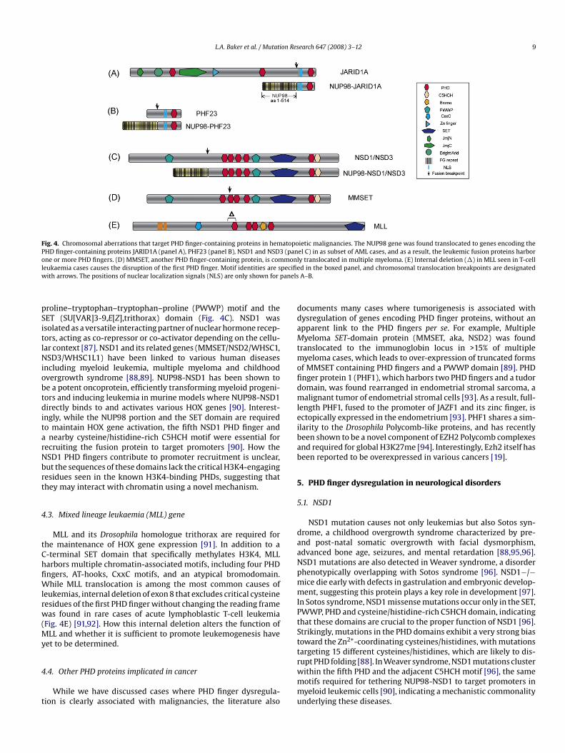
L.A. Baker et al. / Mutation Research 647 (2008) 3–12 9
Fig. 4. Chromosomal aberrations that target PHD finger-containing proteins in hematopoietic malignancies. The NUP98 gene was found translocated to genes encoding thePHD finger-containing proteins JARID1A (panel A), PHF23 (panel B), NSD1 and NSD3 (panel C) in as subset of AML cases, and as a result, the leukemic fusion proteins harboro monll pecifiew anels
pSitlNiobtditarNbrt
4
tChfiWlrw(My
4
t
ddaMtmofidmleibab
5
5
daaNpmmIPtSttargeting 15 different cysteines/histidines, which are likely to dis-
ne or more PHD fingers. (D) MMSET, another PHD finger-containing protein, is comeukaemia cases causes the disruption of the first PHD finger. Motif identities are s
ith arrows. The positions of nuclear localization signals (NLS) are only shown for p
roline–tryptophan–tryptophan–proline (PWWP) motif and theET (SU[VAR]3-9,E[Z],trithorax) domain (Fig. 4C). NSD1 wassolated as a versatile interacting partner of nuclear hormone recep-ors, acting as co-repressor or co-activator depending on the cellu-ar context [87]. NSD1 and its related genes (MMSET/NSD2/WHSC1,SD3/WHSC1L1) have been linked to various human diseases
ncluding myeloid leukemia, multiple myeloma and childhoodvergrowth syndrome [88,89]. NUP98-NSD1 has been shown toe a potent oncoprotein, efficiently transforming myeloid progeni-ors and inducing leukemia in murine models where NUP98-NSD1irectly binds to and activates various HOX genes [90]. Interest-
ngly, while the NUP98 portion and the SET domain are requiredo maintain HOX gene activation, the fifth NSD1 PHD finger and
nearby cysteine/histidine-rich C5HCH motif were essential forecruiting the fusion protein to target promoters [90]. How theSD1 PHD fingers contribute to promoter recruitment is unclear,ut the sequences of these domains lack the critical H3K4-engagingesidues seen in the known H3K4-binding PHDs, suggesting thathey may interact with chromatin using a novel mechanism.
.3. Mixed lineage leukaemia (MLL) gene
MLL and its Drosophila homologue trithorax are required forhe maintenance of HOX gene expression [91]. In addition to a-terminal SET domain that specifically methylates H3K4, MLLarbors multiple chromatin-associated motifs, including four PHDngers, AT-hooks, CxxC motifs, and an atypical bromodomain.hile MLL translocation is among the most common causes of
eukemias, internal deletion of exon 8 that excludes critical cysteineesidues of the first PHD finger without changing the reading frameas found in rare cases of acute lymphoblastic T-cell leukemia
Fig. 4E) [91,92]. How this internal deletion alters the function ofLL and whether it is sufficient to promote leukemogenesis have
et to be determined.
.4. Other PHD proteins implicated in cancer
While we have discussed cases where PHD finger dysregula-ion is clearly associated with malignancies, the literature also
rwmmu
y translocated in multiple myeloma. (E) Internal deletion (�) in MLL seen in T-celld in the boxed panel, and chromosomal translocation breakpoints are designatedA–B.
ocuments many cases where tumorigenesis is associated withysregulation of genes encoding PHD finger proteins, without anpparent link to the PHD fingers per se. For example, Multipleyeloma SET-domain protein (MMSET, aka, NSD2) was found
ranslocated to the immunoglobin locus in >15% of multipleyeloma cases, which leads to over-expression of truncated forms
f MMSET containing PHD fingers and a PWWP domain [89]. PHDnger protein 1 (PHF1), which harbors two PHD fingers and a tudoromain, was found rearranged in endometrial stromal sarcoma, aalignant tumor of endometrial stromal cells [93]. As a result, full-
ength PHF1, fused to the promoter of JAZF1 and its zinc finger, isctopically expressed in the endometrium [93]. PHF1 shares a sim-larity to the Drosophila Polycomb-like proteins, and has recentlyeen shown to be a novel component of EZH2 Polycomb complexesnd required for global H3K27me [94]. Interestingly, Ezh2 itself haseen reported to be overexpressed in various cancers [19].
. PHD finger dysregulation in neurological disorders
.1. NSD1
NSD1 mutation causes not only leukemias but also Sotos syn-rome, a childhood overgrowth syndrome characterized by pre-nd post-natal somatic overgrowth with facial dysmorphism,dvanced bone age, seizures, and mental retardation [88,95,96].SD1 mutations are also detected in Weaver syndrome, a disorderhenotypically overlapping with Sotos syndrome [96]. NSD1−/−ice die early with defects in gastrulation and embryonic develop-ent, suggesting this protein plays a key role in development [97].
n Sotos syndrome, NSD1 missense mutations occur only in the SET,WWP, PHD and cysteine/histidine-rich C5HCH domain, indicatinghat these domains are crucial to the proper function of NSD1 [96].trikingly, mutations in the PHD domains exhibit a very strong biasoward the Zn2+-coordinating cysteines/histidines, with mutations
upt PHD folding [88]. In Weaver syndrome, NSD1 mutations clusterithin the fifth PHD and the adjacent C5HCH motif [96], the sameotifs required for tethering NUP98-NSD1 to target promoters inyeloid leukemic cells [90], indicating a mechanistic commonality
nderlying these diseases.
1 on Res
5X
dmrdmwMrt
ddmimbgdoaofa
5(
daamtBdhmbrn
6
aetPcbwPcpttipro
acaPcdsca
C
A
ftmfwtCAs
R
0 L.A. Baker et al. / Mutati
.2. Alpha Thalassaemia and Mental Retardation Syndrome,-linked (ATRX)
Mutations in ATRX are associated with the ATR-X syndrome, aisorder characterized by severe mental retardation, genital abnor-alities, alpha thalassaemia, microcephaly, seizures, and growth
etardation [98–100]. ATRX is crucial to neuronal survival in theeveloping mouse brain, and this function may explain why ATRXutations associate with mental retardation [101]. ATRX interactsith the heterochromatin-associated proteins HP1alpha, EZH2 andeCP2, and has been implicated in chromatin remodeling and gene
egulation [102–107]. However, its exact function and contributiono ATR-X syndrome remain unclear.
ATRX features an N-terminal ADD (ATRX-DNMT3-DNMT3L)omain, containing a GATA-like zinc finger, an atypical “PHD-like”omain and an alpha helix [102,103]. The ATRX PHD lacks the aro-atic caging residues found in the PHD fingers of BPTF or RAG2,
ndicating that it might interact with chromatin by a differentechanism [102]. Over 40 disease-causing ATRX mutations have
een identified within the ADD domain, including 26 in the PHD fin-er itself [102]. Some mutations may disrupt the folding of the PHDomain and probably destabilize the entire ATRX protein, whilether mutations that lie on the surface of the fold probably do notffect PHD structure, and may cause disease by interfering withther unknown interactions or functions [102]. Future researchocusing on the function of the ATRX PHD finger will provide valu-ble insights of the molecular pathogenesis of ATR-X syndrome.
.3. CREB Binding Protein (CBP/CREBBP) and PHD finger protein 6PHF6)
Haploinsufficiency of CBP leads to Rubenstein–Taybi Syn-rome, a disorder characterized by mental retardation, facialbnormalities, and growth retardation [108]. Many disease-ssociated mutations have been reported in CBP, including pointutations or internal deletions in the PHD domain [109]. Muta-
ions in the PHD finger protein 6 (PHF6) are associated withorjeson–Forssman–Lehmann Syndrome, a recessive X-linkedisorder characterized by mental retardation, hypogonadism,ypometabolism and obesity [110,111], and one disease-associatedutation (C99F) targets the first PHD finger of PHF6 [110]. While
oth CBP and PHF6 are implicated in chromatin regulation and neu-al development, the exact mechanisms underlying their respectiveeurological disorders are still poorly understood [111,112].
. Conclusions
In summary, PHD finger mutations have been associated withwide variety of human diseases. It should be noted that, while
vidence supporting a causal role of PHD finger mutation inhe pathogenesis of some diseases is convincing (such as RAG2-HD finger mutation in immunodeficiency syndrome [23]), directause–effect relationships for some other diseases still remain toe established and beg for further investigation. In this review,e have chosen to focus on diseases linked to aberrations of the
HD domains themselves. However, there are many other PHD-ontaining proteins for which mutations in other regions of therotein or loss of the entire protein associate with diseases, andhe PHD domains of many of these proteins are likely to con-
ribute to appropriate chromatin targeting. For instance, mutationsn SMCX are associated with X-linked mental retardation, and therotein contains several PHD fingers thought to be important foregulating neural development genes [31]. In addition, PHD fingersften occur in conjunction with other chromatin-reading motifs,earch 647 (2008) 3–12
nd such combined action is proposed to be required for pre-isely interpreting complicated chromatin modification patterns,phenomenon known as “multivalency” [7]. The contribution of
HD fingers and other chromatin-“reading” motifs to the functionalonsequences of the epigenetic marks and ultimately, to humaniseases is a topic that has only recently received scrutiny. Under-tanding the regulatory roles of chromatin modifications in theontext of human disease will greatly broaden our mechanisticppreciation of normal and pathological development.
onflict of interest
We declare no conflict of interest.
cknowledgements
We thank Drs. Alexander Ruthenburg, Ping Chi and Chengran Xuor critical reading and advice on this manuscript. Special thankso Dr. Alex Ruthenburg for analyzing disease-associated missense
utations in RAG2 and ING structure, and thanks to Dr. Haotai Lior helping with illustration. We apologize to all those whose worke could not cite due to space constraints. C.D.A. is supported by
he NIH Merit Grant GM 53512, and L.A.B. by NIH training grantA09673. G.W. is supported by a C.H. Li Memorial Scholar Fundward and a Leukemia & Lymphoma Society Postdoctoral Fellow-hip.
eferences
[1] R.D. Kornberg, Y. Lorch, Twenty-five years of the nucleosome, fundamentalparticle of the eukaryote chromosome, Cell 98 (1999) 285–294.
[2] B.E. Bernstein, A. Meissner, E.S. Lander, The mammalian epigenome, Cell 128(2007) 669–681.
[3] T. Kouzarides, Chromatin modifications and their function, Cell 128 (2007)693–705.
[4] M.D. Shahbazian, M. Grunstein, Functions of site-specific histone acetylationand deacetylation, Annu. Rev. Biochem. 76 (2007) 75–100.
[5] A.J. Ruthenburg, C.D. Allis, J. Wysocka, Methylation of lysine 4 on histone H3:intricacy of writing and reading a single epigenetic mark, Mol. Cell 25 (2007)15–30.
[6] T. Kouzarides, SnapShot: histone-modifying enzymes, Cell 131 (2007) 822.[7] A.J. Ruthenburg, H. Li, D.J. Patel, C.D. Allis, Multivalent engagement of chro-
matin modifications by linked binding modules, Nat. Rev. Mol. Cell Biol. 8(2007) 983–994.
[8] S.D. Taverna, H. Li, A.J. Ruthenburg, C.D. Allis, D.J. Patel, How chromatin-binding modules interpret histone modifications: lessons from professionalpocket pickers, Nat. Struct. Mol. Biol. 14 (2007) 1025–1040.
[9] J. Wysocka, T. Swigut, H. Xiao, T.A. Milne, S.Y. Kwon, J. Landry, M. Kauer, A.J.Tackett, B.T. Chait, P. Badenhorst, C. Wu, C.D. Allis, A PHD finger of NURF coupleshistone H3 lysine 4 trimethylation with chromatin remodelling, Nature 442(2006) 86–90.
[10] X. Shi, T. Hong, K.L. Walter, M. Ewalt, E. Michishita, T. Hung, D. Carney, P. Pena,F. Lan, M.R. Kaadige, N. Lacoste, C. Cayrou, F. Davrazou, A. Saha, B.R. Cairns, D.E.Ayer, T.G. Kutateladze, Y. Shi, J. Cote, K.F. Chua, O. Gozani, ING2 PHD domainlinks histone H3 lysine 4 methylation to active gene repression, Nature 442(2006) 96–99.
[11] H. Li, S. Ilin, W. Wang, E.M. Duncan, J. Wysocka, C.D. Allis, D.J. Patel, Molecularbasis for site-specific read-out of histone H3K4me3 by the BPTF PHD finger ofNURF, Nature 442 (2006) 91–95.
[12] H. Li, W. Fischle, W. Wang, E.M. Duncan, L. Liang, S. Murakami-Ishibe, C.D. Allis,D.J. Patel, Structural basis for lower lysine methylation state-specific readoutby MBT repeats of L3MBTL1 and an engineered PHD finger, Mol. Cell 28 (2007)677–691.
[13] P.V. Pena, F. Davrazou, X. Shi, K.L. Walter, V.V. Verkhusha, O. Gozani, R. Zhao,T.G. Kutateladze, Molecular mechanism of histone H3K4me3 recognition byplant homeodomain of ING2, Nature 442 (2006) 100–103.
[14] S.K. Ooi, C. Qiu, E. Bernstein, K. Li, D. Jia, Z. Yang, H. Erdjument-Bromage, P.Tempst, S.P. Lin, C.D. Allis, X. Cheng, T.H. Bestor, DNMT3L connects unmethy-lated lysine 4 of histone H3 to de novo methylation of DNA, Nature 448 (2007)714–717.
[15] F. Lan, R.E. Collins, R. De Cegli, R. Alpatov, J.R. Horton, X. Shi, O. Gozani, X.Cheng, Y. Shi, Recognition of unmethylated histone H3 lysine 4 links BHC80to LSD1-mediated gene repression, Nature 448 (2007) 718–722.
[16] K. Zhang, K. Mosch, W. Fischle, S.I. Grewal, Roles of the Clr4 methyltransferasecomplex in nucleation, spreading and maintenance of heterochromatin, Nat.Struct. Mol. Biol. 15 (2008) 381–388.

on Res
L.A. Baker et al. / Mutati[17] B.D. Strahl, C.D. Allis, The language of covalent histone modifications, Nature403 (2000) 41–45.
[18] P.A. Jones, S.B. Baylin, The epigenomics of cancer, Cell 128 (2007) 683–692.[19] G.G. Wang, C.D. Allis, P. Chi, Chromatin remodeling and cancer. Part I: Covalent
histone modifications, Trends Mol. Med. 13 (2007) 363–372.[20] G.G. Wang, C.D. Allis, P. Chi, Chromatin remodeling and cancer. Part II: ATP-
dependent chromatin remodeling, Trends Mol. Med. 13 (2007) 373–380.[21] T. Bienvenu, J. Chelly, Molecular genetics of Rett syndrome: when DNA methy-
lation goes unrecognized, Nat. Rev. Genet. 7 (2006) 415–426.[22] M. Bienz, The PHD finger, a nuclear protein-interaction domain, Trends
Biochem. Sci. 31 (2006) 35–40.[23] A.G. Matthews, A.J. Kuo, S. Ramon-Maiques, S. Han, K.S. Champagne, D. Ivanov,
M. Gallardo, D. Carney, P. Cheung, D.N. Ciccone, K.L. Walter, P.J. Utz, Y. Shi, T.G.Kutateladze, W. Yang, O. Gozani, M.A. Oettinger, RAG2 PHD finger couples his-tone H3 lysine 4 trimethylation with V(D)J recombination, Nature 450 (2007)1106–1110.
[24] S. Ramon-Maiques, A.J. Kuo, D. Carney, A.G. Matthews, M.A. Oettinger, O.Gozani, W. Yang, The plant homeodomain finger of RAG2 recognizes histoneH3 methylated at both lysine-4 and arginine-2, Proc. Natl. Acad. Sci. U.S.A. 104(2007) 18993–18998.
[25] U. Schindler, H. Beckmann, A.R. Cashmore, HAT3.1, a novel Arabidopsis home-odomain protein containing a conserved cysteine-rich region, Plant J. 4 (1993)137–150.
[26] A. Palacios, I.G. Munoz, D. Pantoja-Uceda, M.J. Marcaida, D. Torres, J.M. Martin-Garcia, I. Luque, G. Montoya, F.J. Blanco, Molecular basis of histone H3K4ME3recognition by ING4, J. Biol. Chem. (2008).
[27] S.D. Taverna, S. Ilin, R.S. Rogers, J.C. Tanny, H. Lavender, H. Li, L. Baker, J. Boyle,L.P. Blair, B.T. Chait, D.J. Patel, J.D. Aitchison, A.J. Tackett, C.D. Allis, Yng1 PHDfinger binding to H3 trimethylated at K4 promotes NuA3 HAT activity at K14 ofH3 and transcription at a subset of targeted ORFs, Mol. Cell 24 (2006) 785–796.
[28] A.N. Iberg, A. Espejo, D. Cheng, D. Kim, J. Michaud-Levesque, S. Richard, M.T.Bedford, Arginine methylation of the histone H3 tail impedes effector binding,J. Biol. Chem. 283 (2008) 3006–3010.
[29] A. Kirmizis, H. Santos-Rosa, C.J. Penkett, M.A. Singer, M. Vermeulen, M. Mann,J. Bahler, R.D. Green, T. Kouzarides, Arginine methylation at histone H3R2controls deposition of H3K4 trimethylation, Nature (2007).
[30] T. Org, F. Chignola, C. Hetenyi, M. Gaetani, A. Rebane, I. Liiv, U. Maran, L. Mollica,M.J. Bottomley, G. Musco, P. Peterson, The autoimmune regulator PHD fingerbinds to non-methylated histone H3K4 to activate gene expression, EMBO Rep.9 (2008) 370–376.
[31] S. Iwase, F. Lan, P. Bayliss, L. de la Torre-Ubieta, M. Huarte, H.H. Qi, J.R. Whet-stine, A. Bonni, T.M. Roberts, Y. Shi, The X-linked mental retardation geneSMCX/JARID1C defines a family of histone H3 lysine 4 demethylases, Cell 128(2007) 1077–1088.
[32] P. Karagianni, L. Amazit, J. Qin, J. Wong, ICBP90, a novel methyl K9 H3 bindingprotein linking protein ubiquitination with heterochromatin formation, Mol.Cell Biol. 28 (2008) 705–717.
[33] X. Shi, I. Kachirskaia, K.L. Walter, J.H. Kuo, A. Lake, F. Davrazou, S.M. Chan, D.G.Martin, I.M. Fingerman, S.D. Briggs, L. Howe, P.J. Utz, T.G. Kutateladze, A.A.Lugovskoy, M.T. Bedford, O. Gozani, Proteome-wide analysis in Saccharomycescerevisiae identifies several PHD fingers as novel direct and selective bindingmodules of histone H3 methylated at either lysine 4 or lysine 36, J. Biol. Chem.282 (2007) 2450–2455.
[34] D. Jung, F.W. Alt, Unraveling V(D)J recombination; insights into gene regula-tion, Cell 116 (2004) 299–311.
[35] M.A. Oettinger, How to keep V(D)J recombination under control, Immunol.Rev. 200 (2004) 165–181.
[36] S.D. Fugmann, A.I. Lee, P.E. Shockett, I.J. Villey, D.G. Schatz, The RAG pro-teins and V(D)J recombination: complexes, ends, and transposition, Annu. Rev.Immunol. 18 (2000) 495–527.
[37] Y. Shinkai, G. Rathbun, K.P. Lam, E.M. Oltz, V. Stewart, M. Mendelsohn, J. Char-ron, M. Datta, F. Young, A.M. Stall, et al., RAG-2-deficient mice lack maturelymphocytes owing to inability to initiate V(D)J rearrangement, Cell 68 (1992)855–867.
[38] K. Schwarz, G.H. Gauss, L. Ludwig, U. Pannicke, Z. Li, D. Lindner, W. Friedrich,R.A. Seger, T.E. Hansen-Hagge, S. Desiderio, M.R. Lieber, C.R. Bartram, RAGmutations in human B cell-negative SCID, Science 274 (1996) 97–99.
[39] V. Marrella, P.L. Poliani, C. Sobacchi, F. Grassi, A. Villa, Of Omenn and mice,Trends Immunol. 29 (2008) 133–140.
[40] C. Sobacchi, V. Marrella, F. Rucci, P. Vezzoni, A. Villa, RAG-dependent primaryimmunodeficiencies, Hum. Mutat. 27 (2006) 1174–1184.
[41] G.S. Omenn, Familial reticuloendotheliosis with eosinophilia, N. Engl. J. Med.273 (1965) 427–432.
[42] M. Goldmit, Y. Ji, J. Skok, E. Roldan, S. Jung, H. Cedar, Y. Bergman, Epigeneticontogeny of the Igk locus during B cell development, Nat. Immunol. 6 (2005)198–203.
[43] K.B. Morshead, D.N. Ciccone, S.D. Taverna, C.D. Allis, M.A. Oettinger, Antigenreceptor loci poised for V(D)J rearrangement are broadly associated with BRG1and flanked by peaks of histone H3 dimethylated at lysine 4, Proc. Natl. Acad.
Sci. U.S.A. 100 (2003) 11577–11582.[44] B.P. Sleckman, J.R. Gorman, F.W. Alt, Accessibility control of antigen-receptorvariable-region gene assembly: role of cis-acting elements, Annu. Rev.Immunol. 14 (1996) 459–481.
[45] M.S. Krangel, Gene segment selection in V(D)J recombination: accessibilityand beyond, Nat. Immunol. 4 (2003) 624–630.
earch 647 (2008) 3–12 11
[46] Y. Liu, R. Subrahmanyam, T. Chakraborty, R. Sen, S. Desiderio, A plant home-odomain in RAG-2 that binds Hypermethylated lysine 4 of histone H3 isnecessary for efficient antigen-receptor-gene rearrangement, Immunity 27(2007) 561–571.
[47] S.K. Elkin, D. Ivanov, M. Ewalt, C.G. Ferguson, S.G. Hyberts, Z.Y. Sun, G.D. Prest-wich, J. Yuan, G. Wagner, M.A. Oettinger, O.P. Gozani, A PHD finger motif inthe C terminus of RAG2 modulates recombination activity, J. Biol. Chem. 280(2005) 28701–28710.
[48] C.A. Gomez, L.M. Ptaszek, A. Villa, F. Bozzi, C. Sobacchi, E.G. Brooks, L.D.Notarangelo, E. Spanopoulou, Z.Q. Pan, P. Vezzoni, P. Cortes, S. Santagata, Muta-tions in conserved regions of the predicted RAG2 kelch repeats block initiationof V(D)J recombination and result in primary immunodeficiencies, Mol. CellBiol. 20 (2000) 5653–5664.
[49] J.G. Noordzij, S. de Bruin-Versteeg, N.S. Verkaik, J.M. Vossen, R. de Groot, E.Bernatowska, A.W. Langerak, D.C. van Gent, J.J. van Dongen, The immunophe-notypic and immunogenotypic B-cell differentiation arrest in bone marrow ofRAG-deficient SCID patients corresponds to residual recombination activitiesof mutated RAG proteins, Blood 100 (2002) 2145–2152.
[50] A. Villa, C. Sobacchi, L.D. Notarangelo, F. Bozzi, M. Abinun, T.G. Abrahamsen,P.D. Arkwright, M. Baniyash, E.G. Brooks, M.E. Conley, P. Cortes, M. Duse, A.Fasth, A.M. Filipovich, A.J. Infante, A. Jones, E. Mazzolari, S.M. Muller, S. Pasic, G.Rechavi, M.G. Sacco, S. Santagata, M.L. Schroeder, R. Seger, D. Strina, A. Ugazio, J.Valiaho, M. Vihinen, L.B. Vogler, H. Ochs, P. Vezzoni, W. Friedrich, K. Schwarz,V(D)J recombination defects in lymphocytes due to RAG mutations: severeimmunodeficiency with a spectrum of clinical presentations, Blood 97 (2001)81–88.
[51] Finnish-German APECED Consortium, An autoimmune disease, APECED,caused by mutations in a novel gene featuring two PHD-type zinc-fingerdomains, Nat. Genet. 17 (1997) 399–403.
[52] K. Nagamine, P. Peterson, H.S. Scott, J. Kudoh, S. Minoshima, M. Heino, K.J.Krohn, M.D. Lalioti, P.E. Mullis, S.E. Antonarakis, K. Kawasaki, S. Asakawa, F.Ito, N. Shimizu, Positional cloning of the APECED gene, Nat. Genet. 17 (1997)393–398.
[53] M.H. Cheng, A.K. Shum, M.S. Anderson, What’s new in the Aire? TrendsImmunol. 28 (2007) 321–327.
[54] M.S. Anderson, E.S. Venanzi, L. Klein, Z. Chen, S.P. Berzins, S.J. Turley, H.von Boehmer, R. Bronson, A. Dierich, C. Benoist, D. Mathis, Projection of animmunological self shadow within the thymus by the aire protein, Science298 (2002) 1395–1401.
[55] J. Derbinski, J. Gabler, B. Brors, S. Tierling, S. Jonnakuty, M. Hergenhahn, L. Pel-tonen, J. Walter, B. Kyewski, Promiscuous gene expression in thymic epithelialcells is regulated at multiple levels, J. Exp. Med. 202 (2005) 33–45.
[56] J. Pitkanen, V. Doucas, T. Sternsdorf, T. Nakajima, S. Aratani, K. Jensen,H. Will, P. Vahamurto, J. Ollila, M. Vihinen, H.S. Scott, S.E. Antonarakis,J. Kudoh, N. Shimizu, K. Krohn, P. Peterson, The autoimmune regulatorprotein has transcriptional transactivating properties and interacts withthe common coactivator CREB-binding protein, J. Biol. Chem. 275 (2000)16802–16809.
[57] P. Bjorses, M. Halonen, J.J. Palvimo, M. Kolmer, J. Aaltonen, P. Ellonen, J.Perheentupa, I. Ulmanen, L. Peltonen, Mutations in the AIRE gene: effectson subcellular location and transactivation function of the autoimmunepolyendocrinopathy-candidiasis-ectodermal dystrophy protein, Am. J. Hum.Genet. 66 (2000) 378–392.
[58] Q.G. Ruan, J.X. She, Autoimmune polyglandular syndrome type 1 and theautoimmune regulator, Clin. Lab. Med. 24 (2004) 305–317.
[59] P. Saugier-Veber, N. Drouot, L.M. Wolf, J.M. Kuhn, T. Frebourg, H. Lefebvre, Iden-tification of a novel mutation in the autoimmune regulator (AIRE-1) gene in aFrench family with autoimmune polyendocrinopathy-candidiasis-ectodermaldystrophy, Eur. J. Endocrinol. 144 (2001) 347–351.
[60] A. Soderbergh, F. Rorsman, M. Halonen, O. Ekwall, P. Bjorses, O. Kampe, E.S.Husebye, Autoantibodies against aromatic l-amino acid decarboxylase iden-tifies a subgroup of patients with Addison’s disease, J. Clin. Endocrinol. Metab.85 (2000) 460–463.
[61] B. Stolarski, E. Pronicka, L. Korniszewski, A. Pollak, G. Kostrzewa, E. Rowin-ska, P. Wlodarski, A. Skorka, M. Gremida, P. Krajewski, R. Ploski, Molecularbackground of polyendocrinopathy-candidiasis-ectodermal dystrophy syn-drome in a Polish population: novel AIRE mutations and an estimate of diseaseprevalence, Clin. Genet. 70 (2006) 348–354.
[62] A.S. Wolff, M.M. Erichsen, A. Meager, N.F. Magitta, A.G. Myhre, J. Bollerslev, K.J.Fougner, K. Lima, P.M. Knappskog, E.S. Husebye, Autoimmune polyendocrinesyndrome type 1 in Norway: phenotypic variation, autoantibodies, and novelmutations in the autoimmune regulator gene, J. Clin. Endocrinol. Metab. 92(2007) 595–603.
[63] P. Peterson, J. Pitkanen, N. Sillanpaa, K. Krohn, Autoimmune polyendocrinopa-thy candidiasis ectodermal dystrophy (APECED): a model disease to studymolecular aspects of endocrine autoimmunity, Clin. Exp. Immunol. 135 (2004)348–357.
[64] M.J. Bottomley, G. Stier, D. Pennacchini, G. Legube, B. Simon, A. Akhtar, M.Sattler, G. Musco, NMR structure of the first PHD finger of autoimmune
regulator protein (AIRE1). Insights into autoimmune polyendocrinopathy-candidiasis-ectodermal dystrophy (APECED) disease, J. Biol. Chem. 280 (2005)11505–11512.[65] I. Garkavtsev, A. Kazarov, A. Gudkov, K. Riabowol, Suppression of the novelgrowth inhibitor p33ING1 promotes neoplastic transformation, Nat. Genet.14 (1996) 415–420.

1 on Res
[
[
[
[
[
[
[
[
[
[
[
2 L.A. Baker et al. / Mutati
[66] J.V. Kichina, M. Zeremski, L. Aris, K.V. Gurova, E. Walker, R. Franks, A.Y. Nikitin,H. Kiyokawa, A.V. Gudkov, Targeted disruption of the mouse ing1 locus resultsin reduced body size, hypersensitivity to radiation and elevated incidence oflymphomas, Oncogene 25 (2006) 857–866.
[67] E.I. Campos, M.Y. Chin, W.H. Kuo, G. Li, Biological functions of the ING familytumor suppressors, Cell Mol. Life Sci. 61 (2004) 2597–2613.
[68] X. Shi, O. Gozani, The fellowships of the INGs, J. Cell Biochem. 96 (2005)1127–1136.
[69] M.A. Soliman, K. Riabowol, After a decade of study—ING, a PHD for a versatilefamily of proteins, Trends Biochem. Sci. 32 (2007) 509–519.
[70] Y. Doyon, C. Cayrou, M. Ullah, A.J. Landry, V. Cote, W. Selleck, W.S. Lane, S.Tan, X.J. Yang, J. Cote, ING tumor suppressor proteins are critical regulatorsof chromatin acetylation required for genome expression and perpetuation,Mol. Cell 21 (2006) 51–64.
[71] Y. Doyon, W. Selleck, W.S. Lane, S. Tan, J. Cote, Structural and functional conser-vation of the NuA4 histone acetyltransferase complex from yeast to humans,Mol. Cell Biol. 24 (2004) 1884–1896.
[72] E.I. Campos, M. Martinka, D.L. Mitchell, D.L. Dai, G. Li, Mutations of the ING1tumor suppressor gene detected in human melanoma abrogate nucleotideexcision repair, Int. J. Oncol. 25 (2004) 73–80.
[73] L. Chen, N. Matsubara, T. Yoshino, T. Nagasaka, N. Hoshizima, Y. Shirakawa, Y.Naomoto, H. Isozaki, K. Riabowol, N. Tanaka, Genetic alterations of candidatetumor suppressor ING1 in human esophageal squamous cell cancer, CancerRes. 61 (2001) 4345–4349.
[74] B. Chen, E.I. Campos, R. Crawford, M. Martinka, G. Li, Analyses of the tumoursuppressor ING1 expression and gene mutation in human basal cell carcinoma,Int. J. Oncol. 22 (2003) 927–931.
[75] W. Gong, K. Suzuki, M. Russell, K. Riabowol, Function of the ING family of PHDproteins in cancer, Int. J. Biochem. Cell Biol. 37 (2005) 1054–1065.
[76] A. Kuzmichev, Y. Zhang, H. Erdjument-Bromage, P. Tempst, D. Reinberg, Role ofthe Sin3-histone deacetylase complex in growth regulation by the candidatetumor suppressor p33(ING1), Mol. Cell Biol. 22 (2002) 835–848.
[77] M. Garate, E.I. Campos, J.A. Bush, H. Xiao, G. Li, Phosphorylation of the tumorsuppressor p33(ING1b) at Ser-126 influences its protein stability and prolif-eration of melanoma cells, FASEB J. 21 (2007) 3705–3716.
[78] M. Takahashi, N. Seki, T. Ozaki, M. Kato, T. Kuno, T. Nakagawa, K. Watan-abe, K. Miyazaki, M. Ohira, S. Hayashi, M. Hosoda, H. Tokita, H. Mizuguchi, T.Hayakawa, S. Todo, A. Nakagawara, Identification of the p33(ING1)-regulatedgenes that include cyclin B1 and proto-oncogene DEK by using cDNA microar-ray in a mouse mammary epithelial cell line NMuMG, Cancer Res. 62 (2002)2203–2209.
[79] M.A. Moore, K.Y. Chung, M. Plasilova, J.J. Schuringa, J.H. Shieh, P. Zhou, G. Mor-rone, NUP98 dysregulation in myeloid leukemogenesis, Ann. N.Y. Acad. Sci.1106 (2007) 114–142.
[80] E.R. Griffis, N. Altan, J. Lippincott-Schwartz, M.A. Powers, Nup98 is a mobilenucleoporin with transcription-dependent dynamics, Mol. Biol. Cell 13 (2002)1282–1297.
[81] L.H. Kasper, P.K. Brindle, C.A. Schnabel, C.E. Pritchard, M.L. Cleary, J.M. vanDeursen, CREB binding protein interacts with nucleoporin-specific FG repeatsthat activate transcription and mediate NUP98-HOXA9 oncogenicity, Mol. CellBiol. 19 (1999) 764–776.
[82] L.J. van Zutven, E. Onen, S.C. Velthuizen, E. van Drunen, A.R. von Bergh,M.M. van den Heuvel-Eibrink, A. Veronese, C. Mecucci, M. Negrini, G.E. deGreef, H.B. Beverloo, Identification of NUP98 abnormalities in acute leukemia:JARID1A (12p13) as a new partner gene, Genes Chromosomes Cancer 45(2006) 437–446.
[83] J.C. Reader, J.S. Meekins, I. Gojo, Y. Ning, A novel NUP98-PHF23 fusion result-ing from a cryptic translocation t(11;17)(p15;p13) in acute myeloid leukemia,Leukemia 21 (2007) 842–844.
[84] R.J. Klose, Q. Yan, Z. Tothova, K. Yamane, H. Erdjument-Bromage, P. Tempst,D.G. Gilliland, Y. Zhang, W.G. Kaelin Jr., The retinoblastoma binding proteinRBP2 is an H3K4 demethylase, Cell 128 (2007) 889–900.
[85] N. Cerveira, C. Correia, S. Doria, S. Bizarro, P. Rocha, P. Gomes, L. Torres,L. Norton, B.S. Borges, S. Castedo, M.R. Teixeira, Frequency of NUP98-NSD1fusion transcript in childhood acute myeloid leukaemia, Leukemia 17 (2003)2244–2247.
[86] R. Rosati, R. La Starza, A. Veronese, A. Aventin, C. Schwienbacher, T. Vallespi,M. Negrini, M.F. Martelli, C. Mecucci, NUP98 is fused to the NSD3 gene inacute myeloid leukemia associated with t(8;11)(p11.2;p15), Blood 99 (2002)3857–3860.
[87] N. Huang, E. vom Baur, J.M. Garnier, T. Lerouge, J.L. Vonesch, Y. Lutz, P. Cham-bon, R. Losson, Two distinct nuclear receptor interaction domains in NSD1,a novel SET protein that exhibits characteristics of both corepressors andcoactivators, EMBO J. 17 (1998) 3398–3412.
[88] K. Tatton-Brown, J. Douglas, K. Coleman, G. Baujat, T.R. Cole, S. Das, D. Horn, H.E.Hughes, I.K. Temple, F. Faravelli, D. Waggoner, S. Turkmen, V. Cormier-Daire,
A. Irrthum, N. Rahman, Genotype-phenotype associations in Sotos syndrome:an analysis of 266 individuals with NSD1 aberrations, Am. J. Hum. Genet. 77(2005) 193–204.[89] J.J. Keats, C.A. Maxwell, B.J. Taylor, M.J. Hendzel, M. Chesi, P.L. Bergsagel,L.M. Larratt, M.J. Mant, T. Reiman, A.R. Belch, L.M. Pilarski, Overexpres-sion of transcripts originating from the MMSET locus characterizes all
[
earch 647 (2008) 3–12
t(4;14)(p16;q32)-positive multiple myeloma patients, Blood 105 (2005)4060–4069.
[90] G.G. Wang, L. Cai, M.P. Pasillas, M.P. Kamps, NUP98-NSD1 links H3K36 methy-lation to Hox-A gene activation and leukaemogenesis, Nat. Cell Biol. 9 (2007)804–812.
[91] P.M. Ayton, M.L. Cleary, Molecular mechanisms of leukemogenesis mediatedby MLL fusion proteins, Oncogene 20 (2001) 5695–5707.
[92] K. Lochner, G. Siegler, M. Fuhrer, J. Greil, J.D. Beck, G.H. Fey, R. Marschalek,A specific deletion in the breakpoint cluster region of the ALL-1 gene isassociated with acute lymphoblastic T-cell leukemias, Cancer Res. 56 (1996)2171–2177.
[93] F. Micci, I. Panagopoulos, B. Bjerkehagen, S. Heim, Consistent rearrangementof chromosomal band 6p21 with generation of fusion genes JAZF1/PHF1 andEPC1/PHF1 in endometrial stromal sarcoma, Cancer Res. 66 (2006) 107–112.
[94] K. Sarma, R. Margueron, A. Ivanov, V. Pirrotta, D. Reinberg, Ezh2 requires PHF1to efficiently catalyze H3 lysine 27 trimethylation in vivo, Mol. Cell Biol. 28(2008) 2718–2731.
[95] N. Kurotaki, K. Imaizumi, N. Harada, M. Masuno, T. Kondoh, T. Nagai, H. Ohashi,K. Naritomi, M. Tsukahara, Y. Makita, T. Sugimoto, T. Sonoda, T. Hasegawa, Y.Chinen, H.A. Tomita Ha, A. Kinoshita, T. Mizuguchi, K. Yoshiura Ki, T. Ohta, T.Kishino, Y. Fukushima, N. Niikawa, N. Matsumoto, Haploinsufficiency of NSD1causes Sotos syndrome, Nat. Genet. 30 (2002) 365–366.
[96] J. Douglas, S. Hanks, I.K. Temple, S. Davies, A. Murray, M. Upadhyaya, S.Tomkins, H.E. Hughes, T.R. Cole, N. Rahman, NSD1 mutations are the majorcause of Sotos syndrome and occur in some cases of Weaver syndrome but arerare in other overgrowth phenotypes, Am. J. Hum. Genet. 72 (2003) 132–143.
[97] G.V. Rayasam, O. Wendling, P.O. Angrand, M. Mark, K. Niederreither, L. Song,T. Lerouge, G.L. Hager, P. Chambon, R. Losson, NSD1 is essential for early post-implantation development and has a catalytically active SET domain, EMBO J.22 (2003) 3153–3163.
[98] R. Gibbons, Alpha thalassaemia-mental retardation, X linked, Orphanet J. RareDis. 1 (2006) 15.
[99] F.L. Raymond, X linked mental retardation: a clinical guide, J. Med. Genet. 43(2006) 193–200.
100] R.J. Gibbons, D.J. Picketts, L. Villard, D.R. Higgs, Mutations in a putativeglobal transcriptional regulator cause X-linked mental retardation with alpha-thalassemia (ATR-X syndrome), Cell 80 (1995) 837–845.
101] N.G. Berube, M. Mangelsdorf, M. Jagla, J. Vanderluit, D. Garrick, R.J. Gibbons,D.R. Higgs, R.S. Slack, D.J. Picketts, The chromatin-remodeling protein ATRX iscritical for neuronal survival during corticogenesis, J. Clin. Invest. 115 (2005)258–267.
102] A. Argentaro, J.C. Yang, L. Chapman, M.S. Kowalczyk, R.J. Gibbons, D.R. Higgs, D.Neuhaus, D. Rhodes, Structural consequences of disease-causing mutations inthe ATRX-DNMT3-DNMT3L (ADD) domain of the chromatin-associated pro-tein ATRX, Proc. Natl. Acad. Sci. U.S.A. 104 (2007) 11939–11944.
103] C.L. Stayton, B. Dabovic, M. Gulisano, J. Gecz, V. Broccoli, S. Giovanazzi, M.Bossolasco, L. Monaco, S. Rastan, E. Boncinelli, et al., Cloning and characteri-zation of a new human Xq13 gene, encoding a putative helicase, Hum. Mol.Genet. 3 (1994) 1957–1964.
104] C. Cardoso, S. Timsit, L. Villard, M. Khrestchatisky, M. Fontes, L. Colleaux, Spe-cific interaction between the XNP/ATR-X gene product and the SET domain ofthe human EZH2 protein, Hum. Mol. Genet. 7 (1998) 679–684.
105] B. Le Douarin, A.L. Nielsen, J.M. Garnier, H. Ichinose, F. Jeanmougin, R. Losson, P.Chambon, A possible involvement of TIF1 alpha and TIF1 beta in the epigeneticcontrol of transcription by nuclear receptors, EMBO J. 15 (1996) 6701–6715.
106] N.G. Berube, C.A. Smeenk, D.J. Picketts, Cell cycle-dependent phosphorylationof the ATRX protein correlates with changes in nuclear matrix and chromatinassociation, Hum. Mol. Genet. 9 (2000) 539–547.
107] X. Nan, J. Hou, A. Maclean, J. Nasir, M.J. Lafuente, X. Shu, S. Kriaucionis, A.Bird, Interaction between chromatin proteins MECP2 and ATRX is disruptedby mutations that cause inherited mental retardation, Proc. Natl. Acad. Sci.U.S.A. 104 (2007) 2709–2714.
108] F. Petrij, R.H. Giles, H.G. Dauwerse, J.J. Saris, R.C. Hennekam, M. Masuno, N.Tommerup, G.J. van Ommen, R.H. Goodman, D.J. Peters, et al., Rubinstein–Taybisyndrome caused by mutations in the transcriptional co-activator CBP, Nature376 (1995) 348–351.
109] E. Kalkhoven, J.H. Roelfsema, H. Teunissen, A. den Boer, Y. Ariyurek, A. Zan-tema, M.H. Breuning, R.C. Hennekam, D.J. Peters, Loss of CBP acetyltransferaseactivity by PHD finger mutations in Rubinstein–Taybi syndrome, Hum. Mol.Genet. 12 (2003) 441–450.
110] K.M. Lower, G. Turner, B.A. Kerr, K.D. Mathews, M.A. Shaw, A.K. Gedeon, S.Schelley, H.E. Hoyme, S.M. White, M.B. Delatycki, A.K. Lampe, J. Clayton-Smith, H. Stewart, C.M. van Ravenswaay, B.B. de Vries, B. Cox, M. Grompe,S. Ross, P. Thomas, J.C. Mulley, J. Gecz, Mutations in PHF6 are associated withBorjeson–Forssman–Lehmann syndrome, Nat. Genet. 32 (2002) 661–665.
[111] A.K. Voss, R. Gamble, C. Collin, C. Shoubridge, M. Corbett, J. Gecz, T. Thomas,
Protein and gene expression analysis of Phf6, the gene mutated in theBorjeson–Forssman–Lehmann Syndrome of intellectual disability and obesity,Gene. Expr. Patterns 7 (2007) 858–871.112] J. Lee, S. Hagerty, K.A. Cormier, R.J. Ferrante, A.L. Kung, H. Ryu, Monoallele dele-tion of CBP leads to pericentromeric heterochromatin condensation throughESET expression and histone H3 (K9) methylation, Hum. Mol. Genet. (2008).