mutations in prdm5 in brittle cornea syndrome identify a pathway
TRANSCRIPT
ARTICLE
Mutations in PRDM5 in Brittle Cornea SyndromeIdentify a Pathway Regulating ExtracellularMatrix Development and Maintenance
Emma M.M. Burkitt Wright,1,13 Helen L. Spencer,1,13 Sarah B. Daly,1 Forbes D.C. Manson,1
Leo A.H. Zeef,2 Jill Urquhart,1 Nicoletta Zoppi,3 Richard Bonshek,4,5 Ioannis Tosounidis,5
Meyyammai Mohan,6 Colm Madden,7 Annabel Dodds,8 Kate E. Chandler,1 Siddharth Banka,1 Leon Au,4
Jill Clayton-Smith,1 Naz Khan,1 Leslie G. Biesecker,9,10 Meredith Wilson,11 Marianne Rohrbach,12
Marina Colombi,3 Cecilia Giunta,12 and Graeme C.M. Black1,*
Extreme corneal fragility and thinning, which have a high risk of catastrophic spontaneous rupture, are the cardinal features of brittle
cornea syndrome (BCS), an autosomal-recessive generalized connective tissue disorder. Enucleation is frequently the only management
option for this condition, resulting in blindness and psychosocial distress. Even when the cornea remains grossly intact, visual function
could also be impaired by a high degree of myopia and keratoconus. Deafness is another common feature and results in combined
sensory deprivation. Using autozygosity mapping, we identified mutations in PRDM5 in families with BCS. We demonstrate that regu-
lation of expression of extracellular matrix components, particularly fibrillar collagens, by PRDM5 is a key molecular mechanism that
underlies corneal fragility in BCS and controls normal corneal development andmaintenance. ZNF469, encoding a zinc finger protein of
hitherto undefined function, has been identified as a quantitative trait locus for central corneal thickness, and mutations in this gene
have been demonstrated in Tunisian Jewish and Palestinian kindreds with BCS.We show that ZNF469 and PRDM5, two genes that when
mutated cause BCS, participate in the same regulatory pathway.
Introduction
Brittle cornea syndrome (BCS) is an autosomal-recessive
generalized connective tissue disorder.1 Individuals with
this condition have a high risk of corneal rupture, which
could occur either spontaneously2 or because of minor
injury.1 It is known that deafness affects many but not all
patients with BCS and results in combined sensory depriva-
tion when present.1 The etiology of this is unclear because
both sensorineural and conductive deafness have been
identified in affected patients.1 Other common clinical
features include joint hypermobility and other features of
connective tissue disorder such as scoliosis,1,3 but for many
patients these aremild in comparison to the dramatic ocular
phenotype.1 Abu et al.4 demonstratedmutations inZNF469,
a 13kbopenreading frameat16q24 (NM_001127464.1) that
is probably a single exon gene, as a cause of BCS. Two
frameshift mutations were identified, a founder mutation,
c.5943delA, in patients of Tunisian Jewish origin, and
c.9527delG in a single large Palestinian family. Both of these
mutationswouldbepredicted toresult inpremature termina-
tion codons, p.Gly1983AlafsX16 and p.Gln3178ArgfsX23,
1Genetic Medicine Research Group, Manchester Biomedical Research Centre, M
Central Manchester Foundation Trust, St Mary’s Hospital, Manchester M13 9W
9PL, UK; 3Division of Biology and Genetics, Department of Biomedical Scienc
Italy; 4Manchester Royal Eye Hospital, Central Manchester Foundation Trust, M
ratory, Manchester Royal Infirmary, Central Manchester Foundation Trust, Ma
Hospital, Blackburn BB2 3HH, UK; 7Department of Paediatric Audiology, Moss
of Audiology, St Peter’s Centre, Church Street, Burnley BB11 2DL, UK; 9Na
Bethesda, MD 20814, USA; 10National Institutes of Health Intramural Seque
USA; 11Department of Clinical Genetics, Children’s Hospital at Westmead, Wes
Tissue Unit, University Children’s Hospital and Children’s Research Center, 813These authors contributed equally to this work
*Correspondence: [email protected]
DOI 10.1016/j.ajhg.2011.05.007. �2011 by The American Society of Human
The Ame
respectively. Homozygous missense and truncating muta-
tions in ZNF469 have subsequently been described in
two further families of Norwegian3 and Syrian5 origins
with BCS, c.10016G>A, predicting p.Cys3339Tyr, and
c.4174G>T, predicting p.Gln1392X. Subsequently, a role
for ZNF469 in normal corneal development has also been
confirmed: genome-wide association studies have each iden-
tified this locus as a determinant of the highly heritable
quantitative trait of corneal thickness.6–8 ZNF469 encodes a
evolutionarily poorly conserved zinc finger protein of
unknown function, and the mechanisms by which it could
influence corneal structure and function and result in the
other features of BCS remain unknown. We demonstrate
that BCS is genetically heterogeneous and can also result
frommutations in thegeneencoding the transcription factor
PRDM5, a determinant of corneal thickness that is critical for
extracellular matrix development andmaintenance.
Subjects and Methods
A family of Pakistani origin (BCS-001, Figure 1D and Table 1) pre-
sented when individual IV:6 suffered an expulsive hemorrhage
anchester Academic Health Sciences Centre, University of Manchester and
L, UK; 2Faculty of Life Sciences, University of Manchester, Manchester M13
es and Biotechnology, Medical Faculty, University of Brescia, 25123 Brescia,
anchester M13 9WL, UK; 5National Specialist Ophthalmic Pathology Labo-
nchester M13 9WL, UK; 6Department of Ophthalmology, Royal Blackburn
Side Health Centre, Monton Street, Manchester M14 4GP, UK; 8Department
tional Human Genome Research Institute, National Institutes of Health,
ncing Center (NISC), National Institutes of Health, Rockville, MD 20892,
tmead Sydney, NSW 2145, Australia; 12Division of Metabolism, Connective
032 Zurich, Switzerland
Genetics. All rights reserved.
rican Journal of Human Genetics 88, 767–777, June 10, 2011 767
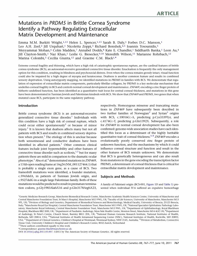
Figure 1. Extreme Corneal Fragility in BCS-001(A and B) Expulsive hemorrhage affecting BCS-001 IV:6. Note also the blue-gray sclera in the remaining eye.(C) 403 magnification of hematoxylin and eosin-stained section through the eviscerated cornea. Note the relative preservation ofperipheral corneal thickness (indicated by double-headed arrow) in contrast to extreme thinning of central cornea in the region ofrupture (indicated by open arrowheads). The stroma (stained pink) is almost absent here: much of the remaining corneal thicknessderives from epithelial and endothelial layers.(D) The pedigree of family BCS-001. The circles indicate females, and the squares indicate males. Filled shapes indicate affected individ-uals, as assessed by history of previous enucleation after minor trauma or phthisis. Blue sclerae and keratoconus in individuals withoutcorneal rupture are also as indicated.(E) Optical coherence tomography demonstrating cross-sectional appearance of remaining eye of individual IV:6 from family BCS-001;note the extreme thinning of central cornea and keratoglobus compared to that in the control image (F).
(Figures 1A and 1B), necessitating emergency enucleation, after
a minor impact when a small child’s shoe, thrown accidentally,
hit her in the face. Three of her sisters had previously lost eyes after
similarly minor trauma, suggesting BCS as the likely diagnosis.1
Another family of Pakistani origin, BCS-002, was also identified
for analysis by the presence of thin corneas, blue sclera, andmixed
sensorineural/conductive deafness (Figure 2). Data on the clinical
features of both affected and unaffected members of these two
families was collected for analysis of the phenotypic spectrum
associated with homozygous and heterozygous mutations. Clin-
ical information was also collated about further individuals with
BCS from six additional affected consanguineous families whose
DNA was available for analysis and in whom mutations in
ZNF469 had previously been excluded. Each of these families
included either a single individual or pair of siblings affected
with BCS (as shown in Table 2).
768 The American Journal of Human Genetics 88, 767–777, June 10,
Clinical AssessmentIndividuals were examined by clinical geneticists, ophthalmolo-
gists, and audiologists. Central corneal thickness was assessed by
pachymetry and optical coherence tomography with the Oculus
Pentacam (Oculus, Lynnwood,WA) and Visante optical coherence
tomography (Carl Zeiss Meditech, Oberkochen) systems. Pure
tone audiometry and tympanometry were carried out with stan-
dard equipment.
Clinical Diagnostic SamplesUrinary pyridinoline crosslinks were analyzed as previously
described.9 Histological analysis of the eviscerated cornea was
carried out in accordance with standard diagnostic protocols:
the tissue was fixed and sectioned for hematoxylin and eosin
staining.
2011
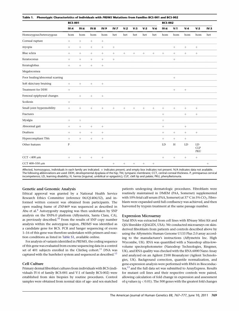
Table 1. Phenotypic Characteristics of Individuals with PRDM5 Mutations from Families BCS-001 and BCS-002
BCS-001 BCS-002
IV:4 IV:6 IV:8 IV:9 IV:7 V:2 V:3 V:5 V:6 IV:6 V:1 V:4 V:5 IV:3
Homozygous/heterozygous hom hom hom hom het het het het het hom hom hom hom het
Corneal rupture þ þ þ þ
myopia þ þ þ þ þ þ þ þ
Blue sclera þ þ þ þ þ þ þ þ þ þ þ þ þ
Keratoconus þ þ þ þ þ þ
Keratoglobus þ þ þ þ
Megalocornea
Poor healing/abnormal scarring þ
Soft skin/easy bruising þ þ þ þ
Treatment for DDH þ þ þ
Femoral epiphyseal changes þ þ þ
Scoliosis þ
Small joint hypermobility þ þ þ þ þ þ þ þ þ þ þ þ þ
Fractures þ þ þ
Myalgia þ þ þ
Abnormal gait þ þ þ þ þ þ
Deafness þ þ þ þ þ þ þ þ
Hypercompliant TMs þ þ þ þ þ þ þ
Other features P LD H LD LDCLPPKU
CCT <400 mm þ þ þ þ þ þ þ þ
CCT 400–550 mm þ þ þ þ þ þ
Affected, homozygous, individuals in each family are indicated. þ indicates present; and empty box indicates not present. N/A indicates data not available.The following abbreviations are used: DDH, developmental dysplasia of the hip; TM, tympanic membrane; CCT, central corneal thickness. P, primiparous cervicalincompetence; LD, learning disability; H, hernia (inguinal, umbilical or epigastric); CLP, cleft lip and palate; PKU, phenylketonuria.
Genetic and Genomic AnalysisEthical approval was granted by a National Health Service
Research Ethics Committee (reference 06/Q1406/52), and in-
formed written consent was obtained from participants. The
open reading frame of ZNF469 was sequenced as described in
Abu et al.4 Autozygosity mapping was then undertaken by SNP
analysis on the SNP6.0 platform (Affymetrix, Santa Clara, CA),
as previously described.10 From the results of SNP copy number
analysis within the autozygous region, PRDM5 was identified as
a candidate gene for BCS. PCR and Sanger sequencing of exons
1–16 of this gene was therefore undertaken with primers and reac-
tion conditions as listed in Table S1, available online.
For analysis of variants identified in PRDM5, the coding sequence
of this genewas evaluated fromexome sequencing data in a control
set of 401 subjects enrolled in the ClinSeq cohort.11 DNA was
captured with the SureSelect system and sequenced as described.12
Cell CulturePrimary dermal fibroblast cultures from individualswith BCS (indi-
viduals IV:4 of family BCS-001 and V:1 of family BCS-002) were
established from skin biopsies by routine procedures. Control
samples were obtained from normal skin of age- and sex-matched
The Ame
patients undergoing dermatologic procedures. Fibroblasts were
routinely maintained in DMEM (PAA, Somerset) supplemented
with10%fetal calf serum(PAA, Somerset) at 37�C in5%CO2. Fibro-
blasts were expanded until full confluency was achieved, and then
harvested by trypsin treatment at the same passage number.
Expression MicroarrayTotal RNA was extracted from cell lines with RNeasy Mini Kit and
QIA Shredder (QIAGEN, USA). We conductedmicroarrays on skin-
derived fibroblasts from patients and controls described above by
using the Affymetrix Human Genome U133 Plus 2.0 array accord-
ing to the manufacturer’s instructions (Affymetrix Inc. High
Wycombe, UK). RNA was quantified with a Nanodrop ultra-low-
volume spectrophotometer (Nanodrop Technologies, Ringmer,
UK), and RNA quality was checked with the RNA 6000 Nano Assay
and analyzed on an Agilent 2100 Bioanalyzer (Agilent Technolo-
gies, UK). Background correction, quantile normalization, and
gene-expression analysis were performed with RMA in Bioconduc-
tor,13 and the full data set was submitted to ArrayExpress. Results
for mutant cell lines and their respective controls were paired,
allowing calculation of fold change in expression and assessment
of q values (q< 0.01). The 500 genes with the greatest fold changes
rican Journal of Human Genetics 88, 767–777, June 10, 2011 769
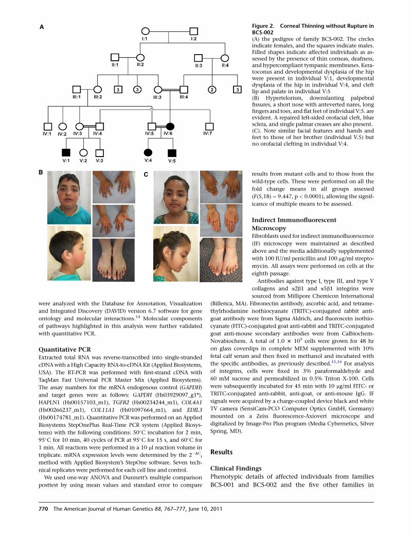
Figure 2. Corneal Thinning without Rupture inBCS-002(A) the pedigree of family BCS-002. The circlesindicate females, and the squares indicate males.Filled shapes indicate affected individuals as as-sessed by the presence of thin corneas, deafness,and hypercompliant tympanic membranes. Kera-toconus and developmental dysplasia of the hipwere present in individual V:1, developmentaldysplasia of the hip in individual V:4, and cleftlip and palate in individual V:5(B) Hypertelorism, downslanting palpebralfissures, a short nose with anteverted nares, longfingers and toes, and flat feet of individual V:5. areevident. A repaired left-sided orofacial cleft, bluesclera, and single palmar creases are also present.(C). Note similar facial features and hands andfeet to those of her brother (individual V.5) butno orofacial clefting in individual V:4.
were analyzed with the Database for Annotation, Visualization
and Integrated Discovery (DAVID) version 6.7 software for gene
ontology and molecular interactions.14 Molecular components
of pathways highlighted in this analysis were further validated
with quantitative PCR.
Quantitative PCRExtracted total RNA was reverse-transcribed into single-stranded
cDNAwith a High Capacity RNA-to-cDNA Kit (Applied Biosystems,
USA). The RT-PCR was performed with first-strand cDNA with
TaqMan Fast Universal PCR Master Mix (Applied Biosystems).
The assay numbers for the mRNA endogenous control (GAPDH)
and target genes were as follows: GAPDH (Hs03929097_g1*),
HAPLN1 (Hs00157103_m1), TGFB2 (Hs00234244_m1), COL4A1
(Hs00266237_m1), COL11A1 (Hs01097664_m1), and EDIL3
(Hs00174781_m1). Quantitative PCR was performed on an Applied
Biosystems StepOnePlus Real-Time PCR system (Applied Biosys-
tems) with the following conditions: 50�C incubation for 2 min,
95�C for 10 min, 40 cycles of PCR at 95�C for 15 s, and 60�C for
1 min. All reactions were performed in a 10 ml reaction volume in
triplicate. mRNA expression levels were determined by the 2�DCt
method with Applied Biosystem’s StepOne software. Seven tech-
nical replicates were performed for each cell line and control.
We used one-way ANOVA and Dunnett’s multiple comparison
posttest by using mean values and standard error to compare
770 The American Journal of Human Genetics 88, 767–777, June 10, 2011
results from mutant cells and to those from the
wild-type cells. These were performed on all the
fold change means in all groups assessed
(F(5,18)¼ 9.447, p< 0.0001), allowing the signif-
icance of multiple means to be assessed.
Indirect Immunofluorescent
MicroscopyFibroblasts used for indirect immunofluorescence
(IF) microscopy were maintained as described
above and the media additionally supplemented
with 100 IU/ml penicillin and 100 mg/ml strepto-
mycin. All assays were performed on cells at the
eighth passage.
Antibodies against type I, type III, and type V
collagens and a2b1 and a5b1 integrins were
sourced from Millipore Chemicon International
(Billerica, MA). Fibronectin antibody, ascorbic acid, and tetrame-
thylrhodamine isothiocyanate (TRITC)-conjugated rabbit anti-
goat antibody were from Sigma Aldrich, and fluoroscein isothio-
cyanate (FITC)-conjugated goat anti-rabbit and TRITC-conjugated
goat anti-mouse secondary antibodies were from Calbiochem-
Novabiochem. A total of 1.0 3 105 cells were grown for 48 hr
on glass coverslips in complete MEM supplemented with 10%
fetal calf serum and then fixed in methanol and incubated with
the specific antibodies, as previously described.15,16 For analysis
of integrins, cells were fixed in 3% paraformaldehyde and
60 mM sucrose and permeabilized in 0.5% Triton X-100. Cells
were subsequently incubated for 45 min with 10 mg/ml FITC- or
TRITC-conjugated anti-rabbit, anti-goat, or anti-mouse IgG. IF
signals were acquired by a charge-coupled device black and white
TV camera (SensiCam-PCO Computer Optics GmbH, Germany)
mounted on a Zeiss fluorescence-Axiovert microscope and
digitalized by Image-Pro Plus program (Media Cybernetics, Silver
Spring, MD).
Results
Clinical Findings
Phenotypic details of affected individuals from families
BCS-001 and BCS-002 and the five other families in
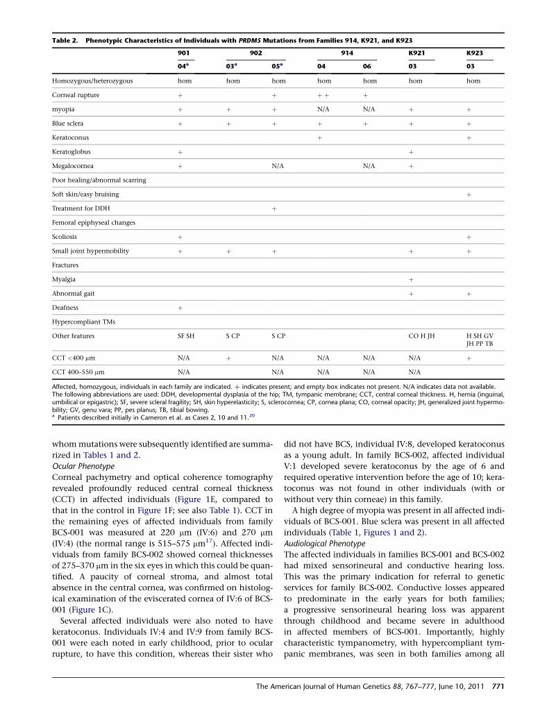
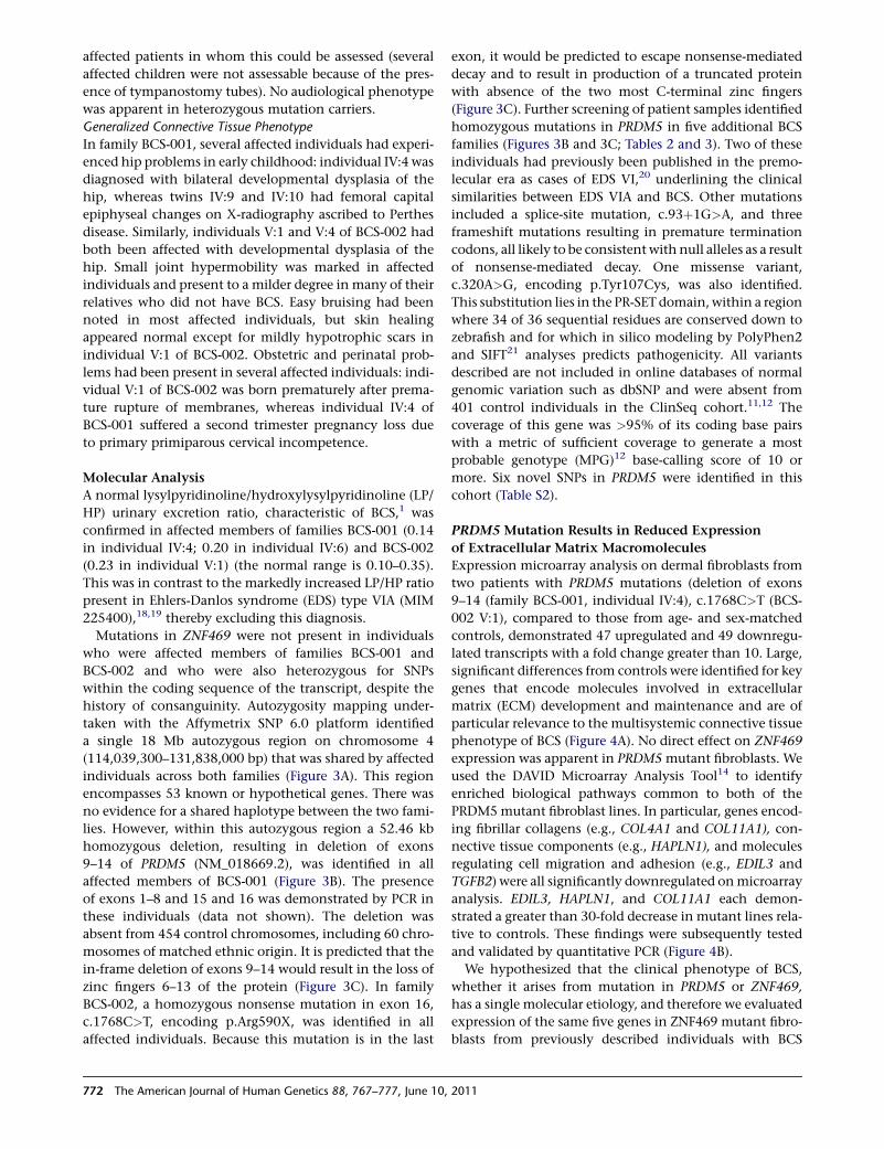
Table 2. Phenotypic Characteristics of Individuals with PRDM5 Mutations from Families 914, K921, and K923
901 902 914 K921 K923
04a 03a 05a 04 06 03 03
Homozygous/heterozygous hom hom hom hom hom hom hom
Corneal rupture þ þ þ þ þ
myopia þ þ þ N/A N/A þ þ
Blue sclera þ þ þ þ þ þ þ
Keratoconus þ þ
Keratoglobus þ þ
Megalocornea þ N/A N/A þ
Poor healing/abnormal scarring
Soft skin/easy bruising þ
Treatment for DDH þ
Femoral epiphyseal changes
Scoliosis þ þ
Small joint hypermobility þ þ þ þ þ
Fractures
Myalgia þ
Abnormal gait þ þ
Deafness þ
Hypercompliant TMs
Other features SF SH S CP S CP CO H JH H SH GVJH PP TB
CCT <400 mm N/A þ N/A N/A N/A N/A þ
CCT 400–550 mm N/A N/A N/A N/A N/A
Affected, homozygous, individuals in each family are indicated. þ indicates present; and empty box indicates not present. N/A indicates data not available.The following abbreviations are used: DDH, developmental dysplasia of the hip; TM, tympanic membrane; CCT, central corneal thickness. H, hernia (inguinal,umbilical or epigastric); SF, severe scleral fragility; SH, skin hyperelasticity; S, sclerocornea; CP, cornea plana; CO, corneal opacity; JH, generalized joint hypermo-bility; GV, genu vara; PP, pes planus; TB, tibial bowing.a Patients described initially in Cameron et al. as Cases 2, 10 and 11.20
whommutations were subsequently identified are summa-
rized in Tables 1 and 2.
Ocular Phenotype
Corneal pachymetry and optical coherence tomography
revealed profoundly reduced central corneal thickness
(CCT) in affected individuals (Figure 1E, compared to
that in the control in Figure 1F; see also Table 1). CCT in
the remaining eyes of affected individuals from family
BCS-001 was measured at 220 mm (IV:6) and 270 mm
(IV:4) (the normal range is 515–575 mm17). Affected indi-
viduals from family BCS-002 showed corneal thicknesses
of 275–370 mm in the six eyes in which this could be quan-
tified. A paucity of corneal stroma, and almost total
absence in the central cornea, was confirmed on histolog-
ical examination of the eviscerated cornea of IV:6 of BCS-
001 (Figure 1C).
Several affected individuals were also noted to have
keratoconus. Individuals IV:4 and IV:9 from family BCS-
001 were each noted in early childhood, prior to ocular
rupture, to have this condition, whereas their sister who
The Ame
did not have BCS, individual IV:8, developed keratoconus
as a young adult. In family BCS-002, affected individual
V:1 developed severe keratoconus by the age of 6 and
required operative intervention before the age of 10; kera-
toconus was not found in other individuals (with or
without very thin corneae) in this family.
A high degree of myopia was present in all affected indi-
viduals of BCS-001. Blue sclera was present in all affected
individuals (Table 1, Figures 1 and 2).
Audiological Phenotype
The affected individuals in families BCS-001 and BCS-002
had mixed sensorineural and conductive hearing loss.
This was the primary indication for referral to genetic
services for family BCS-002. Conductive losses appeared
to predominate in the early years for both families;
a progressive sensorineural hearing loss was apparent
through childhood and became severe in adulthood
in affected members of BCS-001. Importantly, highly
characteristic tympanometry, with hypercompliant tym-
panic membranes, was seen in both families among all
rican Journal of Human Genetics 88, 767–777, June 10, 2011 771
affected patients in whom this could be assessed (several
affected children were not assessable because of the pres-
ence of tympanostomy tubes). No audiological phenotype
was apparent in heterozygous mutation carriers.
Generalized Connective Tissue Phenotype
In family BCS-001, several affected individuals had experi-
enced hip problems in early childhood: individual IV:4 was
diagnosed with bilateral developmental dysplasia of the
hip, whereas twins IV:9 and IV:10 had femoral capital
epiphyseal changes on X-radiography ascribed to Perthes
disease. Similarly, individuals V:1 and V:4 of BCS-002 had
both been affected with developmental dysplasia of the
hip. Small joint hypermobility was marked in affected
individuals and present to a milder degree in many of their
relatives who did not have BCS. Easy bruising had been
noted in most affected individuals, but skin healing
appeared normal except for mildly hypotrophic scars in
individual V:1 of BCS-002. Obstetric and perinatal prob-
lems had been present in several affected individuals: indi-
vidual V:1 of BCS-002 was born prematurely after prema-
ture rupture of membranes, whereas individual IV:4 of
BCS-001 suffered a second trimester pregnancy loss due
to primary primiparous cervical incompetence.
Molecular Analysis
A normal lysylpyridinoline/hydroxylysylpyridinoline (LP/
HP) urinary excretion ratio, characteristic of BCS,1 was
confirmed in affected members of families BCS-001 (0.14
in individual IV:4; 0.20 in individual IV:6) and BCS-002
(0.23 in individual V:1) (the normal range is 0.10–0.35).
This was in contrast to the markedly increased LP/HP ratio
present in Ehlers-Danlos syndrome (EDS) type VIA (MIM
225400),18,19 thereby excluding this diagnosis.
Mutations in ZNF469 were not present in individuals
who were affected members of families BCS-001 and
BCS-002 and who were also heterozygous for SNPs
within the coding sequence of the transcript, despite the
history of consanguinity. Autozygosity mapping under-
taken with the Affymetrix SNP 6.0 platform identified
a single 18 Mb autozygous region on chromosome 4
(114,039,300–131,838,000 bp) that was shared by affected
individuals across both families (Figure 3A). This region
encompasses 53 known or hypothetical genes. There was
no evidence for a shared haplotype between the two fami-
lies. However, within this autozygous region a 52.46 kb
homozygous deletion, resulting in deletion of exons
9–14 of PRDM5 (NM_018669.2), was identified in all
affected members of BCS-001 (Figure 3B). The presence
of exons 1–8 and 15 and 16 was demonstrated by PCR in
these individuals (data not shown). The deletion was
absent from 454 control chromosomes, including 60 chro-
mosomes of matched ethnic origin. It is predicted that the
in-frame deletion of exons 9–14 would result in the loss of
zinc fingers 6–13 of the protein (Figure 3C). In family
BCS-002, a homozygous nonsense mutation in exon 16,
c.1768C>T, encoding p.Arg590X, was identified in all
affected individuals. Because this mutation is in the last
772 The American Journal of Human Genetics 88, 767–777, June 10,
exon, it would be predicted to escape nonsense-mediated
decay and to result in production of a truncated protein
with absence of the two most C-terminal zinc fingers
(Figure 3C). Further screening of patient samples identified
homozygous mutations in PRDM5 in five additional BCS
families (Figures 3B and 3C; Tables 2 and 3). Two of these
individuals had previously been published in the premo-
lecular era as cases of EDS VI,20 underlining the clinical
similarities between EDS VIA and BCS. Other mutations
included a splice-site mutation, c.93þ1G>A, and three
frameshift mutations resulting in premature termination
codons, all likely to be consistent with null alleles as a result
of nonsense-mediated decay. One missense variant,
c.320A>G, encoding p.Tyr107Cys, was also identified.
This substitution lies in the PR-SET domain, within a region
where 34 of 36 sequential residues are conserved down to
zebrafish and for which in silico modeling by PolyPhen2
and SIFT21 analyses predicts pathogenicity. All variants
described are not included in online databases of normal
genomic variation such as dbSNP and were absent from
401 control individuals in the ClinSeq cohort.11,12 The
coverage of this gene was >95% of its coding base pairs
with a metric of sufficient coverage to generate a most
probable genotype (MPG)12 base-calling score of 10 or
more. Six novel SNPs in PRDM5 were identified in this
cohort (Table S2).
PRDM5 Mutation Results in Reduced Expression
of Extracellular Matrix Macromolecules
Expression microarray analysis on dermal fibroblasts from
two patients with PRDM5 mutations (deletion of exons
9–14 (family BCS-001, individual IV:4), c.1768C>T (BCS-
002 V:1), compared to those from age- and sex-matched
controls, demonstrated 47 upregulated and 49 downregu-
lated transcripts with a fold change greater than 10. Large,
significant differences from controls were identified for key
genes that encode molecules involved in extracellular
matrix (ECM) development and maintenance and are of
particular relevance to the multisystemic connective tissue
phenotype of BCS (Figure 4A). No direct effect on ZNF469
expression was apparent in PRDM5 mutant fibroblasts. We
used the DAVID Microarray Analysis Tool14 to identify
enriched biological pathways common to both of the
PRDM5mutant fibroblast lines. In particular, genes encod-
ing fibrillar collagens (e.g., COL4A1 and COL11A1), con-
nective tissue components (e.g., HAPLN1), and molecules
regulating cell migration and adhesion (e.g., EDIL3 and
TGFB2) were all significantly downregulated onmicroarray
analysis. EDIL3, HAPLN1, and COL11A1 each demon-
strated a greater than 30-fold decrease in mutant lines rela-
tive to controls. These findings were subsequently tested
and validated by quantitative PCR (Figure 4B).
We hypothesized that the clinical phenotype of BCS,
whether it arises from mutation in PRDM5 or ZNF469,
has a single molecular etiology, and therefore we evaluated
expression of the same five genes in ZNF469 mutant fibro-
blasts from previously described individuals with BCS
2011
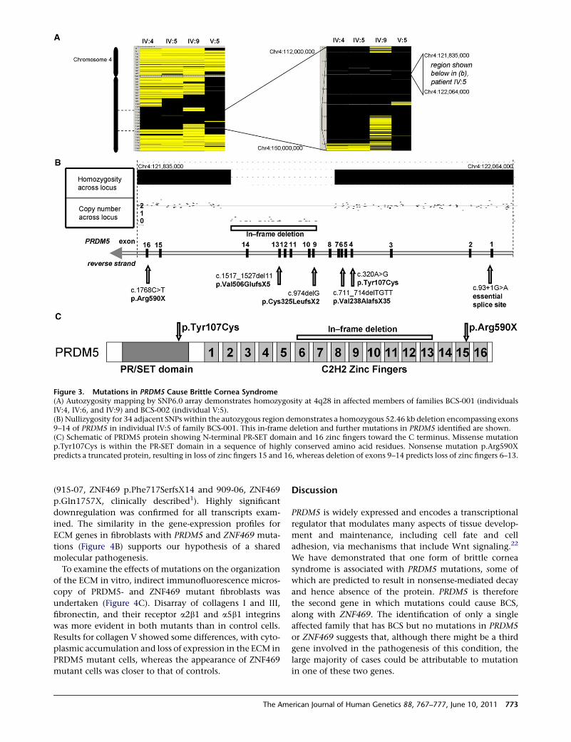
Figure 3. Mutations in PRDM5 Cause Brittle Cornea Syndrome(A) Autozygosity mapping by SNP6.0 array demonstrates homozygosity at 4q28 in affected members of families BCS-001 (individualsIV:4, IV:6, and IV:9) and BCS-002 (individual V:5).(B) Nullizygosity for 34 adjacent SNPs within the autozygous region demonstrates a homozygous 52.46 kb deletion encompassing exons9–14 of PRDM5 in individual IV:5 of family BCS-001. This in-frame deletion and further mutations in PRDM5 identified are shown.(C) Schematic of PRDM5 protein showing N-terminal PR-SET domain and 16 zinc fingers toward the C terminus. Missense mutationp.Tyr107Cys is within the PR-SET domain in a sequence of highly conserved amino acid residues. Nonsense mutation p.Arg590Xpredicts a truncated protein, resulting in loss of zinc fingers 15 and 16, whereas deletion of exons 9–14 predicts loss of zinc fingers 6–13.
(915-07, ZNF469 p.Phe717SerfsX14 and 909-06, ZNF469
p.Gln1757X, clinically described1). Highly significant
downregulation was confirmed for all transcripts exam-
ined. The similarity in the gene-expression profiles for
ECM genes in fibroblasts with PRDM5 and ZNF469 muta-
tions (Figure 4B) supports our hypothesis of a shared
molecular pathogenesis.
To examine the effects of mutations on the organization
of the ECM in vitro, indirect immunofluorescence micros-
copy of PRDM5- and ZNF469 mutant fibroblasts was
undertaken (Figure 4C). Disarray of collagens I and III,
fibronectin, and their receptor a2b1 and a5b1 integrins
was more evident in both mutants than in control cells.
Results for collagen V showed some differences, with cyto-
plasmic accumulation and loss of expression in the ECM in
PRDM5 mutant cells, whereas the appearance of ZNF469
mutant cells was closer to that of controls.
The Ame
Discussion
PRDM5 is widely expressed and encodes a transcriptional
regulator that modulates many aspects of tissue develop-
ment and maintenance, including cell fate and cell
adhesion, via mechanisms that include Wnt signaling.22
We have demonstrated that one form of brittle cornea
syndrome is associated with PRDM5 mutations, some of
which are predicted to result in nonsense-mediated decay
and hence absence of the protein. PRDM5 is therefore
the second gene in which mutations could cause BCS,
along with ZNF469. The identification of only a single
affected family that has BCS but no mutations in PRDM5
or ZNF469 suggests that, although there might be a third
gene involved in the pathogenesis of this condition, the
large majority of cases could be attributable to mutation
in one of these two genes.
rican Journal of Human Genetics 88, 767–777, June 10, 2011 773
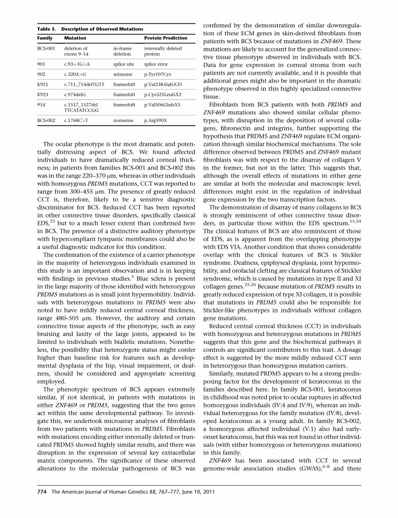
Table 3. Description of Observed Mutations
Family Mutation Protein Prediction
BCS-001 deletion ofexons 9–14
in-framedeletion
internally deletedprotein
901 c.93þ1G>A splice site splice error
902 c.320A>G missense p.Tyr107Cys
K921 c.711_714delTGTT frameshift p.Val238AlafsX35
K923 c.974delG frameshift p.Cys325LeufsX2
914 c.1517_1527delTTCATATCCGG
frameshift p.Val506GlufsX5
BCS-002 c.1768C>T nonsense p.Arg590X
The ocular phenotype is the most dramatic and poten-
tially distressing aspect of BCS. We found affected
individuals to have dramatically reduced corneal thick-
ness; in patients from families BCS-001 and BCS-002 this
was in the range 220–370 mm, whereas in other individuals
with homozygous PRDM5mutations, CCT was reported to
range from 300–455 mm. The presence of greatly reduced
CCT is, therefore, likely to be a sensitive diagnostic
discriminator for BCS. Reduced CCT has been reported
in other connective tissue disorders, specifically classical
EDS,23 but to a much lesser extent than confirmed here
in BCS. The presence of a distinctive auditory phenotype
with hypercompliant tympanic membranes could also be
a useful diagnostic indicator for this condition.
The confirmation of the existence of a carrier phenotype
in the majority of heterozygous individuals examined in
this study is an important observation and is in keeping
with findings in previous studies.5 Blue sclera is present
in the large majority of those identified with heterozygous
PRDM5 mutations as is small joint hypermobility. Individ-
uals with heterozygous mutations in PRDM5 were also
noted to have mildly reduced central corneal thickness,
range 480–505 mm. However, the auditory and certain
connective tissue aspects of the phenotype, such as easy
bruising and laxity of the large joints, appeared to be
limited to individuals with biallelic mutations. Nonethe-
less, the possibility that heterozygote status might confer
higher than baseline risk for features such as develop-
mental dysplasia of the hip, visual impairment, or deaf-
ness, should be considered and appropriate screening
employed.
The phenotypic spectrum of BCS appears extremely
similar, if not identical, in patients with mutations in
either ZNF469 or PRDM5, suggesting that the two genes
act within the same developmental pathway. To investi-
gate this, we undertook microarray analyses of fibroblasts
from two patients with mutations in PRDM5. Fibroblasts
with mutations encoding either internally deleted or trun-
cated PRDM5 showed highly similar results, and there was
disruption in the expression of several key extracellular
matrix components. The significance of these observed
alterations to the molecular pathogenesis of BCS was
774 The American Journal of Human Genetics 88, 767–777, June 10,
confirmed by the demonstration of similar downregula-
tion of these ECM genes in skin-derived fibroblasts from
patients with BCS because of mutations in ZNF469. These
mutations are likely to account for the generalized connec-
tive tissue phenotype observed in individuals with BCS.
Data for gene expression in corneal stroma from such
patients are not currently available, and it is possible that
additional genes might also be important in the dramatic
phenotype observed in this highly specialized connective
tissue.
Fibroblasts from BCS patients with both PRDM5 and
ZNF469 mutations also showed similar cellular pheno-
types, with disruption in the deposition of several colla-
gens, fibronectin and integrins, further supporting the
hypothesis that PRDM5 and ZNF469 regulate ECM organi-
zation through similar biochemical mechanisms. The sole
difference observed between PRDM5 and ZNF469 mutant
fibroblasts was with respect to the disarray of collagen V
in the former, but not in the latter. This suggests that,
although the overall effects of mutations in either gene
are similar at both the molecular and macroscopic level,
differences might exist in the regulation of individual
gene expression by the two transcription factors.
The demonstration of disarray of many collagens in BCS
is strongly reminiscent of other connective tissue disor-
ders, in particular those within the EDS spectrum.15,24
The clinical features of BCS are also reminiscent of those
of EDS, as is apparent from the overlapping phenotype
with EDS VIA. Another condition that shows considerable
overlap with the clinical features of BCS is Stickler
syndrome. Deafness, epiphyseal dysplasia, joint hypermo-
bility, and orofacial clefting are classical features of Stickler
syndrome, which is caused by mutations in type II and XI
collagen genes.25,26 Because mutation of PRDM5 results in
greatly reduced expression of type XI collagen, it is possible
that mutations in PRDM5 could also be responsible for
Stickler-like phenotypes in individuals without collagen
gene mutations.
Reduced central corneal thickness (CCT) in individuals
with homozygous and heterozygous mutations in PRDM5
suggests that this gene and the biochemical pathways it
controls are significant contributors to this trait. A dosage
effect is suggested by the more mildly reduced CCT seen
in heterozygous than homozygous mutation carriers.
Similarly, mutated PRDM5 appears to be a strong predis-
posing factor for the development of keratoconus in the
families described here. In family BCS-001, keratoconus
in childhood was noted prior to ocular ruptures in affected
homozygous individuals (IV:4 and IV:9), whereas an indi-
vidual heterozygous for the family mutation (IV:8), devel-
oped keratoconus as a young adult. In family BCS-002,
a homozygous affected individual (V:1) also had early-
onset keratoconus, but this was not found in other individ-
uals (with either homozygous or heterozygous mutations)
in this family.
ZNF469 has been associated with CCT in several
genome-wide association studies (GWAS),6–8 and there
2011
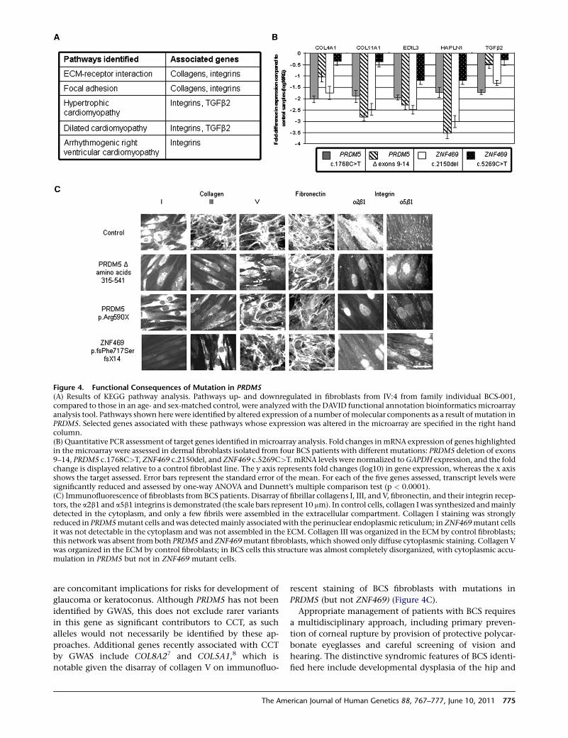
Figure 4. Functional Consequences of Mutation in PRDM5(A) Results of KEGG pathway analysis. Pathways up- and downregulated in fibroblasts from IV:4 from family individual BCS-001,compared to those in an age- and sex-matched control, were analyzed with the DAVID functional annotation bioinformatics microarrayanalysis tool. Pathways shown here were identified by altered expression of a number ofmolecular components as a result of mutation inPRDM5. Selected genes associated with these pathways whose expression was altered in the microarray are specified in the right handcolumn.(B) Quantitative PCR assessment of target genes identified inmicroarray analysis. Fold changes inmRNA expression of genes highlightedin the microarray were assessed in dermal fibroblasts isolated from four BCS patients with different mutations: PRDM5 deletion of exons9–14, PRDM5 c.1768C>T, ZNF469 c.2150del, and ZNF469 c.5269C>T. mRNA levels were normalized toGAPDH expression, and the foldchange is displayed relative to a control fibroblast line. The y axis represents fold changes (log10) in gene expression, whereas the x axisshows the target assessed. Error bars represent the standard error of the mean. For each of the five genes assessed, transcript levels weresignificantly reduced and assessed by one-way ANOVA and Dunnett’s multiple comparison test (p < 0.0001).(C) Immunofluorescence of fibroblasts from BCS patients. Disarray of fibrillar collagens I, III, and V, fibronectin, and their integrin recep-tors, the a2b1 and a5b1 integrins is demonstrated (the scale bars represent 10 mm). In control cells, collagen I was synthesized andmainlydetected in the cytoplasm, and only a few fibrils were assembled in the extracellular compartment. Collagen I staining was stronglyreduced in PRDM5mutant cells andwas detectedmainly associated with the perinuclear endoplasmic reticulum; in ZNF469mutant cellsit was not detectable in the cytoplasm and was not assembled in the ECM. Collagen III was organized in the ECM by control fibroblasts;this network was absent from both PRDM5 and ZNF469mutant fibroblasts, which showed only diffuse cytoplasmic staining. Collagen Vwas organized in the ECM by control fibroblasts; in BCS cells this structure was almost completely disorganized, with cytoplasmic accu-mulation in PRDM5 but not in ZNF469 mutant cells.
are concomitant implications for risks for development of
glaucoma or keratoconus. Although PRDM5 has not been
identified by GWAS, this does not exclude rarer variants
in this gene as significant contributors to CCT, as such
alleles would not necessarily be identified by these ap-
proaches. Additional genes recently associated with CCT
by GWAS include COL8A27 and COL5A1,8 which is
notable given the disarray of collagen V on immunofluo-
The Ame
rescent staining of BCS fibroblasts with mutations in
PRDM5 (but not ZNF469) (Figure 4C).
Appropriate management of patients with BCS requires
a multidisciplinary approach, including primary preven-
tion of corneal rupture by provision of protective polycar-
bonate eyeglasses and careful screening of vision and
hearing. The distinctive syndromic features of BCS identi-
fied here include developmental dysplasia of the hip and
rican Journal of Human Genetics 88, 767–777, June 10, 2011 775
hypercompliant tympanic membranes and serve as impor-
tant diagnostic clues in the early recognition of patients
with this condition, particularly where they are the only
affected individual in their family.
Now that the molecular basis of BCS in the large
majority of affected families ascertained to date has been
clarified, effective genetic testing and further assessment
for genotype-phenotype correlations in this condition
become possible. Significant differences exist between
the phenotypes of families BCS-001 and BCS-002 in this
study, and although the presentation of all affected indi-
viduals in family BCS-001 appears similar, for members
of BCS-002 there is much greater phenotypic variability.
These observations of inter- and intrafamilial variability
emphasize the difficulty inmaking a firm clinical diagnosis
of BCS and hence the importance of molecular diagnostic
testing of individuals at risk for this condition. Early diag-
nosis by presymptomatic genetic testing will help to avert
the devastating physical and psychological sequelae of
corneal rupture, as well as identifying individuals at risk
for the associated features of this condition, whereas
carrier testing will permit effective genetic counseling
and identification of individuals at risk of late-onset
complications, such as keratoconus.
In identifying that mutations in PRDM5 cause extreme
corneal fragility, as well as the related auditory and muscu-
loskeletal connective tissue phenotypes seen in BCS, we
have identified a pathway that is central to corneal devel-
opment and maintenance and that, as a result, profoundly
influences CCT. Further work to determine the role of the
genes mutated in BCS in common ocular disorders associ-
ated with a low CCT, such as glaucoma and keratoconus,
will lead to improved understanding of these disorders
and potentially to improved management. The pathways
in which PRDM5 exerts its effects could also present ther-
apeutic targets for BCS or other connective tissue disorders
in the future.
Supplemental Data
Supplemental Data include two tables and can be found with this
article online at http://www.cell.com/AJHG/.
Acknowledgments
We thank all healthcare professionals contributing to the care of
the families described here, particularly R.W. Paton (Royal Black-
burn Hospital), and J. Teer of the National Human Genome
Research Institute, for sequence coverage analysis in the ClinSeq
cohort. We thank B. Steinmann for useful discussion and support,
H. Al-Hussein for referral of some BCS families, and A. Schwarze
and C. Burer for expert technical assistance. The support of the
Manchester Biomedical Research Centre, funded by the UK
National Institute for Health Research, is gratefully acknowledged.
E.B.W. is funded by a Clinical Research Training Fellowship from
the Wellcome Trust, and F.D.C.M. by Fight for Sight. The NISC
comparative sequencing program and the ClinSeq study are
funded by the National Human Genome Research Institute of
the National Institutes of Health.
776 The American Journal of Human Genetics 88, 767–777, June 10,
Received: February 24, 2011
Revised: April 20, 2011
Accepted: May 6, 2011
Published online: June 9, 2011
Web Resources
The URLs for data presented herein are as follows:
Online Mendelian Inheritance in Man (OMIM), http://www.
omim.org
PolyPhen-2, http://genetics.bwh.harvard.edu/pph2/
NCBI’s Single Nucleotide Polymorphism, http://www.ncbi.nlm.
nih.gov/projects/SNP/
Accession Numbers
The ArrayExpress accession number for the PRDM5 sequence
reported in this paper is E-MEXP-3077.
References
1. Al-Hussain, H., Zeisberger, S.M., Huber, P.R., Giunta, C., and
Steinmann, B. (2004). Brittle cornea syndrome and its delinea-
tion from the kyphoscoliotic type of Ehlers-Danlos syndrome
(EDS VI): Report on 23 patients and review of the literature.
Am. J. Med. Genet. A. 124A, 28–34.
2. Izquierdo, L., Jr., Mannis, M.J., Marsh, P.B., Yang, S.P., and
McCarthy, J.M. (1999). Bilateral spontaneous corneal rupture
in brittle cornea syndrome: A case report. Cornea 18, 621–624.
3. Christensen, A.E., Knappskog, P.M.,Midtbø,M., Gjesdal, C.G.,
Mengel-From, J., Morling, N., Rødahl, E., and Boman, H.
(2010). Brittle cornea syndrome associated with a missense
mutation in the zinc-finger 469 gene. Invest. Ophthalmol.
Vis. Sci. 51, 47–52.
4. Abu, A., Frydman,M., Marek, D., Pras, E., Nir, U., Reznik-Wolf,
H., and Pras, E. (2008). Deleterious mutations in the Zinc-
Finger 469 gene cause brittle cornea syndrome. Am. J. Hum.
Genet. 82, 1217–1222.
5. Khan, A.O., Aldahmesh, M.A., Mohamed, J.N., and Alkuraya,
F.S. (2010). Blue sclera with and without corneal fragility
(brittle cornea syndrome) in a consanguineous family
harboring ZNF469 mutation (p.E1392X). Arch. Ophthalmol.
128, 1376–1379.
6. Lu, Y., Dimasi, D.P., Hysi, P.G., Hewitt, A.W., Burdon, K.P., Toh,
T., Ruddle, J.B., Li, Y.J., Mitchell, P., Healey, P.R., et al. (2010).
Common genetic variants near the Brittle Cornea Syndrome
locus ZNF469 influence the blinding disease risk factor central
corneal thickness. PLoS Genet. 6, e1000947.
7. Vithana, E.N., Aung, T., Khor, C.C., Cornes, B.K., Tay, W.T.,
Sim, X., Lavanya, R., Wu, R., Zheng, Y., Hibberd, M.L., et al.
(2011). Collagen-related genes influence the glaucoma risk
factor, central corneal thickness. Hum. Mol. Genet. 20, 649–
658.
8. Vitart, V., Benci�c, G., Hayward, C., Skunca Herman, J., Huff-
man, J., Campbell, S., Bu�can, K., Navarro, P., Gunjaca, G.,
Marin, J., et al. (2010). New loci associated with central cornea
thickness include COL5A1, AKAP13 and AVGR8. Hum. Mol.
Genet. 19, 4304–4311.
9. Kraenzlin, M.E., Kraenzlin, C.A., Meier, C., Giunta, C., and
Steinmann, B. (2008). Automated HPLC assay for urinary
2011
collagen cross-links: effect of age, menopause, and metabolic
bone diseases. Clin. Chem. 54, 1546–1553.
10. Daly, S.B., Urquhart, J.E., Hilton, E., McKenzie, E.A., Kam-
merer, R.A., Lewis, M., Kerr, B., Stuart, H., Donnai, D., Long,
D.A., et al. (2010). Mutations in HPSE2 cause urofacial
syndrome. Am. J. Hum. Genet. 86, 963–969.
11. Biesecker, L.G., Mullikin, J.C., Facio, F.M., Turner, C., Cheru-
kuri, P.F., Blakesley, R.W., Bouffard, G.G., Chines, P.S., Cruz,
P., Hansen, N.F., et al. (2009). Instrumenting the health care
enterprise for discovery research in the genomic era. Genome
Res. 19, 1675–1681.
12. Teer, J.K., Bonnycastle, L.L., Chines, P.S., Hansen, N.F.,
Aoyama, N., Swift, A.J., Abaan, H.O., Albert, T.J., Margulies,
E.H., Green, E.D., et al; NISC Comparative Sequencing
Program. (2010). Systematic comparison of three genomic
enrichment methods for massively parallel DNA sequencing.
Genome Res. 20, 1420–1431.
13. Bolstad, B.M., Irizarry, R.A., Astrand, M., and Speed, T.P.
(2003). A comparison of normalization methods for high
density oligonucleotide array data based on variance and
bias. Bioinformatics 19, 185–193.
14. Dennis, G., Jr., Sherman, B.T., Hosack, D.A., Yang, J., Gao, W.,
Lane, H.C., and Lempicki, R.A. (2003). DAVID: Database for
Annotation, Visualization, and Integrated Discovery. Genome
Biol. 4, 3.
15. Zoppi, N., Gardella, R., De Paepe, A., Barlati, S., and Colombi,
M. (2004). Human fibroblasts carrying mutations in COL5A1
and COL3A1 genes do not organize collagens and fibronectin
in the extracellular matrix, down-regulate a2b1 integrin, and
recruit a5b3 instead of a5b1 integrin. J. Biol. Chem. 279,
18157–18168.
16. Zoppi, N., Barlati, S., and Colombi, M. (2008). FAK-indepen-
dent a5b3 integrin-paxillin-EGFR complexes rescue from
anoikis matrix-defective fibroblasts. Biochim. Biophys. Acta
1783, 1177–1188.
17. Jahadi Hosseini, H.R., Katbab, A., Khalili, M.R., and Abtahi,
M.B. (2010). Comparison of corneal thickness measurements
The Ame
usingGalilei, HR Pentacam, and ultrasound. Cornea 29, 1091–
1095.
18. Steinmann, B., Eyre, D.R., and Shao, P. (1995). Urinary pyridi-
noline cross-links in Ehlers-Danlos syndrome type VI. Am.
J. Hum. Genet. 57, 1505–1508.
19. Giunta, C., Randolph, A., Al-Gazali, L.I., Brunner, H.G., Kraen-
zlin, M.E., and Steinmann, B. (2005). Nevo syndrome is allelic
to the kyphoscoliotic type of the Ehlers-Danlos syndrome
(EDS VIA). Am. J. Med. Genet. A. 133A, 158–164.
20. Cameron, J.A. (1993). Corneal abnormalities in Ehlers-Danlos
syndrome type VI. Cornea 12, 54–59.
21. Kumar, P., Henikoff, S., and Ng, P.C. (2009). Predicting the
effects of coding non-synonymous variants on protein func-
tion using the SIFT algorithm. Nat. Protoc. 4, 1073–1081.
22. Meani, N., Pezzimenti, F., Deflorian, G., Mione, M., and
Alcalay, M. (2009). The tumor suppressor PRDM5 regulates
Wnt signaling at early stages of zebrafish development. PLoS
ONE 4, e4273.
23. Segev, F., Heon, E., Cole, W.G., Wenstrup, R.J., Young, F.,
Slomovic, A.R., Rootman, D.S., Whitaker-Menezes, D., Cher-
voneva, I., and Birk, D.E. (2006). Structural abnormalities of
the cornea and lid resulting from collagen V mutations.
Invest. Ophthalmol. Vis. Sci. 47, 565–573.
24. Steinmann, B., Royce, P.M., and Superti-Furga, A. (2002). The
Ehlers-Danlos syndrome. In Connective Tissue and its Heri-
table Disorders, 2nd ed, P.M. Royce and B. Steinmann, eds.
(New York: Wiley-Liss), pp. 431–523.
25. Ahmad, N.N., Ala-Kokko, L., Knowlton, R.G., Jimenez, S.A.,
Weaver, E.J., Maguire, J.I., Tasman, W., and Prockop, D.J.
(1991). Stop codon in the procollagen II gene (COL2A1) in
a family with the Stickler syndrome (arthro-ophthalmop-
athy). Proc. Natl. Acad. Sci. USA 88, 6624–6627.
26. Richards, A.J., Yates, J.R., Williams, R., Payne, S.J., Pope, F.M.,
Scott, J.D., and Snead, M.P. (1996). A family with Stickler
syndrome type 2 has a mutation in the COL11A1 gene result-
ing in the substitution of glycine 97 by valine in alpha 1 (XI)
collagen. Hum. Mol. Genet. 5, 1339–1343.
rican Journal of Human Genetics 88, 767–777, June 10, 2011 777