my life and the world of crystals - uclucapikr/sally_physscripta_2015.pdfdownload details: ip...
TRANSCRIPT
This content has been downloaded from IOPscience. Please scroll down to see the full text.
Download details:
IP Address: 144.82.114.112
This content was downloaded on 05/04/2016 at 08:51
Please note that terms and conditions apply.
My life and the world of crystals
View the table of contents for this issue, or go to the journal homepage for more
2015 Phys. Scr. 90 048003
(http://iopscience.iop.org/1402-4896/90/4/048003)
Home Search Collections Journals About Contact us My IOPscience

Invited Comment
My life and the world of crystals
Ian Robinson and Sally Robinson
London Centre for Nanotechnology, University College, London, UKResearch Complex at Harwell, Harwell Campus, Didcot, Oxford, UKMaterials Science and Engineering, TongJi University, Shanghai, People’s Republic of China
E-mail: [email protected]
Received 31 January 2015Accepted for publication 23 February 2015Published 25 March 2015
AbstractThis is an account of my life and my contributions to crystallography which have led to my receivingthe 2015 Aminoff Prize. Periods discussed in this article are childhood influences, formal training atHarvard, life as an independent researcher at Bell Labs, starting the academic routine at Illinois andthen London. Three major discoveries are presented in the form of anecdotes, on the silicon 7×7structure, on crystal truncation rods and coherent x-ray diffraction. Much of my work has centered onthe need for developing the instrumentation behind the intellectual steps, such as beamlines at theBrookhaven, Argonne, and Diamond synchrotron radiation facilities. This trend continues with theemergence of new possibilities for crystallography using x-ray free-electron lasers.
Keywords: beamline, surface, coherence, crystal, structure
(Some figures may appear in colour only in the online journal)
1. In Reading: kings, rivers, and Oscar Wilde
I was born in Reading, England, a town drawn like a leaf or abutterfly on UK maps and sandwiched between the riversKennet and Thames. I like rivers. In my youth, my parents usedto take my siblings and me to many places by the Thames, andin some of our trips I would see dozens of locks, notice howeasy it was to turn the cranks which so interested me that I evendreamed of becoming a lock keeper when I grew up. On ourfree days, my friends and I would go to the Thames, sometimesswim there, or row our canoes checked out from the local club.
I was fascinated by my town’s history especially by thekings who walked its grounds. On some weekends, I wouldroam the ruins of Reading Abbey, adjacent to the rivers,founded by Henry I; when he died, he was buried in front of theabbey’s altar. Among the ruins, it was easy to imagine the gloryof those days when other kings would come to the abbey, kingslike Henry II who met the patriarch of Jerusalem there. HenryIII was also a frequent visitor, and it was there that Edward IV
was married. But if the Abbey was built by King Henry I,another king, King Henry VIII would destroy it when he dis-solved all the monasteries. Today, the ruined abbey still standsat the center of Reading, near the prison house where OscarWilde, the poet who made my town immortal in his poem ‘TheBallad of Reading Gaol’, was once jailed.
My parents are scientists themselves. My late father,Keith, was a physicist and a crystallographer; my mother,Mary, is a botanist. He worked at the Cavendish Laboratory atthe same time as Crick and Watson, and when my parents gotmarried, Francis Crick presented them with a tea table, a jointwedding gift from him and from many of those who workedat the Cavendish. Today, it is still in my mother’s house inReading; we fondly call it the (Nobel) table.
2. In my youth: my first crystals
When I was three years old, my father gave me a ball andstick model of atoms in a crystal. My mother said it becamemy favorite toy. Essentially, it was my first contact withcrystals, but then I was just a toddler interested only in themodel as a toy. I may even have played with the crystals,
| Royal Swedish Academy of Sciences Physica Scripta
Phys. Scr. 90 (2015) 048003 (11pp) doi:10.1088/0031-8949/90/4/048003
Content from this work may be used under the terms of theCreative Commons Attribution 3.0 licence. Any further
distribution of this work must maintain attribution to the author(s) and thetitle of the work, journal citation and DOI.
0031-8949/15/048003+11$33.00 © 2015 The Royal Swedish Academy of Sciences Printed in the UK1

banging them or biting them, or kicking them imagining themas balls. Life really is a marvel. How was I to know at thattime, that one day I would spend a great part of my lifestudying crystals?
One lunch time when I was about eight, my father camehome bringing a diamond sample which he borrowed fromhis colleague Henry Dyer, a large natural octahedron(figure 1), probably of several carats. I clearly rememberedhim showing it to me, but I did not only look at it; my fatherallowed me to hold that precious crystal, and holding it,looking at it, I was mesmerized! It was awesome. It filled halfof my small hand; it felt rather heavy, and it reflected the lightof the Sun. Truly, the look and feel of that Henry Dyer’sSouth African diamond had made a lasting impression on me.My father took it back with him to work, so it was the firstand last time I saw it, but for a long time, I would rememberits almost perfect octahedral shape with natural facets thatwere still rough but were also so transparent that they glis-tened. That was my second encounter with crystals as a child.Later, in school, I would make models of ‘polyhedra’ fromfolded paper templates that I designed myself. The mostdifficult was a ‘Mobius band’ that was a continuous solid ringwith a triangular cross section, but with only one surface,which took drafting skills to construct.
I was privileged to attend Reading School, in the dayswhen it was a traditional ‘grammar’ school. It was very strictwith full-suit uniforms, frequent detentions and mandatoryhaircuts, but the science teachers were excellent motivators ofinterest. Ed Bicknell was the mathematics teacher of the upperyears. His style was very informal and insightful. His monthly‘maths projects’ were so wonderfully creative that I can stillremember half of them to this day: measuring food-containerdimensions to learn calculus, length of daylight calculationsto learn three-dimentional (3D) geometry, and working outthe facet angles of the Platonic solids. The other greatmoment was a school lecture by Art Schawlow, who muchlater won the 1981 Nobel prize, demonstrating the first rubylasers by shooting balloons from the San Francisco Zoo.
3. In Cambridge: a new life
In 1972, after completing the Cambridge entrance exams, Ispent the last eight months in Reading working at ICLDataskil, a branch of International Computers where workwas rather easy, giving me plenty of time to read the first yeartextbooks ahead of my actual placement at Churchill College.I particularly enjoyed the ‘Feynman Lectures’ [1] and learnedthe story of DNA and the ‘Central Dogma’ from the phy-siology text [2]. In September 1973, I went to ChurchillCollege on a scholarship, and in Cambridge, my new lifebegan.
It was a big change … moving away from home, leavingmy family, leaving my school friends, but it was also exciting,meeting new friends. The summer after my first year inCambridge, I went to the USA on a sponsored working visaand had a chance to visit the Massachussetts Institute ofTechnology and Harvard University. I decided right then totake up graduate studies in Harvard, despite strong localadvice that it would be a ‘waste of time’. Back in Cambridge,that school year I took extra courses in Engineering. Whensummer came, I went to the Risø National Laboratory inDenmark to do a 10-week summer research project workingwith Jørgen Kjems on the design of a position sensitiveneutron detector system, which may have been the lastexperiment ever conducted at the DR2 reactor. Later, I wouldwrite my undergraduate dissertation based on the result of thatresearch. Then, I went to ILL in Grenoble, France, spendingsix more weeks doing research with John White on inter-calation compounds using neutron powder diffraction. Mylocal contact person was Christian Riekel, whom I wouldcontinue to know later at ERSF.
But Harvard was still in my mind, and so I applied there,took the GRE, and had an offer and a phone call acceptance.Taking biophysics was a big decision. At that time, I thoughtthat physics was finished and that brain research was thefuture of science. The summer of 1976 was spent at CERN inGeneva, Switzerland, where I divided my time going to thelibrary reading about biophysics, learning about particledetectors from my supervisor, and going on trips to the Swissmountains. My final school year at Cambridge was quiteintensive, with vacations spent studying the Part II Triposwith a 3 week revision period in isolation before 3 days offinal exams. Burnt out after 2 days (4 exams), I could notconcentrate on the last one but still got a ‘first class’ degree.Life was good.
4. Crystals in Harvard
The biophysics curriculum at Harvard, which still existstoday, included three 3 month ‘rotation’ projects, which wasan effective introduction to research. Other universities areonly now starting to realize the value of this form of graduatetraining. Because it was in my original plan, I did the firstrotation in neurophysiology, but immediately found out howhard the experiments were. I did a rotation in populationbiology and found out that theoretical ideas were still very
Figure 1. Natural octahedral diamond of 84 carats. ‘The HarvardDiamond Crystal’ (Wikipedia).
2
Phys. Scr. 90 (2015) 048003 Invited Comment

primitive within biology. Finally I did crystallography where,in a three month rotation project with Don Wiley, I studiedhemaglutinin crystals, freshly grown in the laboratory, andtook some of the first x-ray diffraction patterns. It was mythird contact with crystals and my first time seeing them undera microscope. Don Wiley and Ian Wilson, a postdoc, latersolved the structure, which has had a lasting impact on ourunderstanding of influenza infection and its epidemics.
In the summer of 1977, I went back to Paris where I metSteve Harrison and his group doing the crystallographicphasing calculations of the Tomato Bushy Stunt Virus(TBSV) at the CECAM computation center there in Orsay. Idecided to undertake my PhD with him as my adviser. Mysummer project was at CEA Saclay with Tom Ypsilantis(Berkeley) designing a particle detector, completing the workI started with him previously at CERN the year before.
I discovered that doing research is rather hard and verydifferent from taking courses. So many things could go wrongwithout explanation. For three years, I experienced a generalfeeling of despair at the slow pace of discovery, but finallyovercame it when I realized that I had learned a lot of things andthat I could help others by sharing my accumulated knowledgewith them. In 1978–80, I taught physics courses such as‘Introductory Electromagnetism’ and ‘Electronics’ (the famousPhysics 123) first as a TA (teaching assistant) and later asinstructor. Physics 123 was the pride and joy of Paul Horowitzwho developed it while he was writing The Art of Electronics[3], still a best-seller today. I redesigned the digital part of theirelectronics laboratory and introduced the first exercises to builda computer in class. Our lab notes were written up for pub-lication as Lab Manual for the Art of Electronics in 1981 byCambridge University Press [4].
Summer vacations were mostly taken in USA: Colorado,Utah, California. I bought my first car (a VW Beetle) in 1978,a major American right of passage, which enabled me to doski trips to New Hampshire, often on Wednesdays for the $3special at Mount Sunapee.
In 1979, at 23 and at about the half-way point to my PhD,I took the Qualifier Exam by writing a research proposal onsomething different from my PhD topic (a Förster resonanceenergy transfer study of protein folding of bovine pancreatictrypsin inhibitor). It was examined by Martin Karplus, whocouldn’t have had any inkling that in 2014, 36 years later, hewould win the Nobel Prize. At 26, I had my PhD defense ofmy dissertation on the structure of the expanded state of theTBSV, with Harrison, Wiley and Bill Lipscomb (Nobel 1976)as internal examiners, and with Don Caspar (Brandeis,external). All of these people had a strong influence over mythinking, and I was proud to have learned from them. Thecrystallography community in the Boston area in those dayswas tightly bonded among the various universities and, by theend of my PhD, I felt like I was part of it.
5. From Harvard to Bell Labs
I met David Moncton before my PhD defense in late 1981. Asmember of Bell Labs Technical Staff, he went as a ‘recruiter’
to Harvard seeking graduating students for staff positions.Talking to him for about an hour, I convinced him that I wasworth inviting for interview at Murray Hill. In preparation, Iattended Moncton’s own seminar, read his papers, then gotready for the Murray Hill interview. I also learned how oneshould dress for this type of interview.
At the interview, I remember that Steve Davey, who is stilltoday part of the x-ray community, ran the movie projector forme to show Art Olson’s movie about the structure, assembly,and phase changes of TBSV. Lots of questions were asked byPhil Platzman and Bill Brinkman about whether crystallographywas even possible on proteins. In a heartbeat, I said yes: I haddone it in Harvard for years, so it never occurred to me that itcouldn’t be done! That was my first introduction to the ‘BellLabs Seminar’ style. The discussions went well enough for meto get a job offer on 15 October as a Member of Technical Staff(MTS) at an attractive $34 000 annual salary. Rick Freemanwould be my first department head, a laser physicist, a pioneerin free-electron lasers, also new to the management job. SteveChu (Nobel 1997 and future Secretary of Energy) and PhilBucksbaum were in the same department. I accepted the offerbut delayed my start date so I could take a vacation in Ecuador(Galapagos) and Peru (Machu Pichu), where it is a custom todrink coca tea because of altitude. When I returned from myvacation, I failed the medical test at Bell Labs, because thecocaine from the coca tea was still in my blood. Three weekslater, I was tested again and I passed. On Monday 1 December1981, I started working at Bell Labs in Murray Hill, but thebuildings were like a maze. I had no lunch for three daysbecause I could not find the canteen. Ok, stupid not to ask!
On Friday 5 December 1981, it was arranged for me tofly to Stanford for three weeks of synchrotron experiments.The first one was with David Moncton, Robert Fleming andJohn Axe looking at diffraction from disordered Hg in 1Dlattices within crystals. The second was with Paul Citrindoing surface extended x-ray absorption fine structure(EXAFS) studies with Fabio Comin, his new postdoc. Thethird was a surface x-ray diffraction (SXRD) experiment withPaul Fuoss and Sean Brennan using the vacuum chamber ofPeter Eisenberger, shown in figure 2, taken over by Fuosswhen Eisenberger left, and which he brought to Stanford. Itwas there that I was introduced to the low energy electrondiffraction (LEED) pattern of silicon 7 × 7. All this took placebefore Christmas in my first month on the new job.
In January 1982, David Moncton left Bell Labs to takeover the x-ray physics group at Brookhaven (BNL). PeterEisenberger had left just before and they had all alreadyagreed to take 2 ports (5 beam lines) at the new Brookhavensynchrotron, National Synchrotron Light Source (NSLS). BellLabs now needed new people to pick up this direction andFreeman asked me to build a beamline at Brookhaven. InMarch, Fuoss left for Holmdel and Freeman gave me the oldEisenberger vacuum chamber (figure 2) to start my ownSXRD experiments.
Moncton promptly hired two new postdocs at BNL,Doon Gibbs and Kevin D’Amico, to start surface diffractionthere. They came for a day trip visit at Bell Labs, and theywere impressed that I was completely open about sharing my
3
Phys. Scr. 90 (2015) 048003 Invited Comment

ideas and plans for surface diffraction. Since they too weresupposed to build something similar, they were potentialcompetitors. Looking back, I think that meeting might havehelped start a precedent of openness in the synchrotronradiation (SR) community, which seemed natural to me at thetime. Later on in 1984, when the beamline constructionstarted seriously, I became one of the subtenants in Doon'srented house in Center Moriches. I had a bedroom over-looking the Great South Bay and would watch one of theneighbors take off by seaplane every morning to go to WallStreet.
6. Silicon 7×7 surface: the holy grail of surfacescience
For years, surface scientists had attempted to study thestructure of the silicon (111) crystal surface with its 7 × 7‘reconstruction’ but failed largely because they were usingLEED, which uses electrons to create a diffraction pattern.Though the method results in great surface sensitivity, itcould not solve the structure of silicon 7 × 7 surfaces becausethe necessary dynamical diffraction calculations were toodifficult. Finding a solution for this, then, became the holygrail of surface science. Surely, I thought, there must be amethod. At that time I first learned about the 7 × 7, SXRD wasin its infancy. But I knew that if x-ray diffraction could bemade sufficiently surface sensitive, the accurate kinematicstructure factor measurements it could provide would allowthe powerful methods of crystallography to solve thestructure.
So, after seeing the elegant LEED pattern of the Si(111)7 × 7 surface during my first visit to Stanford SynchrotronRadiation Laboratory (SSRL), I set the solution of its struc-ture as a personal goal. Every three weeks at Bell Labs weheld a seminar devoted to surface science. In those seminars,the 7 × 7, kept coming up with its ‘milkstool’ models andmany more variations, and everyone talked excitedly about it;everyone in the room, it seemed, was applying their ownpersonal techniques to solve its structure. It became clear tome then, why the structure of the 7 × 7 was the ‘holy grail’ ofsurface science, and I decided to set my sights on it with x-raydiffraction. I was quite dedicated to this goal and evendecorated our bathroom in New York with a 7 × 7 design, asshown in figure 3.
By 1985, we were already building the X16A beamline atBrookhaven, but the x-ray ring of the NSLS was badlydelayed in coming on line and had entered another longshutdown. Being at Brookhaven gave me the opportunity tocollaborate with Peter Bennett, who was a postdoc workingwith Jack Rowe at Bell Labs. Peter also spent a lot of time atBrookhaven on the UV ring and I got to know him quite wellthere. During his PhD, Peter had studied the 7 × 7 with BarnieWebb (Wisconsin) and was similarly keen to solve thestructure. Peter had already identified one of the key featuresof the 7 × 7, the ‘stacking fault’ that lies between the recon-structed layer and the rest of the silicon crystal over half of its7 × 7 unit cell [6].
Peter also knew that Kunio Takayanagi had built an ultra-high vacuum (UHV) electron microscope in Yokohama in theearly 1980s and was developing the transmission electrondiffraction (TED) technique that could be performed in suchan electron microscope [7]. For thin enough samples, TEDhad the same kinematical diffraction data providing analo-gous information about the structure as x-ray diffraction.However, Peter and I believed that x-ray diffraction would befundamentally more accurate at measuring the structurefactors.
The other major revolution that swept through the surfacescience community at that time was the invention of thescanning tunneling microscope (STM) [8]. Because of theimportance of the Si(111) 7 × 7 surface [8], it was the secondsample ever looked at by Binnig and Rohrer with STM, andthey saw a symmetric array of 12 bumps in the unit cell,which were later identified as ‘adatoms’. These were thesecond of the three key elements of the final structure.
Figure 2. The vacuum chamber used for the first ever Surface x-rayDiffraction (SXRD) experiments carried out by Eisenberger andMarra just before I came to Bell Labs [5]. The x-ray-transparentBeryllium window can be seen around the waist of the chamber.
Figure 3. Sally Robinson with the 7 × 7 mosaic tile decoration of ourapartment in New York.
4
Phys. Scr. 90 (2015) 048003 Invited Comment
Meanwhile, I started working on the preparation methodfor the 7 × 7 with my Bell Labs colleagues and discovered itcould not only be made on Germanium surfaces [9], it couldalso be preserved underneath an amorphous Si layer thatconveniently avoided the need for UHV and lent itself toeasier x-ray diffraction measurements [10]. I could immedi-ately see the mirror symmetry of the buried 7 × 7 structure thatwas understood to be due to its stacking fault [10]. While wewere waiting for the Brookhaven facilities to be ready, wedesigned and built a small portable UHV chamber for use atSSRL in Stanford, in order to develop the experimentaltechniques and the methods to be used in interpreting theresults. This was in parallel with designing the permanentUHV chamber and beamline to do the routine SXRD mea-surements at Brookhaven.
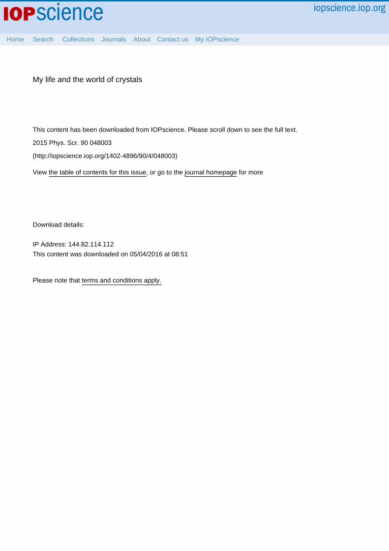
However, while we were performing the Stanfordexperiments on silicon, the structure had already been solvedby Takayanagi, using his TED methods [11]. As we were allhoping, it turned out to be a highly beautiful and extremelyelegant structure, shown in figure 4, which he called thedimer-adatom-stacking-fault (DAS) model. The new featurethat completed the model was the introduction of ‘dimers’:the other features were already known. These were identifiedin a classical crystallographic analysis of the Patterson func-tion [12], which showed new interatomic peaks at short dis-tances, pointing along new directions where bulk silicon hasno bonds. The result caused great excitement in the surface
science community and the paper has been cited over 1000times [11].
We completed our Stanford experiments and confirmedthe ‘adatom’ features of the 7 × 7 [13]. Then in 1988 wecompleted a full crystallographic dataset of the structure asthe debut experiment for the full UHV SXRD setup atbeamline X16A at Brookhaven, once the storage ring andbeamline were fully debugged [14]. These structure factordata were substantially more accurate than the TED mea-surements because our data measurement was not affected bydynamical effects. This allowed us to complete a full crys-tallographic refinement of the atomic positions. There wereclear distortions from the ideal bulk positions of the siliconatoms that yielded a systematic picture of strains in thestructure [14]. Since then, there have been further advances inthe structure refinement both by TED and SXRD, going to 3Ddata with high enough resolution to see bonding charges [15].
7. SXRD methods move out into the world
X-ray diffraction was my first interest, but at Bell Labs, Ifurther developed it by combining it with SR and surfaces;thus I can say that SXRD was born in Bell Labs. As a method,SXRD has become the definitive technique for the determi-nation of the atomic positions at surfaces and interfaces,completely displacing LEED This surface method is still usedtoday at the major SR facilities, NSLS (Brookhaven), ESRF(Grenoble), APS (Chicago), Diamond (Harwell), Soleil(Orsay), DESY (Hamburg), Spring8 (Harima), and SLS(Villigen).
For developing SXRD, I was awarded two prizes for thesurface structure work: the Warren Prize in 2000 and theSurface Structure Prize in 2011.
The years 1982–1990 then, were exciting times for me,years when I started several things simultaneously. At MurrayHill, I did my first SXRD experiment, studying Au(110),using the Eisenberger chamber, shown in figure 2, with the60 kW Rigaku rotating anode generator, with the result pub-lished in Physical Review Letters in 1983 as my first solowork [16]. I collaborated with Paul Fuoss in building the firstpermanent SXRD system for use at Brookhaven, testing itover a period of two years at Holmdel, before moving it toBrookhaven [17]. I started designing pieces of the X16Abeamline at Brookhaven, slowly working towards the struc-ture of the Si(111) 7 × 7 surface. I helped design and build upSXRD at LURE in Paris and then at ESRF in Grenoble. Lastbut not least, I started doing experiments with the Danishgroup at HASYLAB and there discovered the crystal trun-cation rod (CTR).
The first sources of SR were parasitic on high-energyphysics experiments, SSRL on the SPEAR ring at Stanfordand HASYLAB on the DORIS ring at DESY. Both of thesegave me opportunities for methods development in colla-borative experiments. Between 1982 and 1987, I was allo-cated several periods of beam time at SSRL in collaborationwith a number of scientists on separate occasions, includingGabriel Aeppli and Jakob Bohr on nitrogen on graphite;
Figure 4. Full unit cell of the DAS model of the silicon (111) 7 × 7structure, comprising 49 repeats of the underlying bulk crystal.Silicon atom heights are coded by the diameter of the circles. Thecrystallographic asymmetric repeating unit is the lower triangle.[11, 12, 14, 15]. Reproduced with permission from I K Robinson, JVacuum Science Technology A6 1966 (1988). Copyright 1988,American Vacuum Society.
5
Phys. Scr. 90 (2015) 048003 Invited Comment

Robert Feidenhans’l on silicon interfaces; Peter Bennett, andPeder Estrup, Bob Birgeneau, Mike Altman and with WarrenWaskiewicz as technician on Tungsten. The portable vacuumsystem taken to SSRL was also used by Mike Altman to dohis doctoral dissertation.
The NSLS in Brookhaven was the second of the ‘secondgeneration’ storage ring facilities designed specifically forproducing x-rays and Bell Labs was keen to capitalize on theresearch potential offered by a dedicated source. Things cametogether when beamline X16A and the permanent SXRDchamber started operating in 1988, as described below.
In 1987, Michèle Sauvage, a scientist from CNRS,invited me to help her set up her own SXRD chamber at theLURE synchrotron in Paris. Taking advantage of the NSLSshutdown, I went to Paris on a sabbatical leave for 8 months,staying at Place d’Italie and commuting to Orsay by RER tohelp Michèle set up her SXRD. I contributed a version of theSUPER control program [18], originally written by RobertFleming which was ready to install at X16A. This survivedfor many years in both places. We obtained the first SXRDdata on GaAs(100) surface structure using her new beamline.An important paper was published from that experiment inPhysical Review Letters and is widely cited [19].
Then in the winters of 1990–93, I spent a 3 month blockeach year in Grenoble on a Chaire Municipale award to helpdesign the SXRD instrumentation for ESRF during its build-up phase over 1989–94. I worked with Salvador Ferrer indesigning the ID03 UHV surface diffractometer system,played with components for a fast Kappa diffractometer andinteracted with the key team of scientists who would go on tolead the ESRF project.
8. At HASYLAB: the CTR
In the scientific world, new phenomena are always on thehorizon. Sometimes they remain unimportant, unappreciated,until they are discovered. With discerning eyes, we are able todiscover how they work, give names to them, and use them.Such is the case of the CTR. Considered an unimportantcuriosity until I explained it, it has since sprung to lifebecause of its many applications. In 1985, before NSLSturned on, I was collaborating on experiments at HASYLAB(DESY, Hamburg) with the Danish group composed of JakobBohr, Robert Feidenhans’l, Mourits Nielsen, Francois Grey,and Robert Johnson. The ‘baby chamber’ method of surfacediffraction was developed by this group to look at indiumantimonide (InSb) surfaces.
One night during my shift, while measuring InSb sur-faces, the data ran off the page requiring me to extend thelogarithmic graph paper with an extra sheet (on which theintensities were plotted); then it ran off again, and so, I starteda third sheet. The intensities, as I noticed, just kept gettingstronger and stronger as I got closer to the Bragg peak. This isnow understood as a smooth crossing over of the CTR fromthe surface contribution to the bulk. But that night turned outto be an exciting one for me because I then realized that it isthe cutting of the crystal which caused the rods in the
diffraction. The quantitative agreement came a little later, bythe inclusion of surface roughness in the theoretical descrip-tion. I named that phenomenon, which my colleagues at thattime called ‘integer-order reflections’, CTRs; expounded theidea behind it; and carried it forward.
In the morning, Robert Feidenhans’l came in for his shiftand told me (facetiously) that I had just wasted the nightmeasuring integer-order reflections when I could have mea-sured the surface-specific fractional orders. I explained to himwhat I discovered, then tried to make him understand thesignificance of what I saw. We discussed it again over thenext few days of beamtime. Buoyed by my discovery, I laterwrote the ideas into a solo paper ‘CTRs and surface rough-ness’ which was published in 1986 in Physical Review B [20].It was in this article where the name ‘crystal truncation rod’was first mentioned; it is now often called by its acro-nym, CTR.
9. Beamlines X16A and X16C at Brookhaven
It was a great relief when the x-ray ring at Brookhaven finallycompleted its upgrade in 1988, allowing us to start workingthere full-time. The upgrade installed wigglers into thestraight sections to provide higher power beams. Since 1983 Ihad been working on the design and construction of X16A,which was to be dedicated to SXRD. I designed many of theelectronic control systems myself because there was nothingcommercially available. The control computer was a PCrunning a Microsoft C version of SUPER [18], which we alsomaintained ourselves. The construction was a Bell Labs teameffort with contributions from Paul Fuoss, Alastair MacDo-well, Ed Melczer, Rick Levesque, Mike Altman, WarrenWaskiewicz and Steve Davey.
We had completed building X16A at the very end of1986. The NSLS’s planned wiggler shutdown was delayedfor a few months and that gave us the opportunity to testwhether X16A would work. This was when Michèle Sauvage,who had already started building her own SXRD beamline atLURE in Paris, first came to see how the beamline worked.Further collaborations with Francesco Sette, Alastair Mac-Dowell and Robert Feidenhans'l allowed us to complete ourfirst test experiments by February 1987. The X16A beamlineworked!
From 1988–1992, my regular routine was to spend 3 dayblocks of time at BNL, twice per week. Shutdowns wereusually on Tuesdays. Travel back to Bell Labs was once ortwice per month, so I became a bit out of touch with thecoworkers there. Walter Brown, my new department head,was always very supportive of this routine, but other man-agers wanted me to spend more time at Bell Labs.
Those early years 1988–1992 saw a high production ofSXRD results from Brookhaven involving my first Bell Labspostdoc, Elias Vlieg. Many collaborators came to use the newbeam line including Klaus Kern, Randy Headrick, LenFeldman, Ronan McGrath, Salvador Ferrer, Hartmut Zabel,Ed Conrad and Roberto Felici. We solved many crystal sur-face structures and started exploring phase transitions and
6
Phys. Scr. 90 (2015) 048003 Invited Comment

surface defects. One highlight was the study of the role ofsteps in the Pt(110) surface that explained how its missingrow structure evolved from a 1 × 2 to a 1 × 3 reconstructionand were coupled to its phase transitions [21].
In 1992, Peter Eng became my second postdoc, startingfirst at Bell Labs and then overlapping with the Illinois period.Collaborators then at X16A were Detlef Smilgies (for oneyear on fellowship in 1992–93), Rolf Schuster (also for a oneyear fellowship, 1994–95), Peter Bennett, Holger Meyerheim,Harald Reichert, Helmut Dosch, and Ulrich Pietsch. DonWalko was my student who did his thesis work at X16A from1996 to 2000. Then from 2002 to 2005, Sanjit Ghose becamemy third postdoc. Sanjit worked with Bob Averback and PeterBennett on their regular visits to keep using X16A. Peter Engand Detlef Smilgies undertook studies of the Mo(001) surfacewhich was very challenging for the UHV system of X16A,which achieved its peak performance during this period. Withthe help of these collaborators, I studied such crystallographiccuriosities as adsorbate-induced faceting [22], anisotropicsurface vibrations [23] and surface ordering of alloys [24]. Anexample of one of the many surface structures determinedduring these years is the Mo(001)/O √5 ×√5 reconstructedsurface shown in figure 5 [25].
We also built up X16C in 1992, a second beamline atNSLS for EXAFS and general diffraction experiments. Thiswas organized by Alastair MacDowell who used a newmonochromator designed by Paul Fuoss [26]. While I was inGrenoble, in 1990, I started designing a kappa-geometrydiffractometer, then called the ‘Goniomètre a Grande Vitesse’
(GGV) for use at X16C. Its high speed came from the first useof direct-drive servo motors [27]. This was fabricated in theIllinois machine shops in 1993 and delivered to Brookhavenand tested in 1994 [27]. It served well for training studentsand developing SXRD ideas for surfaces and interfaces out-side vacuum. Arunabha Ghosh helped assemble the instru-ment and devised optical methods of aligning it. Yong Chudeveloped x-ray diffraction for electrochemical interfaces andcompleted his PhD studying these [28]. Dave Fanning studiedferroelectric crystals which he grew using strategic dopingand helped develop the diffraction anomolous fine structure(DAFS) technique for studying them [29]. Chinkyo Kimexamined strain transfer effects between thin films of elec-tronic materials [30]. Sébastien Boutet observed the firstCTRs from protein crystals [31]. X16C was a self-organizedproject and was amicably shared with Dave Adler (1993–8),Anatoly Frenkel (1998–2003), Matthew Marcus and AlastairMacDowell (1985–99).
A fact of life is that good things too can end. Around2006, X16C was taken over by Stony Brook for use as apowder diffraction beamline. X16A was taken over by a BNLgroup from NSLS-2 sometime in 2008 and the SXRD activitythere ended. Finally, the whole NSLS facility closed perma-nently in September 2014. One of the last pictures of X16Awhile it was still active is shown in figure 6.
10. Switzerland or USA?
It was in 1987 that I met my future wife, Sally Calong-Robinson, whose doctoral degree is in Literature. I talked toher about a Japanese novelist and asked her if she wanted tosee a Japanese play, and that secured a yes answer from her.We got married in 1989.
Meanwhile, the long-term funding at Bell Labs wasthreatened by the 1984 divestiture of the ‘Baby Bells’ fromthe parent company, AT&T, which lost its virtual monopolyof the telephone network. I decided it was time to look for auniversity position as I had sufficient success to be able toexpect to go straight into a tenured teaching position.
Figure 5. Structure of the Mo(001)/O √5x√5 reconstructed surfacemeasured at X16A. Oxidation of the Mo surface causes theformation of clusters of Mo surrounded by oxygen. One Mo atom islost per √5x√5 unit cell leaving a vacancy behind [25]. © IOPPublishing. Reproduced by permission of IOP Publishing. All rightsreserved.
Figure 6. Peter Bennett and myself standing in front of the X16Acontrol station around 2004.
7
Phys. Scr. 90 (2015) 048003 Invited Comment
In 1992, I got two offers: one from Switzerland andanother from Illinois. After careful consideration, weighingthe pros and the cons for each, we decided to accept the offerfrom Illinois, and we moved to Urbana, a university town, inAugust of the same year. In New York City, we lived in a onebedroom, two-bathroom apartment, as seen in figure 3; inUrbana we bought a huge 400 m2 house for a very reasonableprice.
After moving to Illinois in 1992, Sally and I would go toBrookhaven every summer, driving East in May and West inAugust. We would usually take an apartment on the BNL sitefor 1–2 months. The UHV SXRD diffractometer [17] onX16A would be exchanged with a different one operated byKen Evans-Lutterodt once per year and this equipment couldbe swapped over quickly within a single day. On X16C wewould install the kappa diffractometer [27] for each experi-ment, usually 2–3 weeks at a time. Aligning this was a goodroutine for the students.
At the University of Illinois, I became active in learningto use the high degree of coherence produced by the new‘third generation’ light sources, ESRF in Grenoble starting in1994, APS at Argonne in 1995, and Spring8 in Harima. Thesethird generation sources use magnetic undulators to generatex-ray beams that are thousands of times brighter than thesecond generation, such as Brookhaven. New ideas wereneeded to harness the coherence that follows from thisbrightness and apply it to the study of everyday things likematerials or biology. An early breakthrough, still using asecond generation source, was the observation of x-ray‘speckle’, analogous to its optical equivalent seen with laserbeams [32].
ESRF (Grenoble) came on line in 1994 and I joined witha group of colleagues including Jens Als-Nielsen, Ron Pin-dak, Robert Fleming and Steve Dierker to try its new cap-abilities. We were ready with test samples and motorizedpinholes to look at the ‘speckle’ they would generate, but onthe first occasion we were given just a single day of beamtimeon ID10. In spite of this, already starting with disorderedmultilayer samples, I was able to understand their behavior bymodeling the disorder to obtain crude images from that singlemeasurement [33]. This was the first example of coherentimaging with x-rays, published in 1995.
Moving steadily forwards, my Illinois group started todevelop the slit hardware needed to control the small beamsneeded to achieve coherence and to invent the analysismethods needed to obtain images. Jeff Libbert was the firstpostdoc and John Pitney the first student to try this. IvanVartanyants came to visit us in Illinois from Moscow for3 months in 1997, then every year for 9 months from 1998 to2004. We came to Troika beam line (ID10) of ESRF to imageetched silicon surfaces, looking at their surface morphologyafter treatments that change their structures on the nanoscale[34]. We learned how to invert the coherent diffraction pat-terns by reading the papers of Jim Fienup from the opticsliterature, which showed that they could be inverted, i.e.solving the phase problem, simply because they were over-sampled. These ideas had also been discussed by David Sayre
back in 1952, immediately after the publication of theShannon theorem [35].
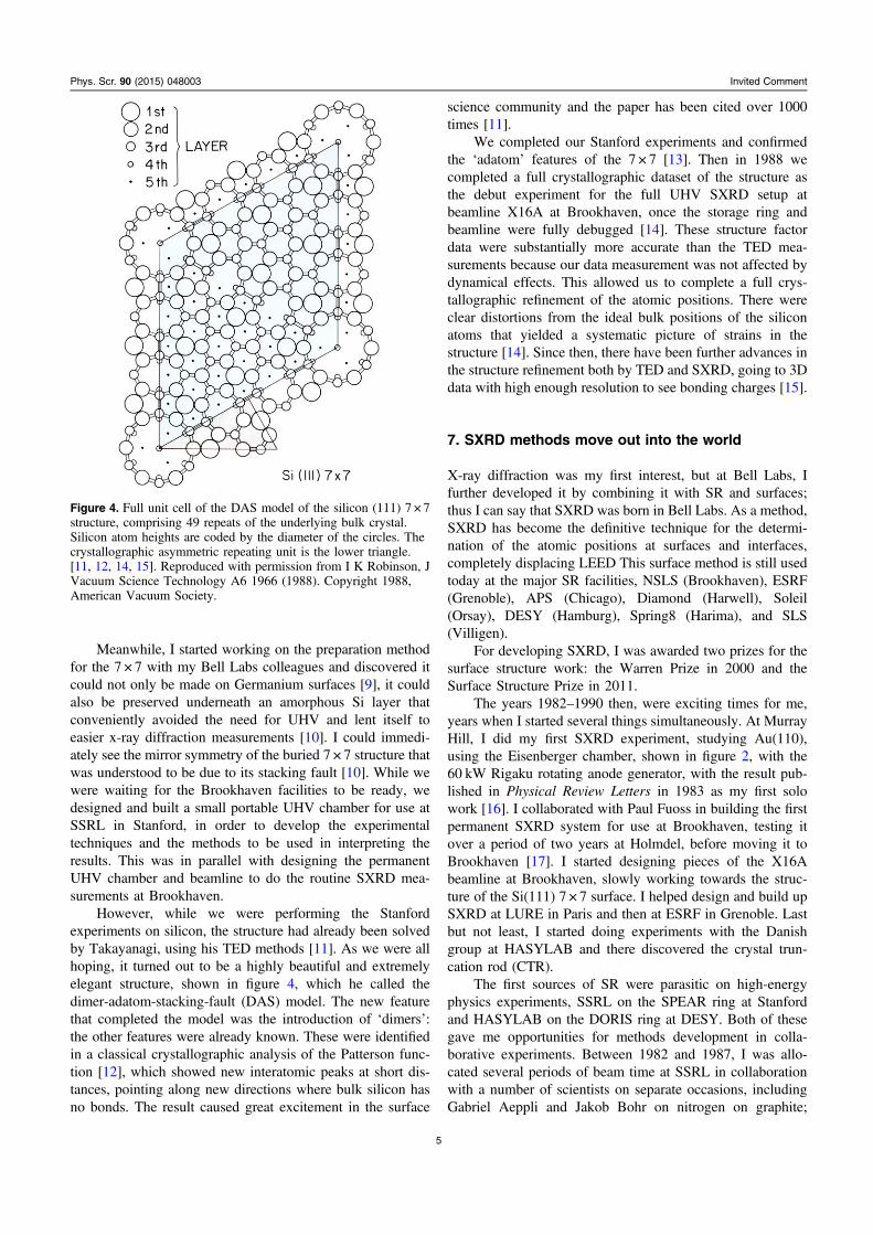
Another breakthrough moment took place in the free-flowing discussions at one of our group meetings in theMaterials Research Laboratory in Urbana. I knew that slitscan diffract a laser beam into a fringed pattern and started towonder what the coherent x-ray diffraction (CXD) pattern ofa small crystal would look like; I concluded it would look thesame and clearly remember sketching this on the blackboard.We decided to test this idea using the new 33-ID beamline runby the University of Illinois at APS. My students GarthWilliams and Mark Pfeifer heated films of gold until theybroke up into small crystals, and when these were placed inthe coherent beam they gave beautiful fringed diffractionpatterns, like the one shown in figure 7. These patterns couldbe inverted into credible images of the crystals, but the qualitywas limited by beam stability and imperfect coherence [36].The CXD method was then born.
Ivan Vartanyants and I had many discussions about thepotential and limitations of CXD. We tried to understand howthe partial coherence of the beam would affect the result andhow dynamical effects would appear in the images. Wethought about new phasing algorithms. Above all, we dis-cussed the sensitivity of CXD to crystal strains [37]. As aresult of those discussions, I started campaigning for fundingto build a dedicated CXD beamline at APS, in order tocontinue developing methods of using the very high coher-ence for direct 3D imaging of structure. The potential appli-cation was its ability to examine strain distributions insidecomplex materials on the nanometer length scale.
One valuable opportunity offered by the University ofIllinois was the chance to take regular sabbatical leaves,typically for 6 months every three years. As Urbana was alittle isolated, this offered me the chance to get back intoactive research environments in other places. In 1996, I spent3 months with Wolfgang Moritz in Munich, playing with aninteresting idea to use CTRs as a reference to solve thecrystallographic phase problem for the surface layer. Thisworked well enough eventually to lead to a publication [38].In 2004, I divided my sabbatical between the Nagoya
Figure 7. Coherent x-ray Diffraction pattern taken from a small goldnanocrystal at the UNICAT 33ID beamline of APS. The dark circlein the center is the shadow of a beamstop used to protect the CCDcamera [36].
8
Phys. Scr. 90 (2015) 048003 Invited Comment
University Venture Business Laboratory (VBL) and the Max-Planck-Institut für Metalforschung, Stuttgart where I wasawarded a Humboldt Foundation Senior Research Fellow-ship. The VBL work with Masao Tabuchi also continued theCTR-based phasing work and led to another publication [39].In Stuttgart, I looked at oxidation of Pt(111) surfaces withAndreas Stierle.
11. The 34-ID beamline at the advanced photonsource
My grant to build sector 34-ID at Argonne was announced in1999 by the National Science Foundation. Howard Birnbaumas MRL director arranged the matching funding and theproject was incorporated into the UNICAT consortiumheaded by Haydn Chen. Curtis Kenney-Benson was hired tohelp me design the beamline, order the components andcoordinate the writing of its ‘Preliminary Design Report’ and‘Technical Design Report’ required by Argonne. My studentsGarth Williams, Mark Pfeifer, Sébastien Boutet, and TommyAngelini spent the summer of 2002 actually building thebeamline. As at Brookhaven before, we bolted all the com-ponents together ourselves and wired up the ‘EquipmentProtection System’, under the guidance of Pete Jemian. PaulZschack, as the manager of UNICAT, interfaced our projectwith the existing sector 33 beamlines and with the Argonnemanagement.
Construction completed, the operation started in 2003.The first experiments were conducted by Sébastien Boutetused the pink beam to look at damage to protein crystals, forsmall-angle scattering by Gerard Wong and Tommy Angelini,and speckle from thin films in the grazing incidence small-angle x-ray scattering (GISAXS) geometry by Wei Zhang. Amonochromator was later added using an economic water-cooled design, which was not in our original budget [26]. Westarted using diamond monochromator crystals, but foundthey were not good at preserving the beam coherence; laterwe switched to silicon. Though these developments, weended up with the only water-cooled silicon monochomator atAPS, a component which is still operating successfully today.
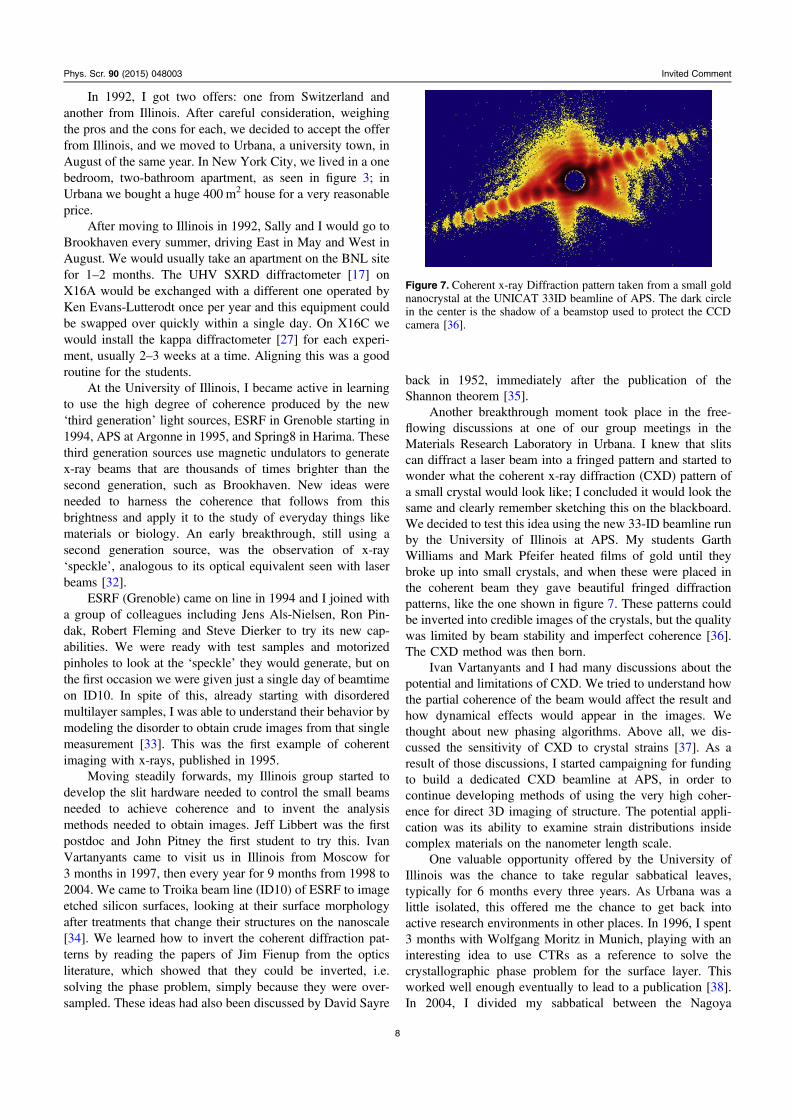
Because of its potential application to the study of sur-faces using CXD, we then designed and built a UHV chamberdirectly into the beamline. We also had to include verydelicate UHV roller-blade slits, fabricated by Alex Nozko, toselect the coherence just in front of the sample. This led up tothe first UHV CXD experiment in 2003 by Mark Pfeifer andGarth Williams in which they grew lead nanocrystals in situin the beamline. This experiment was the first to imagestrains, illustrated in figure 8, a major breakthrough that waspublished in Nature in 2006 [40]. This defined the beamlineand established the Bragg coherent diffraction imaging(BCDI) method.
Initially, the operation of the 34IDC beamline was paidby the University of Illinois through UNICAT, but transferredto the Department of Energy in 2006. Garth Williams, MarkPfeifer, Sébastien Boutet and later student Mengning Liangall continued on in the CXD field. Sébastien Boutet, Garth
Williams and Mengning Liang became founders of the new x-ray free-electron Laser (XFEL) field (below). My postdoc atthat time, Ross Harder (2004-7), continued into the UCLfunding period then transferred onto the Argonne staffin 2007.
This story illustrates the convergence of theoretical ideas,methods development, the involvement of large facilities and,above all, teamwork required to bring about revolutions inscience in the modern era. BCDI was pioneering work aimedat developing new SR-based techniques by using the simplefact that the light waves are in phase with each other. I ori-ginally named this CXD, but the name evolved into BCDI toincorporate the role of imaging and the crystallographicconcept of Bragg diffraction from crystals. Over an entirecentury, starting with the Braggs, diffraction based methodshave succeeded in three-dimensional imaging of materials,under the title of crystallography. My contribution to thisstory was to demonstrate how it is possible to obtain three-dimensional representations of deformations and defects innanomaterials.
12. Nanotechnology in London
In the summer of 2002, Gabriel Aeppli recruited me to moveto London where he was already Director of the LondonCenter for Nanotechnology (LCN). He was my friend andcolleague from Bell Labs, where he had started as MTS at thesame time as me. Sally and I were strolling down ThirdAvenue after dinner one evening that summer and spottedGabriel in another restaurant. This chance encounter was to
Figure 8. Images of strains inside a 700 nm hemispherical leadnanocrystal obtained at sector 34-ID of the Advanced Photon Source(APS). The strain projection (color) is associated with contact withthe silicon substrate upon which the crystals were grown [40].
9
Phys. Scr. 90 (2015) 048003 Invited Comment

change my life, when he told me he was moving to Londonand would have some new faculty positions opening. Iapplied and interviewed in April 2003. The position wasoriginally listed as an opening for neutron diffraction but thiswas broadened to include x-ray diffraction as well. In the endtwo professorial appointments were made, one for myself, theother for Des McMorrow. My appointment was at UniversityCollege London in both the LCN and the Physics andAstronomy Department as Chair of Physics. A 5 year half-time visiting appointment at the Diamond Light Source (DLS)as a ‘Diamond Fellow’ was arranged by Gerd Materlik, itsdirector and CEO. 2003 was an interesting and exciting year,a time to believe that everything is possible!
It was in London where the CXD methods developed.New opportunities lay with the DLS a third generation syn-chrotron located at Rutherford Appleton Laboratory (RAL)near Oxford. My research at UCL has been largely tied to thisdevelopment, although we continue to do experiments atESRF, SLS and APS during the buildup period. Followingthe DLS appointment, I became a founding ‘Diamond Fel-low’ of the Research Complex at Harwell (RCaH), alsolocated at RAL. This is a meeting place for scientists inter-ested in the transfer of methodologies from the physical to thelife sciences. Materials and biological imaging are the maindirections under development there. Through these connec-tions, I have been developing methods of using the very highcoherence of the latest SR sources to enable direct 3D ima-ging of structure.
Three major grants supported the work of my group,divided between the UCL and RCaH centers. The first, enti-tled ‘nanosculpture’, looks at strains induced in nanometer-sized crystals either synthesized from atoms in a‘bottom up’procedure, or else carved by lithography from bulk materialsin a ‘top down’ approach. The second is to study the structureof the human chromosome by coherent x-ray imaging meth-ods. The third is to develop new x-ray imaging methods basedon deliberate modulation of the phase by suitably developedx-ray optics.
My first UCL project was to perform BCDI on nano-wires. Steven Leake, Ross Harder and Marcus Newton lookedat ZnO nanowire structures. Steven’s most important resultwas to find that the apparent coherence was dramaticallydifferent from one Bragg reflection to another, explained as aneffect of the rarer longitudinal coherence of the beam [41].The ‘nanosculpture’ grant was coordinated with a partner userproposal (PUP) running from 2009 to 2011 with Argonne.We introduced a new confocal microscope to the beamline toselect and align known nanostructures in the beam. In thisway, we developed the methods of using multiple Braggpeaks from the same nanocrystal. There were importantresults at 34-ID-C from Moyu Watari, Marcus Newton, RossHarder, and later Jesse Clark and Gang Xiong.
Building on this success, we renewed the PUP to startdeveloping x-ray ptychography [42] during 2011–14. Weprovided new precision piezo scanning stages to 34-ID-C andsupported Xiaojing Huang’s postdoc at Argonne. Many morepapers emerged in the course of developing the method,accounting for the results of the experiments done by Felisa
Berenguer, Jesse Clark, Gang Xiong as postdocs, RichardBean, Laura Shemilt, Xiaowen Shi, Nicolas Burdet, MarianneMonteforte, Maria Civita, Ana Estandate and Chris Lynch asstudents. Like before at Brookhaven, we had the first beam-line dedicated to a promising new technique which attractedprominent visiting scientists during this development period:Franz Pfeiffer (2003), Virginie Chamard (2004), LorenzStadler (2004), Roberto Felici (2005) and Hyunjung Kim(2008-pres), all coming to see what we were up to on the newbeamline and take home ideas. Throughout both PUPs, RossHarder was fully involved with both the science and thetechnical improvements of the 34-ID-C beamline.
Meanwhile back at Diamond, I had prepared BeamlineProposal 048 ‘A CXD and XPCS Beamline for the DLS’ forpresentation to the Science Advisory Committee in November2004. This was accepted as part of its Phase II construction.Christoph Rau was hired as the Principal Beamline Scientistand construction began in 2007. Under his direction, thebeamline evolved into the longest beamline in Europe withtwo parallel branches, one for tomographic imaging and onefor coherent diffractive imaging (CDI). BCDI was cateredfor by the use of giant robots to carry the area detectorsneeded to reach the Bragg peaks with enough distance fromthe sample to allow oversampling. My current group consistsof postdocs Fucai Zhang, Jörg Schwenke, Bo Chen,Mohammed Yusuf and Graeme Morrison. They have usedthe CDI branch on a number of occasions for looking atchromosomes, with fresh samples prepared in the RCaHlaboratory next door, and for developing new modulation-based imaging methods.
Last, but not least, my first XFEL BCDI experiment wasdone at Stanford’s new Linac Coherent Light Source (LCLS)facility in November 2011 with Jesse Clark, Ross Harder anda large group of other scientists and Stanford staff. 60 h ofXFEL beam led to two important results [43]. Using theextremely short x-ray pulses from the LCLS, we were alsoable to show how one can excite motion (phonons) of theatoms in individual nanoparticles and follow how thesemovements propagate in the particles. These new directions,opened up by XFELs, point the way to the future for me: timedomain crystallography for the discovery of new materials.
References
[1] Feynman R P, Leighton R B and Sands M 1963 The FeynmanLectures on Physics (Reading, MA: Addison-Wesley)
[2] Young J Z 1971 An Introduction to the Physiology of Man(Oxford: Oxford University Press)
[3] Horowitz P and Hill W 1981 The Art of Electronics(Cambridge: Cambridge University Press)
[4] Horowitz P and Robinson I 1981 Lab Manual for the Art ofElectronics (Cambridge: Cambridge University Press)
[5] Eisenberger P and Marra W C 1981 X-ray-diffraction study ofthe Ge(001) reconstructed surface Phys. Rev. Lett. 46 1081
[6] Bennett P A, Feldman L C, Kuk Y, McRae E G and Rowe J E1983 Stacking-Fault model for the Si(111)-(7 × 7) surfacePhys. Rev. B 28 3656
[7] Takayanagi K 1984 Surface structure imaging by electronmicroscopy J. Microsc. 136 287
10
Phys. Scr. 90 (2015) 048003 Invited Comment
[8] Binnig G, Rohrer H, Gerber C and Weibel E 1983 7 × 7reconstruction on Si(111) resolved in real space Phys. Rev.Lett. 50 120
[9] Gossman H J, Bean J C, Feldman L C, McRae E G andRobinson I K 1985 7 × 7 reconstruction of Ge(111) surfacesunder compressive strain Phys. Rev. Lett. 55 1106
[10] Robinson I K 1987 The symmetry of Si(111)7 × 7 at an a Siinterface Phys. Rev. B 35 3910
[11] Takayanagi K, Tanishiro Y, Takahashi M and Takahashi S1985 Structural-analysis of Si(111)-7 × 7 by UHV-transmission electron-diffraction and microscopy J. Vac. Sci.Technol. A 3 1502
[12] Takayanagi K, Tanishiro Y, Takahashi S and Takahashi M1985 Structure-analysis of Si(111)-7 × 7 reconstructedsurface by transmission electron-diffraction Surf. Sci.164 367
[13] Robinson I K, Waskiewicz W K, Fuoss P H, Stark J B andBennett P A 1986 X ray diffraction evidence of adatoms inthe Si(111)7 × 7 reconstructed surface Phys. Rev. B 33 7013
[14] Robinson I K, Waskiewicz W K, Fuoss P H and Norton L J1988 Observation of strain in the Si(111) 7 × 7 surface Phys.Rev. B 37 4325
[15] Ciston J, Subramanian A, Robinson I K and Marks L D 2009Diffraction refinement of localized antibonding at the Si(111) 7 × 7 surface Phys. Rev. B 79 193302
[16] Robinson I K 1983 Direct structure determination of the Au(110) reconstruction by x ray diffraction Phys. Rev. Lett.50 1145
[17] Fuoss P H and Robinson I K 1984 Apparatus for x raydiffraction in ultra high vacuum Nucl. Instrum. Methods222 171
[18] Robinson I K 2003 Surface x-ray diffraction Characterizationof Materials ed E N Kaufmann (Hoboken, NJ: Wiley)pp 1007–27
[19] Sauvage Simkin M, Pinchaux R, Massies J, Claverie P,Jedrecy N, Bonnet J and Robinson I K 1989 VariableStoichiometry of the GaAs (001) c(4 × 4) surface: an in situx-ray scattering study Phys. Rev. Lett. 62 563
[20] Robinson I K 1986 Crystal truncation rods and surfaceroughness Phys. Rev. B 33 3830
[21] Robinson I K, Vlieg E and Kern K 1989 Non ising behavior ofthe Pt(110) surface phase transition Phys. Rev. Lett. 63 2578
[22] Walko D A and Robinson I K 2001 Energetics of O-inducedfaceting on Cu(115) Phys. Rev. B 64 045412
[23] Meyerheim H L, Moritz W, Schulz H, Eng P J andRobinson I K 1995 Anharmonic thermal vibrations observedby surface x-ray diffraction for Cs/Cu(001) Surf. Sci.333 1422
[24] Reichert H, Eng P J, Dosch H and Robinson I K 1995Thermodynamics of surface segregation profiles atCu3Au(001) resolved by x-ray scattering Phys. Rev. Lett.74 2006
[25] Robinson I K, Smilgies D M and Eng P J 1992 Clusterformation in the adsorbate-induced reconstruction of the O/Mo(001) surface J. Phys.: Condens. Matter 4 5845
[26] Kupp T, Blank B, Deyhim A, Fuoss P H, Benson C A andRobinson I K 2004 Development of a double crystalmonochromator Proc. of the AIP 8th Int. Conf. CP705 onSynchrotron Radiation Instrumentation pp 651–4
[27] Robinson I K, Graafsma H, Kvick A and Linderholm J 1995First testing of the fast kappa diffractometers at NSLS andESRF Rev. Sci. Instrum. 66 1765
[28] Chu Y S, Robinson I K and Gewirth A A 1999 Comparison ofaqueous and dry oxide formation on Cu(111) J. Chem. Phys.110 5952
[29] Frenkel A I, Cross J O, Fanning D M and Robinson I K 1999DAFS analysis of magnetite J. Synchrotron Radiat. 6 332
[30] Kim C, Robinson I K, Myoung J, Shim K and Kim K 1999Buffer layer strain transfer in AlN/GaN near criticalthickness J. Appl. Phys. 85 4040
[31] Boutet S, Robinson I K, Hu Z, Thomas B R and Chernov A A2002 Surface relaxation in protein crystals Phys. Rev. E 66061914
[32] Sutton M, Mochrie S G J, Greytak T, Nagler S E, Berman L E,Held G A and Stephenson G B 1991 Observation of speckleby diffraction with coherent x-rays Nature 352 608
[33] Robinson I K, Pindak R, Fleming R M, Dierker S B, Ploog K,Grübel G, Abernathy D L and Als-Nielsen J 1995 Observationand explanation of one-dimensional x-ray speckle patternsfrom synthetic multilayers Phys. Rev. B 52 9917–24
[34] Vartanyants I A, Pitney J A, Libbert J L and Robinson I K1997 Reconstruction of surface morphology from coherentx-ray reflectivity Phys. Rev. B 55 13193
[35] Sayre D 1952 Some implications of a theorem due to ShannonActa Crystallogr. 5 843
[36] Robinson I K, Vartanyants I A, Williams G J, Pfeifer M A andPitney J A 2001 Reconstruction of the shapes of goldnanocrystals using coherent x-ray diffraction Phys. Rev. Lett.87 195505
[37] Robinson I K and Vartanyants I A 2001 Use of coherent x-raydiffraction to map strain fields in nanocrystals Appl. Surf.Sci. 182 186–91
[38] Saldin D K, Harder R, Volger H, Moritz W and Robinson I K2001 Solving the structure completion problem in surfacecrystallography Comput. Phys. Commun. 137 12–24
[39] Robinson I K, Tabuchi M, Hisadome S, Oga R and Takeda Y2005 Perturbation method of analysis of crystal truncationrod data J. Appl. Crystallogr. 38 299–305
[40] Pfeifer M A, Williams G J, Vartanyants I A, Harder R andRobinson I K 2006 Three-dimensional mapping of adeformation field inside a nanocrystal Nature 442 63
[41] Leake S J, Newton M C, Harder R and Robinson I K 2009Longitudinal coherence function in x-ray imaging of crystalsOpt. Express 17 15853
[42] Rodenburg J M, Hurst A C, Cullis A G, Dobson B R,Pfeiffer F, Bunk O, David C, Jefimovs K and Johnson I2007 Hard-x-ray lensless imaging of extended objects Phys.Rev. Lett. 98 034801
[43] Clark J N et al 2013 Ultrafast three dimensional imaging oflattice dynamics in gold nanocrystals Science 341 56
11
Phys. Scr. 90 (2015) 048003 Invited Comment