myc—master regulator of the cancer epigenome and … · the importance of myc in human...
TRANSCRIPT

genesG C A T
T A C G
G C A T
Review
MYC—Master Regulator of the Cancer Epigenomeand Transcriptome
Candace J. Poole and Jan van Riggelen *
Augusta University, Department of Biochemistry and Molecular Biology, 1410 Laney-Walker Blvd.,Augusta, GA 30912, USA; [email protected]* Correspondence: [email protected]; Tel.: +1-706-721-0856; Fax: +1-706-721-6608
Academic Editor: Frank BuchholzReceived: 28 February 2017; Accepted: 10 May 2017; Published: 13 May 2017
Abstract: Overexpression of MYC is a hallmark of many human cancers. The MYC oncogene has longbeen thought to execute its neoplastic functions by acting as a classic transcription factor, deregulatingthe expression of a large number of specific target genes. However, MYC’s influence on many ofthese target genes is rather modest and there is little overlap between MYC regulated genes indifferent cell types, leaving many mechanistic questions unanswered. Recent advances in the fieldchallenge the dogma further, revealing a role for MYC that extends beyond the traditional concept ofa sequence-specific transcription factor. In this article, we review MYC’s function as a regulator of thecancer epigenome and transcriptome. We outline our current understanding of how MYC regulateschromatin structure in both a site-specific and genome-wide fashion, and highlight the implicationsfor therapeutic strategies for cancers with high MYC expression.
Keywords: MYC; chromatin remodeling; cancer
1. Introduction
The importance of MYC in human development and disease has generated intense researchinterest over the past 30 years, resulting in numerous original articles and reviews (for example,see [1–3]). The MYC family is comprised of c-MYC (herein referred to as MYC, unless otherwisespecified), N-MYC, and L-MYC, which encode for basic helix–loop–helix leucine zipper (bHLH-Zip)transcription factors [4] that have been found to play a unique role in regulating an extensive range ofbiological processes including stemness, cellular proliferation, and neoplastic transformation. Whilethe activity of MYC family members is tightly regulated in non-malignant cells, their constitutiveexpression is directly linked to the pathogenesis of a wide variety of human cancers [5–8]. The fact thatelevated levels of MYC proteins are found in 60–70% of all cancers (reviewed in [1]) and the discoverythat tumors can be dependent on continuous MYC expression (known as oncogene addiction) [9]have made this family of oncogenes a highly promising therapeutic target. However, even after threedecades of research, both the exact molecular mechanism of how MYC promotes tumorigenesis and apharmacologic inhibitor selectively targeting the oncogene remain elusive.
MYC exerts its neoplastic features by increasing autonomous cellular proliferation, growth,angiogenesis, and genomic destabilization while blocking differentiation (see Figure 1) (reviewedin [10]). However, these diverse cellular functions are attributed to the still not completely understoodability of MYC to control the expression of a large set of genes. MYC proteins have first been describedas sequence-specific transcription factors, forming heterodimeric complexes with MYC-AssociatedFactor X (MAX). MYC–MAX complexes are now known to recognize a consensus sequence knownas Enhancer box (“E-box”), activating the transcription of genes [11–13]. This finding sparkeda comprehensive search for MYC target genes and their function and involvement in neoplastictransformation [14]. However, the scope and complexity of MYC’s action became apparent when
Genes 2017, 8, 142; doi:10.3390/genes8050142 www.mdpi.com/journal/genes

Genes 2017, 8, 142 2 of 28
it was discovered that there are approximately 20,000 E-box sites in the human genome, of whichonly a subset is differentially bound by MYC in a cell type-specific fashion [15]. To further increasethe complexity, MYC was also later found to be capable of repressing the transcription of genesthrough interactions with other transcription factors [16–18]. It turned out that both MYC’s activatingand repressing functions are critical for tumorigenesis and depend on the recruitment of chromatinmodifying co-factors that remodel chromatin structure in the vicinity of the binding sites. However,MYC acts rather weakly at many of its target gene promoters, and even genomic location profiles haveoften proven non-predictive of MYC-dependent transcriptional regulation [15]. Furthermore, despiteconsiderable efforts to identify a MYC target gene signature using comparative gene profiling andgenomic location analyses, little overlap between MYC regulated genes in different cell types has beenfound [14,19]. To explain these discrepancies, the classic mechanistic model has recently been extendedto incorporate MYC’s function as a regulator of global chromatin structure and transcription. In thisarticle, we review the role of MYC as a regulator of the cancer epigenome and transcriptome boththrough site-specific, local mechanisms as well as genome-wide effects, and highlight the potential fornovel therapeutic strategies.
Genes 2017, 8, 142 2 of 28
neoplastic transformation [14]. However, the scope and complexity of MYC’s action became apparent
when it was discovered that there are approximately 20,000 E-box sites in the human genome, of
which only a subset is differentially bound by MYC in a cell type-specific fashion [15]. To further
increase the complexity, MYC was also later found to be capable of repressing the transcription of
genes through interactions with other transcription factors [16–18]. It turned out that both MYC’s
activating and repressing functions are critical for tumorigenesis and depend on the recruitment of
chromatin modifying co-factors that remodel chromatin structure in the vicinity of the binding sites.
However, MYC acts rather weakly at many of its target gene promoters, and even genomic location
profiles have often proven non-predictive of MYC-dependent transcriptional regulation [15].
Furthermore, despite considerable efforts to identify a MYC target gene signature using comparative
gene profiling and genomic location analyses, little overlap between MYC regulated genes in
different cell types has been found [14,19]. To explain these discrepancies, the classic mechanistic
model has recently been extended to incorporate MYC’s function as a regulator of global chromatin
structure and transcription. In this article, we review the role of MYC as a regulator of the cancer
epigenome and transcriptome both through site-specific, local mechanisms as well as genome-wide
effects, and highlight the potential for novel therapeutic strategies.
Figure 1. MYC as a transcription factor and oncogene. Display of the X-ray crystal structure of a MYC–
MAX heterodimer bound to DNA as a site-specific transcription factor complex [20,21].
Overexpression of MYC causes the deregulation of central cellular processes including cell cycle
progression, metabolism, differentiation, and angiogenesis, together contributing to neoplastic
transformation. Deregulated MYC expression is implicated in a wide variety of human cancer types
including Burkitt’s lymphoma, acute lymphoblastic leukemia (ALL), and neuroblastoma. Image
created with: UCSF Chimera; PDB: 1NKP.
2. Recruitment of Chromatin Modifiers for MYC-Dependent Transactivation
The gene-specific transactivation model depends on the ability of MYC–MAX complexes to
recruit chromatin-modifying co-factors. These change the chromatin structure in the vicinity of the
binding site, allowing or preventing the transcription of the corresponding gene by regulating its
accessibility to the basal transcriptional machinery or releasing preloaded RNA Polymerase II (RNA
Pol II) from pausing.
An increasing number of chromatin modifying co-factors including chromatin “writers,”
“readers,” and “erasers” have been found to interact directly or indirectly with MYC–MAX
complexes (for an overview, see Figure 2). Many of these protein–protein interactions are facilitated
through MYC’s N-terminus, which harbors the transcriptional activation domain (TAD) and highly
conserved sequence elements, known as “MYC box” (MB) 0, I, and II, followed by MB III and IV in
the central MYC domain ([22] and reviewed in [23]). MB I, II, and III are essential for all biological
functions of MYC, and are required for site-specific transactivation and transrepression of most, but
Figure 1. MYC as a transcription factor and oncogene. Display of the X-ray crystal structure ofa MYC–MAX heterodimer bound to DNA as a site-specific transcription factor complex [20,21].Overexpression of MYC causes the deregulation of central cellular processes including cell cycleprogression, metabolism, differentiation, and angiogenesis, together contributing to neoplastictransformation. Deregulated MYC expression is implicated in a wide variety of human cancer typesincluding Burkitt’s lymphoma, acute lymphoblastic leukemia (ALL), and neuroblastoma. Image createdwith: UCSF Chimera; PDB: 1NKP.
2. Recruitment of Chromatin Modifiers for MYC-Dependent Transactivation
The gene-specific transactivation model depends on the ability of MYC–MAX complexes torecruit chromatin-modifying co-factors. These change the chromatin structure in the vicinity ofthe binding site, allowing or preventing the transcription of the corresponding gene by regulatingits accessibility to the basal transcriptional machinery or releasing preloaded RNA Polymerase II(RNA Pol II) from pausing.
An increasing number of chromatin modifying co-factors including chromatin “writers”,“readers”, and “erasers” have been found to interact directly or indirectly with MYC–MAX complexes(for an overview, see Figure 2). Many of these protein–protein interactions are facilitated throughMYC’s N-terminus, which harbors the transcriptional activation domain (TAD) and highly conservedsequence elements, known as “MYC box” (MB) 0, I, and II, followed by MB III and IV in the central MYCdomain ([22] and reviewed in [23]). MB I, II, and III are essential for all biological functions of MYC,

Genes 2017, 8, 142 3 of 28
and are required for site-specific transactivation and transrepression of most, but not all, direct targetgenes [24,25]. The exact mechanism of how MYC coordinates all these protein–protein interactions andhow individual chromatin modifying enzymes contribute to MYC’s oncogenic properties is subject tointense research, but still not completely understood. While the recruitment of activating vs. repressingfactors is thought to be determined by additional DNA-binding proteins such as Specificity Protein-1(SP1) or MYC-Interacting Zinc Finger Protein-1 (MIZ-1), it is not clear whether variants of the E-boxmotif found in different sets of target genes influence the quality of protein–protein interactions.Recent research has been focused on local chromatin structure, which facilitates tethered recruitment,as discussed below, but might also play a role in defining the type of co-factor interaction. Besidesthe possibility that specific co-factors are recruited in a context-dependent manner, they also might berecruited in response to stimulation, allowing for an additional level of signal integration to modulatethe transcriptional output of the MYC network.
Genes 2017, 8, 142 3 of 28
not all, direct target genes [24,25]. The exact mechanism of how MYC coordinates all these
protein–protein interactions and how individual chromatin modifying enzymes contribute to MYC’s
oncogenic properties is subject to intense research, but still not completely understood. While the
recruitment of activating vs. repressing factors is thought to be determined by additional DNA-
binding proteins such as Specificity Protein-1 (SP1) or MYC-Interacting Zinc Finger Protein-1 (MIZ-
1), it is not clear whether variants of the E-box motif found in different sets of target genes influence
the quality of protein–protein interactions. Recent research has been focused on local chromatin
structure, which facilitates tethered recruitment, as discussed below, but might also play a role in
defining the type of co-factor interaction. Besides the possibility that specific co-factors are recruited
in a context-dependent manner, they also might be recruited in response to stimulation, allowing for
an additional level of signal integration to modulate the transcriptional output of the MYC network.
Figure 2. Chromatin modifying co-factors interacting with the MYC oncoprotein. MYC’s N-terminus
harbors the transcriptional activation domain (TAD) domain and MYC box (MB) 0, I, and II; MB III
and IV are in the central MYC domain; and MYC’s C-terminus holds the basic helix–loop–helix
leucine zipper (bHLH-Zip) domain. These regions facilitate protein–protein interactions with
multiple chromatin modifiers for activation and repression. The N-terminus associates with co-
activators: Mediator, Transactivation/Transformation-Associated Protein (TRRAP), Positive
Transcription Elongation Factor b (P-TEFb), Bromodomain-Containing Protein 4 (BRD4), and E1A
Binding Protein p300/CREB Binding Protein (p300/CBP) ; while MB II associates with coactivators:
E1A-Binding Protein p400 (p400), General Control of Amino Acid Synthesis Protein 5-Like 2 (GCN5),
60kDa Tat Interacting Protein (TIP60), TRRAP, BRD4; as well as co-repressors: 48 KDa TBP-
Interacting Protein (TIP48)/49 KDa TBP-Interacting Protein (TIP49), Polycomb Repressive Complex 2
(PRC2), and Pim-1 Oncogene/Pro-viral Integration Site 1 (PIM1). The co-repressor, DNA
Methyltransferase 3a (DNMT3A), associates with MB II and MB III while Histone Deacetylase 3
(HDAC3) associates only with the MB III region. WD Repeat Domain 5 (WDR5) interacts with MB III.
The co-activator, Lysine (K)-Specific Demethylase 4 (KDM4B), associates with MYC’s central region.
Coactivators: MYC Associated Factor X (MAX), Interactor 1 Protein (INI1) (part of the SWI/SNF
complex), and p300/CBP associate with the bHLHZip domain, as well as the co-repressor, Myc-
Interacting Zinc Finger Protein 1 (MIZ-1). Dotted lined boxes (Histone Deacetylase 1 (HDAC1) and
Lysine-Specific Histone Demethylase 1A (LSD1) indicate association with MYC, even though the
exact interaction domain is unknown.
2.1. Histone Acetyltransferases and MYC-Dependent Transactivation
The site-specific transactivation of canonical target genes containing one or more E-box
sequences is the best characterized mechanism of how MYC–MAX complexes control transcription
(see Figure 3B). When bound to E-box sites, MYC is known to recruit the adaptor protein,
Transactivation/Transformation-Associated Protein (TRRAP) [26,27]. TRRAP is a component of
many histone acetyltransferase (HAT) complexes including SPT3-TAF(II)31-GCN5L acetylase
coactivator complex (STAGA) (SPT-ADA-GCN5 acetylase coactivator complex (SAGA) in yeast), and
contributes to the transformation activity of MYC through interactions with MB I and II [26,27]. It
serves as a scaffold for assembling multiprotein complexes, recruiting the HATs, General Control of
Figure 2. Chromatin modifying co-factors interacting with the MYC oncoprotein. MYC’s N-terminusharbors the transcriptional activation domain (TAD) domain and MYC box (MB) 0, I, and II; MB IIIand IV are in the central MYC domain; and MYC’s C-terminus holds the basic helix–loop–helixleucine zipper (bHLH-Zip) domain. These regions facilitate protein–protein interactions withmultiple chromatin modifiers for activation and repression. The N-terminus associates withco-activators: Mediator, Transactivation/Transformation-Associated Protein (TRRAP), PositiveTranscription Elongation Factor b (P-TEFb), Bromodomain-Containing Protein 4 (BRD4), and E1ABinding Protein p300/CREB Binding Protein (p300/CBP) ; while MB II associates with coactivators:E1A-Binding Protein p400 (p400), General Control of Amino Acid Synthesis Protein 5-Like 2 (GCN5),60kDa Tat Interacting Protein (TIP60), TRRAP, BRD4; as well as co-repressors: 48 KDa TBP-InteractingProtein (TIP48)/49 KDa TBP-Interacting Protein (TIP49), Polycomb Repressive Complex 2 (PRC2),and Pim-1 Oncogene/Pro-viral Integration Site 1 (PIM1). The co-repressor, DNA Methyltransferase3a (DNMT3A), associates with MB II and MB III while Histone Deacetylase 3 (HDAC3) associatesonly with the MB III region. WD Repeat Domain 5 (WDR5) interacts with MB III. The co-activator,Lysine (K)-Specific Demethylase 4 (KDM4B), associates with MYC’s central region. Coactivators: MYCAssociated Factor X (MAX), Interactor 1 Protein (INI1) (part of the SWI/SNF complex), and p300/CBPassociate with the bHLHZip domain, as well as the co-repressor, Myc-Interacting Zinc Finger Protein 1(MIZ-1). Dotted lined boxes (Histone Deacetylase 1 (HDAC1) and Lysine-Specific Histone Demethylase1A (LSD1) indicate association with MYC, even though the exact interaction domain is unknown.
2.1. Histone Acetyltransferases and MYC-Dependent Transactivation
The site-specific transactivation of canonical target genes containing one or more E-boxsequences is the best characterized mechanism of how MYC–MAX complexes control transcription(see Figure 3B). When bound to E-box sites, MYC is known to recruit the adaptor protein,Transactivation/Transformation-Associated Protein (TRRAP) [26,27]. TRRAP is a component of manyhistone acetyltransferase (HAT) complexes including SPT3-TAF(II)31-GCN5L acetylase coactivatorcomplex (STAGA) (SPT-ADA-GCN5 acetylase coactivator complex (SAGA) in yeast), and contributes
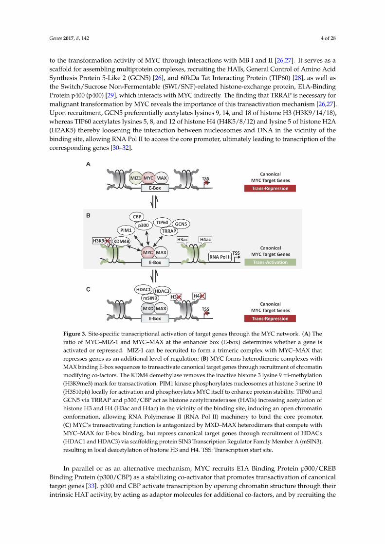
Genes 2017, 8, 142 4 of 28
to the transformation activity of MYC through interactions with MB I and II [26,27]. It serves as ascaffold for assembling multiprotein complexes, recruiting the HATs, General Control of Amino AcidSynthesis Protein 5-Like 2 (GCN5) [26], and 60kDa Tat Interacting Protein (TIP60) [28], as well asthe Switch/Sucrose Non-Fermentable (SWI/SNF)-related histone-exchange protein, E1A-BindingProtein p400 (p400) [29], which interacts with MYC indirectly. The finding that TRRAP is necessary formalignant transformation by MYC reveals the importance of this transactivation mechanism [26,27].Upon recruitment, GCN5 preferentially acetylates lysines 9, 14, and 18 of histone H3 (H3K9/14/18),whereas TIP60 acetylates lysines 5, 8, and 12 of histone H4 (H4K5/8/12) and lysine 5 of histone H2A(H2AK5) thereby loosening the interaction between nucleosomes and DNA in the vicinity of thebinding site, allowing RNA Pol II to access the core promoter, ultimately leading to transcription of thecorresponding genes [30–32].
Genes 2017, 8, 142 4 of 28
Amino Acid Synthesis Protein 5-Like 2 (GCN5) [26], and 60kDa Tat Interacting Protein (TIP60) [28],
as well as the Switch/Sucrose Non-Fermentable (SWI/SNF)-related histone-exchange protein, E1A-
Binding Protein p400 (p400) [29], which interacts with MYC indirectly. The finding that TRRAP is
necessary for malignant transformation by MYC reveals the importance of this transactivation
mechanism [26,27]. Upon recruitment, GCN5 preferentially acetylates lysines 9, 14, and 18 of histone
H3 (H3K9/14/18), whereas TIP60 acetylates lysines 5, 8, and 12 of histone H4 (H4K5/8/12) and lysine
5 of histone H2A (H2AK5) thereby loosening the interaction between nucleosomes and DNA in the
vicinity of the binding site, allowing RNA Pol II to access the core promoter, ultimately leading to
transcription of the corresponding genes [30–32].
Figure 3. Site-specific transcriptional activation of target genes through the MYC network. (A) The
ratio of MYC–MIZ-1 and MYC–MAX at the enhancer box (E-box) determines whether a gene is
activated or repressed. MIZ-1 can be recruited to form a trimeric complex with MYC–MAX that
represses genes as an additional level of regulation; (B) MYC forms heterodimeric complexes with
MAX binding E-box sequences to transactivate canonical target genes through recruitment of
chromatin modifying co-factors. The KDM4 demethylase removes the inactive histone 3 lysine 9 tri-
methylation (H3K9me3) mark for transactivation. PIM1 kinase phosphorylates nucleosomes at
histone 3 serine 10 (H3S10ph) locally for activation and phosphorylates MYC itself to enhance protein
stability. TIP60 and GCN5 via TRRAP and p300/CBP act as histone acetyltransferases (HATs)
increasing acetylation of histone H3 and H4 (H3ac and H4ac) in the vicinity of the binding site,
inducing an open chromatin conformation, allowing RNA Polymerase II (RNA Pol II) machinery to
bind the core promoter. (C) MYC’s transactivating function is antagonized by MXD–MAX
heterodimers that compete with MYC–MAX for E-box binding, but repress canonical target genes
through recruitment of HDACs (HDAC1 and HDAC3) via scaffolding protein SIN3 Transcription
Regulator Family Member A (mSIN3), resulting in local deacetylation of histone H3 and H4. TSS:
Transcription start site.
In parallel or as an alternative mechanism, MYC recruits E1A Binding Protein p300/CREB
Binding Protein (p300/CBP) as a stabilizing co-activator that promotes transactivation of canonical
target genes [33]. p300 and CBP activate transcription by opening chromatin structure through their
intrinsic HAT activity, by acting as adaptor molecules for additional co-factors, and by recruiting the
Figure 3. Site-specific transcriptional activation of target genes through the MYC network. (A) Theratio of MYC–MIZ-1 and MYC–MAX at the enhancer box (E-box) determines whether a gene isactivated or repressed. MIZ-1 can be recruited to form a trimeric complex with MYC–MAX thatrepresses genes as an additional level of regulation; (B) MYC forms heterodimeric complexes withMAX binding E-box sequences to transactivate canonical target genes through recruitment of chromatinmodifying co-factors. The KDM4 demethylase removes the inactive histone 3 lysine 9 tri-methylation(H3K9me3) mark for transactivation. PIM1 kinase phosphorylates nucleosomes at histone 3 serine 10(H3S10ph) locally for activation and phosphorylates MYC itself to enhance protein stability. TIP60 andGCN5 via TRRAP and p300/CBP act as histone acetyltransferases (HATs) increasing acetylation ofhistone H3 and H4 (H3ac and H4ac) in the vicinity of the binding site, inducing an open chromatinconformation, allowing RNA Polymerase II (RNA Pol II) machinery to bind the core promoter.(C) MYC’s transactivating function is antagonized by MXD–MAX heterodimers that compete withMYC–MAX for E-box binding, but repress canonical target genes through recruitment of HDACs(HDAC1 and HDAC3) via scaffolding protein SIN3 Transcription Regulator Family Member A (mSIN3),resulting in local deacetylation of histone H3 and H4. TSS: Transcription start site.
In parallel or as an alternative mechanism, MYC recruits E1A Binding Protein p300/CREBBinding Protein (p300/CBP) as a stabilizing co-activator that promotes transactivation of canonicaltarget genes [33]. p300 and CBP activate transcription by opening chromatin structure through theirintrinsic HAT activity, by acting as adaptor molecules for additional co-factors, and by recruiting the

Genes 2017, 8, 142 5 of 28
basal transcriptional machinery [34,35]. MYC directly recruits p300/CBP via its TAD, independent ofthe adaptor protein, TRRAP [28,33]. However, p300/CBP has also been shown to interact with MYC’sC-terminus, suggesting additional as yet unknown functions of p300/CBP at distinct promoters [36].Moreover, p300 has a dual role in regulating MYC’s activity. In addition to its function as a co-activatorof MYC-dependent transcription, p300 regulates MYC protein turnover [33]. p300 acetylates MYC atseveral lysine residues located between the TAD and DNA-binding domain at the C-terminus. Whilep300/CBP binding has been demonstrated to stabilize MYC protein independently of acetylation,p300-mediated acetylation of MYC results in its increased proteasomal degradation [33,37]. Conversely,MYC protein stability can be increased through expression of GCN5 or TIP60, indicating distinct rolesof HATs in regulating MYC’s functions, even though in the latter case it is not known if this requiresHAT activity [38]. This indicates that MYC not only serves as a hub for co-factors, but that MYC itselfis regulated by these enzymes. Whether any of the acetylation sites facilitate the binding of specificinteraction partners to MYC remains to be seen. While the recruitment of GCN5/TIP60/p300/CBP isassociated with a variety of acetylation marks [19,39], various combinations of these activating markshave been observed at different loci, suggesting that MYC might recruit distinct co-factors to certainpromoters. Their levels generally correlate with transcriptional activity, which is consistent with therecent finding that MYC enhances the expression of already active genes to boost the transcriptome ofa given cell [40,41].
2.2. Histone Demethylases and MYC-Dependent Transactivation
In addition to histone acetyltransferases, there is increasing evidence that MYC recruitslysine-specific histone demethylases (KDM) to activate transcription of canonical target genes, eventhough the exact mechanism of how histone methylation contributes to MYC regulated gene expressionremains unknown. First hints that MYC-dependent transactivation involves KDMs came fromDrosophila MYC (dMYC). The Trithorax group protein dKDM5/LID that belongs to the JARID1family of histone H3 lysine 4 (H3K4) demethylases was found crucial for dMYC-promoted cellgrowth [42]. However, since H3K4 methylation is an active chromatin mark, it seemed counterintuitive that dKDM5/LID is recruited for transactivation. The subsequent finding that dMYC actuallynegatively regulates dKDM5/LID activity, shed some light on this matter and led to the speculation thatdKDM5/LID may facilitate dMYC binding to chromatin or play a role in preserving H3K4 methylationmarks, although this needs further study. More recently, MYC has been reported to directly interactwith Lysine (K)-Specific Demethylase 4 (KDM4B) and recruit the histone demethylase to E-box targetgenes (see Figure 3B) [43,44]. KDM4B interacts with the central region of N-MYC (amino acids99–300) [44]. It specifically demethylates lysine 9 of histone H3 (H3K9me3/me2), removing repressivechromatin marks, thereby contributing to gene activation [45]. This mechanism was reported for MYCin embryonic stem cells (ESCs) and for overexpressed N-MYC in neuroblastoma [43,44], indicating thatthe decreased H3K9me3 deposition plays a role for both MYC’s physiologic as well as its oncogenicfunction. While the elevated expression of KDM4B in N-MYC amplified neuroblastomas is associatedwith poor clinical outcome, inhibition of KDM4B suppresses MYC function. Loss of KDM4B functioncauses downregulation of N-MYC target genes, subsequently inhibits cellular proliferation, inducesdifferentiation, and delays neuroblastoma tumor growth. This indicates that MYC alters histonemethylation patterns in the vicinity of E-box sites, preserving or even accumulating active marks suchas H3K4 methylation, while decreasing inactive marks such as H3K9 methylation.
2.3. Protein Kinases and MYC-Dependent Transactivation
Another chromatin modifying co-factor that MYC recruits to E-box target genes is the ProviralIntegration Site 1 (PIM1) oncogene, a constitutive active serine/threonine kinase (see Figure 3B).After stimulation with growth factors, PIM1 forms a complex with MYC–MAX via MB II [46].Subsequent PIM1-dependent phosphorylation of histone H3 on serine 10 (H3S10ph) in the vicinity ofE-box sites has been shown to contribute to the activation of approximately 20% of the MYC-regulated

Genes 2017, 8, 142 6 of 28
genes and neoplastic transformation of Rat-1 fibroblasts [46]. H3S10ph is thought to stimulate RNA PolII recruitment and release from promoter-proximal pausing [47]. Furthermore, PIM1 can phosphorylateMYC at serine 62 while decreasing threonine 58 phosphorylation, thereby increasing MYC proteinhalf-life [48]. Both residues are known as a main switch controlling MYC protein stability andproteasomal degradation [49,50]. Similarly, Proviral Integration Site 2 (PIM2) synergizes with MYC,stabilizing MYC protein through phosphorylation of serine 329 [48]. PIM kinases were first identifiedas genes that cooperate with Eµ-myc in lymphomagenesis, an observation that later could be extendedto various cancer types including pre-B-cell lymphoma, prostate carcinomas and triple-negative breastcancer [51–53]. Together, this indicates that PIM kinases cooperate with MYC during tumorigenesisby increasing MYC’s transcriptional activity for some target genes through multiple mechanisms,including modifying the phosphorylation status of MYC to enhance its activity and stability, as well asactivating local chromatin structure in the vicinity of MYC binding sites in a signal-dependent fashion.Hence, PIM kinases have sparked interest as a molecular target in multiple cancer types includinglymphomas and prostate cancer.
2.4. The Role of ATP-Dependent Chromatin Remodeling in MYC-Dependent Transactivation
An early connection between MYC and chromatin structure is the interaction with IntegraseInteractor 1 Protein (INI1), a core subunit of the SWI/SNF chromatin remodeling complex [54,55].The SWI/SNF complex mobilizes nucleosomes in an ATP-dependent fashion by catalyzing theexchange of histone octamers allowing for DNA to become accessible to transcriptional machinery(reviewed in [56]). The interaction with the SWI/SNF complex has been shown to be important forMYC-dependent transcription and transformation [54,55]. MYC’s bHLHZip domain directly interactswith INI1 and recruits the SWI/SNF complex to E-boxes for transactivation [54,57]. This interactionwas found independent of MYC–MAX binding despite both binding to MYC’s bHLHZip domain,indicating both activating mechanism occur in parallel. INI1 is a tumor suppressor that interactswith many other proteins, including oncogenes and tumor suppressor genes. INI1 is frequentlymutated in a wide variety of cancers and its loss is associated with neoplastic transformation [58].Interestingly, INI1 and MYC act antagonistically on a subset of target genes including genes involvedin cell cycle progression, metabolism, and ribosomal biogenesis, suggesting that INI1 negativelyregulates MYC binding and/or transcriptional activity. Highlighting the importance of this mechanism,re-expression of INI1 negatively affected proliferation of MYC-positive INI1-deficient rhabdoid tumorcells [55]. Additional investigations are needed to identify MYC- and SWI/SNF-dependent targetgenes and to unravel their molecular mechanisms, specifically how they work together to contributeto neoplastic transformation.
2.5. Models for Antagonizing MYC-Dependent Transactivation
The transactivation of E-box target genes by MYC–MAX can be antagonized by MAX-Dimerization(MXD) proteins. MXD family members such as MXD1 and MAX Network Transcriptional Repressor(MNT) also form heterodimeric complexes with MAX, competing with MYC–MAX for binding tothe same E-box sequences, but subsequently repress the corresponding gene [59,60]. While undernon-malignant conditions an equilibrium exists that is defined by the relative abundance of MYC andMXD proteins, the constitutively elevated expression of MYC shifts the balance toward activation intumor cells. The MXD-dependent repression mechanism relies on the recruitment histone deacetylases(HDACs), such as HDAC1 and HDAC3, which reduce histone acetylation on local chromatin resultingin a more condensed nucleosomal conformation, through the adapter protein SIN3 TranscriptionRegulator Family Member A (mSIN3) (see Figure 3C) [61]. The recruitment of co-repressors is essentialfor all the cellular functions of MXD proteins. The recruitment of HATs by MYC/MAX complexes andHDACs by MXD/MAX complexes can be seen as a transcriptional switch that regulates the activityof canonical MYC target genes through histone acetylation, shifting an equilibrium by “opening” or“closing” the local chromatin structure, allowing or preventing RNA Pol II binding, respectively.

Genes 2017, 8, 142 7 of 28
Adding another level of complexity, the transactivating effect of MYC–MAX on E-box regulatedgenes has recently been found to be antagonized by MIZ-1 [62]. While the interaction between MYCand MIZ-1 has been known to transrepress non-canonical MYC target genes that contain the initiatorelement (INR) sequence motif (see Chapter 3), recent genome-wide location profiling revealed thatMIZ-1 also interacts with MYC–MAX complexes occupying E-box sites [62]. The relative amountsof MYC and MIZ-1 that are bound to the core promoter determine whether the gene is activated orrepressed [62]. While MYC-repressed E-box genes were characterized by a MYC/MIZ-1 ratio close to1, MYC-activated genes showed higher ratios. Interestingly, in tumor cells with high MYC expression,MIZ-1 was found to occupy many more sites in a MYC-dependent manner, indicating a mechanisticdifference between the physiological and oncogenic properties of MYC. Together, this indicates anadditional layer of control, possibly a fine-tuning mechanism through which MYC can modulate thetranscription of E-box containing target genes shaping transcriptional amplification (see Figure 3A).
3. Recruitment of Chromatin Modifiers for MYC-Dependent Transrepression
MYC’s function as a site-specific transcription factor includes not only the activation but also therepression of genes (both protein-coding as well as noncoding RNAs). In fact, both mechanisms areessential for MYC-driven tumor initiation and maintenance [62,63], and both mechanisms dependon the recruitment of chromatin modifying co-factors. An increasing number of non-canonical targetgenes have been identified that MYC represses through protein-protein interactions with zinc fingertranscription factors such MIZ-1 [62], SP1 [16], nuclear factor Y (NF-Y) [61], ying yang 1 (YY1) [64],and transcription factor II I (TFII-I) [65].
The interaction with MIZ-1 is the best characterized example of how MYC represses transcription.MIZ-1 recognizes a consensus sequence known as INR in core promoters and, in the absence of MYC,activates the transcription of the corresponding gene (see Figure 4). However, MYC binding interfereswith the transcriptional activator function of MIZ-1, blocking the interaction with co-activatorswhile facilitating the recruitment of co-repressor complexes [17,66–68]. Evidence for MYC’s functionas a repressor stems from a point mutant, MYCV394D, which is selectively deficient in its abilityto interact with MIZ-1, while still dimerizing with MAX to transactivate canonical target genes.MYCV394D is unable to repress wild-type MYC targets such as Cyclin Dependent Kinase Inhibitor 1A(CDKN1A/p21CIP) and Cyclin Dependent Kinase Inhibitor 2B (CDKN2B/p15INK4B), in a transgeniclymphoma model [69]. Furthermore, protein-protein binding assays suggest that MYC displacesco-activators such as p300/CBP from interacting with MIZ-1 [17]. This switch in transcriptionalactivity can be explained by MIZ-1 playing a role in preventing the association between MYC and theco-activator p300, resulting in a decrease of histone acetylation.
Genes 2017, 8, 142 7 of 28
and MIZ-1 has been known to transrepress non-canonical MYC target genes that contain the initiator
element (INR) sequence motif (see Chapter 3), recent genome-wide location profiling revealed that
MIZ-1 also interacts with MYC–MAX complexes occupying E-box sites [62]. The relative amounts of
MYC and MIZ-1 that are bound to the core promoter determine whether the gene is activated or
repressed [62]. While MYC-repressed E-box genes were characterized by a MYC/MIZ-1 ratio close to
1, MYC-activated genes showed higher ratios. Interestingly, in tumor cells with high MYC
expression, MIZ-1 was found to occupy many more sites in a MYC-dependent manner, indicating a
mechanistic difference between the physiological and oncogenic properties of MYC. Together, this
indicates an additional layer of control, possibly a fine-tuning mechanism through which MYC can
modulate the transcription of E-box containing target genes shaping transcriptional amplification
(see Figure 3A).
3. Recruitment of Chromatin Modifiers for MYC-Dependent Transrepression
MYC’s function as a site-specific transcription factor includes not only the activation but also the
repression of genes (both protein-coding as well as noncoding RNAs). In fact, both mechanisms are
essential for MYC-driven tumor initiation and maintenance [62,63], and both mechanisms depend on
the recruitment of chromatin modifying co-factors. An increasing number of non-canonical target
genes have been identified that MYC represses through protein-protein interactions with zinc finger
transcription factors such MIZ-1 [62], SP1 [16], nuclear factor Y (NF-Y) [61], ying yang 1 (YY1) [64],
and transcription factor II I (TFII-I) [65].
The interaction with MIZ-1 is the best characterized example of how MYC represses
transcription. MIZ-1 recognizes a consensus sequence known as INR in core promoters and, in the
absence of MYC, activates the transcription of the corresponding gene (see Figure 4). However, MYC
binding interferes with the transcriptional activator function of MIZ-1, blocking the interaction with
co-activators while facilitating the recruitment of co-repressor complexes [17,66–68]. Evidence for
MYC’s function as a repressor stems from a point mutant, MYCV394D, which is selectively deficient in
its ability to interact with MIZ-1, while still dimerizing with MAX to transactivate canonical target
genes. MYCV394D is unable to repress wild-type MYC targets such as Cyclin Dependent Kinase
Inhibitor 1A (CDKN1A/p21CIP) and Cyclin Dependent Kinase Inhibitor 2B (CDKN2B/p15INK4B), in
a transgenic lymphoma model [69]. Furthermore, protein-protein binding assays suggest that MYC
displaces co-activators such as p300/CBP from interacting with MIZ-1 [17]. This switch in
transcriptional activity can be explained by MIZ-1 playing a role in preventing the association
between MYC and the co-activator p300, resulting in a decrease of histone acetylation.
Figure 4. Site-specific transcriptional repression of target genes through the MYC network.
MYC–MAX/MIZ-1 mediated transrepression of non-canonical target genes such as Cyclin Dependent
Kinase Inhibitor 1A (CDKN1A/p21CIP) and Cyclin Dependent Kinase Inhibitor 2B
(CDKN2B/p15INK4B) involves recruitment of chromatin co-repressors. DNMT3A suppresses the
corresponding gene through hypermethylation of CpGs in the vicinity. Recruitment of HDAC1 and
HDAC3 contribute to histone deacetylation and thus silencing of certain genes. The demethylase
LSD1 removes active H3K4me marks contributing to gene repression. The PRC2 repressive complex
also interacts with MYC to enhance gene silencing. Filled circles represent methylated CpGs;
transcription start site (TSS).
Figure 4. Site-specific transcriptional repression of target genes through the MYC network. MYC–MAX/MIZ-1 mediated transrepression of non-canonical target genes such as Cyclin Dependent KinaseInhibitor 1A (CDKN1A/p21CIP) and Cyclin Dependent Kinase Inhibitor 2B (CDKN2B/p15INK4B)involves recruitment of chromatin co-repressors. DNMT3A suppresses the corresponding gene throughhypermethylation of CpGs in the vicinity. Recruitment of HDAC1 and HDAC3 contribute to histonedeacetylation and thus silencing of certain genes. The demethylase LSD1 removes active H3K4memarks contributing to gene repression. The PRC2 repressive complex also interacts with MYC toenhance gene silencing. Filled circles represent methylated CpGs; transcription start site (TSS).

Genes 2017, 8, 142 8 of 28
3.1. MYC-Dependent Transrepression via DNA Methyltransferases
In addition to the displacement of co-activators, MYC-mediated gene repression is associatedwith the recruitment of co-repressors. MYC–MAX/MIZ-1 complexes have been shown to recruitthe de novo DNA methyltransferase 3a (DNMT3A) to non-canonical targets such as CDKN1A(p21CIP) and CDKN2B (p15INK4B) [70], increasing the DNA methylation of promoter regions ornearby CpG islands [70]. This is important during tumorigenesis, where MYC suppresses cell cycledependent kinase inhibitors (CDKIs) to antagonize differentiation and cellular senescence, insteadpromoting cell proliferation [17,66,69]. There is some evidence that MYC similarly interacts withDNMT3B [70]. In non-small-cell lung cancer (NSCLC), MYC recruits DNMT3B to the promoter of thetumor suppressor, Ras Association Domain-containing Protein 1 (RASSF1A), silencing its expressionthrough DNA hypermethylation [71,72]. The recruitment of DNMTs is an attractive concept, since itprovides an explanation for the aberrant DNA methylation pattern observed for tumor suppressorgenes in human tumors. Whether transrepression by MYC–MAX/MIZ-1 during tumorigenesisgenerally requires DNMT3A and DNMT3B, and whether this applies also to physiological conditionsis less understood. Nonetheless, the implication that DNMTs are recruited by MYC to generate specificDNA methylation patterns within a cell is intriguing, specifically in the light of DNMT inhibitors beingexploited for therapeutic purposes [73–76].
3.2. MYC-Dependent Transrepression via Histone Deacetylases
While the role of histone acetylation in MYC-dependent transcriptional repression is less wellcharacterized, two HDACs, HDAC1 [77–79] and HDAC3, have been reported to interact with MYC.In both cases, they have been identified as part of co-repressor complexes recruited by MYC tosuppress transcription. HDAC1 was found to be recruited to the promoter of tissue transglutaminase(tTG) [79] and HDAC3 to the promoter of Inhibitor of DNA Binding 2 (ID-2) and Growth Arrestand DNA Damage-Inducible Protein GADD153 (GADD153) [80,81], correlating with a decreasein histone acetylation in these loci and transrepression of the corresponding genes. It would beinteresting to see whether this represents a general mechanism for all INR-regulated or even allMYC-repressed genes. Support for the latter concept comes from the finding that MYC also exploitsHDAC3 for transrepression of microRNAs (MIR-15A/MIR-16-1 and MIR-29). The fact that thosemicroRNAs have tumor-suppressive function in turn strengthens the notion that transrepression ofgenes is critical for MYC-dependent transformation [82]. In the classic model, MYC’s ability to recruiteither HATs or HDACs depends on the DNA binding motif. MYC–MAX complexes interact withHATs for transactivation of canonical target genes, while MYC–MAX/MIZ-1 recruits HDACs fortransrepression of non-canonical target genes. However, whether the recruitment of HDACs extendsto MYC–MAX/MIZ-1 complexes repressing E-box promoters remains to be shown. Nonetheless,the fact that HDAC inhibitors have already been exploited as therapeutic strategies for hematologicmalignancies raises intriguing possibilities for further studies aiming at other cancer types expressingderegulated MYC [83–85].
3.3. MYC-Dependent Transrepression via Histone Demethylases
Histone KDMs are a double-edged sword in MYC-dependent transcriptional regulation, as theyare involved in both repression and activation of MYC-target genes. Lysine-Specific HistoneDemethylase 1A (LSD1) (also known as KDM1A and AOF2) cooperates with N-MYC to represstumor suppressor genes, contributing to tumor maintenance in neuroblastoma [86]. LSD1 has beenknown to remove mono- and di-methyl groups from histone H3 lysine 4 (H3K4me2/me3) andlysine 9 (H3K9me2/me3) [87,88], but can also demethylate non-histone proteins such as p53 andE2F [89,90]. LSD1 has been isolated as a component of several co-repressor complexes including RESTcorepressor 1 (CoREST), C-terminal-binding protein 1 (CtBP), HDAC1, and HDAC2 [91,92], suggestinga role in transcriptional repression. Moreover, H3K4 demethylation and histone deacetylation by
Genes 2017, 8, 142 9 of 28
LSD1-containing complexes seem to be linked, both contributing to MYC’s repressive function.Proteomic analysis revealed that LSD1 directly interacts with N-MYC via MB III [86]. Moreover,LSD1 co-localizes with N-MYC at the promoter of CDKN1A (p21CIP) and Clusterin (CLU), two tumorsuppressor genes, thereby functionally cooperating with N-MYC in neuroblastoma initiation andprogression [86].
Interestingly, by interacting with different co-factors, LSD1 also seems capable of activating genes.MYC-dependent transactivation of E-box genes in rat fibroblasts has been proposed to involveLSD1-mediated demethylation of H3K4 [93]. Given the fact that H3K4 methylation is an activechromatin mark, this seems counter-intuitive at first. However, upon serum stimulation, LSD1 has beenfound to form a complex with MYC at the E-boxes sites of Nucleolin (NCL) and Carbamoyltransferase-dihydroorotase (CAD), causing cycles of methylation and demethylation of lysine 4 in histone H3(H3K4me2/me3), before H4 acetylation increases [93]. These transient LSD1-mediated methylationevents produces H2O2 causing the recruitment of oxidative repair enzymes to E-box genes [93]. Theconcept of linking histone demethylation to oxidation and DNA repair has been proposed to beimplicated in the serum-induced assembly of the transcription initiation complex, although furtherstudies are necessary to validate and generalize this mechanism. It remains to be seen whether thismechanism applies to all MYC-activated promoters, or only a specific set of MYC target genes.
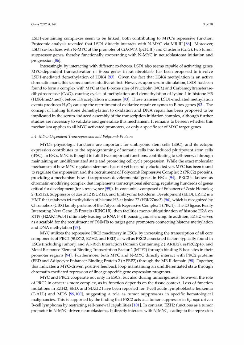
3.4. MYC-Dependent Transrepression and Polycomb Proteins
MYC's physiologic functions are important for embryonic stem cells (ESC), and its ectopicexpression contributes to the reprogramming of somatic cells into induced pluripotent stem cells(iPSC). In ESCs, MYC is thought to fulfill two important functions, contributing to self-renewal throughmaintaining an undifferentiated state and promoting cell cycle progression. While the exact molecularmechanism of how MYC regulates stemness has not yet been fully elucidated yet, MYC has been foundto regulate the expression and the recruitment of Polycomb Repressive Complex 2 (PRC2) proteins,providing a mechanism how it suppresses developmental genes in ESCs [94]. PRC2 is known aschromatin-modifying complex that implements transcriptional silencing, regulating hundreds of genescritical for development (for a review, see [95]). Its core unit is composed of Enhancer of Zeste Homolog2 (EZH2), Suppressor of Zeste 12 (SUZ12), and Embryonic Ectoderm Development (EED). EZH2 is aHMT that catalyzes tri-methylation of histone H3 at lysine 27 (H3K27me3) [96], which is recognized byChromobox (CBX) family proteins of the Polycomb Repressive Complex 1 (PRC1). The E3 ligase, ReallyInteresting New Gene 1B Protein (RING1B), then facilities mono-ubiquitination of histone H2A onK119 (H2AK119ub1) ultimately leading to RNA Pol II pausing and silencing. In addition, EZH2 servesas a scaffold for the recruitment of DNMTs to target gene promoters connecting histone methylationand DNA methylation [97].
MYC utilizes the repressive PRC2 machinery in ESCs, by increasing the transcription of all corecomponents of PRC2 (SUZ12, EZH2, and EED) as well as PRC2-associated factors typically found inESCs (including Jumonji and AT-Rich Interaction Domain Containing 2 (JARID2), esPRC2p48, andMetal Response Element Binding Transcription Factor 2 (MTF2) through binding E-box sites in theirpromoter regions [94]. Furthermore, both MYC and N-MYC directly interact with PRC2 proteins(EED and Adipocyte Enhancer-Binding Protein 2 (AEBP2)) through the MB II domain [98]. Together,this indicates a MYC-driven positive feedback loop maintaining an undifferentiated state throughchromatin-mediated repression of lineage-specific gene expression programs.
MYC and PRC2 cooperate not only in ESCs, but also during tumorigenesis; however, the roleof PRC2 in cancer is more complex, as its function depends on the tissue context. Loss-of-functionmutations in EZH2, EED, and SUZ12 have been reported for T-cell acute lymphoblastic leukemia(T-ALL) and MDS [99,100], suggesting a role as tumor suppressors in specific hematologicalmalignancies. This is supported by the finding that PRC2 acts as a tumor suppressor in Eµ-myc-drivenB-cell lymphoma by restricting self-renewal capabilities [101]. In contrast, EZH2 functions as a tumorpromoter in N-MYC-driven neuroblastoma. It directly interacts with N-MYC, leading to the repression

Genes 2017, 8, 142 10 of 28
of the putative tumor suppressor gene CLU [102]. Furthermore, EZH2 and SUZ12 interact with N-MYCthrough MB III in castration-resistant prostate carcinoma (CRPC), leading to abrogation of androgenreceptor signaling and consequently driving progression to neuroendocrine prostate cancer (NEPC),the aggressive subgroup of late-stage prostate cancer [103]. Together, this indicates that N-MYCcooperates with EZH2 to drive the neuroendocrine phenotype in prostate cancer, thereby providing arationale for therapeutic strategies targeting EZH2 [103].
3.5. MYC-Dependent Transrepression and ATP-Dependent Chromatin Remodeling
Another class of co-repressors that is recruited by MYC are 48 KDa TBP-Interacting Protein(TIP48)/49 KDa TBP-Interacting Protein (TIP49) [104], which function as ATPase/helicase rather thanhistone modifiers. The dependence on their interaction with the MB II domain of MYC already providesa hint for biological activity, including blocking differentiation and oncogenic transformation. Indeed,it has been demonstrated that TIP48 and TIP49 are essential co-factors for MYC-driven neoplastictransformation. Furthermore, their interaction with MYC/MIZ-1 is essential for cell growth andproliferation during normal Drosophila and Xenopus development [105,106]. Even though the precisefunction of their relationship with MYC under physiologic conditions remains unresolved, it hasbeen speculated that TIP48 and TIP49 bridge basic transcription machinery and sequence-specifictranscription factors and act as transcriptional repressors in this context.
4. MYC as a Master Regulator the Cancer Epigenome and Transcriptome
MYC’s role as a site-specific transcription factor regulating the expression of a large set of specifictarget genes by remodeling local chromatin structure has long been thought to be the key to its diversecellular functions. However, a new perspective has been provided by studies that reveal that the MYConcogene influences chromatin structure in a global fashion (Figure 5). These findings indicate anexciting role for MYC that extends beyond the traditional concept of a site-specific transcription factorand promises new directions for therapeutic anti-MYC strategies.
Genes 2017, 8, 142 10 of 28
cancer (NEPC), the aggressive subgroup of late-stage prostate cancer [103]. Together, this indicates
that N-MYC cooperates with EZH2 to drive the neuroendocrine phenotype in prostate cancer,
thereby providing a rationale for therapeutic strategies targeting EZH2 [103].
3.5. MYC-Dependent Transrepression and ATP-Dependent Chromatin Remodeling
Another class of co-repressors that is recruited by MYC are 48 KDa TBP-Interacting Protein
(TIP48)/49 KDa TBP-Interacting Protein (TIP49) [104], which function as ATPase/helicase rather than
histone modifiers. The dependence on their interaction with the MB II domain of MYC already
provides a hint for biological activity, including blocking differentiation and oncogenic
transformation. Indeed, it has been demonstrated that TIP48 and TIP49 are essential co-factors for
MYC-driven neoplastic transformation. Furthermore, their interaction with MYC/MIZ-1 is essential
for cell growth and proliferation during normal Drosophila and Xenopus development [105,106].
Even though the precise function of their relationship with MYC under physiologic conditions
remains unresolved, it has been speculated that TIP48 and TIP49 bridge basic transcription
machinery and sequence-specific transcription factors and act as transcriptional repressors in this
context.
4. MYC as a Master Regulator the Cancer Epigenome and Transcriptome
MYC’s role as a site-specific transcription factor regulating the expression of a large set of
specific target genes by remodeling local chromatin structure has long been thought to be the key to
its diverse cellular functions. However, a new perspective has been provided by studies that reveal
that the MYC oncogene influences chromatin structure in a global fashion (Figure 5). These findings
indicate an exciting role for MYC that extends beyond the traditional concept of a site-specific
transcription factor and promises new directions for therapeutic anti-MYC strategies.
Figure 5. MYC as a master regulator of the cancer epigenome and transcriptome. The MYC oncogene
deregulates histone acetylation and methylation in a global fashion with implication for the cancer
epigenome and transcriptome. MYC–MAX complexes recognize an E-Box sequence in the GCN5
promoter, leading to overexpression of GCN5. The consequently increased histone acetyltransferase
(HAT) activity of GCN5 increases genome-wide acetylation of H3 and H4 (H3ac and H4ac). MYC–
MAX also binds to the HDAC2 promoter and upregulates its transcription, leading to an increase in
HDAC2 activity. MYC activates the transcription of MIR17-92, which represses the histone
methyltransferase (HMT) variegation 4-20 homolog 1 (SUV420H1), implicating that MYC prevents
the methylation of histone H3 at K20 on a genome-wide level.
Figure 5. MYC as a master regulator of the cancer epigenome and transcriptome. The MYC oncogenederegulates histone acetylation and methylation in a global fashion with implication for the cancerepigenome and transcriptome. MYC–MAX complexes recognize an E-Box sequence in the GCN5promoter, leading to overexpression of GCN5. The consequently increased histone acetyltransferase(HAT) activity of GCN5 increases genome-wide acetylation of H3 and H4 (H3ac and H4ac). MYC–MAXalso binds to the HDAC2 promoter and upregulates its transcription, leading to an increase in HDAC2activity. MYC activates the transcription of MIR17-92, which represses the histone methyltransferase(HMT) variegation 4-20 homolog 1 (SUV420H1), implicating that MYC prevents the methylation ofhistone H3 at K20 on a genome-wide level.

Genes 2017, 8, 142 11 of 28
The first evidence of MYC’s global reach stem from neuronal progenitor cells in which thedisruption of N-MYC expression causes widespread changes in chromatin organization, accompaniedby nuclear condensation and heterochromatin formation [107]. These genome-wide changes arecharacterized by a marked decrease in histone H3 and H4 acetylation and an increase in H3K9me3,both indicative of gene silencing. While these observations could not be explained by the sheer numberof MYC binding sites in the genome and MYC’s effect on local chromatin, the oncogene was found toinfluence chromatin in a genome-wide fashion through upregulation of the HAT, GCN5 [107]. N-MYC(subsequently demonstrated also for c-MYC) was found to directly bind to two E-box sequencesin the GCN5 promoter, thereby increasing its transcription in tumor cells. Consequently, GCN5activity increased the widespread acetylation of histones, accumulating active chromatin domains.This established GCN5 as a direct MYC target gene and provided the first evidence for regulation ofgenome-wide chromatin organization by an oncogene.
Subsequently, N- and c-MYC’s influence on global chromatin architecture has been demonstratedfor various additional cell types [107,108]. In human B lymphocytes, serving as a Burkitt’s lymphomamodel (P493-6), the suppression of a conditional c-MYC allele induces global heterochromatic regionsresembling the phenotype described for N-MYC disruption in neuronal progenitor cells [107]. Similarly,the genetic inactivation of c-MYC in conditional mouse models of osteosarcoma, hepatocellularcarcinoma and T-cell ALL triggered a global reduction in histone H4 acetylation and an increase inheterochromatic H3K9me3 [108]. The switchable nature of MYC in these transgenic tumor modelsallowed for analysis of the dynamics in chromatin structure and associated gene expression patterns.Within hours of MYC inactivation, chromatin changes become apparent and develop to widespreadinactive chromatin, including senescence-associated heterochromatic foci (SAHF) [107,108]. In parallelto chromatin, changes in gene expression programs occur in a similarly time-dependent manner [109,110].Further experiments are needed to substantiate a causative relationship between both events.Intriguingly, inactivation followed by the reactivation of MYC in a conditional osteosarcoma mousemodel does not reverse the entire gene expression program controlled by MYC [109]. Furthermore,MYC’s ability to bind to promoter regions was altered, suggesting that permanent changes in thechromatin architecture affect whether certain genes are susceptible to MYC regulation.
In parallel, MYC seems to also control histone deacetylation in a genome-wide fashion. HDAC2expression has been found increased in a MYC-dependent fashion during APC-driven colorectaltumorigenesis [111]. Furthermore, both N-MYC and c-MYC were found to upregulate HDAC2expression in neuroblastoma and pancreatic cancer, respectively, which contributed to MYC-inducedtumor cell proliferation and blocked apoptosis in these models [112]. This depends on a MYCbinding site in the HDAC2 promoter region, which was confirmed by chromatin immunoprecipitationassay [113]. This demonstrates that MYC is capable of upregulating HDAC2 gene expression, thatMYC-induced HDAC2 overexpression contributes to MYC-induced cancer cell proliferation, and thatHDAC2 is likely to be one of the key factors responsible for MYC-induced malignant transformation,tumor initiation and progression in vivo [112]. The notion that MYC upregulates the HAT, GCN5, andHDAC2 raises questions regarding their differential specificity and function on a genome-wide level.
In contrast to histone acetylation, the role of global histone methylation in regulating MYC-mediatedgene expression is more complex. Even though MYC overexpression had been observed to induce bothlocalized and widespread histone H3K4 tri-methylation (and suppressed H3K9me3), these changeswere thought to be a reflection of the proliferative state of the cell rather than a direct consequence ofMYC’s action. Only recently has light been shed on the mechanism by which MYC utilizes the dualfunction of histone methylation as an activator or repressor of transcriptome.
MYC has been uncovered to suppresses the SIN3 Transcription Regulator Family MemberB (SIN3B), HMG-box Transcription Factor 1 (HBP1), suppressor of Variegation 4–20 Homolog 1(SUV420H1), and B Cell Translocation Gene 1 (BTG1) through miR-17-92 [114]. SIN3B interacts withHBP1 and recruits HDACs to silence proliferation-related genes and mediate cell cycle exit andsenescence [115–117]. SUV420H1 catalyzes di- and tri-methylation of histone H4K20 (H4K20me2

Genes 2017, 8, 142 12 of 28
and H4K20me3) [118–120]. H4K20me3 is a marker for heterochromatin and cellular senescence, andis commonly lost in human cancers [121]. Both H4K20me2 and H4K20me3 were found to increaseupon MYC inactivation in lymphoma [114]. BTG1 is a tumor suppressor that is frequently lostin ALL [122,123] and is known to activate the HMT, PRMT1, to di-methylate histone H4 arginine3 (H4R3me2) [124,125]. H4R3me2 is increased upon MYC inactivation in lymphoma [114]. Oncereactivated, these chromatin modifiers promote cell cycle arrest and cellular senescence, supporting thenotion that MYC’s ability to sustain autonomous proliferation, self-renewal, and survival is mediatedthrough chromatin regulatory and survival switches.
The shutoff of this epigenetic switch contributes to MYC oncogene addiction, which is antagonizedby Transforming Growth Factor β (TGF-β) and SMADs. The TGF-β/SMAD pathway can havetumor promoting or suppressing functions depending on the context (reviewed in [126]). Hence,there has been great interest in TGF-β mediated processes in cancer with the goal of providingtherapeutic strategies. During lymphomagenesis TGF-β counteracts MYC’s function by inducing theHMT, SUV39H1, responsible for inactive H3K9me3 resulting in cellular senescence in the absenceof MYC [127]. SUV39H1 deficiency results in loss of H3K9me3, increased genomic instability, andthe development of B- and T- cell lymphomas in mice, indicating its important role for preventingmalignant transformation [127]. However, there is no evidence that MYC directly regulates SUV39H1,but it is speculated that MYC indirectly suppresses SUV39H1 through TGF-β to drive cellularproliferation [128,129].
Furthermore, MYC’s influence on chromatin structure extends beyond gene regulatory or genicregions and also includes expansive intergenic regions. Genomic location analyses suggest that theinactivation of N-MYC depletes large portions of active histone marks in the genome (H3K9 acetylationand H3K4 methylation) [130]. These results implicate that MYC’s ability to induce and maintainwidespread regions of active chromatin is as important as its ability to modulate the transcription ofindividual genes. Indeed, MYC has been shown to have broader effects on chromatin remodeling.MYC changes histone localization patterns through incorporation of the H2A.Z isoform into its targetpromoters, allowing for activation of target genes [39]. Another example for a chromatin-associatedprotein, that has been identified as a direct MYC target is the insulator protein, CCCTC-Binding Factor(CTCF), which is thought to allow for transcriptional alterations of wide regions of the genome byblocking facultative heterochromatin [131,132]. MYC also directly regulates Metastasis-associatedprotein 1 (MTA1), a component of the nucleosome remodeling and deacetylating (NuRD) co-repressorcomplex [133]. Since MTA1 has been linked to epithelial to mesenchymal transition (EMT) [134],it has been speculated that MYC is involved in this process through upregulation of MTA1, ultimatelyleading to increased levels of the SNAIL repressor driving the EMT process.
5. MYC-Driven Transcriptional Amplification
Fueling the oncogene’s enigmatic reputation further and intensifying research interest, recentreports reveal that MYC deregulates large gene expression programs in tumor cells throughtranscriptional amplification. When overexpressed, MYC accumulates in the promoter regions ofactively transcribed genes and acts as a general amplifier, further increasing the transcriptionaloutput, boosting a cell's gene expression profile [41]. Thus, rather than specifying the subset ofgenes that will be actively transcribed in any particular cell in a site-specific manner, MYC amplifiesthe output of the existing gene expression program. MYC facilitates transcription elongation bystimulating the recruitment of the Positive Transcription Elongation Factor b (P-TEFb) and the Mediatorcomplex [40,135]. The large Mediator complex, an essential co-activator of RNA Pol II also involved inchromatin “looping”, which brings distant chromosomal regions into close physical proximity to eachother, interacts through STAGA with amino acids 1–110 of MYC [136]. P-TEFb releases RNA Pol IIfrom pausing by phosphorylating its C-terminal domain (CTD) [137–139]. MB I and II also mediate theassociation with other effectors of MYC activity such as Bromodomain-Containing Protein 4 (BRD4)which is also involved in P-TEFb recruitment [135,137,138]. Therefore, MYC appears to play a critical

Genes 2017, 8, 142 13 of 28
role in RNA Pol II pause release rather than RNA Pol II recruitment, revealing an important facetMYC controls to drive transcriptional amplification. Furthermore, it has been implied that MYC maybe tethered to chromatin domains such as euchromatic islands (active chromatin characterized byH3K4me, H3K79me, and H3ac) and this tethering is crucial for the rate-limiting step of MYC bindingto site-specific DNA consensus sequences [140]. The overall increase of transcriptional output by MYChas a dramatic effect on proliferation and cellular biogenesis. The aspect of transcriptional amplificationmay be able to explain rate-limiting restraints on MYC-driven proliferation and also why MYC hasbroad oncogenic capabilities across a number of tissues and cell types. As an alternative explanationfor the genome-wide upregulation of active genes, a recent study proposes that MYC acts mainlythrough controlling a selective group of genes, which in turn are responsible for global amplificationeither co- or post-transcriptionally [141]. Together these studies support an evolving new dual modelin which MYC’s influence on chromatin is far more complex than previously imagined. The ability ofMYC to act both locally and globally on chromatin may be responsible for its wide-ranging effectson the biology of stem and tumor cells. However, the new dimension of MYC’s reach also raisesthe question of how to integrate the new global and the classic site-specific mechanisms into oneunified model. This becomes specifically apparent when attempting to explain the role of co-repressorrecruitment in the context of genome-wide transcriptional amplification. It has been speculated thatMYC’s ability to repress genes in a site-specific manner might be a negative feedback loop, representinga control mechanism that responds to the unrestrained expression of other MYC targets. Providing amechanistic basis for this, recent reports extend the repressive abilities of MYC–MAX/MIZ-1 to E-boxcontaining genes indicating a fine-tuning mechanism [61]. Given the importance of MYC-mediatedgene repression for tumorigenesis, it would be interesting to see whether MYC is also capable ofrepressing genes in a genome-wide fashion, complementing the global amplifying model by providinga mechanism to further modulate the transcriptional output of a cell.
DNA Sequence-Specific vs. Tethered Recruitment of MYC
The classic model of MYC as a sequence-specific transcription factor leaves important questionsunanswered. It is unable to explain the diversity of transcriptional responses MYC provokes indifferent cell types, indicating an inherent plasticity in MYC’s selection of target genes. Hence,additional mechanisms must exist that determine which genomic loci are occupied by MYC in a givencell type. It has been speculated that the binding of MYC to its target genes is not solely determinedby a specific DNA sequence, but that MYC binding site recognition depends on chromatin structure.Early clues came from large-scale location analyses revealing that the active H3 lysine 4 (K4) and 79(K79) methylation marks are a strict prerequisite for recognition of any target site by MYC, suggestingthat distinct histone variants or modifications can modulate protein recognition [15,140]. This biasfor MYC to bind chromatin enriched for active histone modifications was substantiated by severalgenome-wide location studies [41,61,141]. More recent reports challenge the classic model further,proposing a specific mechanism for a tethered recruitment via WD Repeat Domain 5 (WDR5) [142] (seeFigure 6). WDR5 is a highly conserved chromatin “reader” found in multiple protein complexes [143],including the Myeloid/Lymphoid or Mixed-Lineage Leukemia (MLL) histone methyltransferases thatcatalyze H3K4 methylation and the histone demethylase LSD1 that removes H3K4/K9 marks [144].Genome-wide location analyses reveal that MYC and WDR5 colocalize extensively on active chromatin,with approximately 80% of the genomic loci occupied by MYC also bound by WDR5 [142]. WDR5 wasfound to directly interact with MYC via the MB III motif in the central region [145,146], suggesting ascenario in which WDR5 mediates the recruitment of MYC to active chromatin loci. However, thismodel still needs to be challenged and several important questions remain to be answered. For example,how is WDR5 itself tethered to chromatin, and is MYC competing with other WDR5-binding partnerssuch as MLL? Characterizing the MYC–WDR5 complex biochemically to identify other proteins thatassociate with MYC through WDR5 should provide valuable information about the molecular contextin which the two proteins interact with chromatin. Further studies will be necessary to unravel the

Genes 2017, 8, 142 14 of 28
specific details, but the above results suggest an intriguing role of the WDR5–MYC interaction intethering MYC to chromatin and indicate a critical function of WDR5 for MYC-driven tumorigenesis.
Genes 2017, 8, 142 14 of 28
suggest an intriguing role of the WDR5–MYC interaction in tethering MYC to chromatin and indicate
a critical function of WDR5 for MYC-driven tumorigenesis.
Figure 6. MYC-driven transcriptional amplification. MYC binds actively transcribed genes that are
characterized by histone H3 acetylation (H3ac) and lysine 4 methylation (K4me). MYC binding can
be facilitated either through E-box elements and/or tethered recruitments via WDR5. In turn
Bromodomain-Containing Protein 4 (BDR4) is recruited, which stimulates the Positive Transcription
Elongation Factor b (P-TEFb) via a Mediator complex to trigger RNA Polymerase II (RNA Pol II)
pause release.
6. Therapeutic Strategies: Targeting Epigenetic Mechanisms in MYC-Positive Cancers
The notion that inactivation of MYC can cause tumor regression in animal models by eliciting
oncogene addiction, makes it an attractive target for therapeutic anti-cancer strategies. However,
despite considerable efforts in that regard, a pharmacologic inhibitor that directly targets MYC and
can be used to translate these findings into the clinical setting remains elusive. Recently, novel
therapeutic strategies have emerged that promise to take advantage of the reversibility of chromatin
modifications to adjust the epigenome in ways that slow cell proliferation and induce cellular
senescence or apoptosis in MYC-deregulated cancers. Targeting MYC through chromatin associated
co-factors that affect either the expression and/or the function of the oncogene has proven to be
effective in various cell lines and animal models (for an overview, see Figure 7). Despite the fact that
these strategies can cause off-target effects, some have yielded highly promising results and are tested
in clinical trials, paving the road for epigenetic drugs to treat MYC-associated human cancers.
Figure 7. Targeting the MYC network with epigenetic inhibitors for therapeutic purposes. Schematic
representation of different therapeutic strategies to target the MYC network through chromatin-
modifying co-factors: Histone deacetylase inhibitors (HDACi), histone methyltransferase inhibitors
(HMTi), histone acetyltransferase inhibitors (HATi), lysine-specific histone demethylase inhibitors
(KDMi), DNA methyltransferase inhibitors (DNMTi), and Bromodomain and Extra-Terminal motif
inhibitors (BETi).
6.1. DNA Methyltransferase Inhibitors (DNMTi)
One of the first epigenetic therapeutics brought about and used were the DNA demethylation
agents 5-aza-cytidine (azacytidine) and its analog 5-aza-2′-deoxycytidine (decitabine) [75,76].
Figure 6. MYC-driven transcriptional amplification. MYC binds actively transcribed genes that arecharacterized by histone H3 acetylation (H3ac) and lysine 4 methylation (K4me). MYC bindingcan be facilitated either through E-box elements and/or tethered recruitments via WDR5. In turnBromodomain-Containing Protein 4 (BDR4) is recruited, which stimulates the Positive TranscriptionElongation Factor b (P-TEFb) via a Mediator complex to trigger RNA Polymerase II (RNA Pol II)pause release.
6. Therapeutic Strategies: Targeting Epigenetic Mechanisms in MYC-Positive Cancers
The notion that inactivation of MYC can cause tumor regression in animal models by elicitingoncogene addiction, makes it an attractive target for therapeutic anti-cancer strategies. However,despite considerable efforts in that regard, a pharmacologic inhibitor that directly targets MYC and canbe used to translate these findings into the clinical setting remains elusive. Recently, novel therapeuticstrategies have emerged that promise to take advantage of the reversibility of chromatin modificationsto adjust the epigenome in ways that slow cell proliferation and induce cellular senescence or apoptosisin MYC-deregulated cancers. Targeting MYC through chromatin associated co-factors that affect eitherthe expression and/or the function of the oncogene has proven to be effective in various cell lines andanimal models (for an overview, see Figure 7). Despite the fact that these strategies can cause off-targeteffects, some have yielded highly promising results and are tested in clinical trials, paving the road forepigenetic drugs to treat MYC-associated human cancers.
Genes 2017, 8, 142 14 of 28
suggest an intriguing role of the WDR5–MYC interaction in tethering MYC to chromatin and indicate
a critical function of WDR5 for MYC-driven tumorigenesis.
Figure 6. MYC-driven transcriptional amplification. MYC binds actively transcribed genes that are
characterized by histone H3 acetylation (H3ac) and lysine 4 methylation (K4me). MYC binding can
be facilitated either through E-box elements and/or tethered recruitments via WDR5. In turn
Bromodomain-Containing Protein 4 (BDR4) is recruited, which stimulates the Positive Transcription
Elongation Factor b (P-TEFb) via a Mediator complex to trigger RNA Polymerase II (RNA Pol II)
pause release.
6. Therapeutic Strategies: Targeting Epigenetic Mechanisms in MYC-Positive Cancers
The notion that inactivation of MYC can cause tumor regression in animal models by eliciting
oncogene addiction, makes it an attractive target for therapeutic anti-cancer strategies. However,
despite considerable efforts in that regard, a pharmacologic inhibitor that directly targets MYC and
can be used to translate these findings into the clinical setting remains elusive. Recently, novel
therapeutic strategies have emerged that promise to take advantage of the reversibility of chromatin
modifications to adjust the epigenome in ways that slow cell proliferation and induce cellular
senescence or apoptosis in MYC-deregulated cancers. Targeting MYC through chromatin associated
co-factors that affect either the expression and/or the function of the oncogene has proven to be
effective in various cell lines and animal models (for an overview, see Figure 7). Despite the fact that
these strategies can cause off-target effects, some have yielded highly promising results and are tested
in clinical trials, paving the road for epigenetic drugs to treat MYC-associated human cancers.
Figure 7. Targeting the MYC network with epigenetic inhibitors for therapeutic purposes. Schematic
representation of different therapeutic strategies to target the MYC network through chromatin-
modifying co-factors: Histone deacetylase inhibitors (HDACi), histone methyltransferase inhibitors
(HMTi), histone acetyltransferase inhibitors (HATi), lysine-specific histone demethylase inhibitors
(KDMi), DNA methyltransferase inhibitors (DNMTi), and Bromodomain and Extra-Terminal motif
inhibitors (BETi).
6.1. DNA Methyltransferase Inhibitors (DNMTi)
One of the first epigenetic therapeutics brought about and used were the DNA demethylation
agents 5-aza-cytidine (azacytidine) and its analog 5-aza-2′-deoxycytidine (decitabine) [75,76].
Figure 7. Targeting the MYC network with epigenetic inhibitors for therapeutic purposes. Schematicrepresentation of different therapeutic strategies to target the MYC network through chromatin-modifying co-factors: Histone deacetylase inhibitors (HDACi), histone methyltransferase inhibitors(HMTi), histone acetyltransferase inhibitors (HATi), lysine-specific histone demethylase inhibitors(KDMi), DNA methyltransferase inhibitors (DNMTi), and Bromodomain and Extra-Terminal motifinhibitors (BETi).

Genes 2017, 8, 142 15 of 28
6.1. DNA Methyltransferase Inhibitors (DNMTi)
One of the first epigenetic therapeutics brought about and used were the DNA demethylationagents 5-aza-cytidine (azacytidine) and its analog 5-aza-2′-deoxycytidine (decitabine) [75,76].Azacytidine and decitabine act by inserting nucleoside analogs into DNA and thus inhibitingbinding and enzymatic function of DNMTs, resulting in progressive demethylation throughout celldivisions [73,74]. Low dosages of these inhibitors have been approved by the FDA for use in disorderssuch as myelodysplastic syndromes (MDS) [147]. Both agents can prolong survival and reduce thetime it takes for MDS to progresses into ALL or acute myeloid leukemia (AML) [148–151]. Interestingly,decitabine was able to suppress Eµ-driven translocated MYC through upregulation of NF-kB and ID2and thereby inhibit cell proliferation in Burkitt’s lymphoma [152]. Azacytidine and decitabine might bepromising therapeutic strategies for translocated MYC, found not only in Burkitt’s lymphoma, but alsoin some diffuse large B-cell lymphoma (DLBCL) and other types of non-Hodgkin lymphoma [153,154].Despite promising features, the downside of broad demethylation agents is their cytotoxicity whenadministered in high doses and the lack of cell type specificity, resulting in off-target effects. Moreover,DNA methylation is a double-edged sword in tumorigenesis and loss of function of specific DNMTscan accelerate the onset of cancer in certain circumstances [155]. This provides an avenue of explorationinto inhibitors of specific DNMTs, such as DNMT1, DNMT3A, and DNMT3B, which may prove to bemore beneficial than broad demethylating agents like azacytidine and decitabine.
6.2. Histone Acetyltransferases Inhibitors (HATi)
Acetylation of nucleosomal histones is a key mechanism through which MYC facilitatestransactivation of genes important for tumor development and progression. Consequently, histoneacetyltransferases inhibitors have been explored as therapeutic strategy for MYC-associated tumors.Broadly acting HAT inhibitors such as PU139 and PU141 decreased proliferation and activatedapoptosis in vitro in various cancer types including ovarian carcinoma, hepatocellular carcinoma, andcolon adenocarcinoma through blocking GCN5, p300/CBP, and cAMP-Responsive Element-BindingProtein 1 (CREB) activity. Intriguingly, both inhibitors also affected growth of neuroblastoma inxenograft models [156]. However, the underlying mechanism, whether decreased histone acetylationeffect gene expression programs critical for proliferation or activate fail-safe mechanisms such assenescence or apoptosis, remains unclear. More specific inhibitors for HATs have begun to emerge,such as small molecules that target TIP60. TIP60 inhibitors were efficacious in castrate-resistant prostatecancer and breast cancer in reducing proliferation and inducing apoptosis; however, to our knowledgeTIP60 inhibition has not yet been applied as a therapeutic strategy for cancers that are specificallyMYC-addicted [157,158].
The function of p300/CBP has been linked to the development of various human cancers,including solid tumors and hematological malignancies. p300/CBP is critical for MYC to drivecell proliferation [159]; however, p300/CBP interacts with upwards of 400 different proteins andis involved in multiple cellular processes, making it difficult to target the MYC–p300 interactionspecifically. While many experimental p300 inhibitors have been explored, the most potent andspecific inhibitor described so far is C646. C646 caused suppression of MYC expression and apoptosisin CBP-deficient cancer types, specifically in CBP-mutant non-small cell lung cancer, T-ALL, andfollicular B-cell lymphoma [160]. Even though downregulation of MYC transcription may be the mainmechanism, rather than specifically affecting how MYC regulates its target genes, p300 might be apromising therapeutic target. However, p300 can also act as a tumor suppressor, as engineered loss ofp300 in mice accelerated leukemogenesis and enhanced the transition from MDS to AML [161]. Hence,targeting p300 can be effective but it seems to be context-dependent and further studies are needed tounravel the full possibilities of p300 inhibitors in MYC-positive cancers as well as their translation intothe clinical setting. In addition, a drug that directly affects the MYC–p300 interaction without affectingp300’s interaction with other proteins will be challenging to create.

Genes 2017, 8, 142 16 of 28
6.3. Histone Deacetylase Inhibitors (HDACi)
While inhibiting MYC-associated HAT activity aims at antagonizing MYC-driven transactivationof tumor-promoting genes, the goal of targeting HDAC activity is to release the MYC-driventransrepression of tumor suppressor genes. HDACs have been proven to have multiple functionsin driving proliferation in high MYC-expressing cancers. MYC causes overexpression of HDAC2,contributing to tumor cell proliferation, while inactivating mutations in HDAC2 reduce the frequencyof tumors in mice [162,163]. MYC and HDAC2 repress Cyclin G2 (CCNG2), which ordinarily blocks theprogression of the cell cycle, in neuroblastoma and pancreatic cancer cells to drive proliferation [112].Inhibition of HDAC2 resulted in the downregulation of MYC expression and reactivation of the tumorsuppressor, p53, which contributed to apoptosis and reduced proliferation [164]. Additionally, MYCsuppressed gene transcription of tissue transglutaminase (tTG), which normally promotes apoptosisand induces differentiation, through recruitment of HDAC1 [79]. HDAC inhibitors, such as valproicacid (VA), trichostatin A (TSA), and suberoylanilide hydroxamic acid (SAHA), have come to light aseffective therapeutic strategies for cancers characterized by high MYC expression.
Inhibitors for different classes of HDACs (primarily class I and II due to their overexpressionin various cancers) have been exploited as a broad strategy to reactivate tumor suppressor genes invarious cancer types [84,85,165–168]. Many HDAC inhibitors lack specificity to distinct classes ofHDACs, for example SAHA and TSA inhibit the activity of all HDAC isoforms [169]. There is currentunderstanding that certain HDACs can be targeted based on slight differences in the correspondingactive site to make these molecules more specific to certain HDAC isoforms [169]. SAHA gained FDAapproval for the treatment of cutaneous T-cell lymphoma in the past decade [170], paving the wayfor other HDAC inhibitors to translate into clinic. HDAC inhibitors are thought to exhibit anti-tumorproperties by inducing tumor suppressor genes typically silenced in cancer. These inhibitors induceapoptosis, differentiation and cytoskeletal rearrangements, while inhibiting cellular proliferation,angiogenesis, and EMT [85,164–166,168]. A number of HDAC inhibitors are currently in clinicaltrials and are probably the most encouraging of the emerging epigenetic anti-cancer therapeutics forMYC-associated cancers.
6.4. Histone Methyltransferase Inhibitors (HMTi)
Histone methyltransferases inhibitors have proven to be a successful potential therapeutic strategy.EZH2, a HMT contributor to PRC2, is overexpressed in a variety of human cancers including thosethat express high MYC levels. EZH2 overexpression contributes to colony formation, EMT, inhibitionof DNA repair regulators, and inactive H3K27 methylation marks [171]. The S-adenosylhomocysteinehydrolase inhibitor 3-Deazaneplanocin A (DZNeP) caused a reduction in EZH2, resulting in agenome-wide decrease in H3K27 methylation and apoptosis in a variety of different cancers includingbreast cancer, colorectal cancer, and prostate cancer, while DZNeP left normal cells unaffected;conversely, DZNeP also decreased the methylation of many other histone residues [171–173]. EZH2proves to be a good therapeutic target in MYC-positive cancers; however, DZNeP lacks the neededspecificity to be a promising strategy in clinical applications [172,173]. More recently, a more specificinhibitor of EZH2, GSK126, has been highly effective in treating DLBCL and has emerged as a promisingtherapy for translation into the clinical setting [174].
Loss of another HMT, SUV39H1, in MYC-driven myeloid leukemia prevented chromosomalinstability in mouse models, indicating that it may be a potentially useful therapeutic target [175].NV10, a small molecule inhibitor of SUV39H1, exhibited anti-cancer activity in vivo and in vitro inseveral cancers [176]. Specifically, NV10 was selective in targeting only pancreatic cancer cells whileleaving normal cells unaffected; furthermore, it decreased the tumor volume in melanoma and lungcancer, showing promise for transition into clinic. Further studies are needed to verify if these findingsalso translate into MYC-positive cancers.

Genes 2017, 8, 142 17 of 28
6.5. Histone Demethylases Inhibitors (KDMi)
Similar to targeting HATs or HDACs, the reasoning for inhibiting MYC-associated HMT andKDM activity is based on antagonizing either MYC’s transactivating or transrepressing capabilities.KDMs, particularly those that remove H3K4 and H3K27 methylation are often mutated in cancer,indicating potential therapeutic targets for these enzymes. Histone demethylase inhibitors targetimportant KDMs in MYC-expressing cancers, such as LSD1 (KDM1) and KDM4. H3K4me2 indicatesactive genes and LSD1 is responsible for removing mono- and di-methylated H3K4 and H3K9 marks.Therapeutic potential arose when it was discovered that the silencing of LSD1 reduced the transcriptionof MYC-target genes such as NCL [93]. Inhibitors of LSD, biguanide and bisguanidine polyamineanalogs, could effectively reactivate silenced genes in colorectal cancer; furthermore, LSD inhibitorshave shown promise in MLL patients through inducing differentiation [177,178]. Some LSD inhibitors,ORY-1001, Tranylcypromine, and GSK9552, have reached clinical trials but are pending results fordifferent cancers. Pre-clinical results show exciting results for LSD inhibitors in AML, providing agood possibility of translating into clinic [179,180].
The H3K9 demethylase, KDM4, has shown promise as a therapeutic target due to the discoverythat KDM4B knockout in MYC-driven neuroblastoma cells resulted in downregulation of importantMYC target genes, such as MIR17HG, CDC25A, SOX2, KITLG, VCAN, and SDC1 [44]. Inhibition ofKDM4 via a small molecule inhibitor, SD70, diminished the onset of MLL leukemia, extended survival,exhibited low toxicity and was well tolerated in both mouse and humanized models, implicating itas a promising therapeutic strategy in AML [181]. Another promising inhibitor, NCDM-32B, slowedproliferation, decreased cell viability, and decreased EMT phenotype in basal breast cancer cells.However, NCDM-32B not only inhibited the activity of KDM4 demethylases, but also downregulatedgene expression of several pathways related to proliferation and neoplastic transformation, as well asdownregulation of the MET oncogene [182]. Because of the close homology within the KDM family,identifying small molecule inhibitors that are specific to single KDMs has been challenging and thesedrugs often have off-target effects. Moving forward, HDMs are highly promising for therapeutictargets for MYC-associated cancers but more specific drugs are needed to be developed to overcomethe off-target effects and make these drugs tumor-cell-specific.
6.6. Targeting Epigenetic Readers
More recently, strategies targeting epigenetic “readers” in cancers that harbor MYC overexpressionhave received much attention. The discovery of super enhancers and their role in tumorigenesisled to the emergence of BET bromodomain inhibitors (BETi) as a therapeutic strategy for variouscancer types [183]. BETi, such as JQ1, I-BET151, iBET762, and MS417, block the binding of BRD toacetylated lysine residues on histones, preventing RNA Pol II-mediated transcriptional elongation [184].Super-enhancers are often associated with oncogenes such as MYC, and are more susceptible to BETbromodomain inhibition [183]. BET inhibition by JQ1 blocked BRD4 binding and caused a reductionin MYC expression and consequently in its target genes in multiple myeloma (MM). JQ1 suppressedproliferation through G1 cell cycle arrest and caused tumor regression and cellular senescence,presenting BETi as a successful strategy to target MYC [185–187]. However, despite the promiseBETi show in MYC-positive cancers, these drugs have weak specificity as they downregulate hundredsof other genes, including genes not associated with super-enhancers. To be applicable in the clinicsetting newly emerging molecules need to overcome the high cytotoxicity that the current generationof BETi displays in vivo [188,189]. Combination epigenetic therapy with BETi has shown promisingsynergistic effects, such as the cytotoxic activity of BETi, RVX2135, with the HDAC inhibitors SAHA orLBH-589 in MYC-induced lymphoma in mice [83].
Due to the role of WDR5 in recruiting MYC to E-boxes and in driving DNA replication,the potential of inhibiting WDR5 for therapy has been explored. Recent reports indicate thatWDR5 is needed for proliferation and DNA replication. Two available WDR5 inhibitors with lowefficacy, OICR-9429 and MM-401, show that WDR5 inhibition slowed proliferation in pancreatic

Genes 2017, 8, 142 18 of 28
cancer [190]. OICR-9429 antagonizes the WDR5–MLL interaction, selectively inhibited proliferation,and induced differentiation in human AML cells [191]; in contrast, MM-401 targets the MLL1 H3K4methyltransferase activity and was able to inhibit MLL1 activity by blocking MLL1–WDR5 interactionand complex assembly, specifically blocking proliferation of MLL cells by inducing cell-cycle arrest,apoptosis, and myeloid differentiation without general toxicity to normal bone marrow cells ornon-MLL cells [192]. In this manner, WDR5 is a highly promising therapeutic target; however, morepotent and selective therapeutic options are needed. Specifically targeting the MYC–WDR5 interactionwill be challenging due to the WDR5 structural binding pocket that binds MYC in the same region thatWDR5 utilizes to interact with other proteins.
Thus far, targeting chromatin modifiers associated with MYC’s oncogenic function in cancer hasproven successful and effective; however, there is still much progress to be made to improve inhibitorsas well as clinical efficacy for patients. Targeted strategies against tumor-cell-specific proteins holdthe promise to reduce cytotoxic effects on normal tissue. The difficulty of designing small moleculesthat block protein–protein interactions between MYC and distinct co-factors lies in the complexity ofMYC being a central hub in tumorigenesis and its co-factors’ interactions with other effector molecules.Regardless, the use of chromatin modifying drugs targeting MYC, either through directly affectingMYC expression or through affecting the cofactors MYC requires for its function, can provide anavenue for exploration into promising therapeutic strategies.
7. Future Perspectives
It has become evident that MYC’s ability to deregulate a wide variety of cellular processesculminating in neoplastic transformation cannot be attributed to one single trait, but instead isthe product of multiple molecular mechanisms. MYC acts as a site-specific transcription factor onselective target genes by recruiting chromatin-modifying co-factors that remodel chromatin structurein the vicinity of its binding sites. However, MYC’s role as global regulator of the epigenome andtranscriptome extends its function beyond the traditional concept of a classic transcription factor.MYC has been found to control the expression of an increasing number of epigenetic modifiersthat facilitate widespread changes in chromatin architecture. The resulting epigenetic landscapeprovides the framework for MYC as a global transcriptional amplifier, boosting transcription of alreadyactive genes.
MYC would not be MYC if there were not questions that remain unanswered. There is the chickenor the egg causality dilemma: What came first, MYC or open chromatin? Is a preexisting epigeneticstate the prerequisite for MYC to further remodel the epigenome to a cancer-cell-specific pattern?Despite elusive answers to these mechanistic questions, there is strong evidence that MYC is a highlypromising and effective therapeutic target. Unfortunately, MYC has also been deemed “undruggable”in clinic, so alternative strategies are needed. Targeting MYC indirectly via key chromatin modifiersin its network, taking advantage of the reversibility of epigenetic marks, provides an attractive newapproach. Last but not least, one thing remains the same, regardless of the efforts over the years tounderstand the mechanisms underlying MYC’s neoplastic properties: the oncogene seems to preserveits enigmatic reputation.
Acknowledgments: Due to the vast field of MYC biology, it is challenging to include all authors contributingto these findings and we apologize to those whose work we did not include herein. We would like to thankHeath Robinson for helpful comments, and the American Cancer Society for financial support (#IRG14-193-01).The molecular graphics and analyses were performed using the UCSF Chimera package; Chimera is developedby the Resource for Biocomputing, Visualization, and Informatics at the University of California, San Francisco(supported by NIGMS P41-GM103311).
Conflicts of Interest: The authors declare no conflict of interest.
References
1. Dang, C.V. Myc on the path to cancer. Cell 2012, 149, 22–35. [CrossRef] [PubMed]

Genes 2017, 8, 142 19 of 28
2. Adhikary, S.; Eilers, M. Transcriptional regulation and transformation by myc proteins. Nat. Rev. Mol.Cell Biol. 2005, 6, 635–645. [CrossRef] [PubMed]
3. Kress, T.R.; Sabo, A.; Amati, B. Myc: Connecting selective transcriptional control to global rna production.Nat. Rev. Cancer 2015, 15, 593–607. [CrossRef] [PubMed]
4. Blackwood, E.M.; Eisenman, R.N. Max: A helix-loop-helix zipper protein that forms a sequence-specificDNA-binding complex with myc. Science 1991, 251, 1211–1217. [CrossRef] [PubMed]
5. Little, C.D.; Nau, M.M.; Carney, D.N.; Gazdar, A.F.; Minna, J.D. Amplification and expression of the c-myconcogene in human lung cancer cell lines. Nature 1983, 306, 194–196. [CrossRef] [PubMed]
6. Nau, M.M.; Brooks, B.J.; Battey, J.; Sausville, E.; Gazdar, A.F.; Kirsch, I.R.; McBride, O.W.; Bertness, V.;Hollis, G.F.; Minna, J.D. L-myc, a new myc-related gene amplified and expressed in human small cell lungcancer. Nature 1985, 318, 69–73. [CrossRef] [PubMed]
7. Schwab, M.; Alitalo, K.; Klempnauer, K.H.; Varmus, H.E.; Bishop, J.M.; Gilbert, F.; Brodeur, G.; Goldstein, M.;Trent, J. Amplified DNA with limited homology to myc cellular oncogene is shared by human neuroblastomacell lines and a neuroblastoma tumour. Nature 1983, 305, 245–248. [CrossRef] [PubMed]
8. Dalla-Favera, R.; Wong-Staal, F.; Gallo, R.C. Onc gene amplification in promyelocytic leukaemia cell linehl-60 and primary leukaemic cells of the same patient. Nature 1982, 299, 61–63. [CrossRef] [PubMed]
9. Felsher, D.W.; Bishop, J.M. Reversible tumorigenesis by myc in hematopoietic lineages. Mol. Cell 1999, 4,199–207. [CrossRef]
10. Evan, G.I.; Vousden, K.H. Proliferation, cell cycle and apoptosis in cancer. Nature 2001, 411, 342–348.[CrossRef] [PubMed]
11. Blackwell, T.K.; Kretzner, L.; Blackwood, E.M.; Eisenman, R.N.; Weintraub, H. Sequence-specific DNAbinding by the c-myc protein. Science 1990, 250, 1149–1151. [CrossRef] [PubMed]
12. Amati, B.; Brooks, M.W.; Levy, N.; Littlewood, T.D.; Evan, G.I.; Land, H. Oncogenic activity of the c-mycprotein requires dimerization with max. Cell 1993, 72, 233–245. [CrossRef]
13. Kretzner, L.; Blackwood, E.M.; Eisenman, R.N. Myc and max proteins possess distinct transcriptionalactivities. Nature 1992, 359, 426–429. [CrossRef] [PubMed]
14. Zeller, K.I.; Jegga, A.G.; Aronow, B.J.; O'Donnell, K.A.; Dang, C.V. An integrated database of genes responsiveto the myc oncogenic transcription factor: Identification of direct genomic targets. Genome Biol. 2003, 4, R69.[CrossRef] [PubMed]
15. Zeller, K.I.; Zhao, X.; Lee, C.W.; Chiu, K.P.; Yao, F.; Yustein, J.T.; Ooi, H.S.; Orlov, Y.L.; Shahab, A.;Yong, H.C.; et al. Global mapping of c-myc binding sites and target gene networks in human b cells.Proc. Natl. Acad. Sci. USA 2006, 103, 17834–17839. [CrossRef] [PubMed]
16. Gartel, A.L.; Ye, X.; Goufman, E.; Shianov, P.; Hay, N.; Najmabadi, F.; Tyner, A.L. Myc represses thep21(waf1/cip1) promoter and interacts with sp1/sp3. Proc. Natl. Acad. Sci. USA 2001, 98, 4510–4515.[CrossRef] [PubMed]
17. Staller, P.; Peukert, K.; Kiermaier, A.; Seoane, J.; Lukas, J.; Karsunky, H.; Moroy, T.; Bartek, J.; Massague, J.;Hanel, F.; et al. Repression of p15ink4b expression by myc through association with miz-1. Nat. Cell Biol.2001, 3, 392–399. [CrossRef] [PubMed]
18. Seoane, J.; Pouponnot, C.; Staller, P.; Schader, M.; Eilers, M.; Massague, J. Tgfbeta influences myc, miz-1 andsmad to control the cdk inhibitor p15ink4b. Nat. Cell Biol. 2001, 3, 400–408. [CrossRef] [PubMed]
19. Fernandez, P.C.; Frank, S.R.; Wang, L.; Schroeder, M.; Liu, S.; Greene, J.; Cocito, A.; Amati, B. Genomic targetsof the human c-myc protein. Genes Dev. 2003, 17, 1115–1129. [CrossRef] [PubMed]
20. Nair, S.K.; Burley, S.K. X-ray structures of myc-max and mad-max recognizing DNA. Molecular bases ofregulation by proto-oncogenic transcription factors. Cell 2003, 112, 193–205. [CrossRef]
21. Pettersen, E.F.; Goddard, T.D.; Huang, C.C.; Couch, G.S.; Greenblatt, D.M.; Meng, E.C.; Ferrin, T.E. Ucsfchimera–a visualization system for exploratory research and analysis. J. Comput. Chem. 2004, 25, 1605–1612.[CrossRef] [PubMed]
22. Helander, S.; Montecchio, M.; Pilstal, R.; Su, Y.; Kuruvilla, J.; Elven, M.; Ziauddin, J.M.;Anandapadamanaban, M.; Cristobal, S.; Lundstrom, P.; et al. Pre-anchoring of pin1 to unphosphorylatedc-myc in a fuzzy complex regulates c-myc activity. Structure 2015, 23, 2267–2279. [CrossRef] [PubMed]
23. Tu, W.B.; Helander, S.; Pilstal, R.; Hickman, K.A.; Lourenco, C.; Jurisica, I.; Raught, B.; Wallner, B.;Sunnerhagen, M.; Penn, L.Z. Myc and its interactors take shape. Biochim. Biophys. Acta 2015, 1849, 469–483.[CrossRef] [PubMed]

Genes 2017, 8, 142 20 of 28
24. Herbst, A.; Hemann, M.T.; Tworkowski, K.A.; Salghetti, S.E.; Lowe, S.W.; Tansey, W.P. A conserved elementin myc that negatively regulates its proapoptotic activity. EMBO Rep. 2005, 6, 177–183. [CrossRef] [PubMed]
25. Herbst, A.; Salghetti, S.E.; Kim, S.Y.; Tansey, W.P. Multiple cell-type-specific elements regulate myc proteinstability. Oncogene 2004, 23, 3863–3871. [CrossRef] [PubMed]
26. McMahon, S.B.; Van Buskirk, H.A.; Dugan, K.A.; Copeland, T.D.; Cole, M.D. The novel atm-related proteintrrap is an essential cofactor for the c-myc and e2f oncoproteins. Cell 1998, 94, 363–374. [CrossRef]
27. McMahon, S.B.; Wood, M.A.; Cole, M.D. The essential cofactor trrap recruits the histone acetyltransferasehgcn5 to c-myc. Mol. Cell Biol. 2000, 20, 556–562. [CrossRef] [PubMed]
28. Frank, S.R.; Parisi, T.; Taubert, S.; Fernandez, P.; Fuchs, M.; Chan, H.M.; Livingston, D.M.; Amati, B. Mycrecruits the tip60 histone acetyltransferase complex to chromatin. EMBO Rep. 2003, 4, 575–580. [CrossRef][PubMed]
29. Fuchs, M.; Gerber, J.; Drapkin, R.; Sif, S.; Ikura, T.; Ogryzko, V.; Lane, W.S.; Nakatani, Y.; Livingston, D.M.The p400 complex is an essential e1a transformation target. Cell 2001, 106, 297–307. [CrossRef]
30. Liu, X.; Tesfai, J.; Evrard, Y.A.; Dent, S.Y.; Martinez, E. C-myc transformation domain recruits the humanstaga complex and requires trrap and gcn5 acetylase activity for transcription activation. The J. Biol. Chem.2003, 278, 20405–20412. [CrossRef] [PubMed]
31. Ciurciu, A.; Komonyi, O.; Pankotai, T.; Boros, I.M. The drosophila histone acetyltransferase gcn5 andtranscriptional adaptor ada2a are involved in nucleosomal histone h4 acetylation. Mol. Cell Biol. 2006, 26,9413–9423. [CrossRef] [PubMed]
32. Kenneth, N.S.; Ramsbottom, B.A.; Gomez-Roman, N.; Marshall, L.; Cole, P.A.; White, R.J. Trrap and gcn5 areused by c-myc to activate rna polymerase iii transcription. Proc. Natl. Acad. Sci. USA 2007, 104, 14917–14922.[CrossRef] [PubMed]
33. Faiola, F.; Liu, X.; Lo, S.; Pan, S.; Zhang, K.; Lymar, E.; Farina, A.; Martinez, E. Dual regulation of c-mycby p300 via acetylation-dependent control of myc protein turnover and coactivation of myc-inducedtranscription. Mol. Cell Biol. 2005, 25, 10220–10234. [CrossRef] [PubMed]
34. Utley, R.T.; Ikeda, K.; Grant, P.A.; Cote, J.; Steger, D.J.; Eberharter, A.; John, S.; Workman, J.L. Transcriptionalactivators direct histone acetyltransferase complexes to nucleosomes. Nature 1998, 394, 498–502. [PubMed]
35. Korzus, E.; Torchia, J.; Rose, D.W.; Xu, L.; Kurokawa, R.; McInerney, E.M.; Mullen, T.M.; Glass, C.K.;Rosenfeld, M.G. Transcription factor-specific requirements for coactivators and their acetyltransferasefunctions. Science 1998, 279, 703–707. [CrossRef] [PubMed]
36. Vervoorts, J.; Luscher-Firzlaff, J.M.; Rottmann, S.; Lilischkis, R.; Walsemann, G.; Dohmann, K.; Austen, M.;Luscher, B. Stimulation of c-myc transcriptional activity and acetylation by recruitment of the cofactor cbp.EMBO Rep. 2003, 4, 484–490. [CrossRef] [PubMed]
37. Zhang, K.; Faiola, F.; Martinez, E. Six lysine residues on c-myc are direct substrates for acetylation by p300.Biochem. Biophys. Res. Commun. 2005, 336, 274–280. [CrossRef] [PubMed]
38. Patel, J.H.; Du, Y.; Ard, P.G.; Phillips, C.; Carella, B.; Chen, C.J.; Rakowski, C.; Chatterjee, C.; Lieberman, P.M.;Lane, W.S.; et al. The c-myc oncoprotein is a substrate of the acetyltransferases hgcn5/pcaf and tip60.Mol. Cell Biol. 2004, 24, 10826–10834. [CrossRef] [PubMed]
39. Martinato, F.; Cesaroni, M.; Amati, B.; Guccione, E. Analysis of myc-induced histone modifications on targetchromatin. PLoS ONE 2008, 3, e3650. [CrossRef] [PubMed]
40. Rahl, P.B.; Lin, C.Y.; Seila, A.C.; Flynn, R.A.; McCuine, S.; Burge, C.B.; Sharp, P.A.; Young, R.A. C-mycregulates transcriptional pause release. Cell 2010, 141, 432–445. [CrossRef] [PubMed]
41. Lin, C.Y.; Loven, J.; Rahl, P.B.; Paranal, R.M.; Burge, C.B.; Bradner, J.E.; Lee, T.I.; Young, R.A. Transcriptionalamplification in tumor cells with elevated c-myc. Cell 2012, 151, 56–67. [CrossRef] [PubMed]
42. Secombe, J.; Li, L.; Carlos, L.; Eisenman, R.N. The trithorax group protein lid is a trimethyl histone h3k4demethylase required for dmyc-induced cell growth. Genes Dev. 2007, 21, 537–551. [CrossRef] [PubMed]
43. Das, P.P.; Shao, Z.; Beyaz, S.; Apostolou, E.; Pinello, L.; De Los Angeles, A.; O'Brien, K.; Atsma, J.M.;Fujiwara, Y.; Nguyen, M.; et al. Distinct and combinatorial functions of jmjd2b/kdm4b and jmjd2c/kdm4cin mouse embryonic stem cell identity. Mol. cell 2014, 53, 32–48. [CrossRef] [PubMed]
44. Yang, J.; AlTahan, A.M.; Hu, D.; Wang, Y.; Cheng, P.H.; Morton, C.L.; Qu, C.; Nathwani, A.C.; Shohet, J.M.;Fotsis, T.; et al. The role of histone demethylase kdm4b in myc signaling in neuroblastoma. J. Natl. Cancer Inst.2015, 107, djv080. [CrossRef] [PubMed]

Genes 2017, 8, 142 21 of 28
45. Whetstine, J.R.; Nottke, A.; Lan, F.; Huarte, M.; Smolikov, S.; Chen, Z.; Spooner, E.; Li, E.; Zhang, G.;Colaiacovo, M.; et al. Reversal of histone lysine trimethylation by the jmjd2 family of histone demethylases.Cell 2006, 125, 467–481. [CrossRef] [PubMed]
46. Zippo, A.; De Robertis, A.; Serafini, R.; Oliviero, S. Pim1-dependent phosphorylation of histone h3 at serine10 is required for myc-dependent transcriptional activation and oncogenic transformation. Nat. Cell Biol.2007, 9, 932–944. [CrossRef] [PubMed]
47. Ivaldi, M.S.; Karam, C.S.; Corces, V.G. Phosphorylation of histone h3 at ser10 facilitates rna polymerase iirelease from promoter-proximal pausing in drosophila. Genes Dev. 2007, 21, 2818–2831. [CrossRef] [PubMed]
48. Zhang, Y.; Wang, Z.; Li, X.; Magnuson, N.S. Pim kinase-dependent inhibition of c-myc degradation. Oncogene2008, 27, 4809–4819. [CrossRef] [PubMed]
49. Sears, R.; Nuckolls, F.; Haura, E.; Taya, Y.; Tamai, K.; Nevins, J.R. Multiple ras-dependent phosphorylationpathways regulate myc protein stability. Genes Dev. 2000, 14, 2501–2514. [CrossRef] [PubMed]
50. Yeh, E.; Cunningham, M.; Arnold, H.; Chasse, D.; Monteith, T.; Ivaldi, G.; Hahn, W.C.; Stukenberg, P.T.;Shenolikar, S.; Uchida, T.; et al. A signalling pathway controlling c-myc degradation that impacts oncogenictransformation of human cells. Nat. Cell Biol. 2004, 6, 308–318. [CrossRef] [PubMed]
51. Wang, J.; Kim, J.; Roh, M.; Franco, O.E.; Hayward, S.W.; Wills, M.L.; Abdulkadir, S.A. Pim1 kinase synergizeswith c-myc to induce advanced prostate carcinoma. Oncogene 2010, 29, 2477–2487. [CrossRef] [PubMed]
52. Kim, J.; Roh, M.; Abdulkadir, S.A. Pim1 promotes human prostate cancer cell tumorigenicity and c-myctranscriptional activity. BMC Cancer 2010, 10, 248. [CrossRef] [PubMed]
53. Horiuchi, D.; Camarda, R.; Zhou, A.Y.; Yau, C.; Momcilovic, O.; Balakrishnan, S.; Corella, A.N.; Eyob, H.;Kessenbrock, K.; Lawson, D.A.; et al. Pim1 kinase inhibition as a targeted therapy against triple-negativebreast tumors with elevated myc expression. Nat. Med. 2016, 22, 1321–1329. [CrossRef] [PubMed]
54. Cheng, S.W.; Davies, K.P.; Yung, E.; Beltran, R.J.; Yu, J.; Kalpana, G.V. C-myc interacts with ini1/hsnf5 andrequires the swi/snf complex for transactivation function. Nat. Genet. 1999, 22, 102–105. [PubMed]
55. Stojanova, A.; Tu, W.B.; Ponzielli, R.; Kotlyar, M.; Chan, P.K.; Boutros, P.C.; Khosravi, F.; Jurisica, I.; Raught, B.;Penn, L.Z. Myc interaction with the tumor suppressive swi/snf complex member ini1 regulates transcriptionand cellular transformation. Cell Cycle 2016, 15, 1693–1705. [CrossRef] [PubMed]
56. Wilson, B.G.; Roberts, C.W. Swi/snf nucleosome remodellers and cancer. Nat. Rev. Cancer 2011, 11, 481–492.[CrossRef] [PubMed]
57. Bagnasco, L.; Tortolina, L.; Biasotti, B.; Castagnino, N.; Ponassi, R.; Tomati, V.; Nieddu, E.; Stier, G.;Malacarne, D.; Parodi, S. Inhibition of a protein-protein interaction between ini1 and c-myc by smallpeptidomimetic molecules inspired by helix-1 of c-myc: Identification of a new target of potentialantineoplastic interest. FASEB J. 2007, 21, 1256–1263. [CrossRef] [PubMed]
58. Kadoch, C.; Hargreaves, D.C.; Hodges, C.; Elias, L.; Ho, L.; Ranish, J.; Crabtree, G.R. Proteomic andbioinformatic analysis of mammalian swi/snf complexes identifies extensive roles in human malignancy.Nat. Genet. 2013, 45, 592–601. [CrossRef] [PubMed]
59. Larsson, L.G.; Pettersson, M.; Oberg, F.; Nilsson, K.; Luscher, B. Expression of mad, mxi1, max and c-mycduring induced differentiation of hematopoietic cells: Opposite regulation of mad and c-myc. Oncogene 1994,9, 1247–1252. [PubMed]
60. Ayer, D.E.; Kretzner, L.; Eisenman, R.N. Mad: A heterodimeric partner for max that antagonizes myctranscriptional activity. Cell 1993, 72, 211–222. [CrossRef]
61. Izumi, H.; Molander, C.; Penn, L.Z.; Ishisaki, A.; Kohno, K.; Funa, K. Mechanism for the transcriptionalrepression by c-myc on pdgf beta-receptor. J. Cell Sci. 2001, 114, 1533–1544. [PubMed]
62. Walz, S.; Lorenzin, F.; Morton, J.; Wiese, K.E.; von Eyss, B.; Herold, S.; Rycak, L.; Dumay-Odelot, H.; Karim, S.;Bartkuhn, M.; et al. Activation and repression by oncogenic myc shape tumour-specific gene expressionprofiles. Nature 2014, 511, 483–487. [CrossRef] [PubMed]
63. van de Wetering, M.; Sancho, E.; Verweij, C.; de Lau, W.; Oving, I.; Hurlstone, A.; van der Horn, K.; Batlle, E.;Coudreuse, D.; Haramis, A.P.; et al. The beta-catenin/tcf-4 complex imposes a crypt progenitor phenotypeon colorectal cancer cells. Cell 2002, 111, 241–250. [CrossRef]
64. Shrivastava, A.; Saleque, S.; Kalpana, G.V.; Artandi, S.; Goff, S.P.; Calame, K. Inhibition of transcriptionalregulator yin-yang-1 by association with c-myc. Science 1993, 262, 1889–1892. [CrossRef] [PubMed]
65. Roy, A.L.; Carruthers, C.; Gutjahr, T.; Roeder, R.G. Direct role for myc in transcription initiation mediated byinteractions with tfii-i. Nature 1993, 365, 359–361. [CrossRef] [PubMed]
Genes 2017, 8, 142 22 of 28
66. Wu, S.; Cetinkaya, C.; Munoz-Alonso, M.J.; von der Lehr, N.; Bahram, F.; Beuger, V.; Eilers, M.; Leon, J.;Larsson, L.G. Myc represses differentiation-induced p21cip1 expression via miz-1-dependent interactionwith the p21 core promoter. Oncogene 2003, 22, 351–360. [CrossRef] [PubMed]
67. Wanzel, M.; Herold, S.; Eilers, M. Transcriptional repression by myc. Trends Cell Biol. 2003, 13, 146–150.[CrossRef]
68. Kime, L.; Wright, S.C. Mad4 is regulated by a transcriptional repressor complex that contains miz-1 andc-myc. Biochem. J. 2003, 370, 291–298. [CrossRef] [PubMed]
69. van Riggelen, J.; Muller, J.; Otto, T.; Beuger, V.; Yetil, A.; Choi, P.S.; Kosan, C.; Moroy, T.; Felsher, D.W.;Eilers, M. The interaction between myc and miz1 is required to antagonize tgfbeta-dependent autocrinesignaling during lymphoma formation and maintenance. Genes Dev. 2010, 24, 1281–1294. [CrossRef][PubMed]
70. Brenner, C.; Deplus, R.; Didelot, C.; Loriot, A.; Vire, E.; De Smet, C.; Gutierrez, A.; Danovi, D.; Bernard, D.;Boon, T.; et al. Myc represses transcription through recruitment of DNA methyltransferase corepressor.EMBO J. 2005, 24, 336–346. [CrossRef] [PubMed]
71. Palakurthy, R.K.; Wajapeyee, N.; Santra, M.K.; Gazin, C.; Lin, L.; Gobeil, S.; Green, M.R. Epigenetic silencingof the rassf1a tumor suppressor gene through hoxb3-mediated induction of dnmt3b expression. Mol. Cell2009, 36, 219–230. [CrossRef] [PubMed]
72. Wang, J.; Walsh, G.; Liu, D.D.; Lee, J.J.; Mao, L. Expression of delta dnmt3b variants and its associationwith promoter methylation of p16 and rassf1a in primary non-small cell lung cancer. Cancer Res. 2006, 66,8361–8366. [CrossRef] [PubMed]
73. Momparler, R.L.; Bouchard, J.; Onetto, N.; Rivard, G.E. 5-aza-2’-deoxycytidine therapy in patients with acuteleukemia inhibits DNA methylation. Leuk. Res. 1984, 8, 181–185. [CrossRef]
74. Rivard, G.E.; Momparler, R.L.; Demers, J.; Benoit, P.; Raymond, R.; Lin, K.; Momparler, L.F. Phase i study on5-aza-2’-deoxycytidine in children with acute leukemia. Leuk. Res. 1981, 5, 453–462. [CrossRef]
75. Weiss, A.J.; Stambaugh, J.E.; Mastrangelo, M.J.; Laucius, J.F.; Bellet, R.E. Phase i study of 5-azacytidine(nsc-102816). Cancer Chemother. Rep. 1972, 56, 413–419. [PubMed]
76. Wijermans, P.; Lubbert, M.; Verhoef, G.; Bosly, A.; Ravoet, C.; Andre, M.; Ferrant, A. Low-dose5-aza-2’-deoxycytidine, a DNA hypomethylating agent, for the treatment of high-risk myelodysplasticsyndrome: A multicenter phase ii study in elderly patients. J. Clin. Oncol. 2000, 18, 956–962. [CrossRef][PubMed]
77. Matsuoka, Y.; Fukamachi, K.; Uehara, N.; Tsuda, H.; Tsubura, A. Induction of a novel histone deacetylase1/c-myc/mnt/max complex formation is implicated in parity-induced refractoriness to mammarycarcinogenesis. Cancer Sci. 2008, 99, 309–315. [CrossRef] [PubMed]
78. Huerta, M.; Munoz, R.; Tapia, R.; Soto-Reyes, E.; Ramirez, L.; Recillas-Targa, F.; Gonzalez-Mariscal, L.;Lopez-Bayghen, E. Cyclin d1 is transcriptionally down-regulated by zo-2 via an e box and the transcriptionfactor c-myc. Mol. Biol. Cell 2007, 18, 4826–4836. [CrossRef] [PubMed]
79. Liu, T.; Tee, A.E.; Porro, A.; Smith, S.A.; Dwarte, T.; Liu, P.Y.; Iraci, N.; Sekyere, E.; Haber, M.;Norris, M.D.; et al. Activation of tissue transglutaminase transcription by histone deacetylase inhibition as atherapeutic approach for myc oncogenesis. Proc. Natl. Acad. Sci. USA 2007, 104, 18682–18687. [CrossRef][PubMed]
80. Kurland, J.F.; Tansey, W.P. Myc-mediated transcriptional repression by recruitment of histone deacetylase.Cancer Res. 2008, 68, 3624–3629. [CrossRef] [PubMed]
81. Wang, J.; Elahi, A.; Ajidahun, A.; Clark, W.; Hernandez, J.; Achille, A.; Hao, J.H.; Seto, E.; Shibata, D.The interplay between histone deacetylases and c-myc in the transcriptional suppression of hpp1 in coloncancer. Cancer Biol. Ther. 2014, 15, 1198–1207. [CrossRef] [PubMed]
82. Zhang, X.; Zhao, X.; Fiskus, W.; Lin, J.; Lwin, T.; Rao, R.; Zhang, Y.; Chan, J.C.; Fu, K.; Marquez, V.E.; et al.Coordinated silencing of myc-mediated mir-29 by hdac3 and ezh2 as a therapeutic target of histonemodification in aggressive b-cell lymphomas. Cancer Cell 2012, 22, 506–523. [CrossRef] [PubMed]
83. Bhadury, J.; Nilsson, L.M.; Muralidharan, S.V.; Green, L.C.; Li, Z.; Gesner, E.M.; Hansen, H.C.; Keller, U.B.;McLure, K.G.; Nilsson, J.A. Bet and hdac inhibitors induce similar genes and biological effects and synergizeto kill in myc-induced murine lymphoma. Proc. Natl. Acad. Sci. USA 2014, 111, E2721–2730. [CrossRef][PubMed]

Genes 2017, 8, 142 23 of 28
84. Gottlicher, M.; Minucci, S.; Zhu, P.; Kramer, O.H.; Schimpf, A.; Giavara, S.; Sleeman, J.P.; Lo Coco, F.;Nervi, C.; Pelicci, P.G.; et al. Valproic acid defines a novel class of hdac inhibitors inducing differentiation oftransformed cells. EMBO J. 2001, 20, 6969–6978. [CrossRef] [PubMed]
85. Kramer, O.H.; Zhu, P.; Ostendorff, H.P.; Golebiewski, M.; Tiefenbach, J.; Peters, M.A.; Brill, B.; Groner, B.;Bach, I.; Heinzel, T.; et al. The histone deacetylase inhibitor valproic acid selectively induces proteasomaldegradation of hdac2. EMBO J. 2003, 22, 3411–3420. [CrossRef] [PubMed]
86. Amente, S.; Milazzo, G.; Sorrentino, M.C.; Ambrosio, S.; Di Palo, G.; Lania, L.; Perini, G.; Majello, B.Lysine-specific demethylase (lsd1/kdm1a) and mycn cooperatively repress tumor suppressor genes inneuroblastoma. Oncotarget 2015, 6, 14572–14583. [CrossRef] [PubMed]
87. Shi, Y.; Lan, F.; Matson, C.; Mulligan, P.; Whetstine, J.R.; Cole, P.A.; Casero, R.A.; Shi, Y. Histone demethylationmediated by the nuclear amine oxidase homolog lsd1. Cell 2004, 119, 941–953. [CrossRef] [PubMed]
88. Metzger, E.; Wissmann, M.; Yin, N.; Muller, J.M.; Schneider, R.; Peters, A.H.; Gunther, T.; Buettner, R.;Schule, R. Lsd1 demethylates repressive histone marks to promote androgen-receptor-dependenttranscription. Nature 2005, 437, 436–439. [CrossRef] [PubMed]
89. Huang, J.; Sengupta, R.; Espejo, A.B.; Lee, M.G.; Dorsey, J.A.; Richter, M.; Opravil, S.; Shiekhattar, R.;Bedford, M.T.; Jenuwein, T.; et al. P53 is regulated by the lysine demethylase lsd1. Nature 2007, 449, 105–108.[CrossRef] [PubMed]
90. Kontaki, H.; Talianidis, I. Lysine methylation regulates e2f1-induced cell death. Mol. Cell 2010, 39, 152–160.[CrossRef] [PubMed]
91. Humphrey, G.W.; Wang, Y.; Russanova, V.R.; Hirai, T.; Qin, J.; Nakatani, Y.; Howard, B.H. Stable histonedeacetylase complexes distinguished by the presence of sant domain proteins corest/kiaa0071 and mta-l1.J. Biol. Chem. 2001, 276, 6817–6824. [CrossRef] [PubMed]
92. Shi, Y.J.; Matson, C.; Lan, F.; Iwase, S.; Baba, T.; Shi, Y. Regulation of lsd1 histone demethylase activity by itsassociated factors. Mol. Cell 2005, 19, 857–864. [CrossRef] [PubMed]
93. Amente, S.; Bertoni, A.; Morano, A.; Lania, L.; Avvedimento, E.V.; Majello, B. Lsd1-mediated demethylationof histone h3 lysine 4 triggers myc-induced transcription. Oncogene 2010, 29, 3691–3702. [CrossRef] [PubMed]
94. Neri, F.; Zippo, A.; Krepelova, A.; Cherubini, A.; Rocchigiani, M.; Oliviero, S. Myc regulates the transcriptionof the prc2 gene to control the expression of developmental genes in embryonic stem cells. Mol. Cell Biol.2012, 32, 840–851. [CrossRef] [PubMed]
95. Simon, J.A.; Kingston, R.E. Mechanisms of polycomb gene silencing: Knowns and unknowns. Nat. Rev. Mol.Cell Biol. 2009, 10, 697–708. [CrossRef] [PubMed]
96. Margueron, R.; Li, G.; Sarma, K.; Blais, A.; Zavadil, J.; Woodcock, C.L.; Dynlacht, B.D.; Reinberg, D. Ezh1 andezh2 maintain repressive chromatin through different mechanisms. Mol. Cell 2008, 32, 503–518. [CrossRef][PubMed]
97. Vire, E.; Brenner, C.; Deplus, R.; Blanchon, L.; Fraga, M.; Didelot, C.; Morey, L.; Van Eynde, A.; Bernard, D.;Vanderwinden, J.M.; et al. The polycomb group protein ezh2 directly controls DNA methylation. Nature2006, 439, 871–874. [CrossRef] [PubMed]
98. Fagnocchi, L.; Cherubini, A.; Hatsuda, H.; Fasciani, A.; Mazzoleni, S.; Poli, V.; Berno, V.; Rossi, R.L.;Reinbold, R.; Endele, M.; et al. A myc-driven self-reinforcing regulatory network maintains mouse embryonicstem cell identity. Nat. Commun. 2016, 7, 11903. [CrossRef] [PubMed]
99. Ernst, T.; Chase, A.J.; Score, J.; Hidalgo-Curtis, C.E.; Bryant, C.; Jones, A.V.; Waghorn, K.; Zoi, K.; Ross, F.M.;Reiter, A.; et al. Inactivating mutations of the histone methyltransferase gene ezh2 in myeloid disorders.Nat. Genet. 2010, 42, 722–726. [CrossRef] [PubMed]
100. Zhang, J.; Ding, L.; Holmfeldt, L.; Wu, G.; Heatley, S.L.; Payne-Turner, D.; Easton, J.; Chen, X.; Wang, J.;Rusch, M.; et al. The genetic basis of early t-cell precursor acute lymphoblastic leukaemia. Nature 2012, 481,157–163. [CrossRef] [PubMed]
101. Lee, S.C.; Phipson, B.; Hyland, C.D.; Leong, H.S.; Allan, R.S.; Lun, A.; Hilton, D.J.; Nutt, S.L.; Blewitt, M.E.;Smyth, G.K.; et al. Polycomb repressive complex 2 (prc2) suppresses emu-myc lymphoma. Blood 2013, 122,2654–2663. [CrossRef] [PubMed]
102. Corvetta, D.; Chayka, O.; Gherardi, S.; D'Acunto, C.W.; Cantilena, S.; Valli, E.; Piotrowska, I.; Perini, G.;Sala, A. Physical interaction between mycn oncogene and polycomb repressive complex 2 (prc2) inneuroblastoma: Functional and therapeutic implications. J. Biol. Chem. 2013, 288, 8332–8341. [CrossRef][PubMed]

Genes 2017, 8, 142 24 of 28
103. Dardenne, E.; Beltran, H.; Benelli, M.; Gayvert, K.; Berger, A.; Puca, L.; Cyrta, J.; Sboner, A.; Noorzad, Z.;MacDonald, T.; et al. N-myc induces an ezh2-mediated transcriptional program driving neuroendocrineprostate cancer. Cancer Cell 2016, 30, 563–577. [CrossRef] [PubMed]
104. Wood, M.A.; McMahon, S.B.; Cole, M.D. An atpase/helicase complex is an essential cofactor for oncogenictransformation by c-myc. Mol. Cell 2000, 5, 321–330. [CrossRef]
105. Bellosta, P.; Hulf, T.; Balla Diop, S.; Usseglio, F.; Pradel, J.; Aragnol, D.; Gallant, P. Myc interacts geneticallywith tip48/reptin and tip49/pontin to control growth and proliferation during drosophila development.Proc. Natl. Acad. Sci. USA 2005, 102, 11799–11804. [CrossRef] [PubMed]
106. Etard, C.; Gradl, D.; Kunz, M.; Eilers, M.; Wedlich, D. Pontin and reptin regulate cell proliferation in earlyxenopus embryos in collaboration with c-myc and miz-1. Mech. Dev. 2005, 122, 545–556. [CrossRef][PubMed]
107. Knoepfler, P.S.; Zhang, X.Y.; Cheng, P.F.; Gafken, P.R.; McMahon, S.B.; Eisenman, R.N. Myc influences globalchromatin structure. EMBO J. 2006, 25, 2723–2734. [CrossRef] [PubMed]
108. Wu, C.H.; van Riggelen, J.; Yetil, A.; Fan, A.C.; Bachireddy, P.; Felsher, D.W. Cellular senescence is animportant mechanism of tumor regression upon c-myc inactivation. Proc. Natl. Acad. Sci. USA 2007, 104,13028–13033. [CrossRef] [PubMed]
109. Wu, C.H.; Sahoo, D.; Arvanitis, C.; Bradon, N.; Dill, D.L.; Felsher, D.W. Combined analysis of murineand human microarrays and chip analysis reveals genes associated with the ability of myc to maintaintumorigenesis. PLoS Genet. 2008, 4, e1000090. [CrossRef] [PubMed]
110. Muller, J.; Samans, B.; van Riggelen, J.; Faga, G.; Peh, K.N.R.; Wei, C.L.; Muller, H.; Amati, B.; Felsher, D.;Eilers, M. Tgfbeta-dependent gene expression shows that senescence correlates with abortive differentiationalong several lineages in myc-induced lymphomas. Cell Cycle 2010, 9, 4622–4626. [CrossRef] [PubMed]
111. Zhu, P.; Martin, E.; Mengwasser, J.; Schlag, P.; Janssen, K.P.; Gottlicher, M. Induction of hdac2 expressionupon loss of apc in colorectal tumorigenesis. Cancer Cell 2004, 5, 455–463. [CrossRef]
112. Marshall, G.M.; Gherardi, S.; Xu, N.; Neiron, Z.; Trahair, T.; Scarlett, C.J.; Chang, D.K.; Liu, P.Y.; Jankowski, K.;Iraci, N.; et al. Transcriptional upregulation of histone deacetylase 2 promotes myc-induced oncogeniceffects. Oncogene 2010, 29, 5957–5968. [CrossRef] [PubMed]
113. Bhandari, D.R.; Seo, K.W.; Jung, J.W.; Kim, H.S.; Yang, S.R.; Kang, K.S. The regulatory role of c-myc on hdac2and pcg expression in human multipotent stem cells. J. Cell. Mol. Med. 2011, 15, 1603–1614. [CrossRef][PubMed]
114. Li, Y.; Choi, P.S.; Casey, S.C.; Dill, D.L.; Felsher, D.W. Myc through mir-17-92 suppresses specific targetgenes to maintain survival, autonomous proliferation, and a neoplastic state. Cancer Cell 2014, 26, 262–272.[CrossRef] [PubMed]
115. David, G.; Grandinetti, K.B.; Finnerty, P.M.; Simpson, N.; Chu, G.C.; Depinho, R.A. Specific requirement ofthe chromatin modifier msin3b in cell cycle exit and cellular differentiation. Proc. Natl. Acad. Sci. USA 2008,105, 4168–4172. [CrossRef] [PubMed]
116. Grandinetti, K.B.; Jelinic, P.; DiMauro, T.; Pellegrino, J.; Fernandez Rodriguez, R.; Finnerty, P.M.; Ruoff, R.;Bardeesy, N.; Logan, S.K.; David, G. Sin3b expression is required for cellular senescence and is up-regulatedupon oncogenic stress. Cancer Res. 2009, 69, 6430–6437. [CrossRef] [PubMed]
117. Swanson, K.A.; Knoepfler, P.S.; Huang, K.; Kang, R.S.; Cowley, S.M.; Laherty, C.D.; Eisenman, R.N.;Radhakrishnan, I. Hbp1 and mad1 repressors bind the sin3 corepressor pah2 domain with opposite helicalorientations. Nat. Struct. Mol. Biol. 2004, 11, 738–746. [CrossRef] [PubMed]
118. Pesavento, J.J.; Yang, H.; Kelleher, N.L.; Mizzen, C.A. Certain and progressive methylation of histone h4 atlysine 20 during the cell cycle. Mol. Cell Biol. 2008, 28, 468–486. [CrossRef] [PubMed]
119. Schotta, G.; Lachner, M.; Sarma, K.; Ebert, A.; Sengupta, R.; Reuter, G.; Reinberg, D.; Jenuwein, T. A silencingpathway to induce h3-k9 and h4-k20 trimethylation at constitutive heterochromatin. Genes Dev. 2004, 18,1251–1262. [CrossRef] [PubMed]
120. Schotta, G.; Sengupta, R.; Kubicek, S.; Malin, S.; Kauer, M.; Callen, E.; Celeste, A.; Pagani, M.; Opravil, S.;De La Rosa-Velazquez, I.A.; et al. A chromatin-wide transition to h4k20 monomethylation impairs genomeintegrity and programmed DNA rearrangements in the mouse. Genes Dev. 2008, 22, 2048–2061. [CrossRef][PubMed]

Genes 2017, 8, 142 25 of 28
121. Fraga, M.F.; Ballestar, E.; Villar-Garea, A.; Boix-Chornet, M.; Espada, J.; Schotta, G.; Bonaldi, T.; Haydon, C.;Ropero, S.; Petrie, K.; et al. Loss of acetylation at lys16 and trimethylation at lys20 of histone h4 is a commonhallmark of human cancer. Nat. Genet. 2005, 37, 391–400. [CrossRef] [PubMed]
122. Lundin, C.; Hjorth, L.; Behrendtz, M.; Nordgren, A.; Palmqvist, L.; Andersen, M.K.; Biloglav, A.; Forestier, E.;Paulsson, K.; Johansson, B. High frequency of btg1 deletions in acute lymphoblastic leukemia in childrenwith down syndrome. Genes Chromosomes Cancer 2012, 51, 196–206. [CrossRef] [PubMed]
123. Waanders, E.; Scheijen, B.; van der Meer, L.T.; van Reijmersdal, S.V.; van Emst, L.; Kroeze, Y.; Sonneveld, E.;Hoogerbrugge, P.M.; van Kessel, A.G.; van Leeuwen, F.N.; et al. The origin and nature of tightly clusteredbtg1 deletions in precursor b-cell acute lymphoblastic leukemia support a model of multiclonal evolution.PLoS Genet. 2012, 8, e1002533. [CrossRef] [PubMed]
124. Berthet, C.; Guehenneux, F.; Revol, V.; Samarut, C.; Lukaszewicz, A.; Dehay, C.; Dumontet, C.; Magaud, J.P.;Rouault, J.P. Interaction of prmt1 with btg/tob proteins in cell signalling: Molecular analysis and functionalaspects. Genes Cells 2002, 7, 29–39. [CrossRef] [PubMed]
125. Lin, W.J.; Gary, J.D.; Yang, M.C.; Clarke, S.; Herschman, H.R. The mammalian immediate-early tis21 proteinand the leukemia-associated btg1 protein interact with a protein-arginine n-methyltransferase. J. Biol. Chem.1996, 271, 15034–15044. [PubMed]
126. Lebrun, J.J. The dual role of tgfbeta in human cancer: From tumor suppression to cancer metastasis. ISRN Mol.Biol. 2012, 2012, 381428. [CrossRef] [PubMed]
127. Reimann, M.; Lee, S.; Loddenkemper, C.; Dorr, J.R.; Tabor, V.; Aichele, P.; Stein, H.; Dorken, B.; Jenuwein, T.;Schmitt, C.A. Tumor stroma-derived tgf-beta limits myc-driven lymphomagenesis via suv39h1-dependentsenescence. Cancer Cell 2010, 17, 262–272. [CrossRef] [PubMed]
128. Peters, A.H.; O’Carroll, D.; Scherthan, H.; Mechtler, K.; Sauer, S.; Schofer, C.; Weipoltshammer, K.; Pagani, M.;Lachner, M.; Kohlmaier, A.; et al. Loss of the suv39h histone methyltransferases impairs mammalianheterochromatin and genome stability. Cell 2001, 107, 323–337. [CrossRef]
129. Braig, M.; Lee, S.; Loddenkemper, C.; Rudolph, C.; Peters, A.H.; Schlegelberger, B.; Stein, H.; Dorken, B.;Jenuwein, T.; Schmitt, C.A. Oncogene-induced senescence as an initial barrier in lymphoma development.Nature 2005, 436, 660–665. [CrossRef] [PubMed]
130. Cotterman, R.; Jin, V.X.; Krig, S.R.; Lemen, J.M.; Wey, A.; Farnham, P.J.; Knoepfler, P.S. N-myc regulatesa widespread euchromatic program in the human genome partially independent of its role as a classicaltranscription factor. Cancer Res. 2008, 68, 9654–9662. [CrossRef] [PubMed]
131. Day, L.; Chau, C.M.; Nebozhyn, M.; Rennekamp, A.J.; Showe, M.; Lieberman, P.M. Chromatin profiling ofepstein-barr virus latency control region. J. Virol. 2007, 81, 6389–6401. [CrossRef] [PubMed]
132. Chau, C.M.; Zhang, X.Y.; McMahon, S.B.; Lieberman, P.M. Regulation of epstein-barr virus latency type bythe chromatin boundary factor ctcf. J. Virol. 2006, 80, 5723–5732. [CrossRef] [PubMed]
133. Zhang, X.Y.; DeSalle, L.M.; Patel, J.H.; Capobianco, A.J.; Yu, D.; Thomas-Tikhonenko, A.; McMahon, S.B.Metastasis-associated protein 1 (mta1) is an essential downstream effector of the c-myc oncoprotein.Proc. Natl. Acad. Sci. USA 2005, 102, 13968–13973. [CrossRef] [PubMed]
134. Nawa, A.; Nishimori, K.; Lin, P.; Maki, Y.; Moue, K.; Sawada, H.; Toh, Y.; Fumitaka, K.; Nicolson, G.L. Tumormetastasis-associated human mta1 gene: Its deduced protein sequence, localization, and association withbreast cancer cell proliferation using antisense phosphorothioate oligonucleotides. J. Cell. Biochem. 2000, 79,202–212. [CrossRef]
135. Rahl, P.B.; Young, R.A. Myc and transcription elongation. Cold Spring Harb. Perspect. Med. 2014, 4, a020990.[CrossRef] [PubMed]
136. Liu, X.; Vorontchikhina, M.; Wang, Y.L.; Faiola, F.; Martinez, E. Staga recruits mediator to the myc oncoproteinto stimulate transcription and cell proliferation. Mol. Cell Biol. 2008, 28, 108–121. [CrossRef] [PubMed]
137. Eberhardy, S.R.; Farnham, P.J. Myc recruits p-tefb to mediate the final step in the transcriptional activation ofthe cad promoter. J. Biol. Chem. 2002, 277, 40156–40162. [CrossRef] [PubMed]
138. Kanazawa, S.; Soucek, L.; Evan, G.; Okamoto, T.; Peterlin, B.M. C-myc recruits p-tefb for transcription,cellular proliferation and apoptosis. Oncogene 2003, 22, 5707–5711. [CrossRef] [PubMed]
139. Eberhardy, S.R.; Farnham, P.J. C-myc mediates activation of the cad promoter via a post-RNA polymerase iirecruitment mechanism. J. Biol. Chem. 2001, 276, 48562–48571. [CrossRef] [PubMed]
Genes 2017, 8, 142 26 of 28
140. Guccione, E.; Martinato, F.; Finocchiaro, G.; Luzi, L.; Tizzoni, L.; Dall' Olio, V.; Zardo, G.; Nervi, C.;Bernard, L.; Amati, B. Myc-binding-site recognition in the human genome is determined by chromatincontext. Nat. Cell Biol. 2006, 8, 764–770. [CrossRef] [PubMed]
141. Sabo, A.; Kress, T.R.; Pelizzola, M.; de Pretis, S.; Gorski, M.M.; Tesi, A.; Morelli, M.J.; Bora, P.; Doni, M.;Verrecchia, A.; et al. Selective transcriptional regulation by myc in cellular growth control andlymphomagenesis. Nature 2014, 511, 488–492. [CrossRef] [PubMed]
142. Thomas, L.R.; Wang, Q.; Grieb, B.C.; Phan, J.; Foshage, A.M.; Sun, Q.; Olejniczak, E.T.; Clark, T.; Dey, S.;Lorey, S.; et al. Interaction with wdr5 promotes target gene recognition and tumorigenesis by myc. Mol. Cell2015, 58, 440–452. [CrossRef] [PubMed]
143. Van Nuland, R.; Smits, A.H.; Pallaki, P.; Jansen, P.W.; Vermeulen, M.; Timmers, H.T. Quantitative dissectionand stoichiometry determination of the human set1/mll histone methyltransferase complexes. Mol. Cell Biol.2013, 33, 2067–2077. [CrossRef] [PubMed]
144. Dou, Y.; Milne, T.A.; Ruthenburg, A.J.; Lee, S.; Lee, J.W.; Verdine, G.L.; Allis, C.D.; Roeder, R.G. Regulationof mll1 h3k4 methyltransferase activity by its core components. Nat. Struct. Mol. Biol. 2006, 13, 713–719.[CrossRef] [PubMed]
145. Ullius, A.; Luscher-Firzlaff, J.; Costa, I.G.; Walsemann, G.; Forst, A.H.; Gusmao, E.G.; Kapelle, K.; Kleine, H.;Kremmer, E.; Vervoorts, J.; et al. The interaction of myc with the trithorax protein ash2l promotes genetranscription by regulating h3k27 modification. Nucleic Acids Res. 2014, 42, 6901–6920. [CrossRef] [PubMed]
146. Dingar, D.; Kalkat, M.; Chan, P.K.; Srikumar, T.; Bailey, S.D.; Tu, W.B.; Coyaud, E.; Ponzielli, R.; Kolyar, M.;Jurisica, I.; et al. Bioid identifies novel c-myc interacting partners in cultured cells and xenograft tumors.J. Proteom. 2015, 118, 95–111. [CrossRef] [PubMed]
147. Wang, Y.; Liu, C.; Guo, Q.L.; Yan, J.Q.; Zhu, X.Y.; Huang, C.S.; Zou, W.Y. Intrathecal 5-azacytidine inhibitsglobal DNA methylation and methyl-cpg-binding protein 2 expression and alleviates neuropathic pain inrats following chronic constriction injury. Brain Res. 2011, 1418, 64–69. [CrossRef] [PubMed]
148. Jiang, Y.; Dunbar, A.; Gondek, L.P.; Mohan, S.; Rataul, M.; O'Keefe, C.; Sekeres, M.; Saunthararajah, Y.;Maciejewski, J.P. Aberrant DNA methylation is a dominant mechanism in mds progression to aml. Blood2009, 113, 1315–1325. [CrossRef] [PubMed]
149. Kadia, T.M.; Thomas, X.G.; Dmoszynska, A.; Wierzbowska, A.; Minden, M.; Arthur, C.; Delaunay, J.;Ravandi, F.; Kantarjian, H. Decitabine improves outcomes in older patients with acute myeloid leukemiaand higher blast counts. Am. J. Hematol. 2015, 90, E139–E141. [CrossRef] [PubMed]
150. Lubbert, M.; Suciu, S.; Hagemeijer, A.; Ruter, B.; Platzbecker, U.; Giagounidis, A.; Selleslag, D.; Labar, B.;Germing, U.; Salih, H.R.; et al. Decitabine improves progression-free survival in older high-risk mds patientswith multiple autosomal monosomies: Results of a subgroup analysis of the randomized phase iii study06011 of the eortc leukemia cooperative group and german mds study group. Ann. Hematol. 2016, 95,191–199. [CrossRef] [PubMed]
151. Saba, H.I. Decitabine in the treatment of myelodysplastic syndromes. Ther. Clin. Risk Manag. 2007, 3, 807–817.[PubMed]
152. Kantarjian, H.; Issa, J.P.; Rosenfeld, C.S.; Bennett, J.M.; Albitar, M.; DiPersio, J.; Klimek, V.; Slack, J.;de Castro, C.; Ravandi, F.; et al. Decitabine improves patient outcomes in myelodysplastic syndromes:Results of a phase iii randomized study. Cancer 2006, 106, 1794–1803. [CrossRef] [PubMed]
153. Guan, H.; Xie, L.; Klapproth, K.; Weitzer, C.D.; Wirth, T.; Ushmorov, A. Decitabine represses translocatedmyc oncogene in burkitt lymphoma. J. Pathol. 2013, 229, 775–783. [CrossRef] [PubMed]
154. Yoon, S.O.; Jeon, Y.K.; Paik, J.H.; Kim, W.Y.; Kim, Y.A.; Kim, J.E.; Kim, C.W. Myc translocation and anincreased copy number predict poor prognosis in adult diffuse large b-cell lymphoma (dlbcl), especially ingerminal centre-like b cell (gcb) type. Histopathology 2008, 53, 205–217. [CrossRef] [PubMed]
155. Hlady, R.A.; Novakova, S.; Opavska, J.; Klinkebiel, D.; Peters, S.L.; Bies, J.; Hannah, J.; Iqbal, J.;Anderson, K.M.; Siebler, H.M.; et al. Loss of dnmt3b function upregulates the tumor modifier mentand accelerates mouse lymphomagenesis. J. Clin. Investig. 2012, 122, 163–177. [CrossRef] [PubMed]
156. Gajer, J.M.; Furdas, S.D.; Grunder, A.; Gothwal, M.; Heinicke, U.; Keller, K.; Colland, F.; Fulda, S.; Pahl, H.L.;Fichtner, I.; et al. Histone acetyltransferase inhibitors block neuroblastoma cell growth in vivo. Oncogenesis2015, 4, e137. [CrossRef] [PubMed]

Genes 2017, 8, 142 27 of 28
157. Coffey, K.; Blackburn, T.J.; Cook, S.; Golding, B.T.; Griffin, R.J.; Hardcastle, I.R.; Hewitt, L.; Huberman, K.;McNeill, H.V.; Newell, D.R.; et al. Characterisation of a tip60 specific inhibitor, nu9056, in prostate cancer.PLoS ONE 2012, 7, e45539. [CrossRef] [PubMed]
158. Gao, C.; Bourke, E.; Scobie, M.; Famme, M.A.; Koolmeister, T.; Helleday, T.; Eriksson, L.A.; Lowndes, N.F.;Brown, J.A. Rational design and validation of a tip60 histone acetyltransferase inhibitor. Sci. Rep. 2014,4, 5372. [CrossRef] [PubMed]
159. Adhikary, S.; Marinoni, F.; Hock, A.; Hulleman, E.; Popov, N.; Beier, R.; Bernard, S.; Quarto, M.; Capra, M.;Goettig, S.; et al. The ubiquitin ligase hecth9 regulates transcriptional activation by myc and is essential fortumor cell proliferation. Cell 2005, 123, 409–421. [CrossRef] [PubMed]
160. Ogiwara, H.; Sasaki, M.; Mitachi, T.; Oike, T.; Higuchi, S.; Tominaga, Y.; Kohno, T. Targeting p300 addiction incbp-deficient cancers causes synthetic lethality by apoptotic cell death due to abrogation of myc expression.Cancer Discov. 2016, 6, 430–445. [CrossRef] [PubMed]
161. Cheng, G.; Liu, F.; Asai, T.; Lai, F.; Man, N.; Xu, H.; Chen, S.; Greenblatt, S.; Hamard, P.J.; Ando, K.; et al.Loss of p300 accelerates mds-associated leukemogenesis. Leukemia 2016. [CrossRef] [PubMed]
162. Hrzenjak, A.; Moinfar, F.; Kremser, M.L.; Strohmeier, B.; Staber, P.B.; Zatloukal, K.; Denk, H. Valproateinhibition of histone deacetylase 2 affects differentiation and decreases proliferation of endometrial stromalsarcoma cells. Mol. Cancer Ther. 2006, 5, 2203–2210. [CrossRef] [PubMed]
163. Zimmermann, S.; Kiefer, F.; Prudenziati, M.; Spiller, C.; Hansen, J.; Floss, T.; Wurst, W.; Minucci, S.;Gottlicher, M. Reduced body size and decreased intestinal tumor rates in hdac2-mutant mice. Cancer Res.2007, 67, 9047–9054. [CrossRef] [PubMed]
164. Butler, L.M.; Zhou, X.; Xu, W.S.; Scher, H.I.; Rifkind, R.A.; Marks, P.A.; Richon, V.M. The histone deacetylaseinhibitor saha arrests cancer cell growth, up-regulates thioredoxin-binding protein-2, and down-regulatesthioredoxin. Proc. Natl. Acad. Sci. USA 2002, 99, 11700–11705. [CrossRef] [PubMed]
165. Dalla-Favera, R.; Martinotti, S.; Gallo, R.C.; Erikson, J.; Croce, C.M. Translocation and rearrangements of thec-myc oncogene locus in human undifferentiated b-cell lymphomas. Science 1983, 219, 963–967. [CrossRef][PubMed]
166. Michaelis, M.; Michaelis, U.R.; Fleming, I.; Suhan, T.; Cinatl, J.; Blaheta, R.A.; Hoffmann, K.; Kotchetkov, R.;Busse, R.; Nau, H.; et al. Valproic acid inhibits angiogenesis in vitro and in vivo. Mol. Pharmacol. 2004, 65,520–527. [CrossRef] [PubMed]
167. Dai, W.; Wang, C.; Yu, C.; Yao, J.; Sun, F.; Teng, L.; Li, Y. Preparation of a mixed-matrix hydrogel of vorinostatfor topical administration on the rats as experimental model. Eur. J. Pharm. Sci. 2015, 78, 255–263. [CrossRef][PubMed]
168. Yang, D.H.; Lee, J.W.; Lee, J.; Moon, E.Y. Dynamic rearrangement of f-actin is required to maintain theantitumor effect of trichostatin a. PLoS ONE 2014, 9, e97352. [CrossRef] [PubMed]
169. Khan, N.; Jeffers, M.; Kumar, S.; Hackett, C.; Boldog, F.; Khramtsov, N.; Qian, X.; Mills, E.; Berghs, S.C.;Carey, N.; et al. Determination of the class and isoform selectivity of small-molecule histone deacetylaseinhibitors. Biochem. J. 2008, 409, 581–589. [CrossRef] [PubMed]
170. Vorinostat (zolinza) for cutaneous t-cell lymphoma. Med. Lett. Drugs Ther. 2007, 49, 23–24.171. Sarma, K.; Margueron, R.; Ivanov, A.; Pirrotta, V.; Reinberg, D. Ezh2 requires phf1 to efficiently catalyze h3
lysine 27 trimethylation in vivo. Mol. Cell Biol. 2008, 28, 2718–2731. [CrossRef] [PubMed]172. Miranda, T.B.; Cortez, C.C.; Yoo, C.B.; Liang, G.; Abe, M.; Kelly, T.K.; Marquez, V.E.; Jones, P.A. Dznep is a
global histone methylation inhibitor that reactivates developmental genes not silenced by DNA methylation.Mol. Cancer Ther. 2009, 8, 1579–1588. [CrossRef] [PubMed]
173. Tan, J.; Yang, X.; Zhuang, L.; Jiang, X.; Chen, W.; Lee, P.L.; Karuturi, R.K.; Tan, P.B.; Liu, E.T.; Yu, Q.Pharmacologic disruption of polycomb-repressive complex 2-mediated gene repression selectively inducesapoptosis in cancer cells. Genes Dev. 2007, 21, 1050–1063. [CrossRef] [PubMed]
174. McCabe, M.T.; Ott, H.M.; Ganji, G.; Korenchuk, S.; Thompson, C.; Van Aller, G.S.; Liu, Y.; Graves, A.P.;Della Pietra, A., 3rd; Diaz, E.; et al. Ezh2 inhibition as a therapeutic strategy for lymphoma withezh2-activating mutations. Nature 2012, 492, 108–112. [CrossRef] [PubMed]
175. Vajen, B.; Modlich, U.; Schienke, A.; Wolf, S.; Skawran, B.; Hofmann, W.; Busche, G.; Kreipe, H.;Baum, C.; Santos-Barriopedro, I.; et al. Histone methyltransferase suv39h1 deficiency prevents myc-inducedchromosomal instability in murine myeloid leukemias. Genes Chromosomes Cancer 2013, 52, 423–430.[CrossRef] [PubMed]
Genes 2017, 8, 142 28 of 28
176. Baumann, M.; Dieskau, A.P.; Loertscher, B.M.; Walton, M.C.; Nam, S.; Xie, J.; Horne, D.; Overman, L.E.Tricyclic analogues of epidithiodioxopiperazine alkaloids with promising in vitro and in vivo antitumoractivity. Chem. Sci. 2015, 6, 4451–4457. [CrossRef] [PubMed]
177. Feng, Z.; Yao, Y.; Zhou, C.; Chen, F.; Wu, F.; Wei, L.; Liu, W.; Dong, S.; Redell, M.; Mo, Q.; et al.Pharmacological inhibition of lsd1 for the treatment of mll-rearranged leukemia. J. Hematol. Oncol. 2016,9, 24. [CrossRef] [PubMed]
178. Cheung, N.; Chan, L.C.; Thompson, A.; Cleary, M.L.; So, C.W. Protein arginine-methyltransferase-dependentoncogenesis. Nat. Cell Biol. 2007, 9, 1208–1215. [CrossRef] [PubMed]
179. Fiskus, W.; Sharma, S.; Shah, B.; Portier, B.P.; Devaraj, S.G.; Liu, K.; Iyer, S.P.; Bearss, D.; Bhalla, K.N. Highlyeffective combination of lsd1 (kdm1a) antagonist and pan-histone deacetylase inhibitor against human amlcells. Leukemia 2017. [CrossRef] [PubMed]
180. Ishikawa, Y.; Gamo, K.; Yabuki, M.; Takagi, S.; Toyoshima, K.; Nakayama, K.; Nakayama, A.; Morimoto, M.;Miyashita, H.; Dairiki, R.; et al. A novel lsd1 inhibitor t-3775440 disrupts gfi1b-containing complex leadingto transdifferentiation and impaired growth of aml cells. Mol. Cancer Ther. 2017, 16, 273–284. [CrossRef][PubMed]
181. Cheung, N.; Fung, T.K.; Zeisig, B.B.; Holmes, K.; Rane, J.K.; Mowen, K.A.; Finn, M.G.; Lenhard, B.; Chan, L.C.;So, C.W. Targeting aberrant epigenetic networks mediated by prmt1 and kdm4c in acute myeloid leukemia.Cancer Cell 2016, 29, 32–48. [CrossRef] [PubMed]
182. Ye, Q.; Holowatyj, A.; Wu, J.; Liu, H.; Zhang, L.; Suzuki, T.; Yang, Z.Q. Genetic alterations of kdm4 subfamilyand therapeutic effect of novel demethylase inhibitor in breast cancer. Am. J. Cancer Res. 2015, 5, 1519–1530.[PubMed]
183. Loven, J.; Hoke, H.A.; Lin, C.Y.; Lau, A.; Orlando, D.A.; Vakoc, C.R.; Bradner, J.E.; Lee, T.I.; Young, R.A.Selective inhibition of tumor oncogenes by disruption of super-enhancers. Cell 2013, 153, 320–334. [CrossRef][PubMed]
184. Yang, Z.; He, N.; Zhou, Q. Brd4 recruits p-tefb to chromosomes at late mitosis to promote g1 gene expressionand cell cycle progression. Mol. Cell Biol. 2008, 28, 967–976. [CrossRef] [PubMed]
185. Bandopadhayay, P.; Bergthold, G.; Nguyen, B.; Schubert, S.; Gholamin, S.; Tang, Y.; Bolin, S.; Schumacher, S.E.;Zeid, R.; Masoud, S.; et al. Bet bromodomain inhibition of myc-amplified medulloblastoma. Clin. Cancer Res.2014, 20, 912–925. [CrossRef] [PubMed]
186. Delmore, J.E.; Issa, G.C.; Lemieux, M.E.; Rahl, P.B.; Shi, J.; Jacobs, H.M.; Kastritis, E.; Gilpatrick, T.;Paranal, R.M.; Qi, J.; et al. Bet bromodomain inhibition as a therapeutic strategy to target c-myc. Cell2011, 146, 904–917. [CrossRef] [PubMed]
187. Shao, Q.; Kannan, A.; Lin, Z.; Stack, B.C., Jr.; Suen, J.Y.; Gao, L. Bet protein inhibitor jq1 attenuatesmyc-amplified mcc tumor growth in vivo. Cancer Res. 2014, 74, 7090–7102. [CrossRef] [PubMed]
188. Ambrosini, G.; Sawle, A.D.; Musi, E.; Schwartz, G.K. Brd4-targeted therapy induces myc-independentcytotoxicity in gnaq/11-mutatant uveal melanoma cells. Oncotarget 2015, 6, 33397–33409. [PubMed]
189. Siu, K.T.; Ramachandran, J.; Yee, A.J.; Eda, H.; Santo, L.; Panaroni, C.; Mertz, J.A.; Sims Iii, R.J.; Cooper, M.R.;Raje, N. Preclinical activity of cpi-0610, a novel small-molecule bromodomain and extra-terminal proteininhibitor in the therapy of multiple myeloma. Leukemia 2016. [CrossRef] [PubMed]
190. Carugo, A.; Genovese, G.; Seth, S.; Nezi, L.; Rose, J.L.; Bossi, D.; Cicalese, A.; Shah, P.K.; Viale, A.;Pettazzoni, P.F.; et al. In vivo functional platform targeting patient-derived xenografts identifies wdr5-mycassociation as a critical determinant of pancreatic cancer. Cell Rep. 2016, 16, 133–147. [CrossRef] [PubMed]
191. Grebien, F.; Vedadi, M.; Getlik, M.; Giambruno, R.; Grover, A.; Avellino, R.; Skucha, A.; Vittori, S.;Kuznetsova, E.; Smil, D.; et al. Pharmacological targeting of the wdr5-mll interaction in c/ebpalphan-terminal leukemia. Nat. Chem. Biol. 2015, 11, 571–578. [CrossRef] [PubMed]
192. Cao, F.; Townsend, E.C.; Karatas, H.; Xu, J.; Li, L.; Lee, S.; Liu, L.; Chen, Y.; Ouillette, P.; Zhu, J.; et al. Targetingmll1 h3k4 methyltransferase activity in mixed-lineage leukemia. Mol. Cell 2014, 53, 247–261. [CrossRef][PubMed]
© 2017 by the authors. Licensee MDPI, Basel, Switzerland. This article is an open accessarticle distributed under the terms and conditions of the Creative Commons Attribution(CC BY) license (http://creativecommons.org/licenses/by/4.0/).