mycobacterium tuberculosis complex-restricted … federal do rio de janeiro, rio de janeiro,...
TRANSCRIPT

INFECTION AND IMMUNITY, Dec. 2003, p. 6871–6883 Vol. 71, No. 120019-9567/03/$08.00�0 DOI: 10.1128/IAI.71.12.6871–6883.2003Copyright © 2003, American Society for Microbiology. All Rights Reserved.
The Mycobacterium tuberculosis Complex-Restricted Gene cfp32Encodes an Expressed Protein That Is Detectable in Tuberculosis
Patients and Is Positively Correlated with Pulmonary Interleukin-10Richard C. Huard,1,2 Sadhana Chitale,1 Mary Leung,1 Luiz Claudio Oliveira Lazzarini,1 Hongxia Zhu,1
Elena Shashkina,3 Suman Laal,4 Marcus B. Conde,5 Afranio L. Kritski,5 John T. Belisle,6Barry N. Kreiswirth,3 Jose Roberto Lapa e Silva,5 and John L. Ho1*
Division of International Medicine and Infectious Diseases, Department of Medicine, Joan and Sanford I. Weill Medical College,1
and Graduate School of Medical Sciences,2 Cornell University, and Department of Pathology, New York University School ofMedicine, and Research Center for AIDS and HIV Infection, Veterans Affairs Medical Center,4 New York, New York;
New Jersey Medical School, National Tuberculosis Center, University of Medicine and Dentistry of New Jersey,Newark, New Jersey3; Instituto de Doencas do Torax, Hospital Universitario Clementino Fraga Filho,
Universidade Federal do Rio de Janeiro, Rio de Janeiro, Brazil5; and Mycobacteria ResearchLaboratories, Department of Microbiology, Immunology, and Pathology,
Colorado State University, Fort Collins, Colorado6
Received 7 May 2003/Returned for modification 17 June 2003/Accepted 9 September 2003
Human tuberculosis (TB) is caused by the bacillus Mycobacterium tuberculosis, a subspecies of the M.tuberculosis complex (MTC) of mycobacteria. Postgenomic dissection of the M. tuberculosis proteome is ongoingand critical to furthering our understanding of factors mediating M. tuberculosis pathobiology. Towards thisend, a 32-kDa putative glyoxalase in the culture filtrate (CF) of growing M. tuberculosis (originally annotatedas Rv0577 and hereafter designated CFP32) was identified, cloned, and characterized. The cfp32 gene is MTCrestricted, and the gene product is expressed ex vivo as determined by the respective Southern and Western blottesting of an assortment of mycobacteria. Moreover, the cfp32 gene sequence is conserved within the MTC, asno polymorphisms were found in the tested cfp32 PCR products upon sequence analysis. Western blotting ofM. tuberculosis subcellular fractions localized CFP32 predominantly to the CF and cytosolic compartments.Data to support the in vivo expression of CFP32 were provided by the serum recognition of recombinant CFP32in 32% of TB patients by enzyme-linked immunosorbent assay (ELISA) as well as the direct detection of CFP32by ELISA in the induced sputum samples from 56% of pulmonary TB patients. Of greatest interest was theobservation that, per sample, sputum CFP32 levels (a potential indicator of increasing bacterial burden)correlated with levels of expression in sputum of interleukin-10 (an immunosuppressive cytokine and aputative contributing factor to disease progression) but not levels of gamma interferon (a key cytokine in theprotective immune response in TB), as measured by ELISA. Combined, these data suggest that CFP32 servesa necessary biological function(s) in tubercle bacilli and may contribute to the M. tuberculosis pathogenicmechanism. Overall, CFP32 is an attractive target for drug and vaccine design as well as new diagnosticstrategies.
The Mycobacterium tuberculosis complex (MTC) is a groupof highly related pathogenic mycobacteria that include M. tu-berculosis, Mycobacterium africanum (subtypes I and II), My-cobacterium bovis (along with the attenuated M. bovis bacillusCalmette-Guerin [BCG] vaccine strain), Mycobacterium bovissubsp. caprae, and Mycobacterium microti (13). The MTC taxonis extraordinary in that its members exhibit a restricted numberof fixed single-nucleotide polymorphisms between subspeciesbut differ from one another by the presence or absence of largechromosomal deletion loci, severity of disease, and mammalianhost spectra (13, 43, 66). Of the MTC members, M. tuberculosisis the predominant etiologic agent of human tuberculosis (TB).
M. tuberculosis is arguably the most successful of human
pathogens in having achieved a worldwide penetrance of epi-demic proportions. Estimates based on skin testing indicatethat approximately one-third of the human population havebeen M. tuberculosis infected (21, 74). In most individuals theinfection progresses to a latent phase in which there are noovert signs of disease. However, up to 10% of these persons areexpected to develop life-threatening disease over the course oftheir lifetimes if untreated (21). In fact, TB claims up to 3million lives each year, which is more than any other singlebacterial infectious agent (74). Coupled with the emergence ofdrug-resistant stains and a deadly cooperation with the humanimmunodeficiency virus (HIV) pandemic, the incidence of TBcases worldwide continues to rise (M. Freire and G. Roscigno,Editorial, Bull. W. H. O. 80:429, 2002). Therefore, researchefforts to characterize the unique biology of the tubercle ba-cillus, to develop new pharmacological TB interventions, andto formulate new TB vaccine strategies are of paramount im-portance in order to eliminate this global killer.
M. tuberculosis is remarkable in that it appears to be exquis-
* Corresponding author. Mailing address: Cornell University, Joanand Sanford I. Weill Medical College, Department of Medicine, Di-vision of International Medicine and Infectious Diseases, RoomA-421, 525 East 68th St., New York, NY 10021. Phone: (212) 746-6316.Fax: (212) 746-8675. E-mail: [email protected].
6871
on February 3, 2019 by guest
http://iai.asm.org/
Dow
nloaded from

itely adapted for human parasitization and host immune sys-tem evasion. Following inhalation of aerosolized organisms, M.tuberculosis sets up residence and propagates within the gen-erally hostile environment of the alveolar macrophage. Itavoids sterilization by the subsequent adaptive immune re-sponse that is mounted against it, and it finds sanctuary withinthe inflammatory response-derived granulomas meant to con-tain it. When immunity wanes, after years to decades of per-sistence, M. tuberculosis reactivates and exploits inflammation-mediated lung tissue destruction to enable its transmission tonew persons (26). At present, the correlates of protection fromactive TB and the molecular mechanisms of infection andpathogenesis that account for the success of M. tuberculosisremain largely unknown but are likely to incorporate a com-plex interplay of multiple host and pathogen factors.
A key component of protective immunity to active TB is thetimely and orchestrated production of proinflammatory cyto-kines such as tumor necrosis factor alpha, interleukin-12 (IL-12), IL-1�, and gamma interferon (IFN-�) (16). In order toprevent overzealous proinflammatory responses and to protectagainst undue immune-mediated damage, counteractive im-munosuppressive cytokines are also secreted as part of a bal-anced immune response and include transforming growth fac-tor � and IL-10 (49). However, the premature ordisproportionate secretion of inhibitory cytokines may unde-sirably benefit the pathogen, as elevated IL-10 levels have beenassociated with poor resolution of infections by HIV, humanrhinovirus, Leishmania spp., and Mycobacterium leprae (36, 61,67, 68). Recent studies have suggested that this may also be thecase in M. tuberculosis infection (10, 11, 25, 46, 49).
A major advance for TB research came in 1998 with thepublication of the complete genome sequence of the M. tuber-culosis H37Rv laboratory strain (15). Of the approximately4,000 open reading frames identified, close to 48% have notbeen assigned a function, nor have most been proven to codefor expressed proteins (14). The recent advent of improvedmolecular tools for mycobacteria has allowed the systematicstudy of the M. tuberculosis genomic blueprint in order toidentify genes of importance and to characterize their products(50, 60). Given the astounding success of M. tuberculosis, it isreasonable to anticipate that M. tuberculosis genes devoted todefense against host mycobacteriocidal immune mechanisms,or genes that promote disturbances in effective immune func-tion, will be found. In fact, M. tuberculosis genes implicated inpersistence, resistance to oxidative stress, and immune activa-tion have been identified (18, 28, 30, 42, 57). Several of theseputative virulence factors are secreted or released by growingM. tuberculosis into the culture filtrate (CF) compartment andare thereby strategically positioned as molecular effectors tothe detriment of the host and/or for the benefit of the pathogen(28, 52, 64). A coincident characteristic of many individual CFproteins (as well as the CF as a whole) is their strong immu-nostimulatory capacity. This feature may be important in theM. tuberculosis life cycle strategy but may also contribute toimmune control of infection. Many studies have illustrated thepresence of specific antisera as well as the development ofspecific Th1-like responses (lymphoproliferation and/or IFN-�secretion) and cytotoxic T-cell activity to CF proteins in TBpatients and/or immunized animals (8, 9, 18, 31, 62). Indeed,the production of CF proteins is believed to account for the
heightened efficacy of live, as opposed to killed, M. tuberculosisvaccines in animal models (3, 31). Containing in the range of200 to 800 different proteins (many of which remain unidenti-fied and whose functions are uncharacterized) (35, 53, 64), theCF presents an abundance of candidates for drug intervention,for incorporation into a TB vaccine, or to serve as TB diag-nostic markers. Further systematic dissection and characteriza-tion of the constituents of CF by the TB scientific communitywill undoubtedly uncover useful information about the uniquebiology of M. tuberculosis and will provide fundamental knowl-edge of the immunological parameters associated with protec-tive immunity against M. tuberculosis in humans.
In this study we detail the identification, cloning, and char-acterization of a 32-kDa CF protein that we have designatedCFP32 (originally known as Rv0577). Comparative analysessuggest that the cfp32 gene product may be important to thebiology of the MTC subspecies. Moreover, patient data suggestthat CFP32 is expressed in M. tuberculosis-infected individualsand may be useful as a diagnostic, drug, and/or vaccine target.Surprisingly, levels of CFP32 in TB patient lung sputum werepositively correlated with levels of IL-10 but not of IFN-� inthe same sputum sample, thereby suggesting that a link be-tween M. tuberculosis and IL-10 may play a role in the patho-genic mechanism leading to active TB.
(This study contributed to the fulfillment of the Ph.D. re-quirements of R.C.H.)
MATERIALS AND METHODS
PCR and Southern blotting for CFP32. Primer pairs suited to evaluate thecfp32 (Rv0577) locus were created using the DNASTAR program (DNASTAR,Inc., Madison, Wis.) and GenBank sequence database information (http://www.ncbi.nlm.nih.gov). These primers amplified either the upstream region of cfp32(577proF, 5�-GTG GCT TGG CGG GCA CGG TGG AG-3�; 577proR, 5�-TTTTGG CGG CGG ACT GAT CGG TGG TCT-3�), the full coding region of cfp32(Rv0577F, 5�-ATG CCC AAG AGA AGC GAA TAC AGG-3� [F]; Rv0577R,5�-CTA TTG CTG CGG TGC GGG CTT CAA-3� [R1]), or the extended fullcoding region of cfp32 (577pMS3F, 5�-CCC TTA ATT AAT GTC CGC CACCTA ACG AAA G-3�; 577pMS3R, 5�-CCC AAG CTT CTA GCA TTC TCCGAA-3� [R2]). Each PCR mixture was prepared, each reaction was run usingPCR program 1 (with an initial denaturation step of 5 min at 94°C followed by25 cycles of 1 min at 94°C, 1 min at 60°C, and 1 min at 72°C and ending with afinal elongation step for 10 min at 72°C), and results were analyzed as previouslydescribed (33). Likewise, direct sequencing of PCR fragments was performedand results were analyzed as recently described (33). PCR amplicons weresequenced using their respective amplification primers, and a minimal singleoverlap from two directions for each was usually achieved. Additional sequenc-ing primers internal to cfp32 were also used (�605F, 5�-CGA ATC ATT GGCACG TCT ACT TTG-3�; �281R, 5�-ACC ACC TTG TCC ACC ACC GCAT-3�). Southern blot analysis for cfp32 was done as previously described (37) and,as the hybridization probe, used M. tuberculosis H37Rv cfp32 PCR productsgenerated using the Rv0577F and Rv0577R primer pair.
PAGE followed by gel staining or Western blotting for CFP32. All polyacryl-amide gel electrophoresis (PAGE) and Western blot assays were performed asfollows. NuPage 12% Bis-Tris 10-well gels (Invitrogen, Carlsbad, Calif.) under-went PAGE and transfer using the Xcell II apparatus (Novex, San Diego, Calif.),per the manufacturers’ instructions. In some experiments select samples were notpreboiled or mixed with reducing agent (1 �l of 1 M dithiothreitol) prior to gelloading, as indicated. Full-range rainbow (Amersham, Piscataway, N.J.),midrange (Promega, Madison, Wis.), or kaleidoscope prestained (Bio-Rad, Her-cules, Calif.) molecular weight protein markers were used as standards. Forantibody detection of CFP32, nitrocellulose membranes were first blocked with3% bovine serum albumin in 1� TBSt (Tris-buffered saline with 0.1% Tween 20)for 1 h following transfer. Afterwards, the membranes were probed with a CFP32antiserum for 1 h and then washed three times with TBSt. The membranes werethen probed with either anti-rabbit immunoglobulin (Ig)-horseradish peroxidase(HRP)-linked whole antibody (Amersham; used when anti-recombinant CFP32
6872 HUARD ET AL. INFECT. IMMUN.
on February 3, 2019 by guest
http://iai.asm.org/
Dow
nloaded from

[anti-rCFP32], anti-PepC, or anti-Pep7 was the primary antiserum) or anti-mouse Ig-HRP (Amersham) (used when IT-44 was the primary antibody),washed three times with TBSt, developed using ECL Western blot detectionreagents (Amersham), and then exposed to Kodak BioMax film. Mycobacteriallysates were generated in a mini-BeadBeater (Biospec Products Inc., Bartlesville,Okla.), whereby growing cultures were spun down, the supernatant was removed,the pellet was resuspended with Tris-EDTA buffer, and six 3-mm-diameter glassbeads were added to lyse the bacteria in five 30-s pulses. These lysates weresubsequently heated at 80°C for 30 min and then gamma-irradiated. A total of 37MTC strains and 29 mycobacteria other than the MTC (MOTT) isolates wereevaluated for CFP32 by Western blotting including 8 strains of M. tuberculosis, 7strains of M. bovis, 3 strains of M. bovis BCG, 8 strains of M. microti, 6 strains ofM. africanum subtype I, 4 strains of M. africanum subtype II (Uganda), 1 strainof M. bovis subsp. caprae, 2 strains of Mycobacterium smegmatis, 8 strains ofMycobacterium avium subsp. avium, 2 strains of Mycobacterium avium subsp.intracellulare, 1 isolate of M. leprae, 1 strain of Mycobacterium marinum, 1 strainof Mycobacterium xenopi, 2 strains of Mycobacterium chelonae, 2 strains of My-cobacterium gordonae, 4 strains of Mycobacterium abscessus, and 6 strains ofMycobacterium fortuitum. For each strain, MTC subspecies identity was con-firmed by a recently developed MTC PCR typing protocol (33) and MOTTspecies identity was confirmed by 16S rRNA sequencing, also as describedpreviously (33). M. leprae lysate was kindly provided by P. Brennan as part of theColorado State University (CSU) NIH NIAID Leprosy Research Support Con-tract (http://www.cvmbs.colostate.edu/mip/leprosy). Lysates of pelletedpQE31.577-transformed IPTG (isopropyl-�-D-thiogalactopyranoside)-inducedEscherichia coli were prepared by a method of multiple freeze-thaws with inter-mittent water bath sonication in native condition lysis buffer (50 mM NaH2PO4
[pH 8.0]; 300 mM NaCl; 1 mM phenylmethylsulfonyl fluoride; 1 �g of lysozyme/ml; and 5 �g each of aprotinin, chymostatin, leupeptin, and pepstatin/ml) (Sigma,St. Louis, Mo.). The protein content of mycobacterial and E. coli lysates wasquantified using the Bio-Rad protein assay and an Ultraspec 2100 Pro spectro-photometer (Amersham Pharmacia Biotech, Cambridge, United Kingdom). AllM. tuberculosis subcellular components and CF fractions were generated at CSUas part of the NIH NIAID TB Research Materials and Vaccine Testing Contract(http://www.cvmbs.colostate.edu/microbiology/tb/top.htm). Stocks of the murineIT-44 monoclonal antibody (MAb) (39) are also distributed through CSU. Silverstaining (Invitrogen) and 1% Coomassie blue staining of polyacrylamide gelsfollowed standard protocols. Internal sequencing of a protein band cut from asilver-stained gel that was identified as CFP32 was done by the RockefellerUniversity Protein/DNA Technology Center (23). Computer analysis of CFP32and its homologues employed the GenBank and SwissProt (http://us.expasy.org/sprot) websites. Basic summary information on CFP32 can be found in GenBank(given as Rv0577) as well as the TubercuList website (http://genolist.pasteur.fr/TubercuList/index.html) (given as TB27.3).
CFP32 cloning, expression, and purification. A cfp32 PCR fragment repre-senting the entire open reading frame was generated with PCR program 1 frompurified M. tuberculosis H37Rv DNA by using primers that were engineered tointroduce BamHI and HindIII restriction enzyme sites into the resulting PCRproduct (SMC-1, 5�-GAA AGG ATG AGG ATC CCC AAG AGA AGC G-3�,and SMC-2, 5�-CGG GAT GCT CAA GCT TGC TGC GGT GC-3�). By stan-dard procedures, the amplified product was restriction digested, ligated into thepQE31 vector (Qiagen, Valencia, Calif.) to create the pQE31.577 plasmid, andintroduced into M15 E. coli, and the sequence was confirmed. The production ofN-terminal hexahistidine (His)-tagged rCFP32 followed the methodology de-scribed in the QiaExpressionist handbook (Qiagen) but was further optimized bygrowing the bacteria in Terrific Broth (Sigma) and inducing the pQE31.577transformants with 0.5 mM IPTG (Sigma) for 4.5 h and shaking at 30°C. Thepredicted amino acid sequence of rCFP32 is RGS-6�H-TD-(CFP32)-A. His-tagged rCFP32 was purified by nickel affinity chromatography, using nickel-nitrilotriacetic acid spin columns (Qiagen), under native conditions, per themanufacturer’s protocol. A single difference was that nickel-nitrilotriacetic acid-bound rCFP32 was washed three times using buffer containing 1 mM imidazoleprior to elution. The rCFP32 was then washed free of the imidazole and con-centrated using Centriplus centrifugal filter devices (30-kDa cutoff) (Millipore,Bedford, Mass.). PAGE, followed by 1% Coomassie blue staining and/or silverstaining, was done to qualify the purity of the preparation. A standard Bio-Radprotein assay was done to quantify yield. The identity of the recombinant proteinwas verified by electrospray tandem mass spectrometry of rCFP32 digested withtrypsin and interrogation of the mass spectrometry data against the M. tubercu-losis genome by using Sequest software (22). Rabbit antisera were generated bya commercial provider (Covance Research Products, Denver, Pa.). Candidaterabbits for immunization with rCFP32, or CFP32-derived synthetic peptides,were prescreened for serum reactivity to M. tuberculosis whole-cell lysate by
Western blotting, and only rabbits with low to absent reactivity were chosen. ThePep7 immunogen was generated by a commercial provider (Sigma Genosys,Houston, Tex.) while PepC was kindly provided by Shibo Jiang, New York BloodCenter. These synthetic peptides were covalently linked to keyhole limpet he-mocyanin prior to injection.
Enzyme-linked immunosorbent assay (ELISA) detection of CFP32 and hu-man antibody to CFP32. For the detection of human anti-CFP32 serum spec-ificity, two different ELISAs were fashioned. In the first (Cornell laboratory),the IT-44 murine MAb (1:105 in phosphate-buffered saline [PBS], 50 �l perwell) was used to coat a 96-well ELISA plate (Corning International, Corn-ing, N.Y.) and was incubated overnight at 4°C. PBSt (PBS with 0.1% Tween20) was used to wash the plate four times followed by 2.5 h of incubation with200 �l of blocking buffer (PBS with 10% fetal calf serum) and with shakingat room temperature. Next, rCFP32 (2.5 ng/ml in blocking buffer, 100 �l perwell) was incubated for 2.5 h, with gentle shaking at room temperature, andsubsequently washed four times with PBSt. Duplicate samples of each testhuman serum from a Brazilian cohort (1:5 � 104 in blocking buffer, 100 �l perwell) were then incubated for 2 h, with gentle shaking at room temperature,and subsequently washed four times with PBSt. Biotinylated anti-human Ig(1:104 in blocking buffer, 100 �l per well) (Amersham) was then input andincubated for 1 h, with gentle shaking at room temperature, and washed fourtimes with PBSt. Extravidin peroxidase conjugate (1:2 � 103 in PBS, 100 �lper well) (Sigma) was then applied to the plate, shaken gently for 2 h at roomtemperature, and subsequently washed four times with PBSt. 3,3�,5,5�-Tetra-methylbenzidine (TMB) (Sigma) acted as the enzymatic substrate (100 �l perwell). Once the blue color had sufficiently developed, the reaction wasstopped using 0.5 M H2SO4 (100 �l) and read at 450 nm with an EL 340Biokinetics Reader (BioTek Instruments Inc., Winooski, Vt.). The absor-bance values for each donor sample were then averaged. The detection ofhuman anti-CFP32 antisera from a cohort of patients in India, by the NewYork University laboratory, was performed as previously described (59) usingrCFP32 (2 �g/ml in PBS, 50 �l per well) to coat a 96-well ELISA plate andcapture the specific antibodies. The international standard of �10-mm indu-ration following the injection of 5 TU of M. tuberculosis purified proteinderivative (PPD) was used to define a positive skin test. To get a measure ofCFP32 in the lungs of TB patients, a variation on the ELISA to detectanti-CFP32 antisera (Cornell laboratory) was used. Patients living in Brazilwho presented at the Pulmonary Service with “lung disease suggestive of TB”and who failed to provide a spontaneous sputum sample or for whom asample was negative for acid-fast bacilli (AFB) underwent sputum inductionusing 3% saline in an ultrasonic nebulizer. The induced sputum remainingfrom diagnostic workup was treated with dithiothreitol (Sigma) and centri-fuged, and the supernatant was stored at �80°C prior to use. For the CFP32ELISA, 50 �l of sputum per well, one sample per donor, was input in placeof a single set amount of rCFP32. Duplicate twofold dilutions of rCFP32 (5� 103 to 5 � 101 pg/ml) were also used to establish a standard curve forCFP32 at this stage. As a final difference, anti-rCFP32 (1:104, with gentleshaking at room temperature overnight) was used as the second antiserum (asopposed to the human antisera), thereby necessitating the use of anti-rabbitIg-HRP and TMB substrate for detection. For these experiments, TB casepatients were defined as having a positive solid medium culture or treatmentresponse with resolution of clinical and radiological features of TB. Sus-pected TB cases were defined as patients with clinical and radiological fea-tures compatible with TB for whom cultures were negative, contaminated, ornot available and who had insufficient follow-up or had prior TB withoutsufficient follow-up. Non-TB cases with other lung diseases (OLD) weredefined as those patients who were AFB smear and TB culture negative, forwhom another diagnosis was established, and/or who showed clinical im-provement after a short course of non-TB antibiotics. The detection of CFP32by the testing laboratory was independent of knowledge of the clinical clas-sification of each patient. For the quantification of lung cytokine levels,ELISAs were performed upon the same lung sputum samples as those eval-uated for CFP32. For these experiments, an anti-IL-10 antibody pair (PierceEndogen, Rockford, Ill.) was used in an otherwise identical protocol as givenfor the evaluation of sputum CFP32 levels. IFN-� was measured using acommercial kit (Immunotech, Marseille, France) and converted to picogramsper milliliter using the relationship 1 U of IFN-� � 33.33 pg of IFN-�. Thequantification of CFP32 in M. tuberculosis subcellular compartments followedthe ELISA protocol for sputum CFP32 measurement. All serum and sputumdonors signed informed consent papers, and the study was approved by theInternal Review Boards of Cornell University, Hospital Universitario Clem-entino Fraga Filho, and New York University.
VOL. 71, 2003 M. TUBERCULOSIS PROTEIN CFP32, IL-10, AND TUBERCULOSIS 6873
on February 3, 2019 by guest
http://iai.asm.org/
Dow
nloaded from

RESULTS AND DISCUSSION
Identification of a novel M. tuberculosis CF protein. To iden-tify a novel extracellular M. tuberculosis antigen of prospectivepathological and/or immunological importance, CF proteinswere first separated by anion-exchange chromatography intoanalyzed pools of fractions. Of these pooled fractions, PAGEunder nondenaturing conditions followed by silver stainingrevealed that CF-fraction pool 9 (fx9) contained a predomi-nant band at approximately 24 kDa (Fig. 1A). To determinethe identity of the major CF-fx9 protein, PAGE and silverstaining were repeated for CF-fx9 alone, and the same pre-dominant band was excised (data not shown). N-terminal se-quence analysis of the gel fragment failed; however, internalprotein sequencing identified a peptide that was identical toamino acids 4 to 25 of the predicted product of the M. tuber-culosis gene annotated as Rv0577 (Fig. 1B). Rv0577 was ahypothetical gene of unknown function that was identifiedupon completion of the M. tuberculosis H37Rv genome se-quence (15). Rv0577 also corresponds to the MT0606 locus ofM. tuberculosis strain CDC1551 (GenBank accession no.AE000516). In being a novel CF protein the Rv0577 geneproduct was of sufficient interest that we decided to pursue itscharacterization. In keeping with previous convention (54, 73),
and based upon the PAGE mobility of its gene product underdenaturing conditions (see below), the Rv0577 gene is hereaf-ter designated cfp32 and the protein that it encodes is desig-nated CFP32. While this study was ongoing, a separate groupindependently identified CFP32 by microsequencing CF spotsin silver-stained two-dimensional polyacrylamide gels (given asTB27.3), thereby confirming the synthesis and export of CFP32to the CF (55).
Information predicted by the sequences of cfp32 and CFP32.The gene for CFP32 is transcribed in the forward direction(Fig. 2A), and the start of cfp32 is preceded by an AAGGAputative Shine-Dalgarno ribosomal binding site (RBS) (Fig.2B). The region upstream of cfp32 also contains several puta-tive regulatory elements that are homologous to previouslydescribed mycobacterial �10 and �35 RNA polymerase con-tact sites (n � 3 and 5, respectively), including three �35 sitesidentified for the CF virulence factor katG (44; data notshown). The predicted TAG stop codon of cfp32 is followed by15 intervening codons and a second in-frame TAG. The bio-logical significance of such an arrangement of two stop codonsis not known but has been noted previously for the glutaminesynthetase gene of M. tuberculosis, E. coli, and Salmonellaenterica serovar Typhimurium (27). Rv0576 is the hypothetical
FIG. 1. Identification of CFP32 (Rv0577) from fractionated M. tu-berculosis CF. (A) Silver-stained gel of CF fractions. The CF of grow-ing M. tuberculosis H37Rv was fractionated by anion-exchange chro-matography (using QAE Sepharose resin and an increasing NaClconcentration), and 15 �l of each fraction pool (fx), as well as 1 �g ofunfractionated CF (whole), was subjected to nondenaturing PAGE(without preboiling) followed by silver staining. Molecular mass stan-dards (Bio-Rad; values in kilodaltons) are provided in the lane labeledmarker. (B) Amino acid sequence of CFP32. The predominant band inCF-fx9 was excised and internally sequenced to obtain a peptide (bold-face) matching the hypothetical M. tuberculosis H37Rv gene Rv0577.Synthetic peptides, based upon the underlined amino acid sequences,were used to derive the anti-Pep7 (amino acids 121 to 145) and anti-PepC (amino acids 231 to 161) rabbit antisera.
FIG. 2. Characterization of the cfp32 locus. The predicted codingregion of cfp32 is 786 bp long and located at nucleotide coordinates671166 to 671951 (relative to the M. tuberculosis H37Rv genome se-quence, accession no. AL123456). (A) Illustration of genes in thevicinity of cfp32. (B) Depiction of the DNA sequences upstream anddownstream of cfp32. Shown are the putative RBS, ATG start codon,TAG stop codon, and a second in-frame TAG stop codon, as well as apotential stem-loop structure–transcription stop signal for cfp32.(C) PCR evaluation of the region 3� of cfp32 indicates the presence ofsecondary DNA structure by differences in PCR amplicon intensity.PCR products and a 100-bp ladder (in the first lane) were visualized byagarose gel electrophoresis and ethidium bromide staining. The Fsense primer (Rv0577F) was used in combination with either the R1(Rv0577R; product size, 786 bp) or R2 (577pMS3R; product size, 838bp) antisense primer. Amplification was also done in the presence (�)or absence (�) of DMSO. An additional 2.5 �l of water was includedin the reaction mixtures that purposely excluded DMSO. One repre-sentative example of four experiments is shown.
6874 HUARD ET AL. INFECT. IMMUN.
on February 3, 2019 by guest
http://iai.asm.org/
Dow
nloaded from

open reading frame found upstream of cfp32 (Fig. 2A). Thiselement could cotranscribe with cfp32, as it is the only otherlocal gene also predicted to be transcribed in the forwarddirection. However, the presence of an RBS for cfp32 suggeststhat one level of its regulation is monocistronic. Downstreamof cfp32 is the inversely transcribed PE-PGRS gene Rv0578c aswell as a putative stem-loop structure(s) that may act as thetranscriptional stop signal for both cfp32 and Rv0578c (Fig.2B). Evidence for the presence of secondary DNA structure inthe intergenic region of cfp32 and Rv0578c was provided inPCR amplification experiments (Fig. 2C). Herein, a single for-ward primer (F) was used in combination with either of tworeverse primers that are complementary to sequences flankingeach side of the putative stem-loop. For further comparison,the amplification was done in the presence or absence of di-methyl sulfoxide (DMSO), a PCR recipe additive that helps to
open secondary structure and improve PCR efficiency. As aresult, the PCR product band intensity was significantly re-duced using the reverse primer that was 3� to the putativestem-loop (R2; Fig. 2C) compared to that with the reverseprimer that was 5� to the stem-loop (R1). In addition, theamplification efficiency was further reduced in the absence ofDMSO in the case of the F-R2 but not F-R1 primer pair,thereby indicating that amplification using R2 is inhibited bysecondary DNA structure.
In silico modeling revealed several intriguing insights intothe nature of the cfp32 gene product. Analysis for functionalregions indicated that CFP32 is a bimodular protein with twohomologous domains (N-terminal residues 2 to 129 and C-terminal residues 130 to 261; 26% identity) each with struc-tural similarity to members of the glyoxalase-dioxygenase su-perfamily of enzymes (GenBank; Fig. 3A). CFP32 may
FIG. 3. Alignment of the bimodular CFP32 and its homologues with a divergent glyoxalase reveals conserved amino acids that may be relatedto the catalytic mechanism. The CFP32 polypeptide is predicted to contain 261 amino acids, to have a molecular mass of 27.3 kDa, to have anisoelectric point (pI) of 4.24, and to be a compact globular protein (SwissProt). (A) Cartoon to illustrate the two predicted glyoxylase domains ofeach CFP32 module. (B) M. tuberculosis CFP32 was aligned with its homologues (encoded by sequences with GenBank accession numbers inparentheses; 15 to 58% overall range of homology). The alignment results for CFP32 with two bimodular [R. equi (CorD1, CAC44898) andStreptomyces peucetius (DnrV, AAD04716)] and two unimodular [Mesorhizobium loti (BAB53970) and Caulobacter crescentus (AAK25386)]representative homologues are illustrated. Not shown are the additional CFP32 homologues from M. tuberculosis (Rv0911, CAB08509), Strepto-myces spp. (SgaA, BAA14012; BAA08202; CAA15810; CAB42934; CAB45588; CAB55527; CAB92885; CAB95980; CAC08431), Corynebacteriumglutamicum (CAC26380), M. loti (BAB48973), Vibrio cholerae (AAF96246; AAF96546), C. crescentus (AAK23809), Bacillus halodurans(BAB04023), Pseudomonas aeruginosa (AAG05061), Myxococcus xanthus (AAL56603), Agrobacterium tumefaciens (AAK86662; AAK87322;AAL41869), Brucella melitensis (AAL54026), and Sinorhizobium meliloti (CAC47416). A contrasting glyoxalase from Arabidopsis thaliana(BAB17665) that had low sequence identity (13%) to CFP32 is also provided for comparison. Amino acids that were highly conserved among theset of CFP32-like proteins are shaded. Glutamic acids that substitute for conserved aspartic acids are also shaded. Asterisks above residues indicatethose that are present almost without exception. Homologous residues in the A. thaliana glyoxylase are also specified by shading. Putative asparticacid nucleophiles are shaded black. DnrV, dnrV gene product; CorD1, corD1 gene product; hypoth., hypothetical protein.
VOL. 71, 2003 M. TUBERCULOSIS PROTEIN CFP32, IL-10, AND TUBERCULOSIS 6875
on February 3, 2019 by guest
http://iai.asm.org/
Dow
nloaded from
therefore be a bifunctional enzyme and catalyze more than onereaction. GenBank BLAST searches found that CFP32 showssignificant pairwise homology (15 to 58% identity) to manyother unimodular and bimodular polypeptides from a varietyof microorganisms (described for Fig. 3B). As with CFP32,many of these polypeptides are of unknown function and an-notated as “probable hydrolases” or “hypothetical proteins.”Notably, CFP32 homologues were most plentiful in Streptomy-ces spp., while CorD1 of Rhodococcus equi (a close phyloge-netic relative of M. tuberculosis and an intracellular pathogenthat causes a TB-like pulmonary disease in foals and immuno-compromised patients [47]) had the highest percent identity(58%). Of additional note were the Streptomyces spp. dnrV andsgaA gene product homologues of CFP32. The dnrV-encodedprotein plays a role in the synthesis of the polyketide antibioticdoxorubicin (40), and the sgaA gene encodes a regulatory fac-tor of growth and osmotic stress responses, as well as strepto-mycin production and resistance (4). By analogy, CFP32 maytherefore have similar physiological activities. Remarkably, thealignment of CFP32 homologues revealed several highly con-served amino acids, several of which were also present in asurprising number of ostensibly paralogous glycosyl hydrolases(one example is given in Fig. 3B). Of these residues, the ty-rosines and aspartic acids may be important to the catalyticmechanism with the most significant being CFP32 (module 1)Asp118 and CFP32 (module 2) Asp252. These aspartic acidswere each in the context of a DPXG motif analogous to thatfor the determined enzymatic nucleophile (the residue thatforms the enzyme-substrate intermediate during cleavage) ofthe well-characterized class II (family 38) -mannosidases(32). Compare human Golgi -mannosidase II (PRSGWQIDPFGHSA), jack bean -mannosidase (PRAGWAIDPFGHSP),CFP32 module 1 (GRMSFITDPTGAAV), and CFP32 module2 (GRFAVLSDPQGAIF) whereby the conserved amino acidsare in boldface and the nucleophiles (known and putative) areunderlined. Perhaps residues Asp118 and Asp252 serve similarmechanistic roles for CFP32 and its homologues.
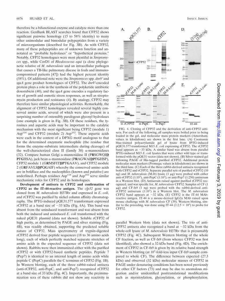
Development of antisera to CFP32 and confirmation ofCFP32 as the IT-44-reactive antigen. The cfp32 gene wascloned from M. tuberculosis H37Rv and expressed in E. coli,and rCFP32 was purified by nickel column affinity chromatog-raphy. The IPTG-induced pQE31.577 transformant expressedrCFP32 at a band size of 33 kDa (Fig. 4A). This band wasabsent from the uninduced transformant and was absent fromboth the induced and uninduced E. coli transformed with thenaked pQE31 plasmid (data not shown). Soluble rCFP32 ofhigh purity, as determined by PAGE and silver staining (Fig.4B), was readily obtained, supporting the predicted solublenature of CFP32. Mass spectrometry of trypsin-digestedrCFP32 derived four peptide sequences (18 to 36 amino acidslong), each of which perfectly matched separate stretches ofamino acids in the expected sequence of CFP32 (data notshown). Rabbits were then immunized either with the purifiedrCFP32 or with CFP32-based synthetic peptides. Peptide 7(Pep7) is identical to an internal length of amino acids whilepeptide C (PepC) parallels the C terminus of CFP32 (Fig. 1B).In Western blotting, each of the three rabbit-raised antisera(anti-rCFP32, anti-PepC, and anti-Pep7) recognized rCFP32at a band size of 33 kDa (Fig. 4C). Importantly, the preimmu-nization sera of these rabbits did not show any reactivity in
parallel Western blots (data not shown). The trio of anti-CFP32 antisera also recognized a band at 32 kDa from thewhole-cell lysate of M. tuberculosis H37Rv that is presumablyCFP32 (Fig. 4C). Subsequent Western blotting of the wholeCF fraction, as well as CF-fx9 (from whence CFP32 was firstidentified), also showed a 32-kDa band (Fig. 4D). The enrich-ment of CFP32 in CF-fx9 is given by its relative band strengthin Western blotting (at 103-fold-less input CF-fx9 sample com-pared to whole CF). The difference between expected (27.3kDa) and observed (32 kDa) molecular masses of CFP32 inPAGE under denaturing conditions has been noted previouslyfor other CF factors (73) and may be due to anomalous mi-gration and/or unidentified posttranslational modificationssuch as myristoylation, glycosylation, or phosphorylation.
FIG. 4. Cloning of CFP32 and the derivation of anti-CFP32 anti-sera. For each of the following, all samples were boiled prior to beingloaded in the gel, and molecular mass protein markers (Amersham;values in kilodaltons) are shown in the first lane. (A) Coomassieblue-stained polyacrylamide gel of lysate from IPTG-inducedpQE31.577-transformed M15 E. coli expressing rCFP32. The rCFP32band appears at 33 kDa. A similar band was absent from parallelIPTG-induced M15 E. coli lysates that were either wild type or trans-formed with the pQE31 vector (data not shown). (B) Silver-stained gelfollowing PAGE of His-tagged purified rCFP32. Additional proteinmolecular mass markers (Promega; values in kilodaltons) are shown inthe third lane. (C) Each of the three rabbit-derived antisera recognizedboth CFP32 and rCFP32. Separate parallel sets of purified rCFP32 (10ng) and M. tuberculosis (M.tb) lysate (1 �g) were probed with eitheranti-rCFP32 (1:103), anti-PepC (1:103), or anti-Pep7 (1:250) antiserumin a Western blot. (D) Antiserum raised against purified rCFP32 rec-ognized, and was specific for, M. tuberculosis CFP32. Samples of CF (1�g) and CF-fx9 (1 ng) were probed with the rabbit-derived anti-rCFP32 antiserum (1:103) in a Western blot. The M. tuberculosisCFP32 band appears at 32 kDa. (E) CFP32 is the IT-44 MAb-reactive antigen. IT-44 is a mouse-derived IgG2a MAb raised uponmouse challenge with M. tuberculosis CF (39). Western blotting, sim-ilar to the preceding, was done using IT-44 (1:2.5 � 104) to probe forCFP32.
6876 HUARD ET AL. INFECT. IMMUN.
on February 3, 2019 by guest
http://iai.asm.org/
Dow
nloaded from

Overall, the combined data confirm the correct cloning andexogenous expression of CFP32 from fractionated CF andargue for the specificity of the developed antisera.
IT-44 (also known as HBT7) is an IgG2a murine MAb thatwas derived from the immunization of inbred mouse strainswith the CF of M. tuberculosis H37Rv (39). A GenBank sub-mission of unpublished data from T. Oettinger (accession no.AJ007737) identified the IT-44-reactive antigen as being thegene product of cfp32 (given as the cfp30B gene). IT-44 wasalso shown previously to react with three spots in two-dimen-sional PAGE of M. tuberculosis CF (64). However, the proteinswere clustered at 32 kDa and migrated within a narrow pIrange of 4.75 to 4.93, thereby suggesting that the antibody wasreacting with multiple isoforms of the same antigen. As a resultof this information, IT-44 was obtained and was evaluated forCFP32 reactivity by Western blotting. Bands for rCFP32 andCFP32 were seen in the same position as in the Western blotsprobed with rabbit anti-rCFP32 antisera (Fig. 4E) while IT-44Western blot reactivity could be blocked by blot preincubationwith anti-rCFP32 (data not shown), thereby verifying CFP32 asthe IT-44-reactive antigen. This finding has been indepen-dently confirmed in CF mapping studies (53, 55). It should alsobe noted that Western blot assays probing for CFP32, similarto previous silver stain gel results (Fig. 1A), suggested thatCFP32 and rCFP32 exist in two states: the respective linearized32- or 33-kDa form that was seen when samples were preparedunder denaturing conditions (by being heated to 100°C for 5min in the presence of dithiothreitol) and a predominant 24-kDa form that was visible in parallel nondenatured samples(data not shown). It was therefore thought that native CFP32maintains a compacted hydrophodynamic volume that is un-folded upon boiling, the likes of which were also noted previ-ously for CFP25 (73). However, the CFP32 sequence containsbut a single cysteine residue, and so forces other than intramo-lecular disulfide bonds must maintain the globular three-di-mensional structure of monomeric CFP32.
Distribution of CFP32 among M. tuberculosis subcellularcompartments. To localize CFP32, M. tuberculosis H37Rv sub-cellular compartments were evaluated for the presence ofCFP32 by Western blotting with the developed antisera (Fig.5). On a per-microgram basis, the greatest quantity of CFP32was found in the CF followed by a very strong CFP32 band inthe cytosolic and whole-cell lysate fractions. Small amountswere also detected in the cell wall, soluble cell wall proteins,and membrane fractions but not in the purified mannosylatedlipoarabinomannan. At least one additional lot of each com-ponent was tested by Western blotting and gave a similar result(data not shown). ELISA measurement of CFP32 levels in theillustrated components supported the Western blot data, indi-cating relative amounts of CFP32 in each by band intensity(Fig. 5). These data suggest a directed movement of CFP32from the cytosol to the CF despite the lack of a clear gener-alized signal peptide for bacterial secretory proteins in theCFP32 N terminus (51; data not shown). It is further notewor-thy that the original sequencing of the CF-fx9 CFP32 band didnot indicate the occurrence of N-terminal cleavage associatedwith export signal peptides. Even so, there are several otherknown CF protein genes that do not code for the classicalsignal peptides, including superoxide dismutase (28), glu-tamine synthetase (27), and CFP29 (54) as well as ESAT-6 and
CFP10 (65). Whether CFP32 or other such proteins are ac-tively exported, excreted, or released during cell division orautolysis is unresolved but may involve an uncharacterizedmycobacterial secretory mechanism (27, 28, 70). Moreover, byanalogy to other CF proteins (27, 28), CFP32 may serve intra-cellular, in addition to extracellular, functions, thus explainingthe necessity of its partial cytoplasmic retention.
CFP32 is MTC restricted. For CFP32 to be characterized asa unique biofactor of tuberculous mycobacteria, it should bepresent in all members of the MTC and absent from MOTT. Ina previous study, 8 MTC subspecies (representing 72 strains)and 12 MOTT species (comprising 46 strains) were evaluatedfor the presence of cfp32 by PCR (given as Rv0577 and usingprimers Rv0577F and Rv0577R [33]). A cfp32 PCR fragmentwas observed only in the MTC groupings, indicating that cfp32is an MTC-restricted gene. To determine the degree of poly-morphism in cfp32 among the MTC subspecies, sequence anal-ysis of PCR products representing the full and/or extended fullcfp32 coding sequence from M. tuberculosis (n � 8), M. africa-num (subtypes I and II, n � 4 and 4, respectively), M. bovis (n� 4), M. bovis BCG (n � 3), M. bovis subsp. caprae (n � 1), andM. microti (n � 4) was done. Likewise, the 321 bp of theputative cfp32 upstream promoter region was also PCR am-plified and sequenced from M. tuberculosis (n � 5), M. africa-num (subtypes I and II, n � 2 and 4, respectively), M. bovis (n� 1), M. bovis BCG (n � 2), M. bovis subsp. caprae (n � 1), andM. microti (n � 3). In all cases, the entire cfp32 locus (1,160 bp,representing nucleotides 670843 to 672002 of the M. tubercu-losis H37Rv genome sequence, accession no. AL123456) wascompletely nonpolymorphic relative to the M. tuberculosisH37Rv genome sequence (data not shown). This observation isin keeping with previous studies that have noted a remarkable
FIG. 5. CFP32 localizes predominantly to the CF and cytosol frac-tions of M. tuberculosis. Western blotting was done to probe the lysate,CF, mannosylated lipoarabinomanan (manLam) glycolipid, cell wall,soluble cell wall proteins (SCWP), membrane, and cytosol componentsof M. tuberculosis (at 1 �g each) for the presence of CFP32 by using theanti-rCFP32 antiserum (1:103). Molecular mass protein markers (Am-ersham; values in kilodaltons) are shown in the first lane. The amountof CFP32, as measured by ELISA (average for duplicate samples inthree experiments), in each sample is given below each respective lane(in picograms per microgram of sample � standard error [SE]). M.tuberculosis PPD was negative for CFP32 by Western blotting (data notshown) and by ELISA was measured as having 51 � 28 pg of CFP32per �g of sample. CF-fx9 was also tested by ELISA and had 610 � 53ng of CFP32 per �g of sample.
VOL. 71, 2003 M. TUBERCULOSIS PROTEIN CFP32, IL-10, AND TUBERCULOSIS 6877
on February 3, 2019 by guest
http://iai.asm.org/
Dow
nloaded from

paucity of single-nucleotide polymorphisms in the structuralgenes of the MTC subspecies (66).
Southern blotting was employed next to verify that cfp32 isrestricted to the MTC organisms. Of the evaluated species,only M. tuberculosis strains H37Rv and W, as well as the ad-ditional MTC subspecies M. africanum subtype I, M. bovis, andM. bovis BCG, were positive for a single copy of cfp32, while all13 MOTT species and strains evaluated were negative (Fig.6A). M. smegmatis was also repeatedly evaluated and found tobe negative for cfp32 by Southern blotting (data not shown).Moreover, a cfp32 homologue could not be found in the M.smegmatis or the M. leprae genome sequences (http://www.tigr.org and http://www.sanger.ac.uk). Further cfp32 Southernblotting probed a comprehensive range of M. tuberculosis clin-ical isolates (n � 70) previously coded by IS6110-restrictionfragment length polymorphism pattern classification (Fig. 6Bsubpanels i to iv) (37). Included in this evaluation were 36strains prototypic for their particular IS6110 fingerprint (Fig.6B subpanels i and ii). Remarkably, every M. tuberculosis strainwas positive for a single band that ran at approximately thesame location for all but two strains, for which it ran slightlylower than the others (Fig. 6B subpanels iii and iv). Thisdifference most likely relates to the emergence of a new a PvuIIcutting site outside cfp32 since sequencing of the strainTN13475 cfp32 did not uncover any polymorphisms. As such, itis impressive that cfp32 was completely conserved within theMTC given that subspecies- and strain-defining large chromo-somal deletions are increasingly found in the MTC genomes(13, 33, 43). These deletions are emerging as potentially sig-nificant determinants of MTC pathobiological diversity but donot appear to include cfp32. Therefore, the complete conser-vation of cfp32 and its sequence for the tested isolates and itsabsence from MOTT species suggest that this gene may play animportant role that is unique to M. tuberculosis and the otherMTC groupings.
To probe for the mycobacterial expression of CFP32, West-ern blotting was done against a panel of MTC subspecies andstrains (n � 37), as well as a range of MOTT species andstrains (n � 29), which are listed in Materials and Methods.Upon completion, a CFP32 band was detected only from M.tuberculosis and the other MTC subspecies and not from any ofthe 10 MOTT species that were evaluated (Fig. 7; select strainsare illustrated). The combined data support the idea that cfp32is an expressed MTC-restricted gene and are in keeping withan integral role for CFP32 in the lifestyle of tubercle bacilli.
Detection of specific antisera to CFP32 in clinical samplesby ELISA. CFP32 is immunogenic for mice as given by thedevelopment of the murine IT-44 MAb following immuniza-tion with M. tuberculosis CF (39). As an indirect indicator thatCFP32 is expressed in TB patients, the human serologic re-sponse to CFP32 was evaluated. Sera from a cohort of patientswith active TB from Brazil (n � 35) and their healthy house-hold contacts (n � 11; four PPD skin test positive, seven PPDskin test negative) were tested for antibodies that recognizerCFP32. Altogether, 34% (12 of 35) of TB case patients haddetectable antibodies to CFP32 while none of the healthy con-trols were positive (P � 0.05, Fisher’s exact test) (Fig. 8A);PPD status did not segregate the healthy household contacts.Notably, only half of these patients had a documented chestX-ray in their medical record, and of these, only 25% of the
CFP32 antiserum-positive patients had cavitary TB, therebyindicating that cavitary TB status did not increase the likeli-hood of having a positive anti-CFP32 serologic response. Theanti-CFP32 positivity rate was similar in those with and thosewithout a documented chest X-ray. To extend these observa-tions to a population from India, a modified ELISA (withoutthe primary coating MAb) was used to test sera from AFBsmear-positive cavitary TB patients (n � 30) and PPD skin
FIG. 6. Southern blot analysis for cfp32. (A) The cfp32 gene isMTC restricted by Zoo blotting. DNA from an assortment of MTCsubspecies (n � 5; namely, M. tuberculosis [M.tb] strains H37Rv andW, M. africanum subtype I, M. bovis, and M. bovis BCG) and myco-bacteria other than MTC (MOTT; n � 13) was evaluated using cfp32PCR fragments as the probe in Southern blotting. (B) Each clinicalisolate of M. tuberculosis tested possesses the cfp32 gene. Subpanels iand ii illustrate the cfp32 Southern blot results for 36 M. tuberculosisisolates with unique IS6110-restriction fragment length polymorphismpatterns prototypical of their lineages. An additional 35 M. tuberculosisclinical isolates were also evaluated (72 M. tuberculosis strains tested intotal), examples of which are shown in subpanels iii and iv.
6878 HUARD ET AL. INFECT. IMMUN.
on February 3, 2019 by guest
http://iai.asm.org/
Dow
nloaded from

test-positive healthy controls (n � 29). In this set, 30% (9 of30) of the cavitary TB patients and 3% (1 of 29) of the PPDskin test-positive controls were reactive to rCFP32 (P � 0.013,Fisher’s exact test) (Fig. 8B). Hence, there was comparativelylimited variation between the two TB patient populations.Combined, 32% of TB case patients (n � 65) and only 2.5% ofhealthy controls (n � 40) exhibited a significant serologicalresponse to E. coli-expressed rCFP32 (P � 0.003, Fisher’sexact test). These data are in the range of those of otherstudies that examined humoral immunity to recombinant M.tuberculosis proteins, whereby, depending upon the antigenevaluated and its method of production, the sera of 12 to 58%of TB patients were found to contain specific antibodies (38,41). Overall, the available data indicate that the spectrum of M.tuberculosis antigens recognized humorally varies dramaticallybetween patients (41, 58). One contributing factor is the ex-pansion of the repertoire of humoral specificities to M. tuber-culosis antigens as TB progresses (2, 58). For example, serumantibodies to the IT-44-reactive antigen (i.e., CFP32) havebeen identified in cavitary TB patients but not in noncavitary
TB patients or PPD-positive individuals by Western blotting(58). (The apparent inconsistency with regard to our detectionof anti-CFP32 specificities in noncavitary TB patients may wellbe a factor of the different means of evaluation.) Moreover, theCFP32 spot was one of only 26 serum-reactive CF spots thatwere seen in the study by Samanich et al. (58). As such, CFP32appears to be a promising humoral antigen for inclusion in amultiantigen serodiagnostic test and may provide a useful in-dicator of worsening disease. It is also important that signifi-cantly fewer patients recognize certain M. tuberculosis proteinswhen they are expressed in E. coli compared to the counterpartnative proteins (59). Since E. coli-expressed rCFP32 was usedin these evaluations as the antibody capture antigen, this mayhave been a factor in reducing the sensitivity of our assays forCFP32 humoral specificity.
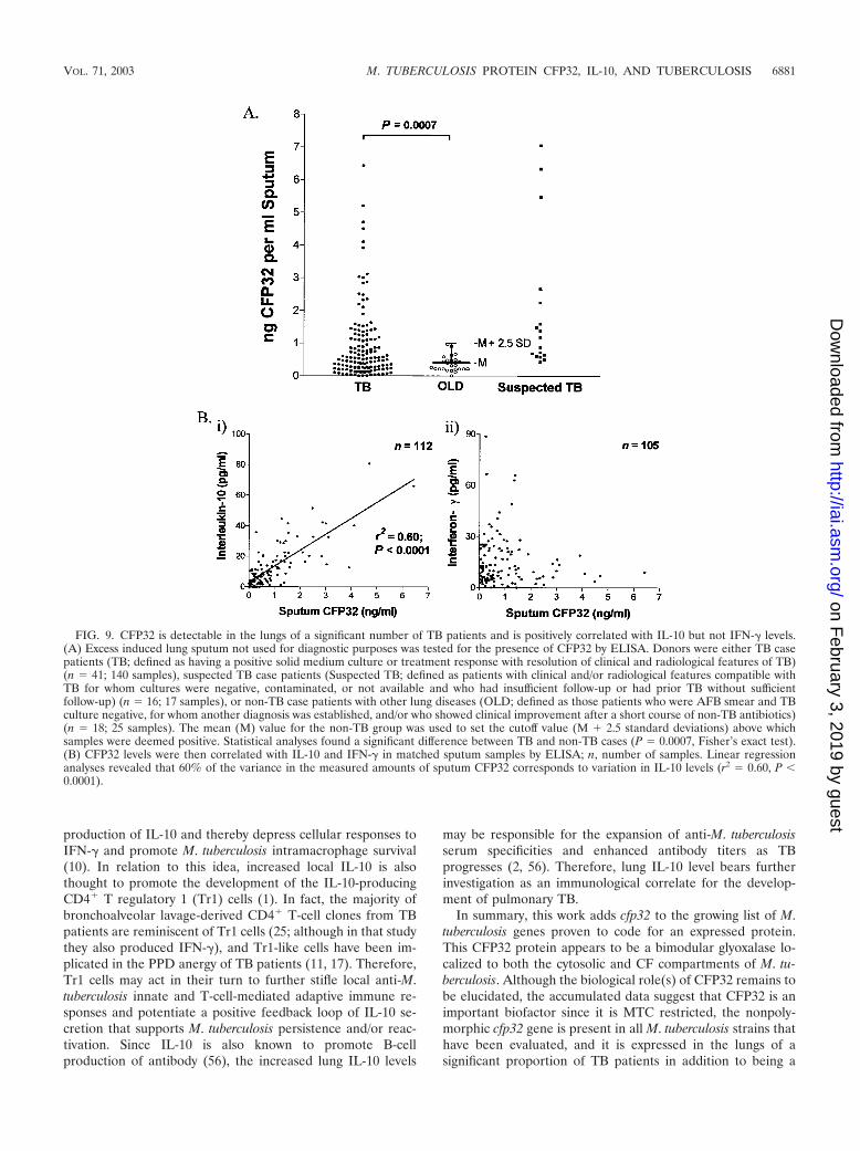
Detection of CFP32 in lung sputum samples of TB patientsby ELISA and positive correlation of CFP32 levels with IL-10.If immunogenic, a candidate in vivo-expressed M. tuberculosisantigen should be present at the site of disease. To detect lungCFP32, the induced sputa from patients in Brazil, who pre-sented for diagnostic workups for “lung disease or suspectedTB,” were tested for the presence of CFP32 by ELISA. Thiscohort included patients with TB (n � 41), suspected TB (n �16), or OLD (n � 18), as defined in Materials and Methods.Sequential samples were taken at days 0, 15, 30, 60, and 180following first presentation with one to five visits per patient. Asignificant number of TB patients had detectable amounts ofCFP32 in their sputum. In contrast to the TB cases, none of thepatients with OLD had detectable CFP32 in their sputum,resulting in a specificity of 100%. In total, 56% (23 of 41) of TBcase patients were positive for CFP32 in at least one sample (P� 0.0001, Fisher’s exact test), of which 32% (13 of 41) werepositive at study entry for CFP32 (P � 0.0057, Fisher’s exacttest). Overall, 59% (10 of 17) of cavitary TB patients wereCFP32 sputum positive in at least one sample and 30% (42 of140) of all samples from TB patients were positive for CFP32(P � 0.0007, Fisher’s exact test) (Fig. 9A). Among TB cases,AFB smear was positive in 54% (22 of 41) while culture waspositive in 80% (33 of 41). Sputum CFP32 was detected in atleast one sample in 61% (20 of 33) of culture-positive TB casesand 50% (3 of 6) culture-negative (n � 5) and culture-unavail-able (n � 1) TB cases. When cross-correlated with AFB smear,64% (14 of 22) of AFB smear-positive and 47% (9 of 19) ofAFB smear-negative case patients had detectable sputumCFP32. There were only five TB case patients who were AFBand culture negative and whose diagnosis was based on treat-ment response with resolution of clinical and radiological fea-tures of TB. Of these case patients, 40% (two of five) werepositive for CFP32. In the suspected TB category, for whomfollow-up data were not available to establish a diagnosis,CFP32 was positive in 56% (9 of 16) of cases. For these ex-periments the standard curve of the CFP32 ELISA was con-sistently linear and sensitive to the level of 5 to 10 pg/ml(data not shown). The data strongly support the idea thatCFP32 is present in the diseased lung. Although the functionof CFP32 here remains unknown, we speculate that it maycontribute to the pathobiology of M. tuberculosis. However, asproposed for other M. tuberculosis CF proteins (5, 12, 52), theactions of CFP32 could include both direct enzymatic activityupon host cells or structures and/or bacterial components and
FIG. 7. Western blot analysis for CFP32. (A) CFP32 is MTC re-stricted. Mycobacterial lysates (7 �g of each sample) and purifiedrCFP32 (10 ng) were probed by Western blotting with the anti-rCFP32antiserum (1:103). A total of 37 MTC isolates (M. tuberculosis H37Rvand one strain each of M. africanum subtype I, M. bovis, M. bovis BCG,and M. microti are shown) and 29 MOTT isolates (one isolate each ofM. avium subsp. avium, M. smegmatis, and M. leprae is illustrated) weretested. A breakdown by MOTT species and MTC subspecies is given inMaterials and Methods. (B) Both laboratory and clinical isolates of M.tuberculosis express CFP32. The lysates of M. tuberculosis strains (3 �gof each sample) were probed by Western blotting with anti-rCFP32antiserum (1:103). For both panels, parallel silver-stained gels (with10-fold-more sample per isolate) confirmed that approximately thesame amount of protein was loaded for each Mycobacterium isolateillustrated (data not shown). Molecular mass protein markers (Amer-sham; values in kilodaltons) are shown in the first lane of each blot.
VOL. 71, 2003 M. TUBERCULOSIS PROTEIN CFP32, IL-10, AND TUBERCULOSIS 6879
on February 3, 2019 by guest
http://iai.asm.org/
Dow
nloaded from

antigenicity-based local tissue damage via immunohyperstimu-lation. By virtue of its expression in vivo and given that CFP32could be detected in the suspected TB subset of patients as wellas a proportion of AFB smear-negative and/or culture-negativepatients, these data further present CFP32 as a strong candi-date antigen for inclusion in a next-generation diagnostic strat-egy as a marker of increasing bacterial burden or as an indi-cation of the effectiveness of TB pharmacologic therapy.
Previously, the coexpression of mRNA for IFN-� with IL-10,IL-2, and inducible nitric oxide synthase, by lung cells frompatients with active pulmonary TB, was described [48; M. D.Bonecini-Almeida, J. R. Lapa e Silva, S. Nicholson, J. Geng, N.Boechat, C. Linhares, L. Rego, and A. L. Kritski, abstract fromthe American Thoracic Society Annual Meeting 1997, Am. J.Respir. Crit. Care Med. 155(Suppl.):A441, 1997]. To furtherdissect the in vivo human immunologic parameters associatedwith TB, ELISA was done to quantify the IL-10 and IFN-� inthe same induced sputum samples from TB patients (n � 34)previously assayed for CFP32. A significant correlation be-tween CFP32 and IL-10 in the sputum of patients was found bylinear regression analysis (n � 112 samples; r2 � 0.60, P �0.0001) (Fig. 9B subpanel i). No convincing association wasidentified between CFP32 and IFN-� (n � 125 samples) (Fig.9B subpanel ii) nor between IL-10 and IFN-� (n � 110 sam-ples; data not shown). IFN-� is regarded as a pivotal cytokinein the protective immune response against M. tuberculosis in-fection, acting as the major mediator of macrophage activationand as a crucial component in the development of specificcounter-M. tuberculosis adaptive immunity (16). IL-10, on theother hand, is a pleiotropic immunosuppressive cytokine thatopposes many IFN-�-mediated effects including macrophage-mediated mycobacteriocidal activity (10, 19, 24). Indeed, evi-
dence is accumulating linking IL-10 to the failure in immunitythat results in the progression to active TB. For example,infection studies with either IL-10 gene-knockout mice orIL-10 transgenic mice have shown that IL-10 is an inhibitor ofanti-tubercle bacillus responses (34, 45, 46, 71). In humans,healthy persons reactive to PPD produce high concentrationsof IFN-� from M. tuberculosis antigen-stimulated peripheralblood mononuclear cells (PBMCs) while TB patients with se-vere disease, and without reactivity to PPD (i.e., anergized),exhibit impaired IFN-� production in association with in-creased IL-10 (11, 17). Moreover, increased levels of IL-10, inthe presence of IFN-�, have been detected in the serum of TBpatients, as well as from ex vivo M. tuberculosis antigen-stim-ulated PBMCs of TB patients (20, 49, 63, 72). In this report, wefound a novel association of bacterial physiological activity (asreflected by CFP32 antigen levels) and IL-10 production in thelungs of patients with the failure of counter-M. tuberculosisimmunity (which was also notably coincident with continuedIFN-� production). These data contrast with those of Barnes etal. (7), who found elevated IFN-� in association with IL-10 inthe pleural fluid of patients with tuberculous pleuritis (a formof TB that resolves without chemotherapy). Together thesedata suggest that the immunosuppressive actions of IL-10 maycome to predominate and eliminate the protective immunesystem-activating properties of IFN-� in the lungs of personswith advanced TB. The mechanism underlying the elevatedIL-10 levels is likely multifactorial and involves contributionsfrom both the host and the pathogen. In this regard, naïvehuman PBMCs, monocytes, and dendritic cells are known toproduce IL-10 when stimulated with M. tuberculosis or with itscell wall constituents (6, 10, 29, 69). Hence, as an immuneevasion strategy, M. tuberculosis may deliberately induce the
FIG. 8. Antiserum specificity to CFP32 is detectable in a significant proportion of human TB patients. (A) Cohort of patients living in Brazil.Sera from 35 active TB case patients, along with the sera of 11 healthy household contacts (seven PPD skin test negative and four PPD skin testpositive), were used in an ELISA to identify human humoral specificity for CFP32. The serologic reactivity of the healthy controls was used to setthe cutoff value above which samples were deemed positive (mean [M] A450 � 1.5 standard deviation). The results of statistical analysis of the dataare shown (P � 0.05, Fisher’s exact test). (B) Cohort of patients living in India. Sera from 30 active TB case patients, as well as from 29PPD-positive controls, were used in a variant ELISA to confirm the existence of human humoral response to CFP32. The serologic reactivity ofthe healthy PPD-positive controls was used to set the cutoff value (mean A490 � 1.5 standard deviation). The results of statistical analysis of thedata are shown (P � 0.013, Fisher’s exact test).
6880 HUARD ET AL. INFECT. IMMUN.
on February 3, 2019 by guest
http://iai.asm.org/
Dow
nloaded from
production of IL-10 and thereby depress cellular responses toIFN-� and promote M. tuberculosis intramacrophage survival(10). In relation to this idea, increased local IL-10 is alsothought to promote the development of the IL-10-producingCD4� T regulatory 1 (Tr1) cells (1). In fact, the majority ofbronchoalveolar lavage-derived CD4� T-cell clones from TBpatients are reminiscent of Tr1 cells (25; although in that studythey also produced IFN-�), and Tr1-like cells have been im-plicated in the PPD anergy of TB patients (11, 17). Therefore,Tr1 cells may act in their turn to further stifle local anti-M.tuberculosis innate and T-cell-mediated adaptive immune re-sponses and potentiate a positive feedback loop of IL-10 se-cretion that supports M. tuberculosis persistence and/or reac-tivation. Since IL-10 is also known to promote B-cellproduction of antibody (56), the increased lung IL-10 levels
may be responsible for the expansion of anti-M. tuberculosisserum specificities and enhanced antibody titers as TBprogresses (2, 56). Therefore, lung IL-10 level bears furtherinvestigation as an immunological correlate for the develop-ment of pulmonary TB.
In summary, this work adds cfp32 to the growing list of M.tuberculosis genes proven to code for an expressed protein.This CFP32 protein appears to be a bimodular glyoxalase lo-calized to both the cytosolic and CF compartments of M. tu-berculosis. Although the biological role(s) of CFP32 remains tobe elucidated, the accumulated data suggest that CFP32 is animportant biofactor since it is MTC restricted, the nonpoly-morphic cfp32 gene is present in all M. tuberculosis strains thathave been evaluated, and it is expressed in the lungs of asignificant proportion of TB patients in addition to being a
FIG. 9. CFP32 is detectable in the lungs of a significant number of TB patients and is positively correlated with IL-10 but not IFN-� levels.(A) Excess induced lung sputum not used for diagnostic purposes was tested for the presence of CFP32 by ELISA. Donors were either TB casepatients (TB; defined as having a positive solid medium culture or treatment response with resolution of clinical and radiological features of TB)(n � 41; 140 samples), suspected TB case patients (Suspected TB; defined as patients with clinical and/or radiological features compatible withTB for whom cultures were negative, contaminated, or not available and who had insufficient follow-up or had prior TB without sufficientfollow-up) (n � 16; 17 samples), or non-TB case patients with other lung diseases (OLD; defined as those patients who were AFB smear and TBculture negative, for whom another diagnosis was established, and/or who showed clinical improvement after a short course of non-TB antibiotics)(n � 18; 25 samples). The mean (M) value for the non-TB group was used to set the cutoff value (M � 2.5 standard deviations) above whichsamples were deemed positive. Statistical analyses found a significant difference between TB and non-TB cases (P � 0.0007, Fisher’s exact test).(B) CFP32 levels were then correlated with IL-10 and IFN-� in matched sputum samples by ELISA; n, number of samples. Linear regressionanalyses revealed that 60% of the variance in the measured amounts of sputum CFP32 corresponds to variation in IL-10 levels (r2 � 0.60, P �0.0001).
VOL. 71, 2003 M. TUBERCULOSIS PROTEIN CFP32, IL-10, AND TUBERCULOSIS 6881
on February 3, 2019 by guest
http://iai.asm.org/
Dow
nloaded from
recognized humoral antigen. That CFP32 levels in the lungs ofactive TB patients correlated with measured IL-10, but notIFN-�, levels supports the hypothesis that M. tuberculosis-stim-ulated local IL-10 secretion precipitates the immunodysregu-lation that contributes to the success of M. tuberculosis as ahuman pathogen. Determining the role of CFP32, if any, in thisparticular pathogenic strategy of M. tuberculosis is a priorityinterest of our laboratory.
ACKNOWLEDGMENTS
We thank Howard Doo, Kelley Sookraj, Krishna Menon, PatriciaLago, Vera Batista, and Brianna Holland for technical assistance andAlbert Ko for aiding the statistical analyses. We are also grateful toLee W. Riley, Sabine Ehrt, and Warren D. Johnson for suggestions,support, and encouragement.
Funding support was provided by NIH grants R0-1 AI39606 andR0-1 HL61960 (J.L.H.), NIH NIAID contract N01 AI-75320 (J.T.B.),NIH Fogarty International Center Training grant (FICTG) D43TW00018, a grant from the Coordenacao de Aperfeicoamento dePessoal de Nivel Superior (CAPES; Ministry of Education-Brazil), anda grant from the Laura Cook Hull Trust Fund (LCHTF) (Warren D.Johnson, Principal Investigator). R.C.H. was supported by theLCHTF, H.Z. was a FICTG trainee, and L.C.O.L. was an FICTG andCAPES trainee. S.L. was supported by a VA merit award. M.B.C.,A.L.K., and J.R.L.S. were funded in Brazil by the following grants:Brazilian TB Research Network 62.0055/01-4-PADCT III/MILLENIUM (CNPq/Brazilian Research Council and World Bank; M.B.C.,A.L.K., J.R.L.S.), “Excellence Research Nuclei for TB Control”66.1028/1998-4 (PRONEX/Brazilian Research Council; A.L.K.,J.R.L.S.), “Scientists of Our State” 2000 and 2003 (Rio de JaneiroResearch Council/FAPERJ; A.L.K., J.R.L.S.), “Small Grants Pro-gram” (Rio de Janeiro Research Council/FAPERJ; M.B.C.), and“Small Grants Program” (Fundacao Universitaria Jose Bonifacio/FUJB; J.R.L.S.).
REFERENCES1. Akbari, O., R. H. DeKruyff, and D. T. Umetsu. 2001. Pulmonary dendritic
cells producing IL-10 mediate tolerance induced by respiratory exposure toantigen. Nat. Immunol. 2:725–731.
2. Amara, R. R., and V. Satchidanandam. 1996. Analysis of a genomic DNAexpression library of Mycobacterium tuberculosis using tuberculosis patientsera: evidence for modulation of host immune response. Infect. Immun.64:3765–3771.
3. Andersen, P. 1997. Host responses and antigens involved in protective im-munity to Mycobacterium tuberculosis. Scand. J. Immunol. 45:115–131.
4. Ando, N., K. Ueda, and S. Horinouchi. 1997. A Streptomyces griseus gene(sgaA) suppresses the growth disturbance caused by high osmolality and ahigh concentration of A-factor during early growth. Microbiology 143:2715–2723.
5. Armitige, L. Y., C. Jagannath, A. R. Wanger, and S. J. Norris. 2000. Dis-ruption of the genes encoding antigen 85A and antigen 85B of Mycobacte-rium tuberculosis H37Rv: effect on growth in culture and in macrophages.Infect. Immun. 68:767–778.
6. Barnes, P. F., D. Chatterjee, J. S. Abrams, S. Lu, E. Wang, M. Yamamura,P. J. Brennan, and R. L. Modlin. 1992. Cytokine production induced byMycobacterium tuberculosis lipoarabinomannan. Relationship to chemicalstructure. J. Immunol. 149:541–547.
7. Barnes, P. F., S. Lu, J. S. Abrams, E. Wang, M. Yamamura, and R. L.Modlin. 1993. Cytokine production at the site of disease in human tubercu-losis. Infect. Immun. 61:3482–3489.
8. Bhaskar, S., S. P. Khanna, and R. Mukherjee. 2000. Isolation, purificationand immunological characterization of novel low molecular weight proteinantigen CFP 6 from culture filtrate of M. tuberculosis. Vaccine 18:2856–2866.
9. Boesen, H., B. N. Jensen, T. Wilcke, and P. Andersen. 1995. Human T-cellresponses to secreted antigen fractions of Mycobacterium tuberculosis. Infect.Immun. 63:1491–1497.
10. Bonecini-Almeida, M. G., S. Chitale, I. Boutsikakis, J. Geng, H. Doo, S. He,and J. L. Ho. 1998. Induction of in vitro human macrophage anti-Mycobac-terium tuberculosis activity: requirement for IFN-gamma and primed lym-phocytes. J. Immunol. 160:4490–4499.
11. Boussiotis, V. A., E. Y. Tsai, E. J. Yunis, S. Thim, J. C. Delgado, C. C.Dascher, A. Berezovskaya, D. Rousset, J. M. Reynes, and A. E. Goldfeld.2000. IL-10-producing T cells suppress immune responses in anergic tuber-culosis patients. J. Clin. Investig. 105:1317–1325.
12. Brodin, P., K. Eiglmeier, M. Marmiesse, A. Billault, T. Garnier, S. Niemann,S. T. Cole, and R. Brosch. 2002. Bacterial artificial chromosome-based com-
parative genomic analysis identifies Mycobacterium microti as a naturalESAT-6 deletion mutant. Infect. Immun. 70:5568–5578.
13. Brosch, R., S. V. Gordon, M. Marmiesse, P. Brodin, C. Buchrieser, K.Eiglmeier, T. Garnier, C. Gutierrez, G. Hewinson, K. Kremer, L. M. Par-sons, A. S. Pym, S. Samper, D. van Soolingen, and S. T. Cole. 2002. A newevolutionary scenario for the Mycobacterium tuberculosis complex. Proc.Natl. Acad. Sci. USA 99:3684–3689.
14. Camus, J. C., M. J. Pryor, C. Medigue, and S. T. Cole. 2002. Re-annotationof the genome sequence of Mycobacterium tuberculosis H37Rv. Microbiology148:2967–2973.
15. Cole, S. T., R. Brosch, J. Parkhill, T. Garnier, C. Churcher, D. Harris, S. V.Gordon, K. Eiglmeier, S. Gas, C. E. Barry III, F. Tekaia, K. Badcock, D.Basham, D. Brown, T. Chillingworth, R. Connor, R. Davies, K. Devlin, T.Feltwell, S. Gentiles, N. Hamlin, S. Holroyd, T. Hornsby, K. Jegels, A.Krogh, J. McLean, S. Moule, L. Murphy, K. Oliver, J. Osborne, M. A. Quail,M.-A. Rajandream, J. Rogers, S. Rutter, K. Seeger, J. Skelton, R. Squares,S. Squares, J. E. Sulston, K. Taylor, S. Whitehead, and B. G. Barrell. 1998.Deciphering the biology of Mycobacterium tuberculosis from the completegenome sequence. Nature 393:537–544.
16. Collins, H. L., and S. H. Kaufmann. 2001. The many faces of host responsesto tuberculosis. Immunology 103:1–9.
17. Delgado, J. C., E. Y. Tsai, S. Thim, A. Baena, V. A. Boussiotis, J. M. Reynes,S. Sath, P. Grosjean, E. J. Yunis, and A. E. Goldfeld. 2002. Antigen-specificand persistent tuberculin anergy in a cohort of pulmonary tuberculosis pa-tients from rural Cambodia. Proc. Natl. Acad. Sci. USA 99:7576–7581.
18. Demissie, A., P. Ravn, J. Olobo, T. M. Doherty, T. Eguale, M. Geletu, W.Hailu, P. Andersen, and S. Britton. 1999. T-cell recognition of Mycobacte-rium tuberculosis culture filtrate fractions in tuberculosis patients and theirhousehold contacts. Infect. Immun. 67:5967–5971.
19. Dickensheets, H. L., S. L. Freeman, M. F. Smith, and R. P. Donnelly. 1997.Interleukin-10 upregulates tumor necrosis factor receptor type-II (p75) geneexpression in endotoxin-stimulated human monocytes. Blood 90:4162–4171.
20. Dlugovitzky, D., A. Torres-Morales, L. Rateni, M. A. Farroni, C. Largacha,O. Molteni, and O. Bottasso. 1997. Circulating profile of Th1 and Th2cytokines in tuberculosis patients with different degrees of pulmonary in-volvement. FEMS Immunol. Med. Microbiol. 18:203–207.
21. Dye, C., S. Scheele, P. Dolin, V. Pathania, and M. C. Raviglione. 1999.Consensus statement. Global burden of tuberculosis: estimated incidence,prevalence, and mortality by country. WHO Global Surveillance and Mon-itoring Project. JAMA 282:677–686.
22. Eng, J. K., A. L. McCormack, and J. R. Yates. 1994. A approach to correlatetandem mass-spectral data of peptides with amino-acid sequences in a pro-tein database. J. Am. Soc. Mass. Spectrom. 5:976–989.
23. Fernandez, J., L. Andrews, and S. M. Mische. 1994. An improved procedurefor enzymatic digestion of polyvinylidene difluoride-bound proteins for in-ternal sequence analysis. Anal. Biochem. 218:112–117.
24. Flesch, I. E., J. H. Hess, I. P. Oswald, and S. H. Kaufmann. 1994. Growthinhibition of Mycobacterium bovis by IFN-gamma stimulated macrophages:regulation by endogenous tumor necrosis factor-alpha and by IL-10. Int.Immunol. 6:693–700.
25. Gerosa, F., C. Nisii, S. Righetti, R. Micciolo, M. Marchesini, A. Cazzadori,and G. Trinchieri. 1999. CD4� T cell clones producing both interferon-gamma and interleukin-10 predominate in bronchoalveolar lavages of activepulmonary tuberculosis patients. Clin. Immunol. 92:224–234.
26. Glickman, M. S., and W. R. Jacobs, Jr. 2001. Microbial pathogenesis ofMycobacterium tuberculosis: dawn of a discipline. Cell 104:477–485.
27. Harth, G., and M. A. Horwitz. 1997. Expression and efficient export ofenzymatically active Mycobacterium tuberculosis glutamine synthetase in My-cobacterium smegmatis and evidence that the information for export is con-tained within the protein. J. Biol. Chem. 272:22728–22735.
28. Harth, G., and M. A. Horwitz. 1999. Export of recombinant Mycobacteriumtuberculosis superoxide dismutase is dependent upon both information in theprotein and mycobacterial export machinery. A model for studying export ofleaderless proteins by pathogenic mycobacteria. J. Biol. Chem. 274:4281–4292.
29. Henderson, R. A., S. C. Watkins, and J. L. Flynn. 1997. Activation of humandendritic cells following infection with Mycobacterium tuberculosis. J. Immu-nol. 159:635–643.
30. Heym, B., Y. Zhang, S. Poulet, D. Young, and S. T. Cole. 1993. Character-ization of the katG gene encoding a catalase-peroxidase required for theisoniazid susceptibility of Mycobacterium tuberculosis. J. Bacteriol. 175:4255–4259.
31. Horwitz, M. A., B. W. Lee, B. J. Dillon, and G. Harth. 1995. Protectiveimmunity against tuberculosis induced by vaccination with major extracellu-lar proteins of Mycobacterium tuberculosis. Proc. Natl. Acad. Sci. USA 92:1530–1534.
32. Howard, S., S. He, and S. G. Withers. 1998. Identification of the active sitenucleophile in jack bean -mannosidase using 5-fluoro-�-L-gulosyl fluoride.J. Biol. Chem. 273:2067–2072.
33. Huard, R. C., L. C. de Oliveira Lazzarini, W. R. Butler, D. van Soolingen,and J. L. Ho. 2003. A PCR-based method to differentiate the subspecies of
6882 HUARD ET AL. INFECT. IMMUN.
on February 3, 2019 by guest
http://iai.asm.org/
Dow
nloaded from

the Mycobacterium tuberculosis complex on the basis of genomic deletions.J. Clin. Microbiol. 41:1637–1650.
34. Jacobs, M., N. Brown, N. Allie, R. Gulert, and B. Ryffel. 2000. Increasedresistance to mycobacterial infection in the absence of interleukin-10. Im-munology 100:494–501.
35. Jungblut, P. R., U. E. Schaible, H. J. Mollenkopf, U. Zimny-Arndt, B.Raupach, J. Mattow, P. Halada, S. Lamer, K. Hagens, and S. H. Kaufmann.1999. Comparative proteome analysis of Mycobacterium tuberculosis andMycobacterium bovis BCG strains: towards functional genomics of microbialpathogens. Mol. Microbiol. 33:1103–1117.
36. Kane, M. M., and D. M. Mosser. 2001. The role of IL-10 in promotingdisease progression in leishmaniasis. J. Immunol. 166:1141–1147.
37. Kurepina, N. E., S. Sreevatsan, B. B. Plikaytis, P. J. Bifani, N. D. Connell,R. J. Donnelly, D. van Soolingen, J. M. Musser, and B. N. Kreiswirth. 1998.Characterization of the phylogenetic distribution and chromosomal insertionsites of five IS6110 elements in Mycobacterium tuberculosis: non-randomintegration in the dnaA-dnaN region. Tuber. Lung Dis. 79:31–42.
38. Lim, R. L., L. K. Tan, W. F. Lau, M. C. Ming, R. Dunn, H. P. Too, and L.Chan. 2000. Cloning and expression of immunoreactive antigens from My-cobacterium tuberculosis. Clin. Diagn. Lab. Immunol. 7:600–606.
39. Ljungqvist, L., A. Worsaae, and I. Heron. 1988. Antibody responses againstMycobacterium tuberculosis in 11 strains of inbred mice: novel monoclonalantibody specificities generated by fusions, using spleens from BALB.B10and CBA/J mice. Infect. Immun. 56:1994–1998.
40. Lomovskaya, N., S. L. Otten, Y. Doi-Katayama, L. Fonstein, X. C. Liu, T.Takatsu, A. Inventi-Solari, S. Filippini, F. Torti, A. L. Colombo, and C. R.Hutchinson. 1999. Doxorubicin overproduction in Streptomyces peucetius:cloning and characterization of the dnrU ketoreductase and dnrV genes andthe doxA cytochrome P-450 hydroxylase gene. J. Bacteriol. 181:305–318.
41. Lyashchenko, K., R. Colangeli, M. Houde, H. Al Jahdali, D. Menzies, andM. L. Gennaro. 1998. Heterogeneous antibody responses in tuberculosis.Infect. Immun. 66:3936–3940.
42. McKinney, J. D., K. Honer zu Bentrup, E. J. Munoz-Elias, A. Miczak, B.Chen, W. T. Chan, D. Swenson, J. C. Sacchettini, W. R. Jacobs, Jr., and D. G.Russell. 2000. Persistence of Mycobacterium tuberculosis in macrophages andmice requires the glyoxylate shunt enzyme isocitrate lyase. Nature 406:735–738.
43. Mostowy, S., D. Cousins, J. Brinkman, A. Aranaz, and M. A. Behr. 2002.Genomic deletions suggest a phylogeny for the Mycobacterium tuberculosiscomplex. J. Infect. Dis. 186:74–80.
44. Mulder, M. A., H. Zappe, and L. M. Steyn. 1997. Mycobacterial promoters.Tuber. Lung Dis. 78:211–223.
45. Murray, P. J., L. Wang, C. Onufryk, R. I. Tepper, and R. A. Young. 1997. Tcell-derived IL-10 antagonizes macrophage function in mycobacterial infec-tion. J. Immunol. 158:315–321.
46. Murray, P. J., and R. A. Young. 1999. Increased antimycobacterial immunityin interleukin-10-deficient mice. Infect. Immun. 67:3087–3095.
47. Navas, J., B. Gonzalez-Zorn, N. Ladron, P. Garrido, and J. A. Vazquez-Boland. 2001. Identification and mutagenesis by allelic exchange of choE,encoding a cholesterol oxidase from the intracellular pathogen Rhodococcusequi. J. Bacteriol. 183:4796–4805.
48. Nicholson, S., M. D. Bonecini-Almeida, J. R. Lapa e Silva, C. Nathan, Q. W.Xie, R. Mumford, J. R. Weidner, J. Calaycay, J. Geng, N. Boechat, C.Linhares, W. Rom, and J. L. Ho. 1996. Inducible nitric oxide synthase inpulmonary alveolar macrophages from patients with tuberculosis. J. Exp.Med. 183:2293–2302.
49. Othieno, C., C. S. Hirsch, B. D. Hamilton, K. Wilkinson, J. J. Ellner, and Z.Toossi. 1999. Interaction of Mycobacterium tuberculosis-induced transform-ing growth factor beta and interleukin-10. Infect. Immun. 67:5730–5735.
50. Parish, T., B. G. Gordhan, R. A. McAdam, K. Duncan, V. Mizrahi, and N. G.Stoker. 1999. Production of mutants in amino acid biosynthesis genes ofMycobacterium tuberculosis by homologous recombination. Microbiology145:3497–3503.
51. Pugsley, A. P. 1993. The complete general secretory pathway in gram-neg-ative bacteria. Microbiol. Rev. 57:50–108.
52. Raynaud, C., G. Etienne, P. Peyron, M. A. Laneelle, and M. Daffe. 1998.Extracellular enzyme activities potentially involved in the pathogenicity ofMycobacterium tuberculosis. Microbiology 144:577–587.
53. Rosenkrands, I., A. King, K. Weldingh, M. Moniatte, E. Moertz, and P.Andersen. 2000. Towards the proteome of Mycobacterium tuberculosis. Elec-trophoresis 21:3740–3756.
54. Rosenkrands, I., P. B. Rasmussen, M. Carnio, S. Jacobsen, M. Theisen, andP. Andersen. 1998. Identification and characterization of a 29-kilodaltonprotein from Mycobacterium tuberculosis culture filtrate recognized by mousememory effector cells. Infect. Immun. 66:2728–2735.
55. Rosenkrands, I., K. Weldingh, S. Jacobsen, C. V. Hansen, W. Florio, I.Gianetri, and P. Andersen. 2000. Mapping and identification of Mycobacte-
rium tuberculosis proteins by two-dimensional gel electrophoresis, microse-quencing and immunodetection. Electrophoresis 21:935–948.
56. Rousset, F., E. Garcia, T. Defrance, C. Peronne, N. Vezzio, D. H. Hsu, R.Kastelein, K. W. Moore, and J. Banchereau. 1992. Interleukin 10 is a potentgrowth and differentiation factor for activated human B lymphocytes. Proc.Natl. Acad. Sci. USA 89:1890–1893.
57. Ruan, J., G. St. John, S. Ehrt, L. Riley, and C. Nathan. 1999. noxR3, a novelgene from Mycobacterium tuberculosis, protects Salmonella typhimuriumfrom nitrosative and oxidative stress. Infect. Immun. 67:3276–3283.
58. Samanich, K. M., J. T. Belisle, M. G. Sonnenberg, M. A. Keen, S. Zolla-Pazner, and S. Laal. 1998. Delineation of human antibody responses toculture filtrate antigens of Mycobacterium tuberculosis. J. Infect. Dis. 178:1534–1538.
59. Samanich, K. M., M. A. Keen, V. D. Vissa, J. D. Harder, J. S. Spencer, J. T.Belisle, S. Zolla-Pazner, and S. Laal. 2000. Serodiagnostic potential of cul-ture filtrate antigens of Mycobacterium tuberculosis. Clin. Diagn. Lab. Im-munol. 7:662–668.
60. Sambandamurthy, V. K., X. Wang, B. Chen, R. G. Russell, S. Derrick, F. M.Collins, S. L. Morris, and W. R. Jacobs. 2002. A pantothenate auxotroph ofMycobacterium tuberculosis is highly attenuated and protects mice againsttuberculosis. Nat. Med. 8:1171–1174.
61. Sieling, P. A., J. S. Abrams, M. Yamamura, P. Salgame, B. R. Bloom, T. H.Rea, and R. L. Modlin. 1993. Immunosuppressive roles for IL-10 and IL-4 inhuman infection. In vitro modulation of T cell responses in leprosy. J. Im-munol. 150:5501–5510.
62. Smith, S. M., M. R. Klein, A. S. Malin, J. Sillah, K. Huygen, P. Andersen,K. P. McAdam, and H. M. Dockrell. 2000. Human CD8� T cells specific forMycobacterium tuberculosis secreted antigens in tuberculosis patients andhealthy BCG-vaccinated controls in The Gambia. Infect. Immun. 68:7144–7148.
63. Song, C. H., H. J. Kim, J. K. Park, J. H. Lim, U. O. Kim, J. S. Kim, T. H.Paik, K. J. Kim, J. W. Suhr, and E. K. Jo. 2000. Depressed interleukin-12(IL-12), but not IL-18, production in response to a 30- or 32-kilodaltonmycobacterial antigen in patients with active pulmonary tuberculosis. Infect.Immun. 68:4477–4484.
64. Sonnenberg, M. G., and J. T. Belisle. 1997. Definition of Mycobacteriumtuberculosis culture filtrate proteins by two-dimensional polyacrylamide gelelectrophoresis, N-terminal amino acid sequencing, and electrospray massspectrometry. Infect. Immun. 65:4515–4524.
65. Sorensen, A. L., S. Nagai, G. Houen, P. Andersen, and A. B. Andersen. 1995.Purification and characterization of a low-molecular-mass T-cell antigensecreted by Mycobacterium tuberculosis. Infect. Immun. 63:1710–1717.
66. Sreevatsan, S., X. Pan, K. E. Stockbauer, N. D. Connell, B. N. Kreiswirth,T. S. Whittam, and J. M. Musser. 1997. Restricted structural gene polymor-phism in the Mycobacterium tuberculosis complex indicates evolutionarilyrecent global dissemination. Proc. Natl. Acad. Sci. USA 94:9869–9874.
67. Stockl, J., H. Vetr, O. Majdic, G. Zlabinger, E. Kuechler, and W. Knapp.1999. Human major group rhinoviruses downmodulate the accessory func-tion of monocytes by inducing IL-10. J. Clin. Investig. 104:957–965.
68. Stylianou, E., P. Aukrust, D. Kvale, F. Muller, and S. S. Froland. 1999. IL-10in HIV infection: increasing serum IL-10 levels with disease progression—down-regulatory effect of potent anti-retroviral therapy. Clin. Exp. Immunol.116:115–120.
69. Thoma-Uszynski, S., S. M. Kiertscher, M. T. Ochoa, D. A. Bouis, M. V.Norgard, K. Miyake, P. J. Godowski, M. D. Roth, and R. L. Modlin. 2000.Activation of toll-like receptor 2 on human dendritic cells triggers inductionof IL-12, but not IL-10. J. Immunol. 165:3804–3810.
70. Tullius, M. V., G. Harth, and M. A. Horwitz. 2001. High extracellular levelsof Mycobacterium tuberculosis glutamine synthetase and superoxide dis-mutase in actively growing cultures are due to high expression and extracel-lular stability rather than to a protein-specific export mechanism. Infect.Immun. 69:6348–6363.
71. Turner, J., M. Gonzalez-Juarrero, D. L. Ellis, R. J. Basaraba, A. Kipnis,I. M. Orme, and A. M. Cooper. 2002. In vivo IL-10 production reactivateschronic pulmonary tuberculosis in C57BL/6 mice. J. Immunol. 169:6343–6351.
72. Verbon, A., N. Juffermans, S. J. Van Deventer, P. Speelman, H. Van Deu-tekom, and T. Van Der Poll. 1999. Serum concentrations of cytokines inpatients with active tuberculosis (TB) and after treatment. Clin. Exp. Immu-nol. 115:110–113.
73. Weldingh, K., I. Rosenkrands, S. Jacobsen, P. B. Rasmussen, M. J. Elhay,and P. Andersen. 1998. Two-dimensional electrophoresis for analysis ofMycobacterium tuberculosis culture filtrate and purification and characteriza-tion of six novel proteins. Infect. Immun. 66:3492–3500.
74. World Health Organization. 1996. Report of the tuberculosis epidemic.World Health Organization, Geneva, Switzerland.
Editor: S. H. E. Kaufmann
VOL. 71, 2003 M. TUBERCULOSIS PROTEIN CFP32, IL-10, AND TUBERCULOSIS 6883
on February 3, 2019 by guest
http://iai.asm.org/
Dow
nloaded from