mycology systemic
TRANSCRIPT
8/8/2019 Mycology Systemic
http://slidepdf.com/reader/full/mycology-systemic 1/12
SYSTEMIC MYCOSESIntroduction
DIMORPHIC FUNGI
y Blastomycosis
o Blastomyces dermatitidis
y Coccidioidomycosis
o Coccidioides immitis
y Histoplasmosis o Histoplasma capsulatum
MONOMORPHIC FUNGI:
y Paracoccidioidomycosis
o Paracoccidioides brasiliensis
y Cryptococcosis
o Cryptococcus neoformans
Various appearances:
y Yeast cells:
o Histoplasma capsulatum
o Cryptococcus neoformans
y Sporangia
o Coccidioides immitis
y Spherules
o Coccidioides immitis
y Granules
o Sporothrix schenkii
Lab Dx:
o KOH and Culture
o Biopsy (Histopath)
o Immunoflourescence
o Immunohistochemistry
o Molecular study: PCR
Different stains used to demonstrate fungi in Histopath
1. PAS: bright red-purple
2. GMS: black
3. H & E: pink to purple
4. Mucicarmine: pink (Cryptococcus
5. Fontana-Masson: dematiaceous fungi
6. Calcoflour-white: fluorescence
7. Gridley: purple to magenta
BLASTOMYCES DERMATITIDIS
Distribution
endemic in
± North America
± Mississippi
± Missouri
± Ohio Rivers
± and their tributaries
zone extends into
± Kentucky
± Carolinas
± Appalachian region
± Canada
± and Wisconsin
Reservoirs
The exact ecologic niche has not been determined
but the fungus has been found in the following:
± moist environments
± wood
± tree bark
± rotting vegetation
± animal habitats
± manure
± wet acid soil from the banks of rivers
MOT
airborne routes
but is only rarely infectious
small threat to people with competent immune
systems
although it can cause fatal infections to immuno-
compromised patients
Human Infection
Blastomycosis
± Gilchrist's disease / blastomycosis
± honoring the first man to publish reports of
the infection
± North American Blastomycosis
± a chronic granulomatous and suppurative
disease that may affect the following:
Skin and mucous membrane
Bones
Lungs
GUT
Cutaneous Blastomycosis
± ( skin) Blastomycosis
- elevated, macerated, ill-defined, scaly
borders, central ulcer
± ( mucous membrane and skin)
Blastomycosis
± Chronic cutaneous blastomycosis (with or
without lung involvement)
verrucose
ulcerated
suppurative
Systemic blastomycosis:
± involve any organ of the body or a
combination of organs
± Pulmonary Blastomycosis
8/8/2019 Mycology Systemic
http://slidepdf.com/reader/full/mycology-systemic 2/12
Inhalation --- lungs ---
disseminated
Round pneumonia
"Mass" like density with
air bronchogram
± Osteoarticular Blastomycosis
Affects the spine, ribs, long bones
painful debilitating arthritis or
osteomyelitis
± GUT Blastomycosis
Affects the prostate and
epidydimis in males
Specimen Sources
skin scrapings
crusts from skin lesions
aspirated pus
Sputum
and other pulmonary specimens
oropharyngeal scrapings
biopsied tissue
blood
prostate secretions
Cerebrospinal Fluid
urine
Specimen Collection and Handling
Specimens should be collected aseptically according
to the standard protocol for the type of specimen
Transported to the laboratory without delay and
plated promptly
Direct examination:
10% KOH added to the mounting fluid to
clear the specimen before it is examined
Mucus and other thick substances should be treated
with N-acetyl-L-cysteine before the wet mount is
done
Tissue specimens:
fixed and stained with
H & E
GMS stain
Giemsa stain
Immunology
two antigens: A and B
A is reported to be the more useful of the
two
Currently no skin test for blastomycosis is
commercially available because of the lack of
purified antigens
Methods
Immunodiffusion (ID) tests - specificity of 84% to
100%, sensitivity of 57% to 62%
Enzyme Immunoassay (EIA) - sensitivity of 80%,
specificity of 98%
Complement Fixation (CF) method
Chemiluminescent DNA probe method
Special Precautions
Laboratory personnel who are working with any
fluffy white colony should take special precautions
and adhere stringently to laboratory procedures.
As a minimum, cultures should always be studied
within a biologic Class II safety cabinet.
Slide cultures should never be made
Culture Media
Modified SDA or BHIA with antibiotics at
temperatures from 25° to 30°C,
SDA without antimicrobials at temperatures above
30°C
Use BHIA containing blood for culture of specimens
from body sites that are normally sterile
Biphasic culture bottles containing both BHI broth
and BHI agar are recommen-ded for blood cultures
PDA or PFA may be used for subculture to encourage
conidia production
Temperature Considerations
Mould form:
Optimal temperature for growth is 25° to
30°C on routine media
Yeast form:
37°C is needed to induce the yeast phase
Primary cultures f or B. dermatitidis should be held
f or 4 to 8 w eeks be f ore being discarded as " no
grow th."
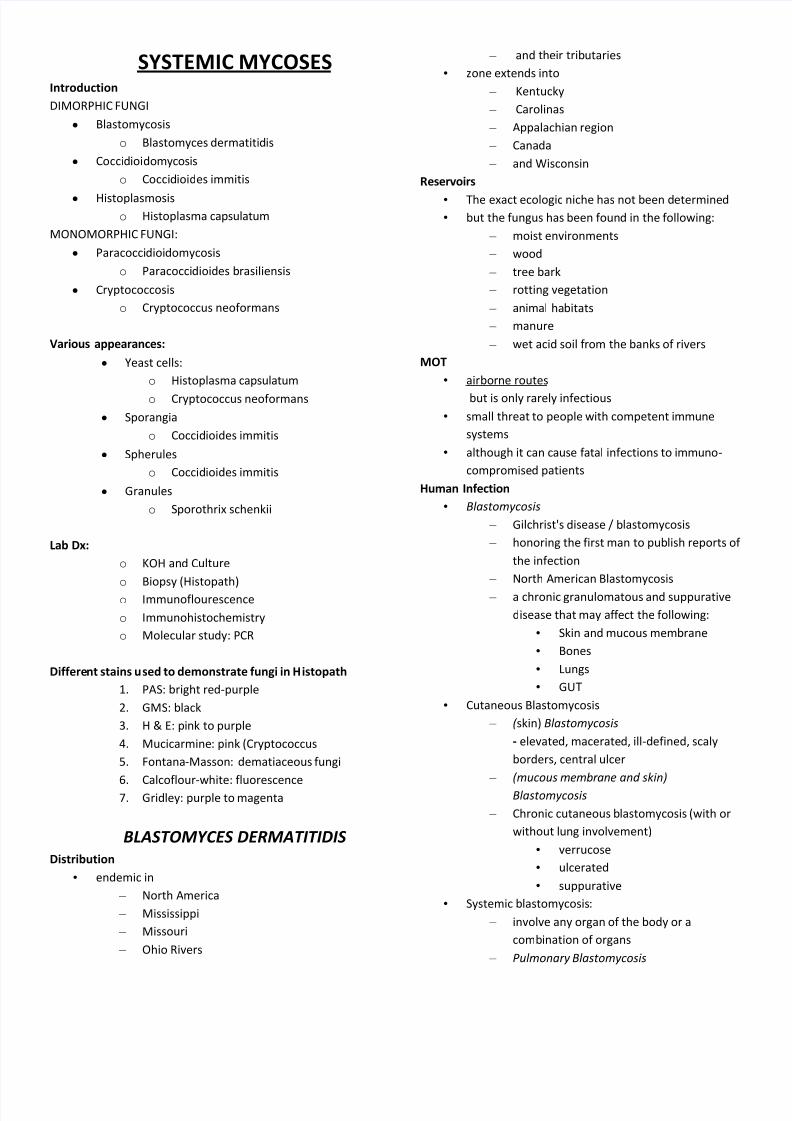
Macroscopic (Colony) Morphology
Mould-form colonies at 25°C
on modified SDA grow slowly
mature colonies typically grow in 6 to 21 days
white or beige to brown at first
with a waxy or glabrous texture
some isolates may be fluffy
are prickly in the center
Reverse pigment of the colonies is tan to brown
8/8/2019 Mycology Systemic
http://slidepdf.com/reader/full/mycology-systemic 3/12
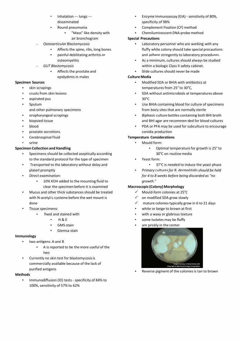
Later, colonies tend to become fluffy or woolly, and
some develop concentric rings.
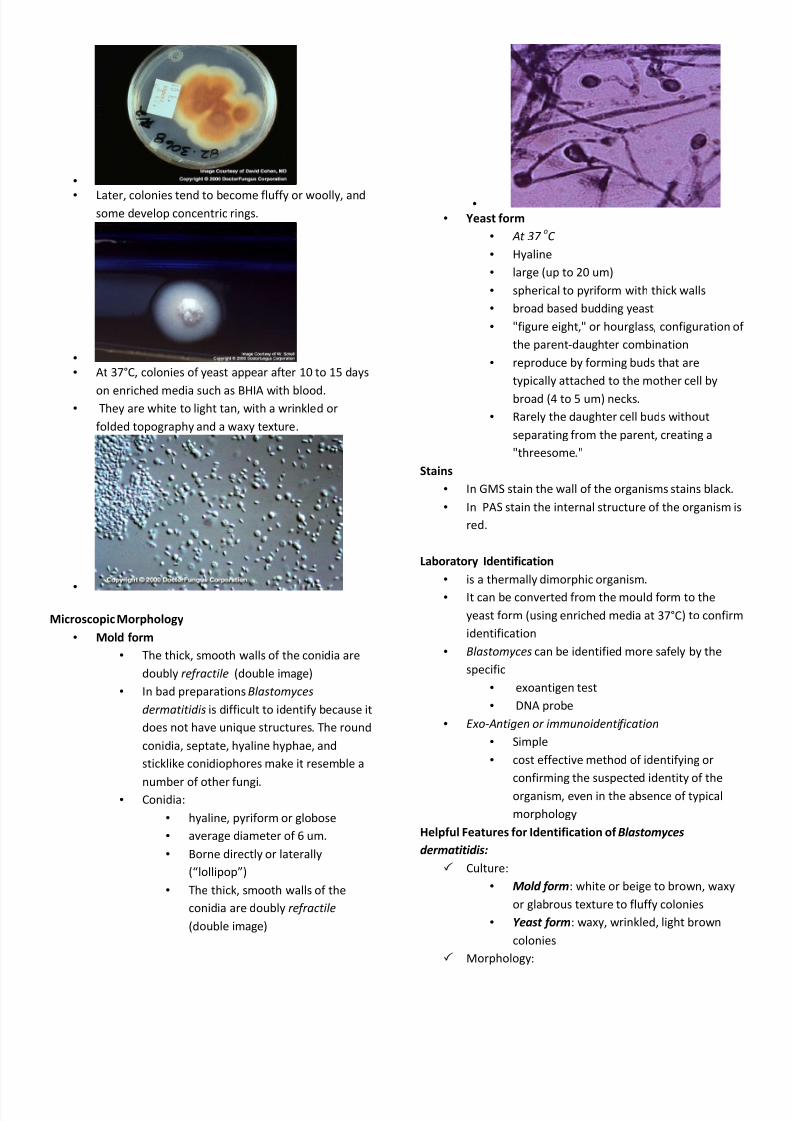
At 37°C, colonies of yeast appear after 10 to 15 days
on enriched media such as BHIA with blood. They are white to light tan, with a wrinkled or
folded topography and a waxy texture.
Microscopic Morphology
Mold form
The thick, smooth walls of the conidia are
doubly re f ractile (double image)
In bad preparations Blastomyces
dermatitidis is difficult to identify because it
does not have unique structures. The round
conidia, septate, hyaline hyphae, and
sticklike conidiophores make it resemble a
number of other fungi. Conidia:
hyaline, pyriform or globose
average diameter of 6 um.
Borne directly or laterally
(lollipop)
The thick, smooth walls of the
conidia are doubly re f ractile
(double image)
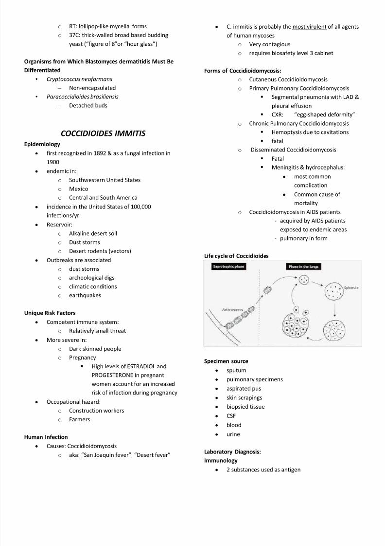
Yeast form
At 37 oC
Hyaline
large (up to 20 um)
spherical to pyriform with thick walls
broad based budding yeast
"figure eight," or hourglass, configuration of
the parent-daughter combination
reproduce by forming buds that are
typically attached to the mother cell by
broad (4 to 5 um) necks.
Rarely the daughter cell buds without
separating from the parent, creating a
"threesome."
Stains
In GMS stain the wall of the organisms stains black.
In PAS stain the internal structure of the organism is
red.
Laboratory Identification
is a thermally dimorphic organism.
It can be converted from the mould form to the
yeast form (using enriched media at 37°C) to confirm
identification
Blastomyces can be identified more safely by the
specific
exoantigen test
DNA probe
E xo- Antigen or immunoidenti f ication
Simple
cost effective method of identifying or
confirming the suspected identity of the
organism, even in the absence of typical
morphology
Helpful Features for Identification of Blastomyces
dermatitidis:
Culture:
Mold form: white or beige to brown, waxy
or glabrous texture to fluffy colonies
Yeast form: waxy, wrinkled, light brown
colonies
Morphology:
8/8/2019 Mycology Systemic
http://slidepdf.com/reader/full/mycology-systemic 4/12
o RT: lollipop-like mycelial forms
o 37C: thick-walled broad based budding
yeast (figure of 8or hour glass)
Organisms from Which Blastomyces dermatitidis Must Be
Differentiated
Cryptococcus neo f ormans
± Non-encapsulated
Paracoccidioides brasiliensis
± Detached buds
COCCIDIOIDES IMMITIS
Epidemiology
y first recognized in 1892 & as a fungal infection in
1900
y endemic in:
o Southwestern United States
o Mexico
o Central and South America
y incidence in the United States of 100,000
infections/yr.
y Reservoir:
o Alkaline desert soil
o Dust storms
o Desert rodents (vectors)
y Outbreaks are associated
o dust storms
o archeological digs
o climatic conditions o earthquakes
Unique Risk Factors
y Competent immune system:
o Relatively small threat
y More severe in:
o Dark skinned people
o Pregnancy
High levels of ESTRADIOL and
PROGESTERONE in pregnant
women account for an increased
risk of infection during pregnancy
y Occupational hazard:
o Construction workers
o Farmers
Human Infection
y Causes: Coccidioidomycosis
o aka: San Joaquin fever; Desert fever
y C. immitis is probably the most virulent of all agents
of human mycoses
o Very contagious
o requires biosafety level 3 cabinet
Forms of Coccidioidomycosis:
o Cutaneous Coccidioidomycosis
o Primary Pulmonary Coccidioidomycosis
Segmental pneumonia with LAD &
pleural effusion
CXR: egg-shaped deformity
o Chronic Pulmonary Coccidioidomycosis
Hemoptysis due to cavitations
fatal
o Disseminated Coccidioidomycosis
Fatal
Meningitis & hydrocephalus:
y most common
complication
y Common cause of
mortality
o Coccidioidomycosis in AIDS patients
- acquired by AIDS patients
exposed to endemic areas
- pulmonary in form
Life cycle of Coccidioides
Specimen source
y sputum
y pulmonary specimens
y aspirated pus
y skin scrapings
y biopsied tissue
y CSF
y blood
y urine
Laboratory Diagnosis:
Immunology
y 2 substances used as antigen
8/8/2019 Mycology Systemic
http://slidepdf.com/reader/full/mycology-systemic 5/12
o Coccidioidin: Is a filtrate prepared from
mould cultures
o Spherulin: Extract from a tissue culture of
the yeast form
y Skin test
o conversion from a negative to a positive
skin test is diagnostic of infection
y CF
o quite specific, but a few cross-reactions
with other mycotic infections
y TP Test
o Highly specific with very few cross-reactions
y LA
o being reevaluated because of reports of up
to 10% false-positive results with CSF and
diluted sera.
y DNA probes
o for culture confirmation
y exoantigen test
o to speed identification of the fungus using
indirect FA
y FA Tests
o rare cross-reaction
o excellent screening test
Culture Media
y Modified SDA
y SDA with antimicrobials
y BHIA
y BHIB
y Temperature
o Optimum temp. = 25-35oC for the mould
form.
o Tissue culture = 37-40oC with increased
CO2.
o Tmax = 54C
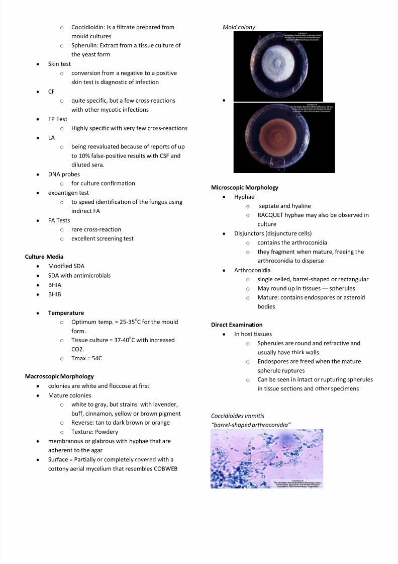
Macroscopic Morphology
y colonies are white and floccose at first
y Mature colonies o white to gray, but strains with lavender,
buff , cinnamon, yellow or brown pigment
o Reverse: tan to dark brown or orange
o Texture: Powdery
y membranous or glabrous with hyphae that are
adherent to the agar
y Surface = Partially or completely covered with a
cottony aerial mycelium that resembles COBWEB
Mold colony
y
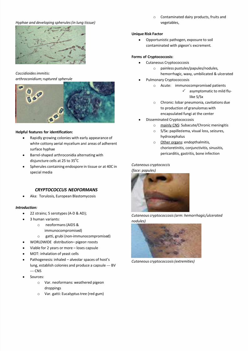
Microscopic Morphology
y Hyphae o septate and hyaline
o RACQUET hyphae may also be observed in
culture
y Disjunctors (disjuncture cells)
o contains the arthroconidia
o they fragment when mature, freeing the
arthroconidia to disperse
y Arthroconidia
o single celled, barrel-shaped or rectangular
o May round up in tissues --- spherules
o Mature: contains endospores or asteroid
bodies
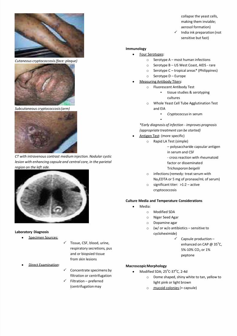
Direct Examination
y In host tissues
o Spherules are round and refractive and
usually have thick walls.
o Endospores are freed when the mature
spherule ruptures
o Can be seen in intact or rupturing spherules
in tissue sections and other specimens
Coccidioides immitis
barrel-shaped arthroconidia
8/8/2019 Mycology Systemic
http://slidepdf.com/reader/full/mycology-systemic 6/12
Hyphae and developing spherules ( in lung tissue)
Coccidioides immitis:
arthroconidium; ruptured spherule
Helpful features for identification:
y Rapidly growing colonies with early appearance of
white cottony aerial mycelium and areas of adherent
surface hyphae
y Barrel-shaped arthroconidia alternating with
disjuncture cells at 25 to 35oC
y Spherules containing endospore in tissue or at 40C in
special media
CRYPTOCOCCUS NEOFORMANS
y Aka: Torulosis, European Blastomycosis
Introduction:
y 22 strains; 5 serotypes (A-D & AD);
y 3 human variants:
o neoformans (AIDS &
immunocompromised)
o gatti, grubi (non-immunocompromised)
y WORLDWIDE distribution pigeon roosts
y Viable for 2 years or more loses capsule
y MOT: inhalation of yeast cells
y Pathogenesis: inhaled alveolar spaces of hosts
lung, establish colonies and produce a capsule --- BV
--- CNS
y Sources:
o Var. neoformans: weathered pigeon
droppings
o Var. gatti: Eucalyptus tree (red gum)
o Contaminated dairy products, fruits and
vegetables,
Unique Risk Factor
y Opportunistic pathogen, exposure to soil
contaminated with pigeons excrement.
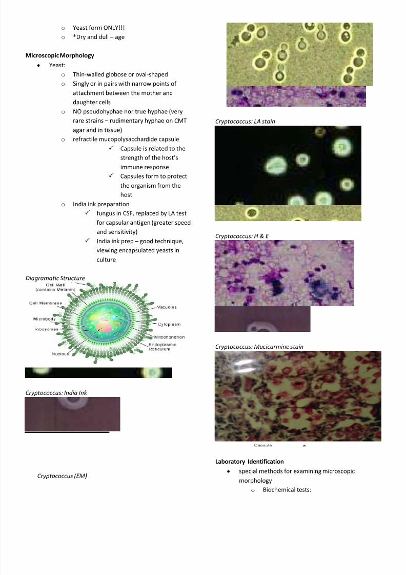
Forms of Cryptococcosis:
y Cutaneous Cryptococcosis
o painless pustules/papules/nodules,
hemorrhagic, waxy, umbilicated & ulcerated
y Pulmonary Cryptococcosis
o Acute: immunocompromised patients
asymptomatic to mild flu-
like S/Sx
o Chronic: lobar pneumonia, cavitations due
to production of granulomas with
encapsulated fungi at the center
y Disseminated Cryptococcosis
o mainly CNS: Subacute/Chronic meningitis
o S/Sx: papilledema, visual loss, seizures,
hydrocephalus
o Other organs: endopthalmitis,
chorioretinitis, conjunctivitis, sinusitis,
pericarditis, gastritis, bone infection
Cutaneous cryptococcis
(f ace: papules)
Cutaneous cryptococcosis ( arm: hemorrhagic/ulcerated
nodules)
Cutaneous cryptococcosis ( extremities)
8/8/2019 Mycology Systemic
http://slidepdf.com/reader/full/mycology-systemic 7/12
Cutaneous cryptococcosis (f ace: plaque)
Subcutaneous cryptococcosis ( arm)
C T w ith intravenous contrast medium injection. Nodular cystic
lesion w ith enhancing capsule and central core, in the parietal
region on the le f t side.
Laboratory Diagnosis
y Specimen Sources:
Tissue, CSF, blood, urine,
respiratory secretions, pus
and or biopsied tissue
from skin lesions
y Direct Examination:
Concentrate specimens by
filtration or centrifugation
Filtration preferred
(centrifugation may
collapse the yeast cells,
making them inviable;
aerosol formation)
India ink preparation (not
sensitive but fast)
Immunology
y Four Serotypes:
o Serotype A most human infections
o Serotype B US West Coast, AIDS - rare
o Serotype C tropical areas* (Philippines)
o Serotype D Europe
y Measuring Antibody Titers:
o Fluorescent Antibody Test
tissue studies & serotyping
cultures
o Whole Yeast Cell Tube Agglutination Test
and EIA
Cryptococcus in serum
*E arly diagnosis o f in f ection - improves prognosis
( appropriate treatment can be started)
y Antigen Test: (more specific)
o Rapid LA Test (simple)
polysaccharide capsular antigen
in serum and CSF
- cross reaction with rheumatoid
factor or disseminated
Trichosporon beigelii
o infections (remedy: treat serum with
Na2EDTA or 5 mg of pronase/mL of serum)
o significant titer: >1:2 active
cryptococcosis
Culture Media and Temperature Considerations
y Media:
o Modified SDA
o Niger Seed Agar
o Dopamine agar
o (w/ or w/o antibiotics sensitive to
cycloheximide)
Capsule production
enhanced on CAP @ 35oC,
5%-10% CO2 or 1%
peptone
Macroscopic Morphology
y Modified SDA; 25oC-37
oC, 2-4d
o Dome shaped, shiny white to tan, yellow to
light pink or light brown
o mucoid colonies (+ capsule)
8/8/2019 Mycology Systemic
http://slidepdf.com/reader/full/mycology-systemic 8/12
o Yeast form ONLY!!!
o *Dry and dull age
Microscopic Morphology
y Yeast:
o Thin-walled globose or oval-shaped
o Singly or in pairs with narrow points of
attachment between the mother and
daughter cells
o NO pseudohyphae nor true hyphae (very
rare strains rudimentary hyphae on CMT
agar and in tissue)
o refractile mucopolysacchardide capsule
Capsule is related to the
strength of the hosts
immune response
Capsules form to protect
the organism from the
host
o India ink preparation
fungus in CSF, replaced by LA test
for capsular antigen (greater speed
and sensitivity)
India ink prep good technique,
viewing encapsulated yeasts in
culture
Diagramatic Structure
Cryptococcus: India Ink
Cryptococcus ( E M )
Cryptococcus: L A stain
Cryptococcus: H & E
Cryptococcus: Mucicarmine stain
Laboratory Identification
y special methods for examining microscopic
morphology
o Biochemical tests:
8/8/2019 Mycology Systemic
http://slidepdf.com/reader/full/mycology-systemic 9/12
Carbohydrate and nitrate
utilization:
dextrose, maltose, sucrose,
galactose cellobiose, inositol,
xylose, raffinose, trehalose,
dulcitol, starch
Urease production +
Phenoloxidase test +
brown colonies in Caffeic acid/bird
seed/thistle/niger seed agar
Treatment
o Non-immunocompromised:
Amphotericin B + Flucytosine
o Immonucompromised:
Amphotericin B + Flucytosine +
Fluconazole
o AIDS px:
longer tx; Fluconazole as
maintenance
PARACOCCIDIOIDES BRASILIENSIS
y A.K.A. Paracoccidioidomycosis or South American
Blastomycosis
y P. brasiliensis, hyaline hyphomycete
y Endemic: Holdridge Life Zones, northwestern,
central, and southeastern South America, Central
America, and southern Mexico
y Reservoir & Unique Risk Factors
o Saprobic mould form acid soil in humidareas (endemic)
o Plants
o Armadillos - carrier
y MOT: Airborne (i.e., plants)
y Risk factors:
o Adult males combined effect of hormonal
makeup and occupations
o Malnutrition and immunocompetence
Human Infection/s
y Pathogenesis:
o Primarily an oral lesion (mouth, palate,
nasal) --- BV & lymphatics --- disseminated
esp to the lungs
y Primary Pulmonary Paracoccidioidomycosis
asymptomatic or subclinical
o self -limiting
subacute primary pulmonary disease mild changes
in the lungs; positive skin test
Secondary asymptomatic infections - pulmonary
disease or dissemination
y Disseminated disease in any organ (esp GIT)
y Pyogenic abscesses and ulcers - granulomatous
y Lymphadenitis - common
Specimen Sources
y Sputum (other pulmonary specimens)
y Pus aspirated from lymph nodes
y Skin scrapings or biopsied tissue edge of ulcers
y Biopsied lung tissue
y Crusts from skin lesions
Specimen Collection and Handling
y Aseptic technique
y Direct examination:
o Simple wet mounts, with added stains
o KOH clear debris
o N-acetyl-L-cysteine (NALC) mucus and
thick substances
o Tissue specimens (fixed and stained)
Papanicolaou stain, H&E, or Giemsa stain
o Specific FA Test detect yeast form in
tissue
Immunology
y Paracoccidioidin - E2 antigen extract from yeast
1. Precipitin Test band 1
2. Skin Tests intradermal injection
epidemiological tool
first serologic test to be positive
do not differentiate between past
exposure and current condition
negative skin test person who was
previously positive indicates the
anergy of disseminated infection
3. CF Test
a. yeast filtrate antigens: recommended
serologic test (titers appear in late and
remain detectable for several months
curedb. cross reactions low titers in patients with
acute histoplasmosis and blastomycosis.
4. ID Test
o concentrated yeast antigens and reference
sera are available
o Sensitivity, 94% and highly specific
o (+) one to three precipitin continuous bands
or identical with the reference sera is
indicative of infection
8/8/2019 Mycology Systemic
http://slidepdf.com/reader/full/mycology-systemic 10/12
o Band 1 is the first to disappear during
treatment
o number of bands is correlated with the CF
titer
o Low titers: localized infection
o high titers: acute infection or dissemination.
o When symptoms are present a combination
of ID and CF tests is 98% specific for
diagnosis of paracoccidioidomycosis.
5. Exoantigen Methods
a. useful for speeding the identification of
cultures
6. Direct FA Tests
a. detects P. brasiliensis cells in smears of
clinical materials
Culture Media and Temperature Considerations
y Modified SDA with antimicrobials except
cycloheximide (primary isolation contaminated with
bacteria; slows fungus growth) at 25oC to 30oC
y SABHI and BHIA with blood (sterile sites)
y No antibiotics above 30C
y Yeast Extract Agar used for primary culture to
encourage initial growth and conidiation
y PDA and PFA for subculture to encourage
conidiation
Macroscopic Morphology
y Mould Form; 25oC, Mod. SDA
o Slow maturation 2 cm in diameter after 2
to 3 weeks
y Young: White to cream, with short, downy aerial
mycelia and elevated centers.
y Mature: Flat, with a membranous or velvety texture
and cerebriform or folded topography.
y Pigment: Beige or brown, with a yellow-brown
reverse in mature colonies.
y Mycelial growth at 24°C on Mycosel Agar, 6 weeks of
incubation. Note multiple colonies.
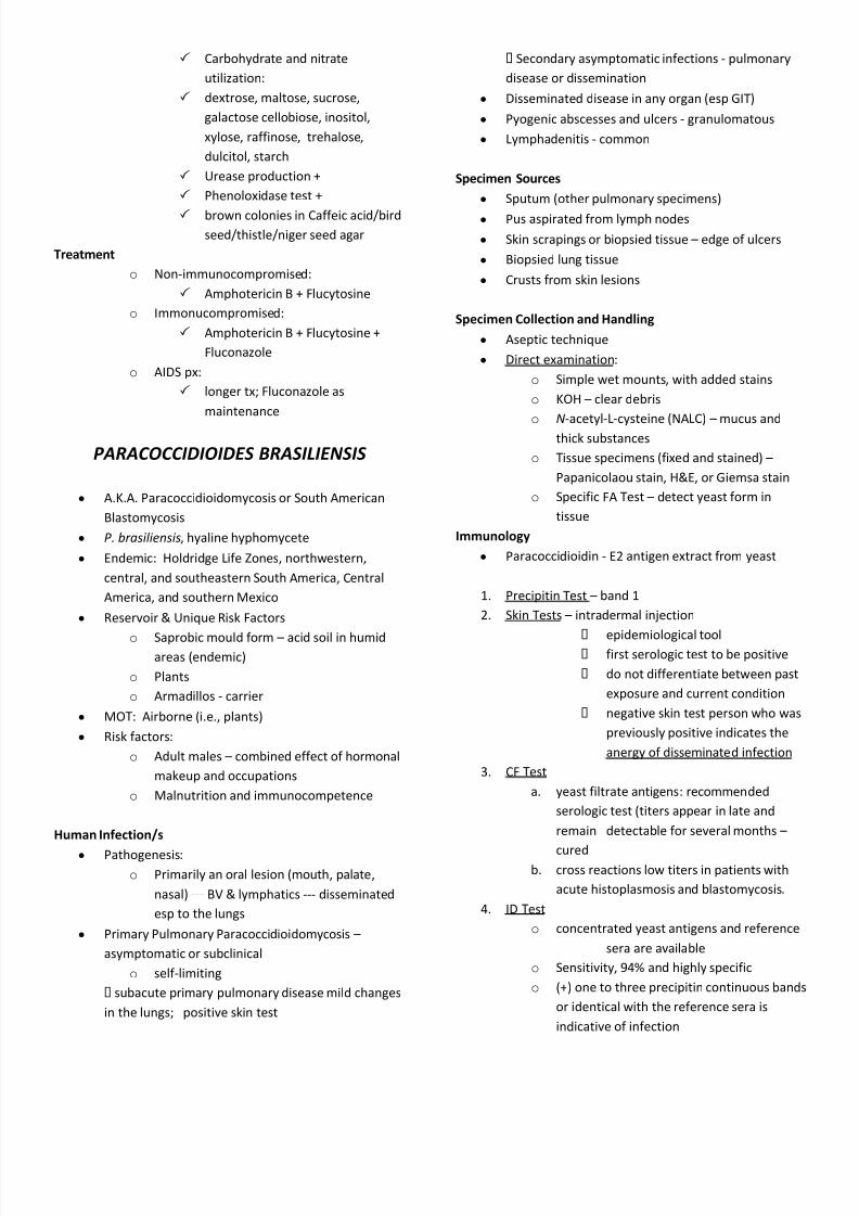
Microscopic MorphologyMould Form; 25
oC to 30
oC
y Modified SDA
y Hyphae: Very fine, hyaline and septate
y Conidia: Few small oval to pyriform truncate on
short conidiophores or sessile hyphae
y Chlamydoconidia: Terminal and intercalary, w/
racquet and coiled hyphae
Yeast Form:
y Yeast Extract Agar (deficient in glucose)
y Conidia: Large number of oval to pyriform with
thick-walled arthroconidia in alternating pattern
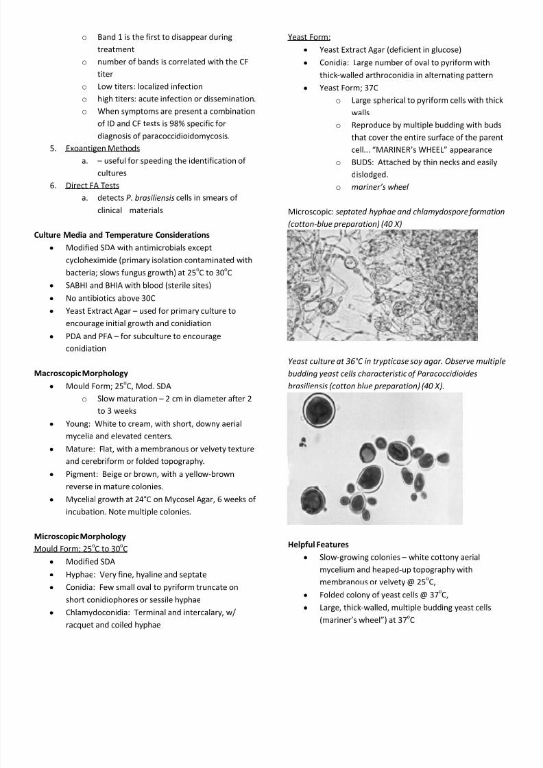
y Yeast Form; 37C
o Large spherical to pyriform cells with thick
walls
o Reproduce by multiple budding with buds
that cover the entire surface of the parent
cell... MARINERs WHEEL appearance
o BUDS: Attached by thin necks and easily
dislodged.
o mariners w heel
Microscopic: septated hyphae and chlamydospore f ormation
( cotton-blue preparation) (40 X )
Y east culture at 36° C in trypticase soy agar. Observe multiple
budding yeast cells characteristic o f Paracoccidioides
brasiliensis ( cotton blue preparation) (40 X ).
Helpful Features
y Slow-growing colonies white cottony aerial
mycelium and heaped-up topography with
membranous or velvety @ 25oC,
y Folded colony of yeast cells @ 37oC,
y Large, thick-walled, multiple budding yeast cells
(mariners wheel) at 37oC
8/8/2019 Mycology Systemic
http://slidepdf.com/reader/full/mycology-systemic 11/12
H ISTOPLASMA CAPSULATUM
Epidemiology
y Phil: 1992 1st reported case
y Prevalent in:
o Central North America
o Central South America
o Africa
o Australia
o India
o Malaysia
y Darlings disease
Reservoir
y Soil with high nitrogen content
y Caves
y birds droppings (birds, chicken, bats)
Human Infection
y Histoplasmosis y MOT: inhalation
y CXR:
o Fibrosis
o coin lesions
Forms of Histoplasmosis
1. Acute Pulmonary Histoplasmosis
y 1-3 wks incubation
y Non-specific flu-like S/Sx; (-) AFS;
y resolves w/o tx
y CXR: infiltrates to pleural effusion
y complications:
o aseptic arthritis/arthralgia
o erythema multiforme
o LAD
2. Chronic Pulmonary Histoplasmosis
y no LAD, no pleural effusion
y assoc with COPD
y Patho: pneumonia --- fibrosis --- cavitation --- lung
destruction --- pleural thickening
y CM: cough, hemoptysis, pleuritic pain
y CXR: interstitial infiltrates in apex of lungs
3. Disseminated Histoplasmosis
y seen in AIDS and immunocompromised patients
y involves the tongue, lungs, liver, GIT, adrenal glands,
blood
y Treatment: Amphotericin B, Itraconazole
Lab Diagnosis
1. Microscopic examination
2. Culture
3. immunology
Specimen
y pulmonary specimen
y pus / abscess
y skin scrapings
y biopsied tissues
y bone marrow
y blood
y csf and urine
Direct Examination
y intracellular
o Wright-Giemsa
o not KOH
o histology lab
H & E
Paps stain
y Mucous (NALC)
y FA tests
Culture Media
y BAP
y mod SDA: moist
y BHIA / BHIB: from sterile sites
y Yeast Extract PO4: inhibits candida
y PDA: encourages conidiation
y Primary cultures should be held for 10 to 12 weeks
before discarding as no growth
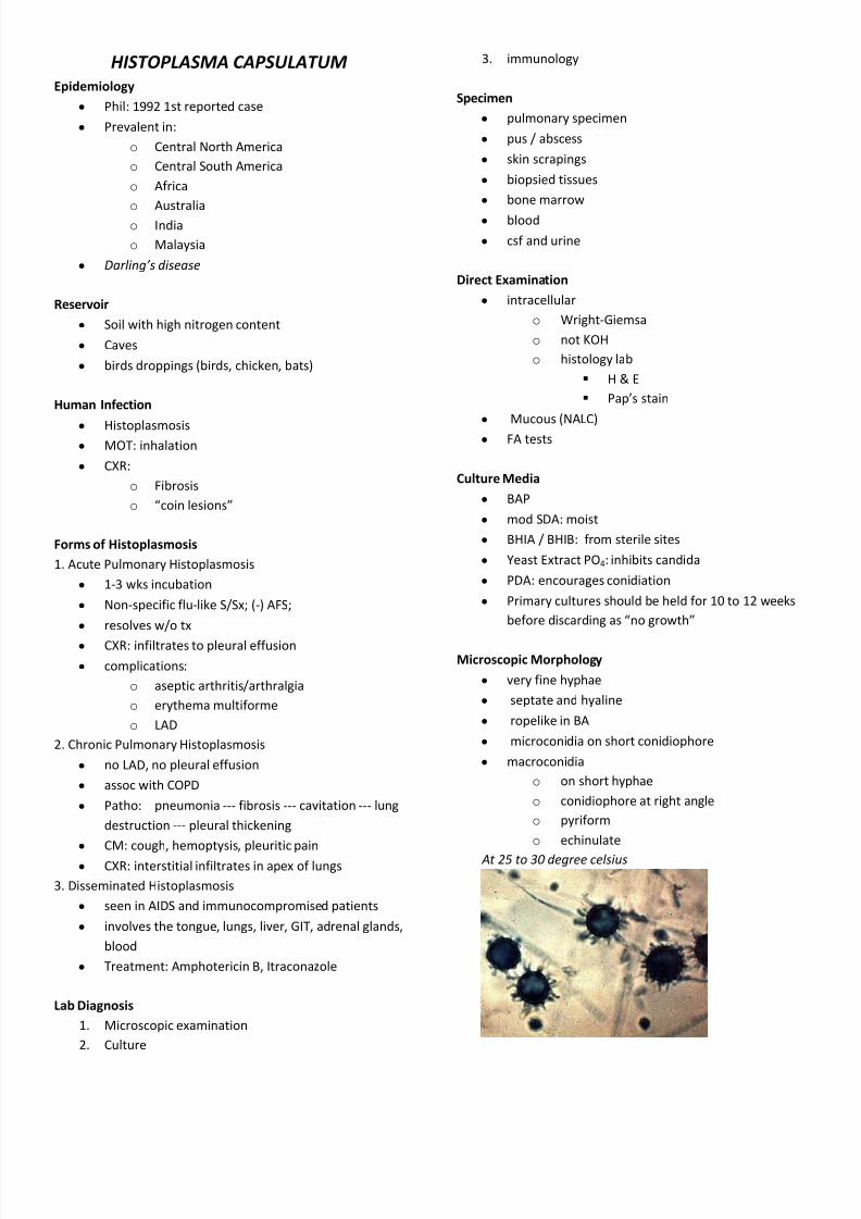
Microscopic Morphology
y very fine hyphae
y septate and hyaline
y ropelike in BA
y microconidia on short conidiophore
y macroconidia
o on short hyphae
o conidiophore at right angle
o pyriform
o echinulate
At 25 to 30 degree celsius
8/8/2019 Mycology Systemic
http://slidepdf.com/reader/full/mycology-systemic 12/12
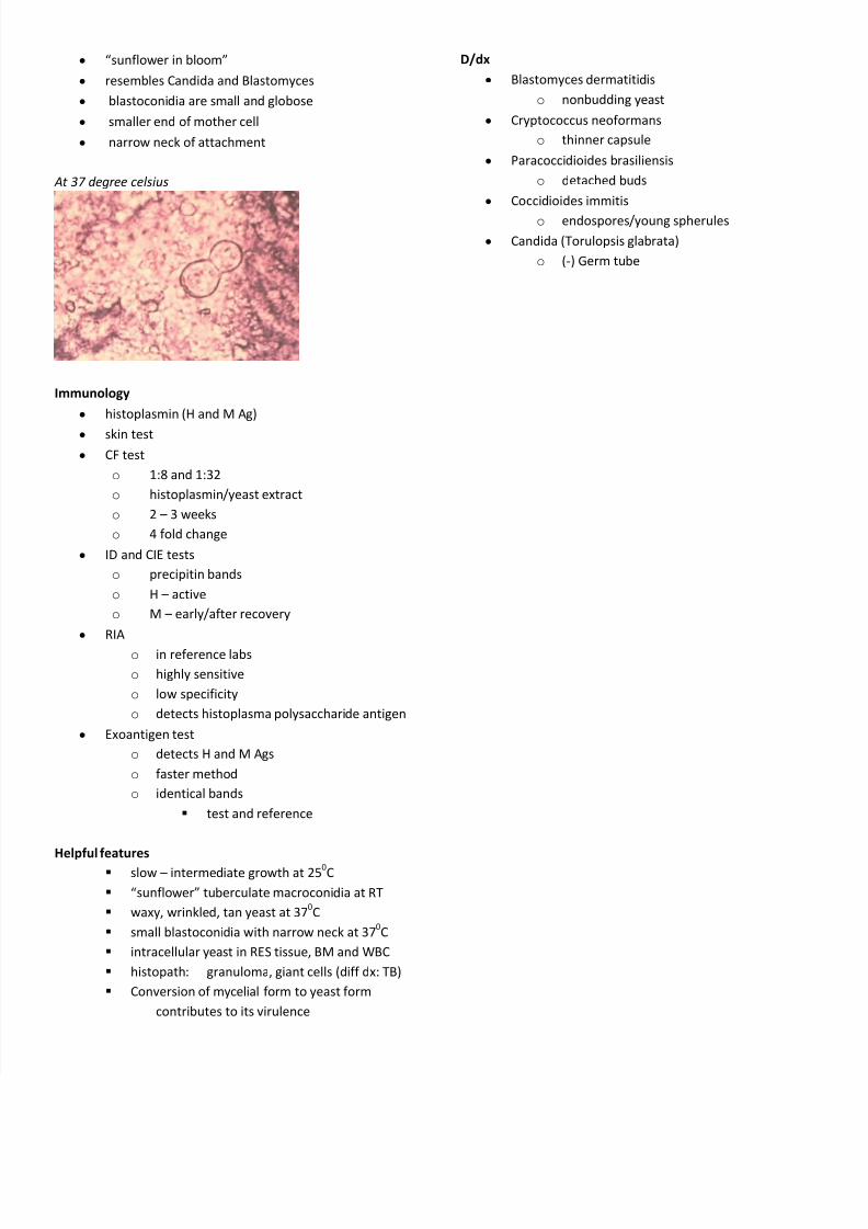
y sunflower in bloom
y resembles Candida and Blastomyces
y blastoconidia are small and globose
y smaller end of mother cell
y narrow neck of attachment
At 37 degree celsius
Immunology
y histoplasmin (H and M Ag)
y skin test
y CF test
o 1:8 and 1:32
o histoplasmin/yeast extract
o 2 3 weeks
o 4 fold change
y ID and CIE tests
o precipitin bands
o H active
o M early/after recovery
y RIA
o in reference labs
o highly sensitive
o low specificity
o detects histoplasma polysaccharide antigen
y Exoantigen test
o detects H and M Ags
o faster method
o identical bands
test and reference
Helpful features
slow intermediate growth at 250C
sunflower tuberculate macroconidia at RT
waxy, wrinkled, tan yeast at 370C
small blastoconidia with narrow neck at 370C
intracellular yeast in RES tissue, BM and WBC
histopath: granuloma, giant cells (diff dx: TB)
Conversion of mycelial form to yeast form
contributes to its virulence
D/dx
y Blastomyces dermatitidis
o nonbudding yeast
y Cryptococcus neoformans
o thinner capsule
y Paracoccidioides brasiliensis
o detached buds
y Coccidioides immitis
o endospores/young spherules
y Candida (Torulopsis glabrata)
o (-) Germ tube