myelogram rad 205-2011
TRANSCRIPT
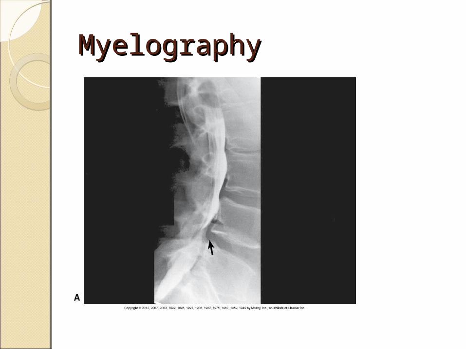
Myelography Myelography
MyelogramMyelogramMyelogram: Myel/o means spinal
cord or bone marrow – gram: …………
Note DO NOT confuse for my/o, which means muscle)
The spinal cord is part of the Nervous System. Therefore, this a study focused on the spinal cord anatomy which could be affected by the bony anatomy of the spine.

Basic Spinal Cord Basic Spinal Cord AnatomyAnatomyFor description, the central
nervous system (CNS) is divided into◦Brain◦Spinal cord
CNS tissue composed of two main portions◦Gray matter = outer portion or cortex◦White matter = inner portion

Spinal CordSpinal CordSlender, elongated
structure Consists of an inner gray
cellular substance and an outer white fibrous substance◦Gray substance forms H-
shape seen on transverse section
Extends from the brain to the L1-L2 space
Conus medullaris = pointed end of spinal cord

SPINAL CORDSPINAL CORDGray substance forms H-shape seen on transverse section

SPINAL CORDSPINAL CORDFilum terminale = delicate fibrous portion that connects the terminal tip to the first coccygeal segment
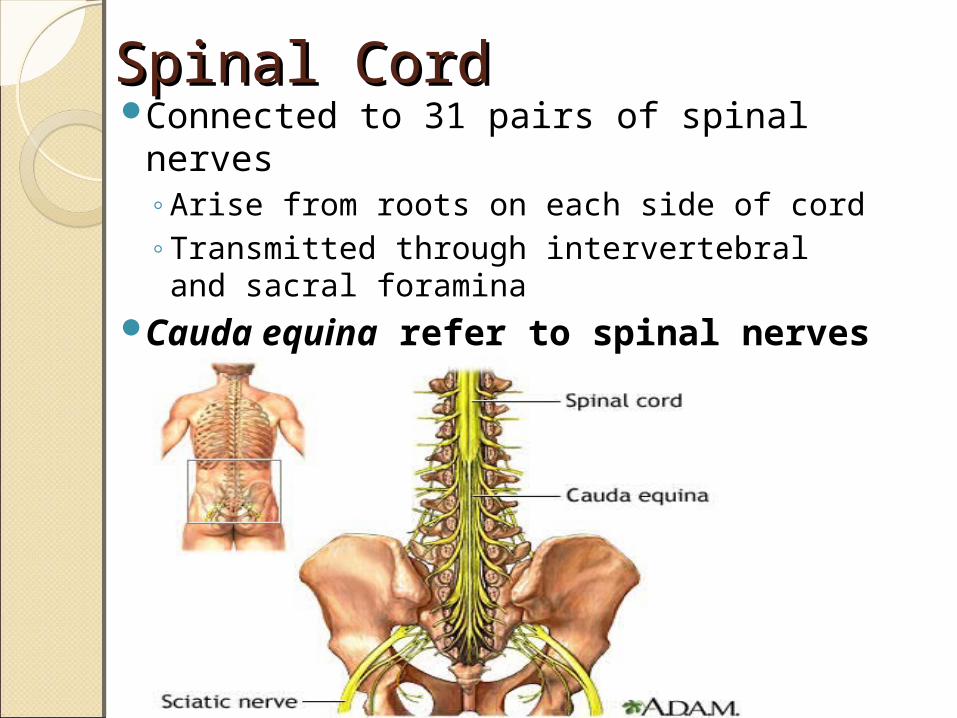
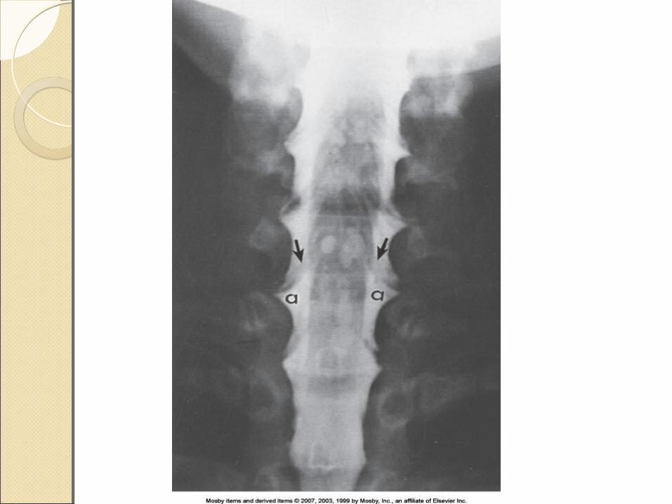
Spinal CordSpinal CordConnected to 31 pairs of spinal nerves
◦Arise from roots on each side of cord◦Transmitted through intervertebral and
sacral foraminaCauda equina refer to spinal
nerves
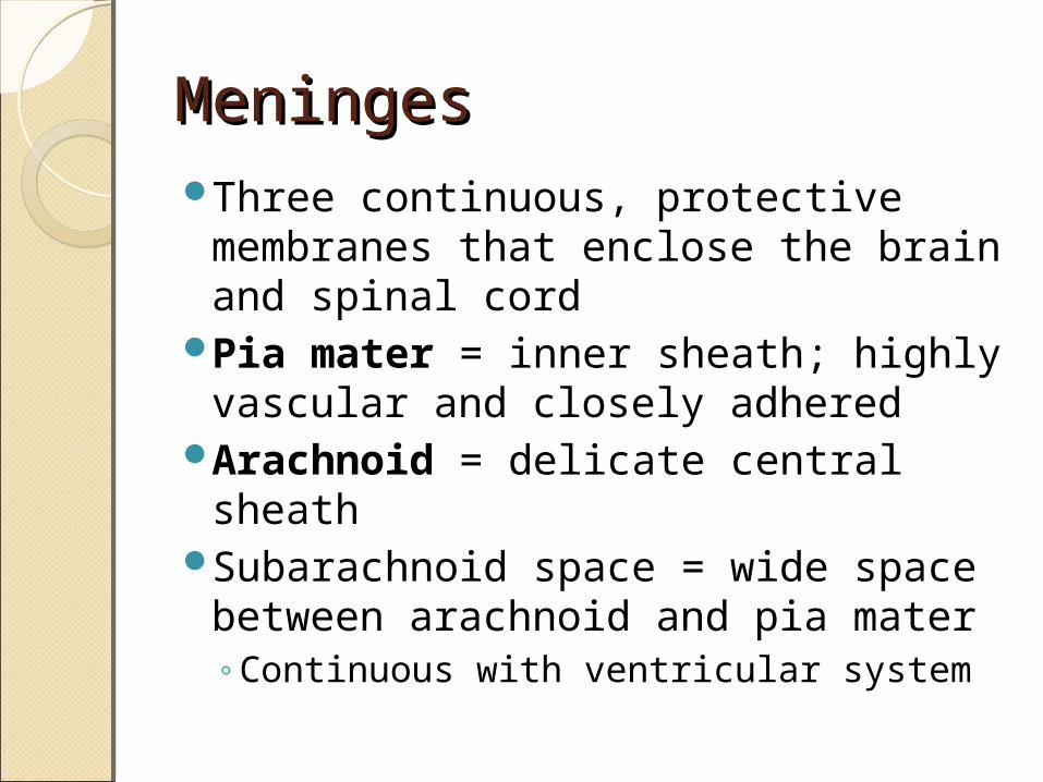
MeningesMeningesThree continuous, protective
membranes that enclose the brain and spinal cord
Pia mater = inner sheath; highly vascular and closely adhered
Arachnoid = delicate central sheath
Subarachnoid space = wide space between arachnoid and pia mater◦Continuous with ventricular system
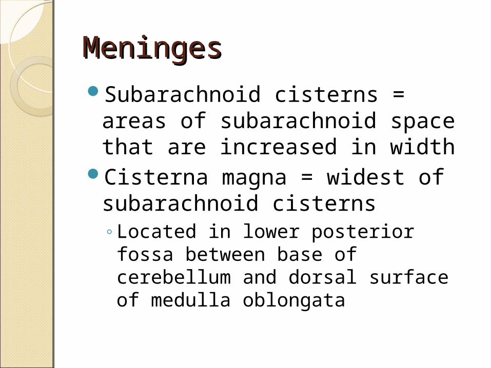
MeningesMeningesSubarachnoid cisterns = areas of
subarachnoid space that are increased in width
Cisterna magna = widest of subarachnoid cisterns◦Located in lower posterior fossa
between base of cerebellum and dorsal surface of medulla oblongata
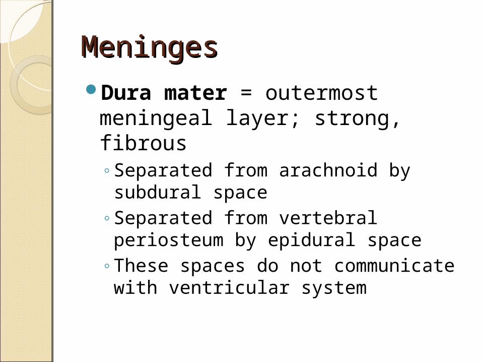
MeningesMeningesDura mater = outermost
meningeal layer; strong, fibrous◦Separated from arachnoid by
subdural space◦Separated from vertebral periosteum
by epidural space◦These spaces do not communicate
with ventricular system

MeningesMeninges◦Dural sac (Thecal sac) = encloses cauda equina
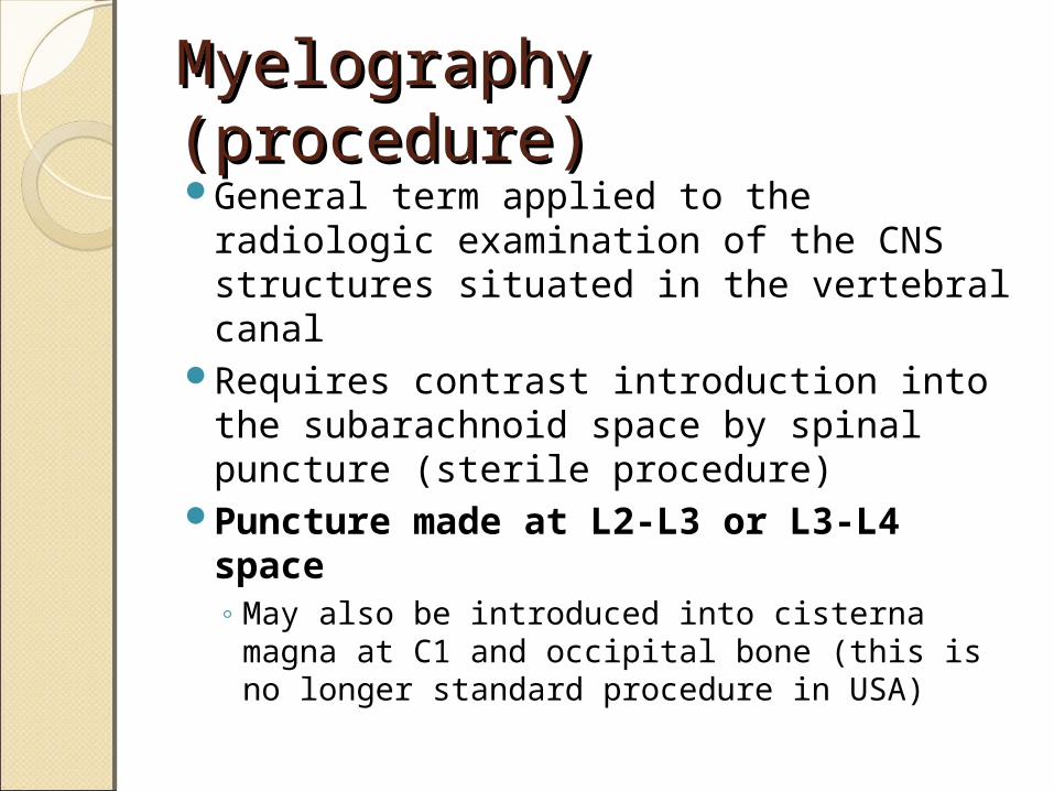
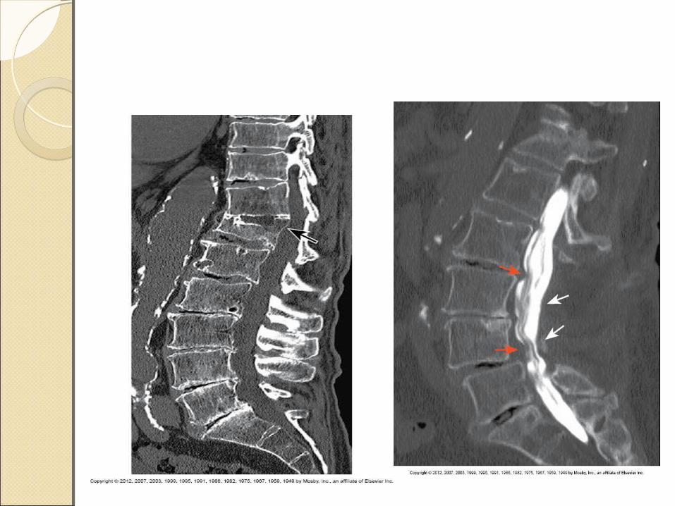
Myelography (procedure)Myelography (procedure)General term applied to the radiologic
examination of the CNS structures situated in the vertebral canal
Requires contrast introduction into the subarachnoid space by spinal puncture (sterile procedure)
Puncture made at L2-L3 or L3-L4 space◦ May also be introduced into cisterna magna
at C1 and occipital bone (this is no longer standard procedure in USA)
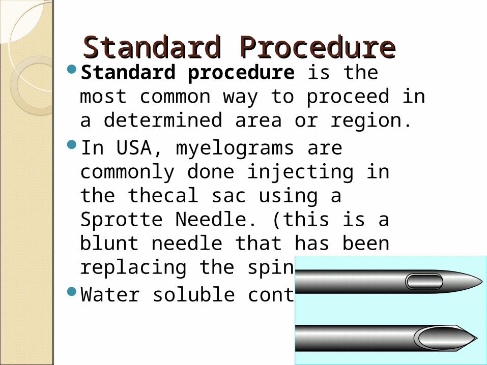
Standard ProcedureStandard ProcedureStandard procedure is the most
common way to proceed in a determined area or region.
In USA, myelograms are commonly done injecting in the thecal sac using a Sprotte Needle. (this is a blunt needle that has been replacing the spinal needle)
Water soluble contrast
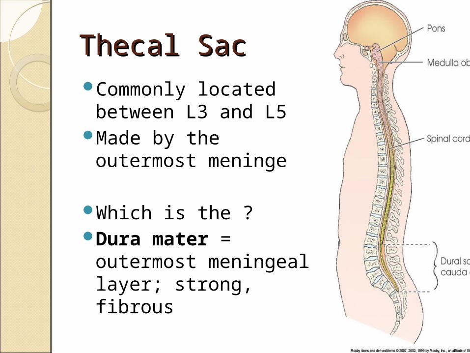
Thecal SacThecal SacCommonly located
between L3 and L5Made by the
outermost meninge
Which is the ?Dura mater =
outermost meningeal layer; strong, fibrous

MyelographyMyelographyUsually performed as outpatient
basis◦Patients held for recovery after
procedure for 4 to 8 hours, then released to go home
MRI often used insteadMyelography still used for patients
with contraindications for MRI◦Pacemakers and metal fusion rods

MyelographyMyelographyContrast is generally water-soluble,
nonionic, and preservative free iodinated medium
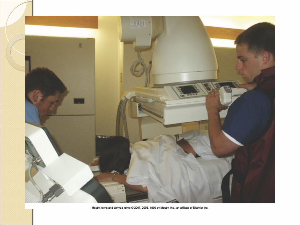
Invasive ProcedureRoom should be prepared by RT before
patient arrival ◦ Table and equipment (FL IR) cleaned◦ Footboard and shoulder supports attached◦ Radiographic equipment checked◦ Image intensifier locked to prevent accidental
contact with sterile field or spinal (Sprote) needle
MyelographyMyelographyPremedication rarely needed
(allergies)Patient should be well hydratedProcedural details, including table
movement and sensations, should be explained
Scout images made: AP & LatProne position usually used for
spinal puncture◦Lateral with spine flexed may also be
used
MyelographyMyelographyLocal anesthesia given at puncture
siteSpinal needle insertedCSF usually withdrawn and sent to
laboratoryContrast injected and needle removedTable angle and gravity used to move
contrast under fluoroscopySpot images taken as needed
(protocol)
MyelographyMyelographyIf contrast is moved into cervical
area, head is positioned in acute extension to prevent contrast from entering ventricular system (varies depending on T, L and/or C spine◦Acute extension compresses cisterna
magna and is the only position that will prevent contrast from entering ventricles
MyelographyMyelographyPostprocedure monitoring
required◦Head and shoulders elevated 30 to 45 degrees
◦Bed rest for several hours (Dr instructions)
◦Fluid encouraged (caffeine not recommended)
◦Puncture site checked before release
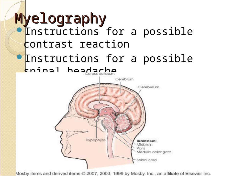
MyelographyMyelographyInstructions for a possible contrast
reactionInstructions for a possible spinal
headache

Spinal Cord Spinal Cord AnatomyAnatomy
Transverse section of spinal cord